Virus-Induced Gene Silencing (VIGS): A Comprehensive Guide to Mechanisms, Applications, and Future Directions in Functional Genomics
This article provides a detailed exploration of Virus-Induced Gene Silencing (VIGS), an RNA-mediated reverse genetics technique that has become an indispensable tool for functional genomics.
Virus-Induced Gene Silencing (VIGS): A Comprehensive Guide to Mechanisms, Applications, and Future Directions in Functional Genomics
Abstract
This article provides a detailed exploration of Virus-Induced Gene Silencing (VIGS), an RNA-mediated reverse genetics technique that has become an indispensable tool for functional genomics. Tailored for researchers, scientists, and drug development professionals, we cover the foundational molecular mechanisms of VIGS, including its basis in post-transcriptional gene silencing (PTGS) and the role of small interfering RNAs (siRNAs). The scope extends to practical methodologies, featuring a comparison of viral vector systems like Tobacco Rattle Virus (TRV) and Bean Pod Mottle Virus (BPMV), and their application in gene function analysis across diverse species, from model plants to crops. We critically address troubleshooting, optimization strategies, and the limitations of the technology. Finally, the article validates VIGS by comparing it with other functional genomics tools and discusses its advanced evolution into areas like epigenetic modification and genome editing, highlighting its significant implications for biomedical and agricultural research.
The Core Principles of VIGS: Unraveling the RNAi-Based Silencing Mechanism
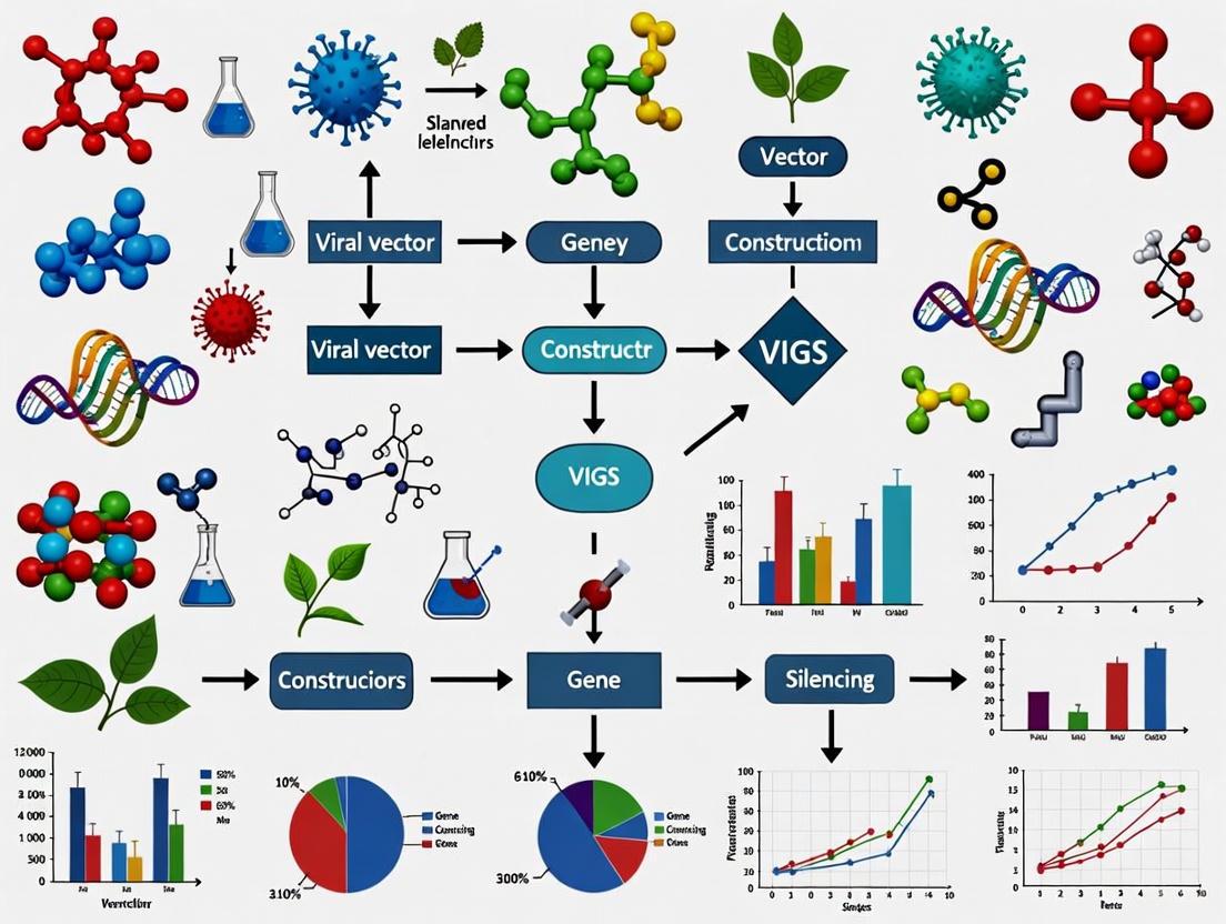
Virus-induced gene silencing (VIGS) is an RNA-mediated reverse genetics technique that has evolved from a fundamental antiviral defense mechanism into an indispensable tool for plant functional genomics. This technology leverages the plant's innate post-transcriptional gene silencing (PTGS) machinery to target specific endogenous genes for knockdown, enabling rapid functional characterization without the need for stable transformation. This review comprehensively examines the molecular mechanisms underpinning VIGS, its experimental applications across diverse plant species, recent methodological advances, and its emerging role in epigenetic studies and crop improvement programs. We also present standardized protocols, quantitative data comparisons, and visual workflow representations to facilitate implementation by researchers and biotechnology professionals.
Originally characterized by van Kammen as 'recovery from viral infection' [1], VIGS was first experimentally demonstrated as a functional genomics tool by Kumagai et al. (1995) using Tobacco mosaic virus (TMV) to silence the phytoene desaturase (PDS) gene in Nicotiana benthamiana, resulting in a visible albino phenotype [1]. This pioneering work established that recombinant viruses could be harnessed to suppress endogenous gene expression through sequence homology [1]. Since then, VIGS has transformed into a high-throughput functional genomics platform applicable to numerous plant species, including horticultural crops, forest trees, and recalcitrant perennial species that are not amenable to traditional genetic transformation [1] [2] [3].
The fundamental significance of VIGS lies in its ability to provide rapid gene function characterization through transient silencing, typically manifesting within 1-4 weeks post-inoculation depending on the host species and viral vector system [2]. This approach effectively bridges the gap between genomic sequencing and functional annotation, particularly valuable in species with long life cycles or complex genetics. Furthermore, VIGS has recently expanded beyond traditional gene knockdown applications to include heritable epigenetic modifications and virus-induced gene editing (VIGE), positioning it at the forefront of modern plant biotechnology and molecular breeding initiatives [1] [2].
Molecular Mechanisms of VIGS
The Core Post-Transcriptional Gene Silencing Pathway
VIGS operates through the plant's conserved RNA interference (RNAi) machinery, which naturally functions as an antiviral defense mechanism. The process initiates when a recombinant virus containing a fragment of a host gene (typically 200-500 bp) is introduced into the plant tissue [3]. The molecular events unfold through a coordinated sequence:
- Viral Replication and dsRNA Formation: Following infection, viral replication in the plant cell cytoplasm produces double-stranded RNA (dsRNA) intermediates, a hallmark of viral replication cycles [2].
- dicer-mediated processing: These dsRNA molecules are recognized and cleaved by plant Dicer-like (DCL) nucleases into small interfering RNA (siRNA) duplexes of 21-24 nucleotides in length [1] [2].
- RISC Assembly and mRNA Cleavage: The siRNAs are incorporated into the RNA-induced silencing complex (RISC), where the guide strand directs the complex to complementary endogenous mRNA transcripts through sequence homology. The catalytic component of RISC, typically an Argonaute (AGO) protein, then mediates the endonucleolytic cleavage or translational inhibition of the target mRNA [1] [2].
- Amplification and Systemic Spread: Plant RNA-dependent RNA polymerases (RDRPs) amplify the silencing signal by using the cleaved mRNA fragments as templates to generate secondary dsRNAs, which are subsequently processed into additional siRNAs. These secondary siRNAs facilitate the systemic spread of silencing throughout the plant, enabling whole-plant phenotypic analysis [1].
Figure 1: Molecular mechanism of VIGS-mediated post-transcriptional gene silencing. The recombinant viral vector introduces target sequence, leading to siRNA production and ultimately degradation of complementary mRNA.
Transcriptional Gene Silencing and Epigenetic Modifications
Beyond the cytoplasmic PTGS mechanism, VIGS can also induce transcriptional gene silencing (TGS) through RNA-directed DNA methylation (RdDM) in the nucleus. When siRNAs derived from the viral vector exhibit complementarity to gene promoter regions, they can guide epigenetic modifiers to these loci, resulting in cytosine methylation at CG, CHG, and CHH contexts [1]. This methylation, particularly when established in promoter sequences, can lead to stable, heritable gene silencing that persists even after the viral vector has been cleared from the plant [1]. Bond et al. (2015) demonstrated this phenomenon by using VIGS to target the FWA promoter in Arabidopsis, establishing transgenerational epigenetic silencing that was maintained through multiple generations [1]. This epigenetic dimension significantly expands VIGS applications beyond transient knockdown to include the creation of stable epi-alleles with modified gene expression patterns.
Essential VIGS Toolbox: Vectors and Research Reagents
Viral Vector Systems
The effectiveness of VIGS depends critically on selecting appropriate viral vectors tailored to the host plant species. Different vector systems offer distinct advantages and limitations based on their host range, symptom severity, and silencing efficiency.
Table 1: Comparison of Major Viral Vectors Used in VIGS
| Vector Type | Virus Name | Genome Type | Host Range Examples | Key Advantages | Primary Limitations |
|---|---|---|---|---|---|
| RNA Virus | Tobacco Rattle Virus (TRV) | Bipartite RNA | Nicotiana benthamiana, tomato, pepper, Arabidopsis, soybean [4] [5] | Mild symptoms, efficient systemic movement, meristem penetration [4] | Limited efficiency in some monocots |
| RNA Virus | Bean Pod Mottle Virus (BPMV) | RNA | Soybean [4] | Highly efficient in soybean; well-established system | Requires particle bombardment; can cause leaf symptoms [4] |
| RNA Virus | Barley Stripe Mosaic Virus (BSMV) | RNA | Barley, wheat, other monocots [2] | Effective in monocotyledonous species | Host range primarily limited to monocots |
| DNA Virus | Tomato Yellow Leaf Curl Virus (TYLCV) | Single-stranded DNA | Cabbage (Brassica rapa) [2] | Useful for dicot species recalcitrant to RNA viruses | Limited host range compared to TRV |
| DNA Virus | Cotton Leaf Crumple Virus (CLCrV) | Single-stranded DNA | Cotton, Nicotiana benthamiana [5] | Effective for gene silencing in cotton | Relatively narrow host range |
Essential Research Reagents and Solutions
Successful implementation of VIGS requires a comprehensive suite of molecular biology reagents and plant growth materials optimized for the target species.
Table 2: Key Research Reagent Solutions for VIGS Experiments
| Reagent Category | Specific Examples | Function/Purpose | Application Notes |
|---|---|---|---|
| Vector Systems | pTRV1, pTRV2, pBPMV, pTYLCV [2] [4] | Viral genome components for silencing construct | TRV system requires both pTRV1 (replication proteins) and pTRV2 (target insert) [4] |
| Agrobacterium Strains | GV3101, LBA4404, AGL1 [3] [4] | Delivery of viral vectors into plant cells | GV3101 commonly used with pTRV system; requires appropriate antibiotic resistance [4] |
| Induction Solution | Acetosyringone (100-200 μM), MES buffer (10 mM, pH 5.6) [3] | Activates Agrobacterium virulence genes; maintains pH during infiltration | Critical for efficient T-DNA transfer; fresh preparation recommended |
| Culture Media | YEB, LB with appropriate antibiotics (kanamycin, rifampicin) [3] | Growth and selection of Agrobacterium carrying viral vectors | Optical density (OD600 = 0.5-1.0) critical for infiltration efficiency [6] |
| Infiltration Buffers | MgCl2 (10 mM), MES (10 mM, pH 5.6) [3] | Dilution of bacterial cultures for infiltration | Maintains bacterial viability and facilitates plant cell entry |
Experimental Implementation: Protocols and Methodologies
Standard TRV-Based VIGS Protocol
The Tobacco Rattle Virus (TRV) system has emerged as one of the most versatile and widely adopted VIGS platforms due to its broad host range and mild symptomology. The following protocol outlines the key steps for implementing TRV-mediated silencing:
Vector Construction Phase (7-10 days)
- Target Gene Fragment Selection: Identify a 200-300 bp gene-specific fragment with minimal off-target potential using tools like the SGN VIGS Tool (https://vigs.solgenomics.net/) [3]. Verify sequence specificity through BLAST analysis against the host genome.
- Molecular Cloning: Amplify the target fragment using gene-specific primers incorporating appropriate restriction sites (e.g., EcoRI, XhoI). Ligate into the pTRV2 vector and transform into E. coli DH5α competent cells [4].
- Sequence Verification: Isolate plasmid DNA from positive colonies and verify insert sequence fidelity through Sanger sequencing.
Agrobacterium Preparation Phase (4-5 days)
- Agrobacterium Transformation: Introduce verified recombinant pTRV2 and helper pTRV1 plasmids into Agrobacterium tumefaciens strain GV3101 using freeze-thaw or electroporation methods [4].
- Culture Expansion: Plate transformed Agrobacterium on selective media (YEB/LB with kanamycin 50 μg/mL and rifampicin 50 μg/mL) and incubate at 28°C for 2 days [3].
- Liquid Culture Preparation: Inoculate single colonies into 4-5 mL of liquid YEB medium with appropriate antibiotics and incubate with shaking (200-240 rpm) at 28°C for 24 hours [3].
- Induction Culture: Dilute the culture 1:50 into fresh YEB medium containing antibiotics, 10 mM MES buffer (pH 5.6), and 200 μM acetosyringone. Grow until OD600 reaches 0.8-1.0 [3] [4].
- Harvest and Resuspension: Pellet bacteria by centrifugation (5000 rpm, 15 minutes) and resuspend in infiltration medium (10 mM MgCl2, 10 mM MES, pH 5.6, 200 μM acetosyringone) to final OD600 of 0.5-1.5, depending on plant species sensitivity [6] [4].
Plant Infiltration Phase (1 day)
- Bacterial Mixture Preparation: Combine pTRV1 and pTRV2-derived Agrobacterium cultures in 1:1 ratio and incubate at room temperature for 3-4 hours [4].
- Plant Material Preparation: Select appropriate plant developmental stage (typically 2-4 leaf stage for herbaceous plants) [2] [4].
- Inoculation Method Selection:
- Leaf Infiltration: Use needleless syringe to infiltrate bacterial mixture into abaxial side of leaves (optimal for N. benthamiana, tomato) [2].
- Vacuum Infiltration: Submerge entire seedlings in bacterial solution and apply vacuum (400-500 mmHg) for 2 minutes, then release slowly (effective for Arabidopsis, taro) [6].
- Stem/Cotyledonary Node Injection: Inject 10-20 μL bacterial solution into stem nodes or cotyledonary petioles (suitable for soybean, pepper) [4].
- Pericarp Cutting Immersion: For recalcitrant fruits, make superficial cuts on pericarp and immerse in bacterial solution (developed for Camellia drupifera capsules) [3].
Post-Inoculation Phase (2-6 weeks)
- Incubation Conditions: Maintain infiltrated plants under moderate temperature (20-22°C) and high humidity for 48-72 hours to facilitate infection, then transfer to standard growth conditions [4] [5].
- Phenotype Monitoring: Observe plants for silencing phenotypes beginning at 10-21 days post-infiltration, depending on species and target gene [2] [4].
- Efficiency Validation: Quantify silencing efficiency through qRT-PCR analysis of target gene expression and/or observe visible markers (e.g., photobleaching for PDS silencing) [6] [4].
Figure 2: VIGS experimental workflow. The process begins with target gene selection and proceeds through vector construction, plant inoculation, and final analysis of silencing effects.
Species-Specific Protocol Modifications
Successful application of VIGS in recalcitrant species often requires customization of standard protocols:
For Soybean: The thick cuticle and dense trichomes of soybean leaves necessitate alternative inoculation methods. An optimized approach involves:
- Using half-seed explants with bisected cotyledons
- Immersion in Agrobacterium suspension (OD600 = 1.0) for 20-30 minutes
- Achieving infection efficiency exceeding 80% in cultivar Tianlong 1 [4]
For Tea Oil Camellia (Camellia drupifera): Lignified capsules require specialized infiltration:
- Pericarp cutting immersion method achieves ~94% infiltration efficiency
- Optimal silencing varies with developmental stage: early stage (69.8% for CdCRY1) and mid stage (90.9% for CdLAC15) [3]
For Taro: Optimization of bacterial concentration significantly improves efficiency:
- Leaf injection at OD600 = 0.6 achieves 12.23% silencing plant rate
- Increasing to OD600 = 1.0 enhances silencing rate to 27.77% [6]
- Bulb vacuum treatment shows comparable efficiency to leaf injection [6]
Quantitative Analysis of VIGS Efficiency
The effectiveness of VIGS systems is quantified through both phenotypic observations and molecular metrics. The following data compiled from recent studies demonstrates the range of silencing efficiencies achievable across different plant species and experimental conditions.
Table 3: Quantitative Assessment of VIGS Efficiency Across Plant Systems
| Plant Species | Target Gene | Vector System | Inoculation Method | Silencing Efficiency | Key Optimization Factors |
|---|---|---|---|---|---|
| Soybean (Tianlong 1) | GmPDS | TRV | Cotyledon node immersion | 65-95% [4] | Explant preparation, bacterial density (OD600 = 1.0) |
| Taro (Ganyu No.1) | CePDS | TRV | Leaf injection | 59-77% (transcript reduction) [6] | Bacterial concentration (OD600 = 1.0) |
| Taro (Ganyu No.1) | CePDS | TRV | Leaf injection | 12.23-27.77% (silenced plants) [6] | Increased bacterial density |
| Taro (Ganyu No.2) | CeTCP14 | TRV | Bulb vacuum treatment | 44-63% (transcript reduction) [6] | Bacterial concentration optimization |
| Camellia drupifera | CdCRY1 | TRV | Pericarp cutting immersion | 69.8% (early stage) [3] | Developmental stage specificity |
| Camellia drupifera | CdLAC15 | TRV | Pericarp cutting immersion | 90.9% (mid stage) [3] | Developmental stage specificity |
| Nicotiana benthamiana | NbPDS | BSMV | Leaf infiltration | Visible at 9-10 dpi [2] | Optimal at 4-leaf stage |
Applications and Advances in Plant Research
Functional Gene Characterization
VIGS has become an indispensable tool for determining gene function through reverse genetics approaches:
Overcoming Genetic Redundancy: VIGS can simultaneously silence multiple members of gene families by targeting conserved regions, overcoming functional redundancy that often complicates traditional knockout approaches. For example, silencing of the highly conserved Heat Shock Protein 90 (HSP90) family in tomato using Potato Virus X (PVX) resulted in stunted growth and leaf deformation phenotypes that revealed the essential role of this gene family in plant development [2].
Essential Gene Analysis: VIGS enables functional analysis of essential genes that would be lethal in stable knockout mutants. The transient nature of VIGS allows researchers to study the effects of gene knockdown without permanent genetic disruption, as demonstrated in studies of Proliferating Cell Nuclear Antigen (PCNA) in tomato [2].
Breeding and Biotechnology Applications
The implementation of VIGS has accelerated crop improvement programs through multiple applications:
Disease Resistance Gene Identification: In soybean, TRV-based VIGS successfully silenced candidate resistance genes (GmRpp6907 and GmRPT4), enabling rapid validation of their roles in defense responses against pathogens [4]. This approach facilitates high-throughput screening of candidate genes without the need for stable transformation.
Metabolic Pathway Engineering: VIGS has been employed to characterize genes involved in important metabolic pathways. In taro, silencing of CeTCP14 led to significantly reduced starch content (70.88-80.61% of control), identifying this transcription factor as a key regulator of starch accumulation in corms [6].
Epigenetic Breeding: The discovery that VIGS can induce heritable epigenetic modifications opens new avenues for crop improvement. Fei et al. (2021) demonstrated that VIGS-mediated DNA methylation can be maintained over multiple generations, creating stable epialleles with modified gene expression patterns [1].
Current Challenges and Future Perspectives
Despite its significant advantages, VIGS implementation faces several challenges that require continued methodological development. Efficiency variability across plant species and tissues remains a constraint, particularly in monocots and woody plants with complex architecture [3]. Additionally, off-target effects necessitate careful bioinformatic design of silencing fragments, while the host immune response to viral vectors can sometimes confound phenotypic analysis [2] [5].
Future developments in VIGS technology are focusing on several promising directions. The integration of VIGS with CRISPR/Cas9 systems through virus-induced gene editing (VIGE) enables targeted genome modification without stable transformation [2]. Tissue-specific promoters are being incorporated into viral vectors to achieve spatially controlled silencing, addressing limitations in systemic silencing approaches [5]. Additionally, the combination of VIGS with multi-omics approaches (transcriptomics, metabolomics, proteomics) provides comprehensive functional characterization beyond single-gene analysis [5].
As these technical advancements continue, VIGS is poised to remain a cornerstone technology in plant functional genomics, increasingly bridging the gap between gene discovery and applied crop improvement in both model and non-model plant species.
Virus-induced gene silencing (VIGS) is an RNA-mediated reverse genetics technology that exploits the innate antiviral defense mechanism of plants for functional genomics research [1]. As a powerful tool for studying gene function, VIGS enables researchers to downregulate endogenous genes by utilizing the post-transcriptional gene silencing (PTGS) machinery of plants, which naturally targets viral RNA for sequence-specific degradation [7] [1]. The foundational principle of VIGS lies in its ability to trigger sequence-specific degradation of both viral and homologous host plant mRNAs, thereby creating loss-of-function phenotypes that facilitate gene characterization without the need for stable transformation [7] [8].
The significance of VIGS technology within biological research stems from its unique advantages over traditional functional genomics approaches. VIGS provides an exceptionally rapid experimental timeline, with knockdown phenotypes typically observable within 1 to 2 months of target sequence identification—significantly faster than production and analysis of knockout mutants or stably transformed RNAi plants [7]. Furthermore, VIGS does not require full-length cDNA sequences, enables transient silencing that circumvents lethal phenotypes, and can effectively target multiple homeologous genes in polyploid plants due to its homology-dependent mechanism [7] [1]. These characteristics have established VIGS as an indispensable approach for high-throughput functional screening, particularly in species that are difficult to transform or have complex genomes [5].
The Foundational Work of Kumagai et al. (1995)
The seminal research conducted by Kumagai et al. in 1995 marked the birth of VIGS as a purposeful genetic tool [1] [5]. This pioneering team developed the first VIGS vector using Tobacco mosaic virus (TMV) and demonstrated its application in Nicotiana benthamiana by targeting the phytoene desaturase (PDS) gene [1] [5]. Their experimental approach involved constructing a recombinant TMV vector containing a fragment of the NbPDS gene and inoculating in vitro RNA transcripts into plants [1].
The critical breakthrough came from their observations of the resulting albino phenotype in silenced plants, which provided clear visual evidence of successful gene knockdown [1] [5]. This photo-bleaching phenomenon occurred because silencing PDS, a key enzyme in carotenoid biosynthesis, led to chlorophyll degradation upon light exposure [5]. The work established several fundamental principles of VIGS: (1) recombinant viruses could be engineered to carry plant gene fragments, (2) these constructs could systemically silence homologous host genes, and (3) the resulting phenotypes could be used to infer gene function [5].
Kumagai et al.'s methodology created the paradigm for subsequent VIGS vector development and applications. Their demonstration that VIGS could efficiently silence endogenous genes without stable transformation opened new possibilities for rapid functional genomics in plants, establishing a platform that would be adapted and refined for numerous plant species in the following decades [1] [5].
Evolution of VIGS Vector Systems
Following the pioneering work with TMV, the VIGS toolbox has expanded significantly with the development of diverse viral vectors capable of infecting both dicotyledonous and monocotyledonous plants [5]. These advancements have broadened the range of amenable host plants and improved silencing efficiency across various species.
Key VIGS Vector Systems
Table 1: Major VIGS Vector Systems and Their Applications
| Vector Name | Virus Type | Host Range | Key Applications | Notable Features |
|---|---|---|---|---|
| TMV (Kumagai et al.) | RNA virus/Tobamovirus | Nicotiana benthamiana | First demonstration of VIGS; PDS silencing | In vitro RNA transcripts; induced albino phenotype [1] [5] |
| BSMV | RNA virus/Hordeivirus | Barley, wheat, other grasses | Functional genomics in monocots; disease resistance studies | Tripartite genome (RNAα, RNAβ, RNAγ); 120-500bp insert size [7] [8] |
| TRV | RNA virus/Tobravirus | Solanaceae family (tomato, pepper, tobacco) | Broadest host range; fruit development studies | Bipartite system (TRV1, TRV2); targets meristematic tissues [5] |
| CGMMV | RNA virus/Tobamovirus | Cucurbits (cucumber, luffa, watermelon) | Gene function in cucurbit species; tendril development | Recently established system; effective in Luffa acutangula [9] |
| DNA virus vectors (Geminiviruses, CLCrV, ACMV) | DNA virus | Various dicot species | Epigenetic studies; persistent silencing | Nuclear replication; potential for transcriptional silencing [1] [5] |
BSMV Vectors for Grass Species
The development of Barley stripe mosaic virus (BSMV)-based vectors represented a significant milestone in extending VIGS to monocotyledonous plants, particularly grasses [7]. BSMV has a tripartite RNA genome composed of α, β, and γ RNAs, with the γ RNA plasmid serving as the primary insertion site for plant gene fragments [7] [8]. The optimal insert size for BSMV-VIGS ranges from 120-500 bp, with fragments smaller than 120 bp being significantly less effective [7]. BSMV infection is typically initiated by mixing in vitro transcripts from the α, β, and γ DNA plasmids and rub-inoculating them onto susceptible host plants [7]. When BSMV infection begins on the second leaf of barley or wheat seedlings, the virus moves systemically into the third leaf where significant silencing can be detected within 3 days post-inoculation and persists for at least 21 days [7].
TRV Vectors for Solanaceous Plants
Tobacco rattle virus (TRV)-based vectors have emerged as particularly versatile tools, especially for plants in the Solanaceae family [5]. The TRV system utilizes two plasmid vectors: TRV1, which encodes replicase proteins and movement proteins ensuring virus replication and systemic spread; and TRV2, which contains the capsid protein gene and a multiple cloning site for inserting target sequences [5]. This bipartite system has proven effective in model plants like Arabidopsis thaliana and Nicotiana benthamiana, as well as crops such as tomato and pepper [5]. The broad host range, efficient systemic movement, and ability to target meristematic tissues make TRV one of the most widely adopted VIGS systems across diverse plant families [5].
Molecular Mechanisms of VIGS
The biological foundation of VIGS lies in the natural plant defense mechanism of post-transcriptional gene silencing (PTGS), which is triggered by viral infection [1] [5]. The process begins when plants recognize and process double-stranded RNA (dsRNA) intermediates produced during viral replication [9]. Cellular Dicer-like enzymes (DCL) cleave these long dsRNA molecules into 21-24 nucleotide small interfering RNAs (siRNAs) [1] [5]. These virus-derived siRNAs are then incorporated into an RNA-induced silencing complex (RISC) containing Argonaute (AGO) endonuclease, which guides the sequence-specific degradation of complementary viral mRNA sequences [1] [8] [5].
When a recombinant viral vector carries a fragment of a plant gene, the siRNAs generated target not only the viral RNA but also homologous endogenous plant mRNA transcripts for degradation [8]. Secondary siRNAs, produced through the cleavage of dsRNA synthesized by the host RNA-directed RNA polymerase (RDRP) using primary siRNA as a template, enhance VIGS maintenance and dissemination throughout the plant [1]. Simultaneously, the AGO complex can interact with target DNA molecules in the nucleus, leading to transcriptional repression via DNA methylation—a process known as RNA-directed DNA methylation (RdDM) [1]. This epigenetic dimension of VIGS has enabled its application in inducing heritable epigenetic modifications that can be transmitted to subsequent generations [1].
Diagram 1: Molecular mechanism of VIGS and epigenetic modifications
Technical Protocols and Methodological Advances
Target Sequence Selection and Vector Construction
Effective VIGS requires careful selection of target sequences and appropriate vector construction. For BSMV-VIGS in wheat, the coding sequence of the target gene should be analyzed using software such as si-Fi (siRNA Finder) to identify 250-400 nucleotide regions predicted to generate high numbers of silencing-effective siRNAs [8]. This analysis helps evaluate the likelihood of off-target silencing by comparing candidate sequences against comprehensive databases of wheat mRNAs or gene coding sequences [8]. Researchers typically select at least two non-overlapping regions of the gene of interest for VIGS analysis; observation of the same phenotype with multiple independent constructs provides stronger evidence that the phenotype results from specific silencing of the intended target [8].
When silencing individual members of gene families, sequences from the 3'- or 5'-untranslated regions (UTRs) are preferable due to their generally higher variability compared to coding sequences, thus minimizing off-target effects [8]. Conversely, when addressing functional redundancy among gene family members, conserved regions can be targeted to simultaneously silence multiple related genes [8]. Standard controls should include negative control constructs containing fragments of non-plant origin genes (e.g., GFP or GUS) and positive controls such as BSMV::asTaPDS or BSMV::asTaChlH, which induce photobleaching or yellow-orange coloration due to depletion of carotenoid pigments or chlorophyll, respectively [8].
Agroinfiltration and Delivery Methods
The development of Agrobacterium tumefaciens-mediated delivery systems represents a significant advancement in VIGS methodology [8] [5] [9]. Binary BSMV VIGS vectors can be delivered into host plant cells via Agrobacterium, simplifying the inoculation process [8]. A standard protocol involves transforming the recombinant plasmid into Agrobacterium tumefaciens strain GV3101, growing the bacteria in YEP liquid medium with appropriate antibiotics to an OD600 of 0.6-0.8, then collecting cells by centrifugation and resuspending them in infiltration buffer containing 10 mM MgCl2, 10 mM MES, and 200 μM acetosyringone (AS) [9].
For agroinfiltration, the bacterial suspension is adjusted to an OD600 of 0.8-1.0 and maintained at room temperature for at least 2 hours before inoculation [9]. The suspension is typically infiltrated into seedlings with two true leaves using a needleless syringe, sometimes after gently creating small holes on cotyledons and true leaves with a syringe needle to facilitate infiltration [9]. After infiltration, plants are covered with clear polyethylene covers to maintain high humidity, cultured for one day in dark conditions at 24°C, then transferred to standard growth conditions (e.g., 28°C/24°C with 16h light/8h dark photoperiod) [9].
Experimental Parameters Optimization
Research has identified critical factors that significantly influence VIGS efficiency. Environmental conditions, particularly temperature and humidity, play crucial roles in silencing effectiveness [10]. Studies in tomato have demonstrated that silencing of the PDS gene is enhanced by low temperature (15°C) and low humidity (30%) [10]. The developmental stage of plants at inoculation also affects results, with most protocols targeting seedlings at specific growth stages—typically with two true leaves for optimal systemic silencing [5] [9].
Table 2: Key Parameters for Optimizing VIGS Efficiency
| Parameter | Optimal Conditions | Impact on Silencing | Experimental Considerations |
|---|---|---|---|
| Temperature | 15-24°C (varies by species) | Lower temperatures generally enhance silencing | Temperature affects viral replication and movement [10] |
| Humidity | 30-70% (species-dependent) | Low humidity (30%) improves silencing in some species | High humidity immediately post-inoculation beneficial [10] [9] |
| Plant developmental stage | Seedlings with 2 true leaves | Younger tissue generally more amenable to silencing | Must balance susceptibility with need for phenotypic assessment [9] |
| Agroinoculum concentration | OD600 0.8-1.0 | Higher concentrations may improve infection but increase stress | Species- and vector-dependent optimization required [9] |
| Insert size | 120-500 bp for BSMV; 250-400 bp optimal | Fragments <120 bp significantly less effective | Larger fragments may be unstable in viral vectors [7] [8] |
| Insert orientation | Antisense possibly more efficient | Direction may affect siRNA production | Both orientations typically functional [8] |
Diagram 2: Standard VIGS experimental workflow
The Scientist's Toolkit: Essential Research Reagents
Table 3: Essential Research Reagents for VIGS Experiments
| Reagent/Resource | Function/Purpose | Specific Examples |
|---|---|---|
| Viral Vectors | Delivery of target gene fragments into host plants | TMV, BSMV, TRV, CGMMV, DNA virus vectors (CLCrV, ACMV) [7] [5] |
| Agrobacterium Strains | Delivery of binary VIGS vectors into plant cells | GV3101 for dicot transformation [9] |
| Enzymes for Molecular Cloning | Construction of recombinant VIGS vectors | Restriction enzymes (BamHI), ligases, Phanta Max Super-Fidelity DNA Polymerase [9] |
| Selection Antibiotics | Selection of bacterial transformants | Kanamycin (50 mg/L), Rifampicin (25 mg/L) [9] |
| Infiltration Buffer | Medium for agroinfiltration | 10 mM MgCl₂, 10 mM MES, 200 μM acetosyringone [9] |
| Positive Control Constructs | Validation of VIGS system functionality | BSMV::PDS (photobleaching), BSMV::ChlH (chlorosis) [8] [9] |
| Negative Control Constructs | Control for virus and inoculation effects | BSMV::GFP, BSMV::GUS (non-plant genes) [8] |
| RNA Extraction Kits | Assessment of silencing efficiency | Commercial kits for plant RNA extraction [9] |
| RT-qPCR Reagents | Quantitative measurement of gene expression | Reverse transcriptase, SYBR Green master mix, gene-specific primers [9] |
Applications and Case Studies in Plant Research
VIGS technology has enabled functional gene analysis across numerous plant species, contributing significantly to our understanding of plant biology. In wheat, BSMV-VIGS has been instrumental in studying interactions with pathogens such as Zymoseptoria tritici (causing Septoria tritici leaf blotch), leaf rust, stripe rust, and powdery mildew [8]. This application has accelerated the identification of wheat genes involved in disease resistance pathways, informing development of new control strategies for important pathogens [8].
In pepper (Capsicum annuum L.), VIGS has emerged as a particularly valuable tool due to the difficulty of stable genetic transformation in this species [5]. Researchers have successfully applied VIGS to identify genes governing fruit quality traits including color, biochemical composition, and pungency (capsaicinoid biosynthesis), as well as resistance to bacterial pathogens, oomycetes, insects, and abiotic stresses such as temperature and salt stress [5]. The ability to perform high-throughput functional screening in pepper has significantly accelerated gene discovery in this economically important crop [5].
Recent research has expanded VIGS applications to additional species, including the establishment of a CGMMV-based VIGS system in Luffa acutangula (ridge gourd) [9]. This system successfully silenced the LaPDS gene, producing the characteristic photobleaching phenotype, and the LaTEN gene, which encodes a CYC/TB1-like transcription factor involved in tendril development [9]. Plants inoculated with pV190-TEN exhibited shorter tendril length and higher nodal positions where tendrils appeared, demonstrating the utility of VIGS for studying developmental genes in non-model species [9].
Contemporary Advances and Future Perspectives
Recent technological innovations have significantly expanded VIGS applications beyond traditional gene silencing. The discovery that VIGS can induce heritable epigenetic modifications represents a paradigm shift in its potential applications [1]. Studies in Arabidopsis have demonstrated that TRV-based vectors carrying promoter sequences (e.g., TRV:FWAtr) can trigger RNA-directed DNA methylation (RdDM) that leads to transgenerational epigenetic silencing [1]. This epigenetic silencing persists over multiple generations and involves both RNA-independent maintenance dependent on DNA methyltransferases MET1 and CMT3, and RNA-dependent maintenance through canonical PolIV-RdDM pathways [1].
The integration of VIGS with emerging technologies has created powerful new research platforms. Virus-induced genome editing (VIGE) combines VIGS with CRISPR-Cas systems to enable targeted genome editing [9]. Similarly, virus-induced overexpression (VOX) utilizes viral vectors to overexpress genes of interest, complementing loss-of-function approaches [9]. These developments, coupled with the combination of VIGS with multi-omics technologies, provide unprecedented opportunities for comprehensive functional genomics studies [5].
Future directions for VIGS research include further optimization of vector systems for recalcitrant species, enhancement of silencing efficiency and persistence, development of inducible and tissue-specific systems, and application in synthetic biology approaches for trait engineering [1] [5] [9]. As VIGS continues to evolve, it promises to remain an indispensable tool for plant functional genomics, potentially playing increasingly important roles in accelerated crop breeding programs and the development of sustainable agricultural solutions [1] [5].
Post-Transcriptional Gene Silencing (PTGS) is a conserved RNA-based defensive mechanism that degrades target messenger RNA (mRNA) with sequence specificity, leading to suppressed gene expression. As a cornerstone of antiviral defense in plants, this cellular process forms the foundation for powerful functional genomics tools like Virus-Induced Gene Silencing (VIGS), enabling researchers to probe gene function on a large scale [11] [12]. This guide provides an in-depth technical examination of the PTGS machinery, its experimental applications, and its pivotal role in modern VIGS research.
The Core Mechanism of PTGS
PTGS is an RNA degradation pathway activated when double-stranded RNA (dsRNA) molecules are present in the cell. The process can be broken down into a series of distinct, sequential steps.
Initiation and Effector Phases
The PTGS mechanism begins with the recognition of dsRNA, a key molecular signature often associated with viral replication or endogenous triggers.
- Trigger Recognition and dicing: The core enzyme Dicer or plant-specific Dicer-like (DCL) enzymes, which function as RNase III-type ribonucleases, recognize and cleave long dsRNA molecules. This processing generates short RNA duplexes of 21–24 nucleotides in length, known as small interfering RNAs (siRNAs) [5] [13].
- RISC Assembly: These siRNAs are then loaded into a multi-protein complex known as the RNA-Induced Silencing Complex (RISC). A key component of RISC is a member of the Argonaute (AGO) protein family, which acts as a catalytic core [13].
- Target Cleavage: Within RISC, the siRNA duplex is unwound, and the guide strand is retained. This strand directs RISC to complementary mRNA sequences through base-pairing. Once bound, the AGO protein cleaves the target mRNA, leading to its degradation [11] [13].
- Amplification and Systemic Spread: In plants, the silencing signal can be amplified and spread systemically. This involves RNA-dependent RNA Polymerases (RdRPs), which use the target mRNA as a template to synthesize secondary dsRNA, which is in turn processed into more siRNAs. This amplifies the silencing response and allows it to move throughout the plant [13].
The following diagram visualizes this coordinated molecular pathway.
PTGS in Action: Virus-Induced Gene Silencing (VIGS)
VIGS is a reverse genetics technique that co-opts the plant's innate PTGS-based antiviral defense. Researchers use recombinant viral vectors to deliberately trigger silencing of endogenous plant genes, allowing for rapid functional analysis [5] [12].
The VIGS Workflow
A standard VIGS experiment involves a clear, multi-stage protocol. The following workflow outlines the key steps from vector preparation to phenotypic analysis.
Detailed Experimental Methodology
The following section expands on the critical technical procedures for conducting a VIGS experiment, using the widely adopted Tobacco Rattle Virus (TRV) system as a model [5] [14].
Step 1: Vector Construction and Clone Verification A fragment (200–500 base pairs) of the candidate plant gene is cloned into the VIGS vector in the sense orientation [3] [15]. For the bipartite TRV system, the fragment is inserted into the TRV2 plasmid, while TRV1 contains genes for viral replication and movement [5] [14]. The recombinant plasmid is then transformed into Agrobacterium tumefaciens strain GV3101 [14] [16]. Positive clones are verified by colony PCR and sequencing.
Step 2: Agrobacterium Culture Preparation Single colonies of Agrobacterium harboring pTRV1 and pTRV2-derived vectors are cultured in Luria-Bertani (LB) medium with appropriate antibiotics (e.g., kanamycin, rifampicin) [3]. The main culture is grown to an Optical Density at 600 nm (OD600) of 0.9–1.0 [15]. Cells are harvested by centrifugation and resuspended in an infiltration buffer (10 mM MES, 10 mM MgCl₂, 200 µM acetosyringone, pH 5.6) [14] [16]. The suspensions for pTRV1 and pTRV2 are mixed in a 1:1 ratio before inoculation.
Step 3: Plant Inoculation via Agroinfiltration The optimal inoculation method depends on the plant species. Common techniques include:
- Syringe Infiltration: Using a needleless syringe to press the bacterial suspension into the abaxial side of leaves [16].
- Vacuum Infiltration: Submerging entire seedlings or tissues in bacterial suspension and applying a vacuum, often for 20-30 minutes, to draw the solution into the plant [16].
- Cotyledon-Based Methods: Infiltrating young, etiolated seedlings for faster, more efficient silencing, as optimized for medicinal plants like Catharanthus roseus [16].
- Cutting Immersion: Using cut stems or cotyledon nodes to draw up the bacterial suspension, effective for species like soybean and woody plants [14] [3].
Step 4: Post-Inoculation Management and Analysis Inoculated plants are maintained under high humidity for 2-3 days, then grown under standard conditions. Silencing phenotypes typically manifest 2–4 weeks post-inoculation [14] [16]. Knockdown efficiency is validated using:
The Scientist's Toolkit: Essential Reagents for VIGS
Table 1: Key research reagents and their applications in VIGS experiments.
| Reagent / Solution | Function / Application in VIGS | Example Use Case |
|---|---|---|
| TRV-based Vectors (pTRV1, pTRV2) | Bipartite RNA virus vector system; pTRV1 for replication/movement, pTRV2 for inserting target gene fragment. | Widely used for Solanaceae species (pepper, tomato), Arabidopsis, and an increasing number of crops [5] [14]. |
| Agrobacterium tumefaciens (GV3101) | Delivery vehicle for transferring T-DNA containing the VIGS vector from plasmid to plant cells. | Standard strain for agroinfiltration in multiple plant species due to high transformation efficiency [14] [16]. |
| Infiltration Buffer (MgCl₂, MES, Acetosyringone) | Facilitates Agrobacterium attachment to plant cells and T-DNA transfer. | Used to resuspend bacterial pellets to a final OD600 of 0.5-2.0 for inoculation [14] [16]. |
| Marker Genes (PDS, ChlH) | Visual reporter genes whose silencing causes photobleaching (white/yellow tissues), confirming VIGS efficiency. | Phytoene desaturase (PDS) is a standard marker for optimizing VIGS protocols in new species [14] [15] [16]. |
| Viral Suppressors of RNAi (VSRs) | Proteins like P19 or HC-Pro that inhibit host silencing, can be co-expressed to enhance VIGS efficiency. | Co-expression of P19 to prevent premature degradation of viral RNAs, strengthening silencing signals [5]. |
Quantitative Factors Influencing VIGS Efficiency
The success of a VIGS experiment is governed by several critical parameters that must be optimized for each plant system.
Table 2: Key factors affecting VIGS efficiency and their optimization guidelines.
| Factor | Impact on Efficiency | Optimization Strategy |
|---|---|---|
| Insert Fragment | Longer fragments (≥300 bp) often yield higher specificity and efficiency. | Design a 200-500 bp fragment with <40% similarity to non-target genes to avoid off-target silencing [3]. |
| Plant Genotype & Stage | Efficiency varies with genetic background and tissue development stage. | Use young, rapidly growing tissues; for cotyledon-VIGS, use 5-day-old etiolated seedlings [16]. |
| Agrobacterium OD600 & Culture Density | Critical for balancing infection efficacy and plant cell survival. | Optimize OD600 between 0.5 and 2.0; 1.0 is commonly used for vacuum infiltration [15] [16]. |
| Environmental Conditions | Temperature and light intensity directly affect viral replication and plant defense. | Maintain plants at ~20-22°C after inoculation to optimize viral spread and silencing [5]. |
Advanced Applications and Recent Advancements
The application of VIGS has moved beyond basic gene knockdown. Recent research highlights its power in sophisticated experimental contexts.
- High-Throughput Functional Screening: VIGS serves as a powerful tool for large-scale gene function validation, allowing researchers to rapidly silence hundreds of candidate genes and identify those involved in agronomically valuable traits such as disease resistance and stress tolerance in crops like pepper and soybean [5] [14].
- Elucidating Biosynthetic Pathways: VIGS is indispensable for characterizing genes involved in specialized metabolism in non-model medicinal plants. For example, silencing the transcription factor CrGATA1 in Catharanthus roseus led to the downregulation of vindoline pathway genes and reduced alkaloid accumulation, confirming its regulatory role [16].
- Inducing Heritable Epigenetic Modifications: A cutting-edge application of VIGS is its use to induce RNA-directed DNA methylation (RdDM) at specific genomic loci. This can lead to stable, transgenerational epigenetic modifications, opening new avenues for plant breeding and trait manipulation without altering the underlying DNA sequence [17].
Post-Transcriptional Gene Silencing represents a fundamental cellular defense mechanism that has been ingeniously repurposed by scientists into the versatile VIGS technology. A detailed understanding of the step-by-step molecular machinery of PTGS—from dsRNA trigger to RISC-mediated mRNA cleavage—is essential for effectively designing and troubleshooting VIGS experiments. As a rapid, cost-effective, and powerful reverse genetics tool, VIGS continues to be instrumental in accelerating functional genomics, decoding complex metabolic pathways, and contributing to the development of improved crop varieties. Its ongoing evolution, particularly through integration with epigenetics and high-throughput screening, promises to further expand its utility in plant research and biotechnology.
Virus-induced gene silencing (VIGS) has emerged as a powerful reverse genetics tool for functional genomics research, enabling rapid characterization of gene function without stable transformation. This technical guide examines the core molecular machinery—Dicer, RNA-dependent RNA polymerase (RDRP), RNA-induced silencing complex (RISC), and small interfering RNAs (siRNAs)—that orchestrates targeted gene knockdown in VIGS. Within the context of plant antiviral defense mechanisms, we detail how these components collectively mediate sequence-specific mRNA degradation through the RNA interference (RNAi) pathway. The whitepaper provides quantitative binding data, standardized experimental protocols for functional gene validation, and visual workflow representations to facilitate implementation in research settings. For drug development professionals, understanding these mechanisms is crucial for harnessing VIGS in therapeutic target identification and metabolic engineering in medicinal plants.
Virus-induced gene silencing (VIGS) is a sequence-specific post-transcriptional gene silencing (PTGS) mechanism that leverages the plant's innate antiviral RNA interference (RNAi) pathway to knock down endogenous gene expression [5] [1]. The process begins when viral vectors introduce double-stranded RNA (dsRNA) into plant cells, triggering a conserved cellular defense response that ultimately leads to degradation of complementary mRNA sequences [1] [18]. This sophisticated molecular immunity system involves four core components: Dicer-like (DCL) enzymes that initiate the process by processing dsRNA into small interfering RNAs (siRNAs), RNA-dependent RNA polymerases (RDRPs) that amplify the silencing signal, Argonaute (AGO) proteins that serve as the catalytic engine of the RNA-induced silencing complex (RISC), and siRNAs that guide sequence-specific target recognition [1] [18] [19]. The coordinated activity of these players enables researchers to transiently silence genes of interest by simply engineering viral vectors to carry fragments of target genes, making VIGS an indispensable tool for high-throughput functional genomics, especially in non-model organisms and species recalcitrant to stable transformation [5] [14] [15].
Table 1: Core Components of the RNAi Pathway in VIGS
| Component | Primary Function | Key Characteristics | Role in VIGS |
|---|---|---|---|
| Dicer | RNase III enzyme that cleaves dsRNA into siRNAs | Processes dsRNA into 21-24 nucleotide siRNAs; recognizes 3' overhangs via PAZ domain [18] [19] | Initiates silencing by generating virus-derived siRNAs from viral replication intermediates [1] |
| RDRP | Amplifies RNAi signal by synthesizing secondary dsRNA | Uses primary siRNAs as templates to produce secondary siRNAs; enhances systemic silencing [1] [18] | Critical for maintaining and spreading silencing signals beyond initial infection sites [1] |
| RISC | Effector complex that executes mRNA cleavage | Contains AGO proteins with catalytic "Slicer" activity; guided by siRNAs to complementary targets [18] [19] | Mediates degradation of viral and endogenous mRNAs with sequence complementarity to guide strand [20] |
| siRNAs | Guide molecules for sequence recognition | 21-24 nucleotide duplexes with 2-nucleotide 3' overhangs; guide strand incorporated into RISC [1] [18] | Serve as sequence-specific guides that direct RISC to cleave complementary viral and target mRNAs [5] [1] |
Molecular Mechanisms of Gene Knockdown
Initiation: Dicer Processing of Double-Stranded RNA
The VIGS pathway initiates when the ribonuclease III enzyme Dicer recognizes and cleaves long double-stranded RNA (dsRNA) molecules into short interfering RNAs (siRNAs) of defined lengths [18] [19]. During viral infection, dsRNA forms either as replication intermediates of RNA viruses or through host RDRP activity on viral templates [1]. Dicer contains several critical domains including PAZ, RNase IIIa, RNase IIIb, and dsRNA-binding domains that collectively facilitate substrate recognition and precise processing [19]. The PAZ domain specifically recognizes the 3' overhangs of dsRNA substrates, while the tandem RNase III domains catalyze cleavage to produce siRNAs typically 21-24 nucleotides in length with characteristic 2-nucleotide 3' overhangs and 5' phosphate groups [18] [19]. In plants such as Arabidopsis thaliana, multiple Dicer-like (DCL) enzymes with specialized functions have evolved; DCL1 primarily processes microRNAs, while DCL2, DCL3, and DCL4 generate different classes of viral and endogenous siRNAs [19]. This specialized processing is crucial for antiviral defense and establishes the foundation for sequence-specific gene silencing in VIGS applications.
Amplification: RDRP and Secondary siRNA Production
Following the initial processing by Dicer, the RNAi signal undergoes substantial amplification through the activity of RNA-dependent RNA polymerases (RDRPs) [1] [18]. These enzymes recognize the cleaved mRNA fragments generated by RISC-mediated cleavage and use them as templates to synthesize secondary dsRNA molecules [1]. This secondary dsRNA is subsequently processed by Dicer into additional populations of secondary siRNAs, which dramatically amplifies the silencing signal and facilitates systemic spread of silencing throughout the organism [1] [18]. The amplification mechanism is particularly important for the effectiveness of VIGS as it enables sustained and robust gene silencing from initially limited viral replication events. In C. elegans, this amplification process generates secondary siRNAs that are structurally distinct from the primary Dicer-produced siRNAs, being predominantly 22 nucleotides long with 5' triphosphate groups and showing a strong bias for the guide strand [18]. This efficient amplification system explains how VIGS can achieve comprehensive systemic silencing despite the initially limited molar concentrations of primary siRNAs.
Execution: RISC Assembly and Target Cleavage
The effector stage of RNAi involves the assembly and activation of the RNA-induced silencing complex (RISC), a multi-protein complex that executes sequence-specific mRNA cleavage [18] [19]. RISC assembly begins with the loading of the siRNA duplex into the complex, a process facilitated by the Dicer-loading complex that includes Dicer and its cofactors such as TRBP (transactivating response RNA-binding protein) in humans [19]. Within RISC, the Argonaute (AGO) protein family serves as the catalytic core, with AGO2 in humans retaining endonuclease ("Slicer") activity [18] [19]. During RISC activation, the siRNA duplex is unwound, and the passenger (sense) strand is cleaved and discarded by AGO2, while the guide (antisense) strand is retained to direct target recognition [18] [19]. The activated RISC complex then scans cytoplasmic mRNAs for complementarity to the guide strand. When perfect or near-perfect complementarity is identified, the PIWI domain of AGO2 mediates endonucleolytic cleavage of the target mRNA between nucleotides 10 and 11 relative to the 5' end of the guide strand [19]. The cleaved mRNA fragments are subsequently degraded by cellular exonucleases, preventing translation and effectively knocking down gene expression [19]. For viral mRNAs and perfectly complementary targets, this results in destructive cleavage, while for endogenous targets with imperfect complementarity (such as those targeted by miRNAs), RISC typically mediates translational repression without substantial mRNA degradation [18].
Diagram 1: RNAi Pathway for Gene Knockdown. The process initiates with dsRNA processing by Dicer, followed by RISC assembly and activation, culminating in target mRNA cleavage and degradation, with signal amplification via RDRP.
Quantitative Analysis of Molecular Interactions
The efficiency of gene knockdown in VIGS depends on precise molecular interactions between core RNAi components, particularly the binding kinetics between siRNAs and their protein partners. Quantitative studies of the p19 viral suppressor protein from Carnation Italian Ringspot Virus (CIRV) have revealed detailed binding parameters that influence siRNA sequestration and RNAi suppression efficiency [21]. Fluorescence quenching and electrophoretic mobility shift assays have demonstrated that the siRNA:p19 interaction is characterized by rapid binding and marked dissociation, with a bimolecular binding rate constant of (1.69 ± 0.07) × 10⁸ M⁻¹s⁻¹ and a dissociation rate constant of 0.062 ± 0.002 s⁻¹ [21]. These kinetic parameters yield a solution-based dissociation equilibrium constant (KP,sol) of 0.37 ± 0.08 nM, indicating high-affinity yet reversible binding that enables multiple-turnover suppression of RNAi [21]. Competitive binding studies between p19 and human Dicer have further elucidated the dynamic interplay between viral suppressors and host RNAi machinery, showing that even low concentrations of p19 (0.17 nM) can weaken the siRNA:Dicer interaction by more than 25-fold [21].
Table 2: Quantitative Binding Parameters in RNAi Pathway
| Molecular Interaction | Binding Rate Constant (kₒₙ) | Dissociation Rate Constant (kₒff) | Equilibrium Constant (KD) | Experimental Method |
|---|---|---|---|---|
| siRNA:p19 binding | (1.69 ± 0.07) × 10⁸ M⁻¹s⁻¹ | 0.062 ± 0.002 s⁻¹ | 0.37 ± 0.08 nM | Fluorescence quenching assays [21] |
| siRNA:Human Dicer | Not specified | Not specified | 3.7 ± 0.4 nM | EMSA [21] |
| siRNA:p19 competition | Not applicable | Not applicable | >25-fold weakening with 0.17 nM p19 | Competitive EMSA [21] |
| RISC target recognition | Not specified | Not specified | Dependent on complementarity | mRNA cleavage assays [19] |
Experimental Protocols for VIGS Implementation
TRV Vector Construction and Agrobacterium Preparation
The tobacco rattle virus (TRV) has emerged as the most widely adopted viral vector for VIGS due to its broad host range, efficient systemic movement, and mild symptom development [5] [14] [15]. The bipartite TRV genome requires two plasmid vectors: TRV1 (pYL192) encoding replicase and movement proteins, and TRV2 (pYL156) containing the coat protein and multiple cloning site for inserting target gene fragments [14] [22]. To construct a functional VIGS vector, a 255-500 bp fragment of the target gene coding sequence is amplified using gene-specific primers with appropriate restriction sites (e.g., EcoRI and XhoI) and cloned into the TRV2 vector [14] [15]. The recombinant plasmids are then transformed into Agrobacterium tumefaciens strain GV3101 through electroporation or freeze-thaw methods [14] [16]. For plant infiltration, single colonies of Agrobacterium harboring TRV1 or recombinant TRV2 are inoculated in liquid LB media containing appropriate antibiotics (kanamycin 50 μg/mL, gentamicin 25 μg/mL) and grown overnight at 28°C with shaking [22]. The bacterial cultures are then diluted to an OD600 of 0.8-1.2 in induction media (10 mM MES, 10 mM MgCl₂, 200 μM acetosyringone) and incubated for 3-4 hours at room temperature to activate virulence genes [14] [22]. Finally, the TRV1 and TRV2 cultures are mixed in a 1:1 ratio immediately before plant infiltration.
Plant Infiltration and Silencing Validation
Multiple infiltration methods have been optimized for different plant species, with Agrobacterium-mediated delivery being the most common approach [14] [16]. For cotyledon-based VIGS in species like soybean, Catharanthus roseus, and walnut, vacuum infiltration of 5-10 day old seedlings has proven highly effective [14] [15] [16]. Seedlings are immersed in Agrobacterium suspension (OD600 = 0.8-1.5) and subjected to vacuum pressure (250-500 mbar) for 2-5 minutes, followed by rapid release to facilitate bacterial entry through stomata [16]. Alternatively, for established plants, syringe infiltration can be performed by gently pressing a needleless syringe containing Agrobacterium suspension against the abaxial leaf surface [5] [16]. Following infiltration, plants are maintained under high humidity conditions for 24-48 hours, then transferred to normal growth conditions. Silencing phenotypes typically appear 2-4 weeks post-infiltration, depending on the target gene and plant species [14] [15]. Validation of gene knockdown is essential and is typically performed using reverse-transcription quantitative PCR (RT-qPCR) with stable reference genes appropriate for the specific experimental conditions [22]. For visible marker genes like phytoene desaturase (PDS), photobleaching provides direct visual confirmation of silencing efficiency, which can reach 65-95% in optimized systems [14] [15].
Diagram 2: VIGS Experimental Workflow. Key steps from target gene selection to silencing validation, highlighting critical parameters for successful implementation.
The Scientist's Toolkit: Essential Research Reagents
Table 3: Key Research Reagents for VIGS Experiments
| Reagent/Resource | Function/Application | Examples/Specifications |
|---|---|---|
| TRV Vectors | Bipartite viral vector system for VIGS | pYL192 (TRV1), pYL156 (TRV2) with multiple cloning sites [14] [22] |
| Agrobacterium Strain | Delivery vehicle for TRV vectors | GV3101 with appropriate antibiotic resistance [14] [16] |
| Antibiotics | Selection for transformed Agrobacterium | Kanamycin (50 μg/mL), Gentamicin (25 μg/mL) [22] |
| Induction Buffer | Activate Agrobacterium virulence genes | 10 mM MES, 10 mM MgCl₂, 200 μM acetosyringone [14] [22] |
| Marker Genes | Visual assessment of silencing efficiency | Phytoene desaturase (PDS), Chlorophyll H (ChlH) [15] [16] |
| Reference Genes | RT-qPCR normalization for silencing validation | GhACT7, GhPP2A1 in cotton; species-specific stable genes [22] |
The coordinated activities of Dicer, RDRP, RISC, and siRNAs form the molecular foundation of virus-induced gene silencing, enabling precise and efficient gene knockdown in functional genomics research. Dicer initiates the pathway by processing dsRNA into siRNAs, which are then loaded into RISC to guide sequence-specific mRNA cleavage. RDRP amplifies the silencing signal, ensuring robust and systemic gene silencing throughout the organism. The quantitative parameters governing these interactions, particularly the binding kinetics between siRNAs and RNAi machinery components, directly impact the efficiency of gene knockdown. Standardized protocols for TRV-based VIGS implementation, coupled with appropriate validation methods, provide researchers with powerful tools for functional gene characterization in diverse plant species. For drug development professionals, understanding these core mechanisms is essential for leveraging VIGS in therapeutic target identification, metabolic engineering of medicinal compounds, and advancing agricultural biotechnology. As VIGS technology continues to evolve with improvements in vector design, delivery methods, and applications in non-model species, its utility in both basic research and applied biotechnology will undoubtedly expand.
Virus-induced gene silencing (VIGS) has emerged as a powerful reverse genetics tool that leverages the plant's innate antiviral defense machinery to study gene function. This technology utilizes recombinant viral vectors to carry host-derived gene fragments, triggering sequence-specific suppression of target gene expression. The foundation of VIGS was established in 1995 when Kumagai et al. used a Tobacco mosaic virus vector carrying a phytoene desaturase (PDS) gene fragment to induce silencing in Nicotiana benthamiana, resulting in a characteristic photo-bleaching phenotype [5]. Since this pioneering work, VIGS has evolved into a versatile platform for functional genomics across diverse plant species.
The plant's silencing machinery operates through two fundamentally distinct yet interconnected mechanisms: cytoplasmic post-transcriptional gene silencing (PTGS) and nuclear transcriptional gene silencing (TGS). While PTGS targets RNA molecules for degradation in the cytoplasm, TGS induces epigenetic modifications that suppress transcription in the nucleus. Understanding the molecular nuances between these pathways is crucial for optimizing VIGS applications in basic research and crop improvement. This review provides a comprehensive analysis of both silencing mechanisms, their experimental parameters, and their expanding applications in plant biotechnology.
Molecular Mechanisms of Silencing Pathways
Cytoplasmic Post-Transcriptional Gene Silencing (PTGS)
PTGS represents an RNA degradation mechanism that occurs in the plant cell cytoplasm, serving as a primary defense against viral pathogens [1]. The process initiates when viral replicases generate double-stranded RNA (dsRNA) molecules during viral replication [2]. These dsRNA structures are recognized and cleaved by Dicer-like (DCL) enzymes, primarily DCL2 and DCL4, which process them into 21-24 nucleotide small interfering RNAs (siRNAs) [1].
These virus-derived siRNAs are then incorporated into an RNA-induced silencing complex (RISC) where the Argonaute (AGO) protein serves as the catalytic core [1]. The siRNA acts as a guide, directing RISC to complementary viral mRNA sequences through base-pairing interactions. Once bound, AGO proteins cleave the target mRNA, preventing viral protein translation and inhibiting infection [5] [23]. The silencing signal amplifies through the action of host RNA-dependent RNA polymerases (RDRPs), which use the cleaved RNA fragments as templates to generate secondary dsRNAs, thereby reinforcing and systemically spreading the silencing effect [1].
In VIGS applications, researchers exploit this pathway by engineering viral vectors to carry fragments of endogenous plant genes. When these recombinant viruses infect plants, the PTGS machinery mistakenly targets corresponding host mRNAs for degradation, effectively knocking down gene expression and enabling functional studies [5] [2].
Transcriptional Gene Silencing (TGS)
In contrast to PTGS, TGS operates at the epigenetic level in the nucleus by establishing heritable chromatin modifications that suppress gene transcription [1]. This pathway shares initial steps with PTGS until the siRNA biogenesis stage, but then diverges significantly through the involvement of RNA-directed DNA methylation (RdDM).
In TGS, 24-nucleotide siRNAs generated through DCL3 processing associate with AGO proteins, primarily AGO4 and AGO6 [1]. These AGO-siRNA complexes interact with nascent scaffold transcripts produced by RNA Polymerase V (Pol V) at target genomic loci [1]. This recruitment brings DNA methyltransferases, including DOMAINS REARRANGED METHYLTRANSFERASE 2 (DRM2), to specific gene promoter regions [24].
The methyltransferases catalyze the addition of methyl groups to cytosine bases in all sequence contexts (CG, CHG, and CHH), establishing de novo DNA methylation patterns [1]. When methylation occurs densely in gene promoter regions, it creates a transcriptionally repressive chromatin state that inhibits RNA Polymerase II binding and transcription initiation, effectively silencing gene expression without altering the underlying DNA sequence [1] [24].
The pioneering work by Bond et al. demonstrated that VIGS can induce TGS by targeting viral vectors to promoter sequences, leading to heritable epigenetic modifications that persist across multiple generations even after the viral vector is no longer present [1].
Figure 1: Comparative overview of cytoplasmic PTGS and nuclear TGS pathways in plants, highlighting key molecular components and cellular locations.
Comparative Analysis: PTGS vs. TGS
Table 1: Fundamental characteristics of PTGS and TGS pathways
| Parameter | Cytoplasmic PTGS | Transcriptional TGS |
|---|---|---|
| Primary Mechanism | mRNA degradation & translational inhibition | Chromatin modification & transcriptional repression |
| Cellular Location | Cytoplasm | Nucleus |
| Key Effector Molecules | 21-22 nt siRNAs, AGO1, AGO2 | 24 nt siRNAs, AGO4, AGO6, Pol V |
| Epigenetic Modifications | Limited or none | DNA methylation, histone modifications |
| Inheritance Pattern | Transient, non-heritable | Potentially stable and heritable |
| Typical Target Regions | Coding sequences | Promoter/regulatory regions |
| Onset Timing | Rapid (days to weeks) | Slower (weeks to generations) |
| Duration of Effect | Transient (weeks to months) | Persistent across generations |
Key Distinctions and Experimental Considerations
The choice between PTGS and TGS approaches depends heavily on research objectives and experimental constraints. PTGS is particularly valuable for rapid functional characterization of genes where transient knockdown suffices to observe phenotypes [2]. Its cytoplasmic localization and mRNA targeting make it ideal for studying non-coding genes, analyzing lethal genes that would be embryonically fatal in stable knockouts, and investigating genes with functional redundancy by targeting conserved domains across gene families [5] [2].
Conversely, TGS enables permanent epigenetic silencing that persists across generations without continued presence of the triggering vector [1]. This approach is invaluable for studying genomic imprinting, paramutation, and developmental programming. The landmark study by Bond and colleagues demonstrated that TRV-based vectors targeting the FWA promoter induced DNA methylation and stable transcriptional silencing that was inherited independently of the viral trigger in subsequent generations [1]. Similarly, Fei et al. showed that virus-induced TGS-mediated DNA methylation was fully established in parental lines and stably transmitted to progeny [1].
Experimental Parameters for VIGS Implementation
Table 2: Critical optimization parameters for effective VIGS
| Parameter | Optimal Conditions | Impact on Silencing Efficiency |
|---|---|---|
| Insert Fragment Length | 200-500 bp [3] | Longer fragments may trigger recombination; shorter fragments reduce specificity |
| Insert Identity | >80% identity to target; <40% to non-targets [3] | High specificity prevents off-target silencing |
| Agroinoculum Concentration (OD₆₀₀) | 0.6-1.0 [9] | Lower OD reduces efficiency; higher OD causes phytotoxicity |
| Plant Developmental Stage | Species-dependent: 2-4 true leaves for herbs [9]; specific fruit stages for woody plants [3] | Younger tissues generally more amenable to silencing |
| Environmental Conditions | Temperature: 20-25°C; High humidity post-inoculation [5] | Temperature affects viral replication; humidity reduces inoculation stress |
| Inoculation Method | Cotyledon node immersion [14], leaf infiltration [9], pericarp injection [3] | Dependent on species and tissue type |
Vector Systems and Delivery Optimization
The effectiveness of VIGS depends critically on selecting appropriate viral vectors and delivery methods matched to the target plant species. Tobacco Rattle Virus (TRV) has emerged as one of the most versatile and widely used VIGS vectors due to its broad host range, efficient systemic movement, and mild infection symptoms [5] [14]. The bipartite TRV genome requires two vectors: TRV1 encoding replicase and movement proteins, and TRV2 containing the coat protein and cloning site for target gene insertion [5].
For difficult-to-transform species and recalcitrant tissues, implementation of tissue-specific optimization is crucial. In walnut, comparative studies identified that vacuum infiltration of germinated seeds achieved significantly higher silencing efficiency (48%) than leaf spray or injection methods [15]. Similarly, for soybean, conventional methods like misting and direct injection showed low efficiency due to thick cuticles and dense trichomes; optimized protocols using cotyledon node immersion for 20-30 minutes achieved up to 95% silencing efficiency [14].
For woody plants with lignified tissues, such as Camellia drupifera capsules, standard infiltration methods often fail. Researchers successfully established VIGS by testing four infiltration approaches across five developmental stages, finding that pericarp cutting immersion at specific developmental windows achieved up to 93.94% efficiency for genes involved in pigmentation [3].
The Scientist's Toolkit: Essential Research Reagents
Table 3: Key research reagents and their applications in VIGS studies
| Reagent/Resource | Function/Application | Examples/Specifications |
|---|---|---|
| Viral Vectors | Delivery of target gene fragments into host plants | TRV [5], BSMV [23], CGMMV [9], TGMV [23] |
| Agrobacterium Strains | Mediate vector delivery via T-DNA transfer | GV3101 [14] [3] [9], LBA4404 |
| Marker Genes | Visual assessment of silencing efficiency | PDS (photobleaching) [14] [15] [25], RbcS (pale yellow) [25] |
| Suppressor Proteins | Enhance silencing efficiency in certain hosts | P19 [5], HC-Pro, C2b [5] |
| Infiltration Buffers | Maintain Agrobacterium viability during inoculation | 10 mM MgCl₂, 10 mM MES, 200 μM AS [9] |
| Online Design Tools | Target fragment selection and specificity verification | SGN VIGS Tool [3], Primer3web [3] |
Advanced Methodologies and Protocols
TRV-Based VIGS Protocol for Soybean Functional Genomics
The following optimized protocol demonstrates a high-efficiency VIGS system for soybean, achieving 65-95% silencing efficiency [14]:
Vector Construction:
- Amplify 200-500 bp target gene fragment from cDNA using gene-specific primers with engineered restriction sites (EcoRI and XhoI)
- Digest pTRV2 vector and purify linearized plasmid
- Ligate target fragment into pTRV2 using T4 DNA ligase
- Transform ligation product into DH5α competent cells and verify by sequencing
- Introduce confirmed recombinant plasmids into Agrobacterium tumefaciens GV3101
Plant Material Preparation:
- Surface-sterilize soybean seeds and soak in sterile water until swollen
- Bisect seeds longitudinally to obtain half-seed explants
- Use explants with intact cotyledonary nodes for agroinfiltration
Agroinfiltration:
- Culture recombinant Agrobacterium strains in YEP medium with appropriate antibiotics
- Resuspend bacterial pellets in infiltration buffer (10 mM MgCl₂, 10 mM MES, 200 μM acetosyringone) to OD₆₀₀ = 0.8-1.0
- Mix TRV1 and TRV2-derived cultures in 1:1 ratio
- Immerse explants in bacterial suspension for 20-30 minutes with gentle agitation
- Transfer treated explants to regeneration medium and maintain at 22°C with 16/8h photoperiod
Efficiency Validation:
- Monitor GFP fluorescence at inoculation sites 4 days post-infection
- Observe phenotypic changes (e.g., photobleaching for GmPDS) from 21 dpi
- Quantify gene expression reduction via RT-qPCR using the 2^(-ΔΔCT) method
Figure 2: Generalized workflow for implementing VIGS in plants, highlighting critical optimization points at each experimental stage.
CGMMV-Based VIGS System for Luffa Functional Studies
For cucurbit species like ridge gourd (Luffa acutangula), researchers have established a CGMMV-based VIGS protocol that effectively silences genes in leaves and stems [9]:
Vector Design and Preparation:
- Clone 300 bp fragments of target genes (LaPDS or LaTEN) into pV190 CGMMV vector using BamHI restriction sites
- Transform recombinant plasmids into Agrobacterium tumefaciens GV3101
- Culture positive clones in YEP medium with kanamycin and rifampicin
- Resuspend bacterial pellets in infiltration buffer (10 mM MgCl₂, 10 mM MES, 200 μM AS) to OD₆₀₀ = 0.8-1.0
Plant Inoculation:
- Grow Luffa seedlings to two-true-leaf stage under controlled conditions
- Gently puncture cotyledons and true leaves with syringe needle
- Infiltrate bacterial suspension from abaxial leaf surface using needleless syringe
- Maintain high humidity for 24 hours post-inoculation using plastic covers
- Culture plants at 28°C/24°C (day/night) with 16/8h photoperiod
Phenotypic and Molecular Analysis:
- For PDS silencing, observe photobleaching symptoms within 2-3 weeks post-inoculation
- For TEN silencing, measure tendril length and nodal position of tendril emergence
- Extract total RNA from silenced tissues and quantify gene expression reduction via RT-qPCR
This protocol successfully achieved significant downregulation of both PDS and TEN genes, with TEN silencing resulting in shorter tendrils and higher nodal positions for tendril appearance, confirming the role of TEN in tendril development [9].
The distinction between cytoplasmic PTGS and nuclear TGS represents a fundamental dichotomy in the plant silencing landscape, with each pathway offering unique experimental advantages. While PTGS provides rapid, transient knockdown for rapid functional screening, TGS enables stable, heritable epigenetic modifications that open new avenues for crop improvement.
Recent advances have expanded VIGS technology beyond basic gene silencing to include virus-induced gene editing (VIGE) and virus-induced overexpression (VOX) systems [2]. The integration of VIGS with CRISPR/Cas9 platforms is particularly promising, enabling targeted genome editing without stable transformation [2]. Furthermore, the demonstration that VIGS can induce heritable epigenetic changes through RdDM provides unprecedented opportunities for epigenetic engineering and breeding [1].
As sequencing technologies continue to generate massive genomic data for non-model crops and orphan species, VIGS stands as an indispensable tool for functional validation. The ongoing development of optimized viral vectors, delivery methods, and tissue-specific protocols will further democratize functional genomics, enabling researchers to bridge genotype-phenotype gaps across diverse plant species. Through continued refinement and integration with multi-omics approaches, VIGS will remain a cornerstone technology for plant functional genomics and accelerated crop improvement.
VIGS in Practice: Vector Systems, Protocols, and High-Throughput Functional Screening
Virus-induced gene silencing (VIGS) is a powerful reverse genetics tool that leverages the plant's innate antiviral RNA silencing defense mechanism to suppress target gene expression. As a rapid, transient alternative to stable genetic transformation, VIGS enables high-throughput functional genomics studies by introducing modified viral vectors carrying partial fragments of host genes, triggering sequence-specific mRNA degradation through post-transcriptional gene silencing (PTGS) [5] [1]. Since its initial development using Tobacco mosaic virus in 1995, VIGS technology has expanded to encompass numerous viral vectors with diverse host ranges and applications [1] [2]. The core mechanism involves processing double-stranded RNA replication intermediates into 21-24 nucleotide small interfering RNAs (siRNAs) by Dicer-like enzymes, which guide RNA-induced silencing complexes (RISC) to cleave complementary mRNA targets [5] [1]. This review provides a comprehensive comparative analysis of major VIGS vectors, including Tobacco Rattle Virus (TRV), Bean Pod Mottle Virus (BPMV), and Cucumber Green Mottle Mosaic Virus (CGMMV), focusing on their molecular characteristics, host suitability, silencing efficiency, and experimental applications to inform appropriate vector selection for diverse research needs.
Molecular Mechanisms of VIGS
The VIGS process initiates when a recombinant viral vector containing a fragment of a plant gene is introduced into the host plant. The molecular pathway unfolds through several key stages that exploit the plant's natural defense systems, ultimately leading to targeted gene silencing.
Figure 1: The core molecular mechanism of Virus-Induced Gene Silencing (VIGS)
Following viral replication and double-stranded RNA (dsRNA) formation, plant Dicer-like (DCL) enzymes process these dsRNA molecules into 21-24 nucleotide small interfering RNAs (siRNAs) [1]. These siRNAs are then loaded into Argonaute (AGO) protein-containing effector complexes to form the RNA-induced silencing complex (RISC), which directs sequence-specific cleavage and degradation of complementary endogenous mRNA targets [1]. The silencing signal amplifies and spreads systemically throughout the plant, resulting in observable phenotypic changes that enable functional characterization of the targeted gene [2]. This process represents post-transcriptional gene silencing (PTGS) at the RNA level, but certain VIGS systems can also induce epigenetic modifications through RNA-directed DNA methylation (RdDM) that may be heritable across generations [1].
Comprehensive Vector Comparison
Various viral vectors have been developed for VIGS applications, each with distinct structural characteristics, host ranges, and operational parameters. The optimal vector choice depends on multiple factors including target plant species, tissue type, and research objectives.
Table 1: Comparative Analysis of Major VIGS Vectors
| Vector | Virus Type | Primary Host Range | Insert Size (bp) | Key Advantages | Major Limitations |
|---|---|---|---|---|---|
| TRV (Tobacco Rattle Virus) | Bipartite RNA virus | Solanaceae, Arabidopsis, Soybean, Woody plants [14] [3] [15] | 200-500 [3] | Broad host range, efficient meristem silencing [5] [15] | Variable efficiency in monocots |
| BPMV (Bean Pod Mottle Virus) | RNA virus | Soybean [14] | Not specified | Well-established for soybean [14] | Requires particle bombardment [14] |
| CGMMV (Cucumber Green Mottle Mosaic Virus) | Tobamovirus | Cucurbits (watermelon, melon, cucumber) [26] | 69-300 [26] | Efficient in recalcitrant cucurbits [26] | Limited to cucurbit hosts |
| TrMMV (Trichosanthes Mottle Mosaic Virus) | Tobamovirus | Cucurbits (C. pepo, C. sativus, C. lanatus, C. melo) [27] | 90-400 [27] | High silencing efficiency in multiple cucurbit species [27] | Newer system, less validation |
| ALSV (Apple Latent Spherical Virus) | - | Cucurbits, Soybean [27] [14] | Not specified | Wide host range | Requires particle bombardment [27] |
Technical Specifications and Performance Metrics
Beyond host range considerations, the practical performance of VIGS vectors varies significantly in terms of silencing efficiency, duration, and operational parameters. These technical factors critically influence experimental design and vector selection.
Table 2: Technical Performance Metrics of VIGS Vectors
| Vector | Silencing Efficiency | Silencing Duration | Delivery Methods | Optimal Infiltration Conditions |
|---|---|---|---|---|
| TRV | 65-95% in soybean [14] | Several weeks | Agrobacterium-mediated, cotyledon node immersion [14] | OD~600~ 0.9-1.0 [3] |
| CGMMV | High in cucurbits [26] | >2 months [26] | Agrobacterium infiltration [26] | Not specified |
| TrMMV | Notably high in C. melo [27] | Persistent in flowers [27] | Agrobacterium infiltration [27] | Not specified |
| TRV (Walnut) | Up to 48% [15] | Not specified | Spraying, injection, immersion [15] | OD~600~ 1.0-1.5 [15] |
Species-Specific Vector Applications
VIGS in Dicotyledonous Crops
Solanaceous Plants: TRV-based vectors represent the gold standard for VIGS in solanaceous crops due to their broad host compatibility and efficient systemic movement. In pepper (Capsicum annuum L.), TRV-VIGS has successfully identified genes governing fruit quality traits (color, biochemical composition, pungency), disease resistance, and abiotic stress tolerance [5]. The system has been optimized through factors including insert design, agroinfiltration methodology, plant developmental stage, and environmental conditions to maximize silencing efficiency [5].
Cucurbits: The recalcitrance of cucurbit species to genetic transformation has driven the development of specialized VIGS vectors. CGMMV-based vectors have demonstrated efficacy across multiple cucurbit species including watermelon, melon, cucumber, and bottle gourd [26]. More recently, the TrMMV-VIGS system has shown remarkable efficiency in Cucumis melo, inducing pronounced photobleaching phenotypes in flowers and highlighting its potential for functional genomic research of floral traits [27]. Fragment sizes of 90-400 bp have proven effective, with optimal results observed with 150 bp inserts [27].
Legumes: In soybean (Glycine max), both BPMV and TRV vectors have been successfully employed. While BPMV has been the traditional choice, recent advances have established highly efficient TRV-VIGS protocols achieving 65-95% silencing efficiency through cotyledon node immersion [14]. This system has successfully silenced key genes including GmPDS, the rust resistance gene GmRpp6907, and the defense-related gene GmRPT4 [14].
VIGS in Woody and Recalcitrant Species
Application of VIGS in woody species presents unique challenges due to lignified tissues and persistent infections, yet successful systems have been developed. In walnut (Juglans regia L.), TRV-mediated VIGS has been optimized through careful consideration of infiltration method, fragment length, plant cultivar, and Agrobacterium cell density [15]. A 255 bp fragment of the JrPDS gene delivered via injection or immersion at OD~600~ 1.0-1.5 achieved up to 48% silencing efficiency [15]. Similarly, TRV-VIGS has been successfully implemented in Camellia drupifera capsules using pericarp cutting immersion, achieving remarkable 93.94% infiltration efficiency for genes involved in pericarp pigmentation (CdCRY1 and CdLAC15) [3].
Advanced Vector Engineering and Emerging Applications
Multiplexing and Enhanced Functionality
Recent advances in VIGS vector engineering have expanded their capabilities beyond single gene silencing. A novel Turnip crinkle virus (TCV)-derived vector (CPB1B) enables simultaneous silencing of two different Arabidopsis genes while incorporating a visualizable PDS silencing indicator to assess silencing penetrance [28]. This system demonstrates that optimal insertion size is approximately 100 nt and opens avenues for synthetic fragments containing multiple predicted siRNA sequences to silence several functional genes concurrently [28]. Similar advancements include the development of all-in-one plant virus-based vector toolkits that support not only gene silencing but also overexpression and genome editing applications [29].
Epigenetic Applications
Beyond transient transcript suppression, VIGS can induce heritable epigenetic modifications through RNA-directed DNA methylation (RdDM). When viral vector inserts target promoter sequences rather than coding regions, they can trigger DNA methylation that persists transgenerationally [1]. This VIGS-induced epigenetic silencing has been demonstrated in Arabidopsis, where TRV:FWA~tr~ infection led to transgenerational epigenetic silencing of the FWA promoter [1]. This application provides unprecedented opportunities for studying the relationship between epigenome regulation and phenotypic diversity, with significant implications for crop improvement strategies.
Experimental Design and Protocols
Vector Selection and Insert Design
The foundation of successful VIGS experiments lies in appropriate vector selection and meticulous insert design. Key considerations include:
- Host-Vector Compatibility: Select vectors with documented efficacy in your target species. TRV offers the broadest host range, while CGMMV and TrMMV are specialized for cucurbits [27] [26] [14].
- Insert Specificity: Design inserts with 200-500 bp fragments that exhibit high specificity to target genes. Tools like the SGN VIGS Tool can assist in specificity verification [3].
- Fragment Optimization: Test multiple fragment sizes when possible. Research indicates optimal sizes vary: 150 bp for TrMMV [27], 255 bp for walnut TRV [15], and 69-300 bp for CGMMV [26].
- Sequence Verification: Ensure inserts are cloned in antisense orientation for some vectors (e.g., CPB1B [28]) and verify sequences through comprehensive analysis.
Standardized TRV-VIGS Protocol
The following optimized protocol for TRV-mediated VIGS in soybean demonstrates key principles applicable across systems with modifications for specific species:
Figure 2: Standardized TRV-VIGS experimental workflow
Critical Optimization Parameters:
- Agrobacterium Density: OD~600~ 0.9-1.0 for most applications [3], though some systems (walnut) perform better at OD~600~ 1.0-1.5 [15].
- Infiltration Method: Cotyledon node immersion outperforms spraying or injection in difficult-to-transform species [14].
- Developmental Stage: Silencing efficiency varies with tissue age and developmental stage [3].
- Environmental Conditions: Temperature, humidity, and photoperiod significantly impact silencing efficiency [5].
Essential Research Reagent Solutions
Table 3: Key Research Reagents for VIGS Experiments
| Reagent/Vector | Function/Purpose | Key Features |
|---|---|---|
| pTRV1/pTRV2 System | Bipartite TRV vector system | RNA1 (replication/movement), RNA2 (insert cloning) [5] [14] |
| CGMMV-based Vectors | Cucurbit-specific silencing | Contains duplicated CP subgenomic promoter [26] |
| TrMMV-MCS Vector | Advanced cucurbit silencing | Multiple cloning sites between MP and CP genes [27] |
| CPB1B (TCV-derived) | Multiplex silencing in Arabidopsis | Visual PDS silencing indicator, dual-gene capacity [28] |
| Agrobacterium GV3101 | Vector delivery | Standard strain for plant transformation [14] [3] |
| Phytoene Desaturase (PDS) | Silencing reporter gene | Visible photobleaching phenotype [27] [26] [15] |
VIGS technology has evolved from a specialized tool into a versatile platform for gene function analysis across diverse plant species. The optimal vector choice depends on multiple factors including host species compatibility, required silencing efficiency, tissue specificity, and experimental timeframe. TRV remains the most versatile vector for broad applications, while BPMV, CGMMV, and TrMMV offer specialized solutions for specific plant families. Emerging trends include the development of multiplex silencing systems, virus-induced genome editing, and epigenetic modification capabilities that collectively expand VIGS beyond traditional knockdown studies. As sequencing technologies continue to reveal novel candidate genes in non-model species, the refinement and specialization of VIGS vectors will play an increasingly critical role in accelerating functional genomics and crop improvement programs.
Virus-induced gene silencing (VIGS) has emerged as an indispensable reverse genetics tool in plant functional genomics, enabling researchers to rapidly elucidate gene function by knocking down target gene expression. This technology exploits the plant's innate RNA interference (RNAi) antiviral defense mechanism, where recombinant viral vectors carrying host gene fragments trigger sequence-specific degradation of complementary mRNA [1] [30]. The effectiveness of any VIGS experiment hinges critically on the structural design and engineering of the viral vector, which must successfully navigate the competing demands of efficient viral replication, systemic movement, and potent induction of the host's silencing machinery [5]. A well-designed vector achieves robust silencing with minimal viral symptomology, thereby avoiding phenotypic confusion. This technical guide examines the core structural elements and design principles underlying effective VIGS vectors, providing a framework for researchers to select, optimize, and implement these powerful tools for gene function analysis across diverse plant species, including recalcitrant crops and woody perennials [3].
Fundamental Structural Components of VIGS Vectors
The architecture of a VIGS vector is fundamentally dictated by the genome of the parent virus from which it is derived. Despite significant diversity among viral families, all functional VIGS vectors share several core structural components that enable their dual role as both infectious agent and gene silencing trigger.
Viral Backbone Elements
The viral backbone provides the essential framework for vector replication, movement, and encapsidation. These elements are typically derived directly from the wild-type virus and are indispensable for the viral life cycle.
- Replication-Associated Genes: These genes, often encoding RNA-dependent RNA polymerase (RdRp) for RNA viruses or replication-associated proteins (Rep) for DNA viruses, are essential for amplifying the viral genome within host cells [5]. In bipartite systems like Tobacco Rattle Virus (TRV), these genes are often separated onto different segments (e.g., TRV1 encodes the 134K and 194K replicase proteins) [14] [5].
- Movement Proteins (MP): These proteins facilitate the cell-to-cell and long-distance systemic movement of the virus through plasmodesmata and the phloem, respectively. This function is crucial for establishing silencing throughout the plant, especially in tissues distant from the initial inoculation site [5].
- Capsid Proteins (CP): The CP encapsulates the viral genome, providing protection from degradation and often playing a role in vector transmission and systemic movement. In some VIGS vectors, CP genes can be deleted or modified to reduce pathogenicity while retaining silencing efficiency [5].
- Origin of Replication: A specific genomic sequence that initiates viral replication, which must be maintained in the vector design.
Silencing Cassette Components
The silencing cassette is the engineered component of the vector responsible for inducing sequence-specific gene silencing. Its design is critical for determining silencing efficiency and specificity.
- Multiple Cloning Site (MCS): A polylinker region containing restriction enzyme recognition sequences that allows for the precise insertion of target gene fragments into the viral genome [14] [5].
- Target Gene Insert: A fragment (typically 200-500 nucleotides) from the host gene to be silenced. This insert must be carefully selected to ensure specificity and minimize off-target effects [9] [3]. The insert size is a critical parameter—too short may reduce efficiency, while too long may destabilize the vector [3].
- Promoter Elements: For Agrobacterium-delivered vectors, strong constitutive promoters (e.g., Cauliflower Mosaic Virus 35S promoter) drive the transcription of the viral genome from binary plasmids [14] [9].
- Terminator Sequences: Transcription termination signals, such as the nopaline synthase (NOS) terminator, ensure proper processing of viral transcripts [14].
Table 1: Core Structural Components of VIGS Vectors
| Component Category | Specific Elements | Function in Vector System | Design Considerations |
|---|---|---|---|
| Viral Backbone | Replication-associated genes (RdRp, Rep) | Viral genome amplification | Essential for vector replication and spread |
| Movement Proteins (MP) | Cell-to-cell and systemic movement | Determines tissue specificity and silencing spread | |
| Capsid Proteins (CP) | Genome protection, transmission | Can be modified to attenuate symptoms | |
| Silencing Cassette | Multiple Cloning Site (MCS) | Insertion point for target gene fragment | Flexibility for cloning different inserts |
| Target Gene Insert | Triggers sequence-specific silencing | 200-500 bp; specific to avoid off-target effects | |
| Regulatory Elements | Promoter (e.g., 35S) | Drives viral transcript production | Strong constitutive promoters typically used |
| Terminator (e.g., NOS) | Proper transcription termination | Ensures transcript stability |
Major Classes of VIGS Vectors and Their Architectures
VIGS vectors are primarily categorized based on the nature of their parent viral genome—RNA viruses, DNA viruses, or satellite viruses. Each class possesses distinct structural features, advantages, and limitations that make them suitable for particular applications or host species.
RNA Virus-Based Vectors
Vectors derived from RNA viruses represent the most widely used and versatile platforms for VIGS. Their replication occurs in the cytoplasm, bypassing the nuclear stage, which can lead to more rapid onset of silencing.
Tobacco Rattle Virus (TRV): The TRV-based system is arguably the most versatile and widely adopted VIGS vector, particularly effective in Solanaceae species but also adapted for use in diverse plant families [14] [31]. TRV has a bipartite genome requiring two separate vectors for effective silencing:
- TRV1: Contains genes encoding the 134K and 194K replicase proteins, a movement protein (29K), and a weak RNA silencing suppressor (16K) that facilitates viral spread without completely shutting down the host silencing machinery [5].
- TRV2: Harbors the capsid protein gene and features a multiple cloning site for insertion of target gene fragments, serving as the primary vehicle for initiating silencing [14] [5].
- The bipartite nature allows for flexibility in engineering and reduces the risk of vector recombination, while providing extensive systemic movement capable of reaching meristematic tissues [5].
Bean Pod Mottle Virus (BPMV): Particularly valuable for legume species, especially soybean, where it has been extensively used to study disease resistance genes [14]. While highly efficient, its implementation often relies on particle bombardment, which can cause leaf damage that complicates phenotypic analysis [14].
Cucumber Green Mottle Mosaic Virus (CGMMV): A tobamovirus-based vector that has shown effectiveness in cucurbit species, including ridge gourd (Luffa acutangula) [9]. Its single-component RNA genome simplifies vector construction, though host range is primarily restricted to cucurbits.
DNA Virus-Based Vectors
Vectors derived from DNA viruses, particularly geminiviruses with their small, single-stranded DNA genomes, offer distinct advantages for certain applications.
- Geminivirus-Based Vectors (CLCrV, ACMV): These vectors, based on viruses such as Cotton Leaf Crumple Virus (CLCrV) and African Cassava Mosaic Virus (ACMV), replicate in the nucleus through double-stranded DNA intermediates [5]. Their circular genomes can be more easily manipulated in standard bacterial plasmid systems, and they often cause milder symptoms compared to some RNA viruses [5].
- Geminivirus Vectors for Epigenetic Silencing: DNA virus-based vectors are particularly suited for inducing transcriptional gene silencing (TGS) via DNA methylation, as they access the nuclear machinery responsible for epigenetic modifications [1]. This capability enables heritable epigenetic modifications, expanding VIGS applications beyond transient post-transcriptional silencing.
Satellite Virus-Based Systems
Satellite viruses depend on helper viruses for replication and movement but can be engineered to carry host-derived sequences. These systems can achieve efficient silencing while minimizing pathological effects because the satellite virus typically attenuates the symptoms caused by the helper virus [5].
Table 2: Major VIGS Vector Classes and Their Characteristics
| Vector Class | Examples | Genome Structure | Primary Host Range | Advantages | Limitations |
|---|---|---|---|---|---|
| RNA Viruses | Tobacco Rattle Virus (TRV) | Bipartite (+)ssRNA | Broad (Solanaceae, Brassicaceae, etc.) | Efficient systemic movement; mild symptoms | Requires two components (TRV1 & TRV2) |
| Bean Pod Mottle Virus (BPMV) | Bipartite (+)ssRNA | Legumes (especially soybean) | Highly efficient in compatible hosts | Often requires particle bombardment | |
| Cucumber Green Mottle Mosaic Virus (CGMMV) | (+)ssRNA | Cucurbits | Effective in recalcitrant cucurbits | Narrow host range | |
| DNA Viruses | Geminiviruses (CLCrV, ACMV) | Circular ssDNA | Dependent on specific virus | Suitable for transcriptional silencing; milder symptoms | Limited host range compared to TRV |
| Satellite Viruses | Satellite Virus-Based Vectors | Dependent on helper virus | Varies | Can attenuate helper virus symptoms | Requires co-infection with helper virus |
Molecular Mechanism of VIGS: From Vector Delivery to Gene Silencing
The process of virus-induced gene silencing engages a sophisticated interplay between the engineered vector and the host's RNA interference machinery. Understanding this mechanism is essential for rational vector design and troubleshooting silencing efficiency.
The VIGS mechanism begins with the delivery of the recombinant viral vector into plant cells, typically via Agrobacterium tumefaciens-mediated transformation (agroinfiltration) or, less commonly, through in vitro transcript inoculation or particle bombardment [14] [9]. Once inside the cell, the vector undergoes replication and transcription according to its genomic type—RNA viruses replicate in the cytoplasm using viral RNA-dependent RNA polymerases, while DNA viruses like geminiviruses move to the nucleus and utilize host DNA polymerases for replication [5].
During replication, the viral RNA forms double-stranded RNA (dsRNA) intermediates, which are recognized by the plant as pathogen-associated molecular patterns (PAMPs) [1] [5]. This dsRNA is cleaved by host Dicer-like (DCL) enzymes, primarily DCL2 and DCL4, into small interfering RNAs (siRNAs) of 21-24 nucleotides in length [1]. These siRNAs are then incorporated into the RNA-induced silencing complex (RISC), where the guide strand directs the complex to complementary viral RNAs for sequence-specific cleavage via the catalytic component Argonaute (AGO) [1].
Crucially, when the viral vector carries inserts derived from host genes, the siRNA population includes sequences complementary to endogenous mRNAs, leading to their degradation as well [1] [9]. The silencing signal is systemically amplified and spread throughout the plant through the action of host RNA-directed RNA polymerases (RDRPs) that synthesize secondary dsRNA using the target mRNA as a template, and through movement of siRNAs via plasmodesmata and the phloem [1]. This results in a visible silencing phenotype in tissues distal to the inoculation site, typically appearing within 1-3 weeks post-inoculation [14] [31].
For DNA virus-based vectors, an additional epigenetic dimension exists: siRNAs can direct RNA-directed DNA methylation (RdDM) to homologous promoter sequences, leading to transcriptional gene silencing (TGS) that can be meiotically heritable across generations, opening possibilities for epigenetic breeding applications [1].
Essential Research Reagents and Experimental Protocols
Research Reagent Solutions
Successful implementation of VIGS requires a comprehensive toolkit of molecular biology reagents, microbial strains, and plant materials.
Table 3: Essential Research Reagents for VIGS Experiments
| Reagent Category | Specific Examples | Function in VIGS Workflow |
|---|---|---|
| Vector Systems | pTRV1, pTRV2, pBPMV, pCGMMV | Backbone plasmids for constructing VIGS vectors |
| Agrobacterium Strains | GV3101, LBA4404 | Delivery of VIGS vectors into plant tissues |
| Plant Growth Media | YEP, LB with appropriate antibiotics | Culturing Agrobacterium for inoculation |
| Inoculation Buffers | 10 mM MgCl₂, 10 mM MES, 200 μM AS | Resuspension medium for agroinfiltration |
| Selection Antibiotics | Kanamycin, Rifampicin | Selection of transformed Agrobacterium |
| Target Gene Cloning Kits | Restriction enzymes, ligases, recombinant kits | Insertion of target fragments into VIGS vectors |
| Detection Reagents | GFP fluorescence markers, RNA extraction kits | Validation of infection and silencing efficiency |
Detailed Experimental Protocol: TRV-Based VIGS
The following protocol outlines the established methodology for implementing TRV-based VIGS, with optimizations for challenging plant species as demonstrated in recent studies.
Vector Construction and Clone Verification
Step 1: Target Fragment Selection and Amplification
- Identify a unique 200-500 bp fragment from the target gene coding sequence with <40% similarity to other genes in the genome to minimize off-target silencing [3].
- Design gene-specific primers incorporating appropriate restriction sites (e.g., EcoRI and XhoI) or homologous recombination arms for seamless cloning [14].
- Amplify the fragment using high-fidelity DNA polymerase to minimize mutations [9].
Step 2: Vector Ligation and Transformation
- Digest the pTRV2 vector with appropriate restriction enzymes and purify the linearized backbone [14].
- Ligate the target fragment into the vector using traditional ligation or advanced recombination systems (e.g., Gibson Assembly, Golden Gate) [9].
- Transform the ligation product into E. coli DH5α competent cells and select on LB agar plates containing kanamycin (50 mg/L) [14] [9].
- Verify positive clones by colony PCR and Sanger sequencing to ensure correct insert orientation and sequence fidelity [9].
Step 3: Agrobacterium Transformation
Plant Inoculation and Tissue Processing
Step 4: Agrobacterium Culture Preparation
- Inoculate a single colony of each Agrobacterium strain (containing pTRV1 or recombinant pTRV2) in 4-5 mL of YEP medium with appropriate antibiotics and incubate at 28°C with shaking (200 rpm) for 24-48 hours [9] [3].
- Subculture the bacterial suspension into 50-100 mL of fresh YEP medium (1:20 dilution) with antibiotics, 10 mM MES (pH 5.6), and 200 μM acetosyringone [3].
- Harvest bacteria when OD600 reaches 0.8-1.0 by centrifugation at 5000 × g for 15 minutes [3].
- Resuspend the pellet in infiltration buffer (10 mM MgCl₂, 10 mM MES, 200 μM acetosyringone) to adjust the final OD600 to 0.8-1.0 [9] [3].
- Incubate the bacterial suspension at room temperature for 2-4 hours before inoculation [9].
Step 5: Plant Material Preparation and Inoculation
- For optimal silencing, use young plants at specific developmental stages—typically at the 2-4 true leaf stage for herbaceous plants [14] [9].
- For plants with dense trichomes or thick cuticles that impede infiltration (e.g., soybean), alternative methods such as cotyledon node immersion may significantly improve efficiency [14].
- Mix the pTRV1 and recombinant pTRV2 Agrobacterium suspensions in a 1:1 ratio immediately before inoculation [9].
- For leaf infiltration, use a needleless syringe to gently press against the abaxial side of leaves and infiltrate the bacterial suspension [9]. For recalcitrant tissues like lignified capsules, pericarp cutting immersion may achieve >90% efficiency [3].
- Maintain inoculated plants in high humidity conditions (covered with clear polyethylene) for 24 hours in darkness, then transfer to normal growth conditions (22-25°C, 16h light/8h dark photoperiod) [9].
Silencing Validation and Phenotypic Analysis
- Step 6: Efficiency Assessment and Phenotyping
- Monitor plants for visual silencing phenotypes beginning at 10-14 days post-inoculation, with maximal silencing typically observed at 3-4 weeks [14] [9].
- For quantitative assessment of silencing efficiency, extract total RNA from silenced tissues using established protocols [9].
- Perform reverse transcription quantitative PCR (RT-qPCR) to measure transcript levels of the target gene, comparing to empty vector controls and non-inoculated plants [14] [9].
- Calculate silencing efficiency as the percentage reduction in target transcript abundance compared to controls, with effective systems typically achieving 65-95% reduction [14].
The engineering of viral vectors for effective gene silencing represents a sophisticated intersection of virology, molecular biology, and plant physiology. The structural design of a VIGS vector must balance multiple competing requirements: maintaining essential viral functions for replication and movement while incorporating host-derived sequences that effectively trigger silencing without compromising vector stability. The choice of vector system—whether RNA virus-based like the versatile TRV system, DNA virus-based for epigenetic applications, or specialized vectors for particular plant families—must be guided by the target species, tissue type, and specific research objectives [14] [1] [5].
Recent advances continue to expand the capabilities of VIGS technology. The integration of VIGS with CRISPR/Cas9-mediated genome editing creates powerful complementary approaches for gene function validation [31]. The development of vectors capable of inducing heritable epigenetic modifications opens new avenues for crop improvement without permanent genetic alteration [1]. Furthermore, optimization of delivery methods for recalcitrant species, including woody plants with lignified tissues, continues to extend the reach of this technology [3]. As these vector systems become increasingly refined and specialized, VIGS will undoubtedly maintain its position as a cornerstone technology in plant functional genomics, enabling rapid characterization of gene function across an ever-expanding range of plant species.
Virus-Induced Gene Silencing (VIGS) is a powerful reverse genetics technique that exploits the plant's innate RNA-mediated antiviral defense mechanism, known as Post-Transcriptional Gene Silencing (PTGS), to study gene function [5] [1] [32]. The foundation of VIGS was laid in 1995 when Kumagai et al. used a Tobacco mosaic virus (TMV) vector carrying a fragment of the phytoene desaturase (PDS) gene to induce silencing in Nicotiana benthamiana, resulting in a characteristic photo-bleaching phenotype [5] [1]. Since this pioneering work, VIGS has evolved into an indispensable tool for functional genomics, enabling rapid characterization of hundreds of genes involved in disease resistance, abiotic stress responses, and metabolism across diverse plant species [5].
The core mechanism of VIGS involves several key steps. First, a recombinant viral vector is engineered to carry a fragment (typically 200-500 bp) of the plant's target gene [1] [3]. This vector is then delivered into the plant cell, where it is transcribed, and the plant's RNA-dependent RNA polymerase (RdRP) replicates the viral RNA, producing double-stranded RNA (dsRNA) [1] [32]. The plant recognizes this dsRNA as aberrant, triggering the RNA interference (RNAi) pathway. Cellular Dicer-like (DCL) enzymes cleave the dsRNA into 21-24 nucleotide small interfering RNAs (siRNAs) [33] [1]. These siRNAs are incorporated into the RNA-induced silencing complex (RISC), which uses them as guides to identify and catalyze the sequence-specific degradation of complementary endogenous mRNA, thereby silencing the target gene [5] [1] [32]. A crucial feature of VIGS is the systemic spread of the silencing signal, often mediated by secondary siRNAs, leading to observable phenotypic changes in organs distant from the initial infection site [1].
The following diagram illustrates this core mechanism and the central role of the delivery method in initiating the process.
VIGS offers significant advantages over stable genetic transformation, including speed, cost-effectiveness, and the ability to study gene function in species that are recalcitrant to transformation [14] [3]. Its application has been successfully demonstrated in over 50 plant species, including major crops like tomato, barley, soybean, and cotton [5].
VIGS Delivery Methods
The efficacy of a VIGS experiment is profoundly influenced by the chosen method of delivery, which must efficiently introduce the recombinant viral vector into the plant system. The primary methodologies can be categorized into Agrobacterium-mediated delivery and direct delivery methods like particle bombardment.
Agrobacterium-Mediated Delivery
This is the most widely used approach for VIGS, leveraging the natural ability of Agrobacterium tumefaciens to transfer DNA into plant cells. The gene of interest is cloned into a viral vector placed between the T-DNA borders of a binary plasmid, which is then transformed into an Agrobacterium strain [34]. The prepared Agrobacterium culture is infiltrated into plants using various techniques.
Syringe Agroinfiltration: This is a common laboratory-scale method where a needleless syringe is used to press an Agrobacterium suspension through a small nick made in the leaf epidermis, often on the abaxial side where stomata are more abundant [34]. The infiltrated area darkens as the intercellular spaces fill with the suspension. Its advantages include simplicity, no need for specialized equipment, and the ability to test multiple constructs on a single leaf [34]. A key limitation is its difficulty in scaling up for high-throughput studies or large-scale protein production.
Vacuum Agroinfiltration: This method involves submerging entire above-ground parts of a plant (e.g., seedlings) in an Agrobacterium suspension and applying a vacuum. The vacuum forces air out of the intercellular spaces, and upon its release, the bacterial suspension is drawn into the plant [34]. This method is highly efficient and scalable, allowing for the simultaneous infiltration of hundreds of plants, making it suitable for industrial-scale production of pharmaceutical proteins [34]. However, it requires specialized equipment like vacuum chambers and pumps and uses larger volumes of culture.
Novel and Optimized Agroinfiltration Techniques: Recent research has focused on developing more efficient and less destructive infiltration methods, particularly for recalcitrant species.
- INABS (Injection of No-Apical-Bud Stem Section): This method involves injecting 100-200 µL of agroinfiltration liquid directly into the bare stem of a "Y-type" stem section that contains an axillary bud (1-3 cm in length) [35]. It achieves high transformation (56.7%) and inoculation efficiency (68.3%), generating VIGS transformants in a very short period (8 days post-inoculation, dpi) and is applicable to crops like tomato, sweet potato, and potato [35].
- Root Wounding-Immersion: This innovative technique involves cutting off approximately one-third of the root length of a 3-4 week old seedling and immersing the wounded root system in an Agrobacterium suspension for 30 minutes [36]. This method has achieved silencing rates of 95-100% in N. benthamiana and tomato and has been successfully applied to pepper, eggplant, and Arabidopsis thaliana [36]. It allows for high-throughput, batch-wise inoculation.
Agro-drench: This soil-based method involves pouring the Agrobacterium suspension directly onto the soil around the base of the plant, targeting the root system [36]. The bacteria are thought to infect through natural root wounds or sites of lateral root emergence. It is a simple, non-invasive technique suitable for larger plants, though its efficiency can be variable and is often lower than direct infiltration methods.
Particle Bombardment (Biolistics)
Particle bombardment is a direct physical method for gene delivery that does not rely on biological vectors. In this technique, tungsten or gold microparticles (approximately 1-2 µm in diameter) are coated with the plasmid DNA containing the viral vector [34]. These particles are then accelerated to high speed using a gene gun (helium-driven propulsion) to penetrate the plant cell wall and membrane, directly delivering the DNA into the cytoplasm or nucleus [37] [34].
A key application of biolistics in VIGS is with the Bean Pod Mottle Virus (BPMV)-based silencing system in soybean, where it is a frequently used method [14]. The main advantage of biolistics is its host versatility; it can, in theory, be used to transform any plant species and can target both nuclear and chloroplast genomes [34]. However, significant drawbacks include the frequent integration of multiple transgene copies, potential for significant tissue damage, lower transformation efficiency compared to Agrobacterium-based methods, and the high cost of equipment and consumables [37] [34].
Comparative Analysis of Delivery Methods
The table below provides a structured comparison of the key delivery methods discussed, summarizing their core principles, applications, and performance characteristics.
Table 1: Comparative Analysis of VIGS Delivery Methods
| Delivery Method | Core Principle | Typical Applications / Hosts | Key Advantages | Key Limitations / Challenges |
|---|---|---|---|---|
| Syringe Agroinfiltration [34] | Mechanical pressure forces Agrobacterium suspension into leaf intercellular spaces. | Lab-scale studies in amenable species (e.g., N. benthamiana, tomato). | Simple, no special equipment; multiple assays on one leaf. | Low throughput; not scalable; difficult for some species. |
| Vacuum Agroinfiltration [34] | Vacuum draws Agrobacterium suspension into plant tissues. | Scalable production (e.g., pharmaceuticals); species hard to syringe-infiltrate. | High efficiency; highly scalable for large batches. | Requires vacuum equipment; large culture volumes; less flexible. |
| INABS [35] | Injection of Agrobacterium into a specific, exposed stem section with an axillary bud. | High-throughput screens in tomato, potato, tobacco. | Very fast (symptoms in ~8 dpi); high efficiency (~57-68%); saves space. | Requires specific plant tissue (no-apical-bud stem section). |
| Root Wounding-Immersion [36] | Immersion of wounded root system in Agrobacterium suspension. | High-throughput functional screens in N. benthamiana, tomato, pepper, eggplant. | Batch processing; high efficiency (up to 100%); non-destructive to shoots. | Requires root wounding; optimized for seedlings. |
| Agro-drench [36] | Pouring Agrobacterium suspension onto soil/roots. | Larger plants; studies where tissue damage is undesirable. | Simple and non-invasive to aerial tissues. | Variable and often lower efficiency. |
| Particle Bombardment [14] [34] | High-velocity DNA-coated microparticles penetrate cells. | BPMV-VIGS in soybean; species recalcitrant to Agrobacterium. | Host-independent; targets organelles. | Tissue damage; low efficiency; high cost; complex transgene integration. |
Factors Influencing Delivery and Silencing Efficiency
The success of VIGS is not determined by the delivery method alone. Multiple biological, environmental, and molecular factors interact to influence the final silencing efficiency.
Plant Genotype and Developmental Stage: The plant's genotype significantly impacts VIGS efficiency due to natural variation in RNAi machinery components, such as Argonaute proteins, and the systemic movement of siRNAs [5]. The developmental stage is also critical; for example, in Camellia drupifera, optimal VIGS was achieved at specific early and mid stages of capsule development [3]. Seedlings are often more amenable to silencing than mature plants.
Agroinoculum Preparation: The optical density (OD600) of the Agrobacterium culture used for infiltration is a crucial parameter. Both excessively high and low OD600 can reduce efficiency. For instance, an OD600 > 1.0 can cause leaf necrosis in N. benthamiana, while an OD600 of 1.5 is effective in tomatoes [36]. The use of chemical inducers like acetosyringone during culture preparation is standard practice to enhance T-DNA transfer [3] [36].
Environmental Conditions: Post-inoculation environmental conditions are critical for robust silencing. Studies have shown that lower temperatures (e.g., 18-21°C) and high humidity can significantly enhance the efficiency and persistence of VIGS, likely by reducing the plant's growth rate and mitigating biotic stress [36].
Viral Vector and Insert Design: The choice of viral vector is paramount. Tobacco Rattle Virus (TRV) is one of the most widely used vectors due to its broad host range, efficient systemic movement, ability to infect meristematic tissues, and mild symptomatic infection [5] [32] [36]. The inserted target gene fragment must be specific (200-500 bp), devoid of homopolymeric regions, and designed to avoid off-target silencing of homologous genes [3] [32].
Suppression of Host Silencing: To maximize transient expression and silencing efficiency, Viral Suppressors of RNA silencing (VSRs) like the tombusviral P19 protein are often co-expressed [5] [33]. P19 acts by sequestering 21-22 nt siRNA duplexes, preventing their incorporation into RISC and thereby delaying the plant's silencing of the viral vector [33].
Detailed Experimental Protocols
This section provides detailed, step-by-step protocols for two highly efficient VIGS delivery methods: the established vacuum agroinfiltration and the novel root wounding-immersion technique.
Protocol: Vacuum Agroinfiltration for Large-Scale VIGS
This protocol is adapted for scalability and high efficiency, ideal for processing large batches of plants [34].
- Vector Construction and Agrobacterium Transformation: Clone a 200-500 bp fragment of the target gene (e.g., PDS) into a VIGS vector (e.g., pTRV2). Transform the recombinant plasmid and the corresponding helper plasmid (e.g., pTRV1) into an appropriate Agrobacterium strain (e.g., GV3101) [14] [32].
- Agrobacterium Culture Preparation:
- Inoculate single colonies of Agrobacterium harboring pTRV1 and pTRV2-target gene into liquid LB media containing appropriate antibiotics (e.g., kanamycin, rifampicin) and 20 µM acetosyringone.
- Incubate at 28°C with shaking (200 rpm) for ~24 hours until the OD600 reaches 0.9-1.0 [3].
- Harvesting and Resuspension:
- Pellet the bacterial cultures by centrifugation (e.g., 5000 rpm for 15 minutes).
- Resuspend the pellets in an infiltration buffer (e.g., 10 mM MgCl₂, 10 mM MES pH 5.6, 150 µM acetosyringone) to a final OD600 of 0.8-1.5, depending on the plant species [36].
- Mix the pTRV1 and pTRV2-target suspensions in a 1:1 ratio and incubate the mixture in the dark at room temperature for 3-4 hours.
- Vacuum Infiltration:
- Submerge the above-ground parts of the plants (e.g., 2-3 week-old seedlings) completely in the Agrobacterium suspension in a suitable container.
- Place the container inside a vacuum desiccator. Apply a vacuum (e.g., 0.8-1.0 bar) for 1-2 minutes, ensuring the infiltration solution thoroughly infiltrates the leaves (indicated by the darkening of tissue).
- Slowly release the vacuum. Remove the plants from the suspension and gently lay them on their sides in a tray.
- Post-Infiltration Care and Analysis:
- Maintain the infiltrated plants in high-humidity conditions and at a lower temperature (e.g., 18-21°C) for 2-3 days to facilitate infection [36].
- Return plants to normal growth conditions and monitor for silencing phenotypes, which typically appear 2-4 weeks post-infiltration.
- Confirm silencing efficiency via quantitative RT-PCR (qRT-PCR) and phenotypic analysis.
Protocol: Root Wounding-Immersion for High-Throughput VIGS
This protocol describes a rapid and highly efficient method suitable for high-throughput functional screening [36].
- Plant Material and Agrobacterium Preparation:
- Grow plants (e.g., N. benthamiana, tomato) to the 3-4 leaf stage (approximately 3 weeks old).
- Prepare the Agrobacterium suspension as described in Steps 1-3 of the vacuum infiltration protocol, adjusting the final OD600 to 0.8.
- Root Wounding:
- Carefully remove seedlings from the soil, ensuring the root system remains intact.
- Gently wash the roots with pure water to remove soil and impurities.
- Using a sterilized blade or scissors, cut off approximately one-third of the root system lengthwise.
- Immersion Inoculation:
- For the "concurrent inoculation" method, mix the pTRV1 and pTRV2-target Agrobacterium suspensions in a 1:1 ratio.
- Immerse the wounded roots of the seedlings in the mixed bacterial suspension for 30 minutes.
- Transplanting and Post-Inoculation Care:
- After immersion, immediately transplant the treated seedlings into fresh soil or a sterile planting medium.
- Maintain the plants under high humidity for 1-2 days to aid recovery.
- Grow plants under standard conditions and observe for silencing phenotypes, which can appear as early as 10-12 days post-inoculation [36].
- Efficiency Assessment:
- Silencing efficiency can be evaluated by the percentage of plants showing the expected phenotype (e.g., photo-bleaching for PDS) and validated by qRT-PCR. This method has reported efficiencies of 95-100% in N. benthamiana and tomato [36].
The Scientist's Toolkit: Essential Research Reagents
Successful implementation of VIGS relies on a core set of reagents and materials. The table below details these essential components and their functions.
Table 2: Essential Reagents and Materials for VIGS Experiments
| Reagent / Material | Function / Role in VIGS | Examples & Notes |
|---|---|---|
| Binary VIGS Vectors [5] [32] | Engineered viral genomes in T-DNA plasmids for delivery by Agrobacterium. | pTRV1/pTRV2 (most common, broad host range); BPMV (soybean); TYLCV (DNA virus). |
| Agrobacterium Strain [14] [36] | Mediates the transfer of T-DNA from the binary vector into the plant cell. | GV3101, GV2260, EHA105; disarmed, virulent strains. |
| Infiltration Buffer [36] | Medium for suspending Agrobacterium; induces virulence. | 10 mM MgCl₂, 10 mM MES (pH 5.6), 150 µM acetosyringone. |
| Viral Suppressor of RNAi (VSR) [5] [33] | Enhances transient expression by inhibiting the plant's RNA silencing machinery. | Tombusviral P19 protein (binds siRNAs); often co-infiltrated. |
| Selection Antibiotics [3] | Maintains selective pressure for the binary vector in Agrobacterium. | Kanamycin, Rifampicin; concentration is strain- and plasmid-dependent. |
| Target Gene Fragment [3] [32] | A unique sequence inserted into the viral vector that directs silencing to the endogenous gene. | 200-500 bp fragment; must be specific to avoid off-target effects. |
| Reporter Genes [14] [32] | Visual markers for assessing infection efficiency and silencing spread. | GFP (green fluorescence), PDS (photo-bleaching phenotype). |
The choice of delivery method is a critical determinant in the design and success of any VIGS study. From the simple syringe to scalable vacuum infiltration, and further to innovative techniques like INABS and root wounding-immersion, each method offers a unique balance of efficiency, throughput, and applicability. As VIGS continues to evolve, its integration with cutting-edge technologies like CRISPR/Cas genome editing—where viral vectors are being exploited to deliver editing reagents—promises to further revolutionize functional genomics and molecular breeding in plants [37]. Understanding the principles, advantages, and limitations of each delivery strategy empowers researchers to select the optimal tool for their specific biological question and plant system, thereby accelerating the pace of gene discovery and crop improvement.
Virus-induced gene silencing (VIGS) has emerged as a powerful reverse genetics technology for characterizing gene function in plants. This RNA-mediated approach utilizes the plant's innate antiviral defense mechanism to achieve sequence-specific downregulation of endogenous genes [1]. The fundamental principle involves a plant's post-transcriptional gene silencing (PTGS) machinery, which normally degrades viral RNA, being co-opted to target complementary host mRNAs when recombinant viral vectors carrying plant gene fragments are introduced [38]. Since its initial development using Tobacco mosaic virus (TMV) to silence the phytoene desaturase (PDS) gene in Nicotiana benthamiana [1], VIGS has evolved into an indispensable tool for functional genomics. Its key advantage lies in bypassing the need for stable transformation, enabling rapid gene function analysis—often within weeks—compared to months or years required for traditional mutant generation or transgenic approaches [38] [39]. This technical whitpaper examines the application of VIGS in identifying genes governing agronomically crucial traits, with particular focus on disease resistance, abiotic stress tolerance, and specialized metabolism.
The molecular mechanism of VIGS begins with the introduction of a recombinant virus carrying a fragment (typically 200-500 bp) of the target plant gene. Within the plant cell, viral replication generates double-stranded RNA (dsRNA) intermediates, which are recognized by Dicer-like (DCL) enzymes and processed into 21-24 nucleotide small interfering RNAs (siRNAs). These siRNAs are incorporated into the RNA-induced silencing complex (RISC), which guides sequence-specific cleavage and degradation of complementary endogenous mRNA transcripts [1] [38]. The following diagram illustrates this process:
Technical Foundations of VIGS
Key Viral Vector Systems
The efficacy of VIGS experiments depends critically on selecting appropriate viral vectors. Different vectors offer distinct advantages based on host range, systemic movement, and symptom severity. Tobacco rattle virus (TRV) has emerged as one of the most versatile and widely used VIGS vectors, particularly for Solanaceous plants [38] [5]. TRV's bipartite genome organization requires two plasmid components: TRV1, encoding replicase and movement proteins, and TRV2, containing the coat protein gene and a multiple cloning site for inserting target sequences [5]. Other notable vectors include Barley stripe mosaic virus (BSMV), predominantly used for monocotyledonous species like wheat and barley [38], and Bean pod mottle virus (BPMV) for legumes. Satellite virus-based systems, such as those utilizing DNAβ satellite with Tomato yellow leaf curl china virus (TYLCCNV) as a helper virus, can produce stronger silencing phenotypes compared to satellite viruses alone [38].
Essential Research Reagents and Solutions
Successful implementation of VIGS requires carefully selected research reagents and methodological optimization. The table below details key components of the VIGS experimental toolkit:
Table 1: Essential Research Reagent Solutions for VIGS Experiments
| Reagent/Component | Function & Importance | Examples & Specifications |
|---|---|---|
| Viral Vectors | Deliver target gene fragments to plant cells; different vectors have varying host ranges | TRV (broad host range), BSMV (monocots), CLCrV (dicots) [38] [5] |
| Agrobacterium tumefaciens Strains | Mediate vector delivery via T-DNA transfer | GV3101, GV2260, LBA4404 [3] [36] |
| Induction Buffers | Activate Agrobacterium vir genes and facilitate plant transformation | 10 mM MgCl₂, 10 mM MES (pH 5.6), 150 μM acetosyringone [36] |
| Selection Antibiotics | Maintain plasmid stability in bacterial cultures | Kanamycin (50 μg/mL), rifampicin (25-50 μg/mL) [3] [36] |
| Infiltration Media | Support Agrobacterium viability during inoculation | LB medium with appropriate antibiotics, MES, acetosyringone [36] |
Methodological Optimization for Efficient Silencing
Optimizing delivery methodology significantly enhances VIGS efficiency. While agroinfiltration remains the most common delivery method, different approaches offer advantages for specific species and tissues. Recent innovations include pericarp cutting immersion for recalcitrant woody tissues like Camellia drupifera capsules, achieving ~94% infiltration efficiency [3], and root wounding-immersion, which enables high-throughput silencing in multiple species with 95-100% efficiency in N. benthamiana and tomato [36]. Critical parameters requiring optimization include plant developmental stage (early to mid stages often show optimal silencing), agroinoculum concentration (OD₆₀₀ typically 0.5-1.0), and environmental conditions (lower temperatures and humidity often prolong and intensify silencing) [38] [5] [36].
Application Showcase: Identifying Disease Resistance Genes
VIGS has dramatically accelerated the identification and validation of plant disease resistance (R) genes and components of defense signaling pathways. In pepper (Capsicum annuum L.), VIGS has enabled high-throughput functional screening of candidate R genes against bacterial, oomycete, and viral pathogens [5]. For example, silencing the SITL5 and SITL6 disease-resistance genes in tomato cultivar CLN2037E successfully decreased disease resistance, confirming their functional role in plant immunity [36]. The following experimental workflow illustrates a typical VIGS approach for identifying disease resistance genes:
The table below summarizes key disease resistance genes identified and validated using VIGS in various crop species:
Table 2: Disease Resistance Genes Identified Using VIGS
| Gene Identified | Plant Species | Pathogen Resistance | Experimental Confirmation |
|---|---|---|---|
| SITL5 & SITL6 | Tomato (Solanum lycopersicum) | Multiple pathogens | Decreased resistance upon silencing [36] |
| R Genes | Pepper (Capsicum annuum) | Bacteria, oomycetes, insects | Susceptibility increased after silencing [5] |
| CaMLO2 | Pepper | Powdery mildew | Loss of function enhanced resistance [5] |
Application Showcase: Unraveling Abiotic Stress Tolerance Mechanisms
VIGS has proven particularly valuable for characterizing genes involved in complex abiotic stress responses, including drought, salinity, oxidative stress, and nutrient deficiencies [38]. The transient nature of VIGS enables functional analysis of genes whose permanent knockout would be lethal during early plant development. In wheat and barley, BSMV-based VIGS has identified critical genes involved in salt and drought tolerance pathways [38]. For instance, silencing specific abiotic-stress-responsive genes in these cereals has revealed their roles in osmotic adjustment, reactive oxygen species scavenging, and ion homeostasis [38]. The application of VIGS to abiotic stress gene discovery typically involves subjecting silenced plants to controlled stress conditions and quantifying physiological and biochemical parameters alongside molecular analyses to establish gene function.
Table 3: Abiotic Stress Tolerance Genes Characterized Using VIGS
| Gene Function | Plant Species | Stress Tolerance Role | VIGS Vector Used |
|---|---|---|---|
| Reactive Oxygen Species (ROS) Scavenging | Various crop plants | Oxidative stress tolerance | TRV, BSMV [38] |
| Osmoprotectant Biosynthesis | Cereals (wheat, barley) | Drought and salt tolerance | BSMV [38] |
| Ion Transporters/Channels | Nicotiana benthamiana | Ion homeostasis under salt stress | TRV [38] |
| Nutrient Deficiency Responsive | Tomato, Pepper | Nutrient use efficiency | TRV [38] [5] |
Application Showcase: Elucidating Metabolic Pathways
VIGS has revolutionized the study of specialized metabolism in medicinal and crop plants, particularly for pathways that are developmentally regulated, tissue-specific, or absent in standard in vitro culture systems [39]. A seminal application involves the terpenoid indole alkaloid (TIA) pathway in Madagascar periwinkle (Catharanthus roseus), which produces the valuable anticancer compounds vinblastine and vincristine [39]. VIGS enabled functional characterization of vindoline biosynthetic genes in planta, overcoming the limitation that cell suspension and hairy root cultures do not produce this essential TIA precursor [39]. Similarly, in California poppy (Eschscholzia californica), TRV-based VIGS identified the role of the SEEDSTICK (STK) ortholog in fruit development and demonstrated that the dehiscence zone gene regulatory network differs significantly from the well-characterized Arabidopsis model [40].
The experimental protocol for metabolic pathway analysis typically involves:
- Selecting target genes from transcriptome data or homology-based searches
- Designing gene-specific fragments (300-500 bp) with careful avoidance of conserved domains to ensure specificity
- Cloning into appropriate VIGS vectors (e.g., pTRV2 for C. roseus and E. californica)
- Plant inoculation using optimized methods (e.g., pinch-wounding for C. roseus)
- Metabolite profiling of silenced tissues using HPLC, LC-MS, or GC-MS
- Correlating gene expression knockdown with metabolic changes to establish function
Advanced VIGS Methodologies and Protocols
Root Wounding-Immersion Method
The recently developed root wounding-immersion method represents a significant advancement for high-throughput VIGS applications [36]. This protocol involves cutting approximately one-third of the root length and immersing the wounded roots in Agrobacterium suspension containing TRV1 and TRV2 vectors for 30 minutes. This method achieves 95-100% silencing efficiency in N. benthamiana and tomato, and has been successfully applied to pepper, eggplant, and Arabidopsis [36]. Key advantages include the ability to process large batches of plants simultaneously, reuse of bacterial suspensions, and applicability to seedlings at early growth stages. The protocol includes critical optimization steps such as controlling bacterial density (OD₆₀₀ = 0.8), using appropriate induction agents (acetosyringone), and maintaining proper temperature during immersion.
VIGS in Recalcitrant Species: Camellia drupifera Case Study
Implementing VIGS in recalcitrant woody species requires specialized optimization, as demonstrated in Camellia drupifera [3]. Researchers employed an orthogonal design testing three factors: silencing target, inoculation approach, and capsule developmental stage. They identified pericarp cutting immersion as the most effective delivery method (~94% efficiency) and determined optimal silencing periods at specific developmental stages (early stage for CdCRY1 and mid stage for CdLAC15) [3]. This systematic approach provides a template for adapting VIGS to other challenging plant species with lignified tissues.
VIGS has established itself as an indispensable tool for functional genomics, enabling rapid characterization of genes involved in disease resistance, abiotic stress tolerance, and specialized metabolism. Its unique advantages—including bypassing stable transformation, rapid results, and applicability to non-model species—make it particularly valuable for connecting genomic sequences to biological function. Future developments will likely focus on integrating VIGS with emerging technologies such as CRISPR/Cas for complementary functional analysis, improving vector systems for more persistent and tissue-specific silencing, and developing standardized protocols for recalcitrant species. As genomic sequencing continues to advance, VIGS will play an increasingly critical role in bridging the gap between gene sequence information and biological function, ultimately accelerating crop improvement programs and enhancing our understanding of plant biology.
Virus-induced gene silencing (VIGS) has emerged as a powerful reverse genetics tool for functional genomics in plants, enabling rapid characterization of gene function without the need for stable transformation. This technology exploits the plant's natural antiviral defense mechanism of post-transcriptional gene silencing (PTGS). When a recombinant virus carrying a fragment of a host gene infects the plant, the plant's RNA interference (RNAi) machinery processes the viral RNA into small interfering RNAs (siRNAs) that guide the sequence-specific degradation of complementary endogenous mRNA transcripts [5] [2]. The significance of VIGS lies in its ability to provide a rapid, cost-effective alternative to traditional genetic transformation methods, particularly for plant species that are recalcitrant to transformation or have long life cycles [3] [2]. Since its initial demonstration in 1995 using a Tobacco mosaic virus vector carrying a phytoene desaturase (PDS) fragment, the VIGS toolkit has expanded considerably, with vectors now developed for over 50 plant species [5].
While VIGS has been successfully implemented in model plants like Arabidopsis thaliana and Nicotiana benthamiana, its application in crops with complex genomes, redundant gene families, or challenging transformation systems presents unique opportunities for functional genomics research [5] [2]. This technical guide examines the implementation of VIGS in three non-model species—soybean, pepper, and luffa—detailing the optimized protocols, vector systems, and key considerations that enable successful gene function analysis in these agriculturally important crops.
Molecular Mechanisms and Workflow of VIGS Technology
The biological foundation of VIGS is the plant's RNAi machinery, which provides defense against viral pathogens. The process initiates when double-stranded RNA (dsRNA) replication intermediates of the recombinant virus are recognized by the host plant's Dicer or Dicer-like (DCL) nucleases [5] [2]. These enzymes cleave the dsRNA into 21-24 nucleotide small interfering RNAs (siRNAs), which are then incorporated into an RNA-induced silencing complex (RISC). The siRNA acts as a guide, directing RISC to complementary viral RNA sequences for cleavage and degradation. Critically, this silencing effect extends to the host's endogenous mRNAs that share sequence similarity with the fragment inserted into the viral vector, resulting in knockdown of the target gene and observable phenotypic changes [5].
The following diagram illustrates the systematic workflow and underlying molecular mechanisms of a typical VIGS experiment:
Species-Specific VIGS Implementation Case Studies
Soybean (Glycine max)
Soybean presents particular challenges for VIGS due to its thick leaf cuticle and dense trichomes, which impede conventional infiltration methods [14]. Recent research has established a highly efficient tobacco rattle virus (TRV)-based VIGS system using an optimized cotyledon node transformation method. This approach involves bisecting surface-sterilized soybean seeds to create half-seed explants, then infecting fresh explants by immersion in Agrobacterium tumefaciens GV3101 suspensions containing pTRV1 or pTRV2 derivatives for 20-30 minutes [14]. This method achieved remarkable infection efficiency exceeding 80%, reaching up to 95% for the soybean cultivar Tianlong 1, as confirmed by GFP fluorescence observations [14].
The TRV-VIGS system demonstrated high efficacy in silencing key soybean genes, including the phytoene desaturase (GmPDS), rust resistance gene (GmRpp6907), and defense-related gene (GmRPT4) [14]. Silencing efficiency ranged from 65% to 95%, inducing significant phenotypic changes observable within 21 days post-inoculation (dpi) [14]. This protocol represents a significant advancement over previous bean pod mottle virus (BPMV)-based systems that often relied on particle bombardment, which could cause leaf phenotypic alterations interfering with accurate phenotypic evaluation [14].
Pepper (Capsicum annuumL.)
Pepper is renowned for its high genetic diversity and complex biochemistry, including unique capsaicinoid biosynthesis pathways [5]. However, stable genetic transformation of pepper remains challenging due to low regeneration efficiency and strong genotype dependence, making VIGS an essential tool for high-throughput functional screening in this crop [5]. TRV-based vectors have emerged as the most versatile system for pepper, characterized by broad host range, efficient systemic movement, and ability to target meristematic tissues [5].
Successful VIGS implementation in pepper requires careful optimization of multiple factors. Agroinfiltration methodology, plant developmental stage, agrobacterial concentration (typically OD~600~ = 0.8-1.0), and environmental conditions (temperature, humidity, photoperiod) significantly influence silencing efficiency [5]. Pepper genotypes exhibit varying susceptibility to VIGS, necessitating genotype-specific protocol optimization [5]. Researchers have successfully employed VIGS to identify pepper genes governing critical agronomic traits, including fruit quality (color, biochemical composition, pungency), resistance to biotic stresses (bacteria, oomycetes, insects), and tolerance to abiotic stresses (temperature, salt, osmotic stress) [5].
Luffa (Luffa acutangula)
The establishment of a VIGS system in luffa represents a significant advancement for functional genomics in cucurbitaceous crops. Researchers developed a TRV-based VIGS protocol using the phytoene desaturase (PDS) gene as a visual marker and successfully applied it to silence a tendril synthesis-related gene (TEN) [41]. The photobleaching phenotype resulting from PDS silencing serves as a valuable visual indicator of successful VIGS implementation, allowing researchers to optimize delivery methods and assess silencing efficiency [41].
This technical breakthrough enables functional studies of genes involved in luffa's unique morphological characteristics, particularly tendril development, which has implications for plant architecture and productivity. The establishment of a reliable VIGS system in luffa provides researchers with a powerful tool for rapid gene function analysis in this economically important vegetable crop, overcoming limitations posed by traditional transformation methods [41].
Comparative Analysis of VIGS Implementation
Table 1: Comparative Analysis of VIGS Implementation Across Three Crop Species
| Parameter | Soybean | Pepper | Luffa |
|---|---|---|---|
| Primary Vector System | TRV [14] | TRV [5] | TRV [41] |
| Delivery Method | Cotyledon node immersion [14] | Agroinfiltration [5] | Agroinfiltration [41] |
| Key Optimized Parameters | Explant preparation, immersion duration (20-30 min) [14] | Developmental stage, agrobacterial concentration, environmental conditions [5] | Infiltration technique, plant growth stage [41] |
| Efficiency Range | 65-95% [14] | Varies by genotype and protocol optimization [5] | Confirmed by visible phenotypes [41] |
| Silencing Onset | 21 dpi [14] | Varies | Not specified |
| Reported Applications | Disease resistance genes (GmRpp6907, GmRPT4) [14] | Fruit quality, biotic/abiotic stress resistance, plant architecture [5] | Tendril development [41] |
| Special Challenges | Thick cuticle, dense trichomes [14] | Genotype-dependent efficiency, low regeneration capacity [5] | Limited established protocols [41] |
Advanced VIGS Methodologies and Protocol Optimization
Innovative Delivery Methods
Recent advancements in VIGS delivery methodologies have significantly improved efficiency across various plant species. The root wounding-immersion method represents a particularly promising approach, especially for plant species resistant to above-ground infection techniques [36]. This protocol involves removing approximately one-third of the root length and immersing the wounded root system in a TRV1:TRV2 mixed Agrobacterium solution for 30 minutes [36]. Research demonstrates this method achieves 95-100% silencing efficiency for PDS in N. benthamiana and tomato, and has been successfully applied to pepper, eggplant, and Arabidopsis thaliana [36]. The key advantages of this system include the ability to inoculate large batches of plants rapidly, reuse fresh bacterial infusions multiple times, and apply the technique to seedlings at early growth stages [36].
Vector Engineering Considerations
Vector engineering plays a crucial role in optimizing VIGS efficiency. Recent research has explored modifying movement proteins (MPs) of the 30K family to create novel VIGS vectors [42]. This approach enables the insertion of smaller gene fragments (54 bp or less) compared to conventional vectors (typically 100-500 bp), allowing calibration of silencing levels based on insert size [42]. Studies demonstrate that fragments of 21-39 nucleotides achieve approximately 45% silencing efficiency, 42-nucleotide fragments reach 65% efficiency, and fragments of 45 nucleotides or larger achieve 75-90% efficiency [42]. This strategy has been successfully implemented with alfalfa mosaic virus (AMV), cucumber mosaic virus (CMV), and tobacco mosaic virus (TMV), and could potentially be applied to over 500 viral species within the MP 30K family that infect agronomically important plants [42].
Essential Research Reagent Solutions
Table 2: Key Research Reagents for VIGS Implementation
| Reagent/Resource | Function/Application | Examples/Specifications |
|---|---|---|
| Viral Vectors | Delivery of target gene fragments into plant cells | TRV, BPMV, AMV, CMV, TMV [14] [42] [5] |
| Agrobacterium Strains | Mediate vector transfer into plant tissues | GV3101, GV1301 [14] [36] |
| Selection Antibiotics | Selective maintenance of vectors in bacterial systems | Kanamycin (50 μg/mL), Rifampicin (25-50 μg/mL) [14] [3] [36] |
| Induction Compounds | Enhance T-DNA transfer during agroinfiltration | Acetosyringone (150-200 μM), MES (10 mM) [14] [36] |
| Marker Genes | Visual assessment of silencing efficiency | PDS (photobleaching), GFP (fluorescence) [14] [3] [41] |
| Infiltration Buffers | Medium for agroinfiltration | MgCl~2~ (10 mM), MES (10 mM, pH 5.6) [36] |
The successful implementation of VIGS technology in soybean, pepper, and luffa demonstrates its expanding utility beyond model plant species. The optimized protocols, vector systems, and delivery methods detailed in this technical guide provide researchers with powerful tools for functional genomics in agriculturally important crops. As VIGS technology continues to evolve, emerging approaches including virus-mediated genome editing (VIGE) and combinatorial screening platforms promise to further accelerate crop improvement efforts [5] [2]. The integration of VIGS with multi-omics technologies will likely enhance our understanding of gene function networks and facilitate the development of novel crop varieties with improved yield, quality, and stress tolerance [5].
Maximizing VIGS Efficiency: Critical Factors, Common Challenges, and Proven Solutions
Virus-Induced Gene Silencing (VIGS) is a powerful reverse genetics tool that harnesses the plant's innate RNA interference (RNAi) machinery to transiently knock down target gene expression [1]. The technique utilizes recombinant viral vectors carrying host-derived gene fragments to trigger sequence-specific degradation of complementary mRNA transcripts [43]. As a rapid, cost-effective alternative to stable transformation, VIGS has become indispensable for functional genomics studies in numerous plant species, from model organisms to agriculturally important crops with complex genomes like cotton and pepper [5]. The efficiency of this process is profoundly influenced by the strategic design of the insert fragment incorporated into the viral vector, making optimization of insert parameters a cornerstone of successful VIGS experimentation.
The fundamental mechanism involves double-stranded RNA (dsRNA) formation during viral replication, which is recognized and cleaved by Dicer-like (DCL) enzymes into 21-24 nucleotide small interfering RNAs (siRNAs) [1]. These siRNAs are incorporated into the RNA-induced silencing complex (RISC), guiding it to cleave homologous endogenous mRNA transcripts, thereby preventing their translation [1]. The design of the insert fragment directly impacts each stage of this process, from dsRNA formation and siRNA biogenesis to the efficiency and specificity of target mRNA degradation.
Core Parameters for Optimal VIGS Insert Design
Insert Length
Extensive empirical studies have established clear guidelines for optimal insert length, which balances the need for sufficient siRNA generation with the constraints of viral vector capacity and replication efficiency.
Table 1: Optimal VIGS Insert Length Ranges from Experimental Studies
| Plant Species | Target Gene | Tested Length Range (bp) | Optimal/Effective Length (bp) | Silencing Efficiency | Citation |
|---|---|---|---|---|---|
| Nicotiana benthamiana | Phytoene desaturase (PDS) | 54 - 1661 bp | 192 - 1304 bp | Efficient silencing across this range | [44] |
| Camellia drupifera | CdCRY1, CdLAC15 | 200 - 300 bp | ~200 - 300 bp | Infiltration efficiency ~93.94% | [45] |
| Sunflower (Helianthus annuus) | Phytoene desaturase (HaPDS) | N/A | 193 bp | Successful silencing confirmed | [46] |
| Nicotiana benthamiana | Putrescine N-methyltransferase (PMT) | 122 - 517 bp | 122 - 517 bp | >90% nicotine reduction | [44] |
The data demonstrates that while a broad range of insert lengths can be functional, the 200-500 base pair (bp) range is most consistently reliable. Very short fragments (<100 bp) may generate insufficient siRNAs, while excessively long inserts (>1300 bp) can impair viral replication or movement, reducing silencing efficiency and coverage [44].
Insert Position and Specificity
The position of the fragment within the target gene's coding sequence is a critical determinant of silencing efficiency. Fragments derived from the central regions of the cDNA consistently outperform those from the 5' or 3' ends [44]. This is likely because terminal regions may contain regulatory sequences or exhibit lower accessibility to the silencing machinery.
Specificity is paramount to avoid unintended off-target effects. The insert sequence must be rigorously analyzed for homology to other genes in the plant's genome. This is achieved by:
- BLAST Analysis: Using the insert sequence to search against the plant's genome database to identify and avoid regions with significant homology to non-target genes [45].
- SGN VIGS Tool: Leveraging online tools like the one provided by the Sol Genomics Network (SGN) to screen for potential off-target sites [45].
- Specificity Threshold: Selecting sequences with high similarity to the target gene (>80%) but low similarity (<40%) to other genes in the genome is recommended to ensure specific silencing [45].
Furthermore, inserts must avoid homopolymeric regions, such as poly(A) or poly(G) tracts, as these can drastically reduce silencing efficiency by interfering with siRNA processing or viral replication [44].
Insert Orientation
The orientation of the insert within the viral vector is a key design consideration. Inserts are typically cloned in the antisense orientation relative to the viral coat protein promoter [44]. When the virus replicates, it produces transcripts that are complementary to the endogenous target mRNA. This antisense RNA can directly bind to the sense mRNA to form dsRNA, or it can be replicated by viral or host RNA-dependent RNA polymerases (RDRPs) to generate dsRNA, which is the primary trigger for the RNAi pathway [1].
Experimental Workflow for Insert Design and Validation
The following diagram summarizes the key steps involved in designing, constructing, and validating an effective VIGS construct.
Detailed Methodologies for Key Experiments
Protocol 1: Insert Preparation and TRV Vector Cloning (Adapted from [45] [46])
- Template and Primer Design: Use high-fidelity cDNA from the target plant species as a PCR template. Design primers with added restriction enzyme sites (e.g., XbaI and BamHI) for directional cloning and ensure they amplify a fragment within the 200-500 bp optimal range from the central region of the CDS.
- PCR Amplification: Perform PCR using a high-fidelity DNA polymerase. A typical reaction includes: 2 μL cDNA, 2.5 μL each of forward and reverse primers (10 μM), 25 μL of 2× Hieff Robust PCR Master Mix, and ddH₂O to 50 μL. Cycling parameters: 98°C for 4 min; 30 cycles of 98°C for 10 s, 59°C for 15 s, 72°C for 20 s; final extension at 72°C for 5 min.
- Gel Extraction and Purification: Separate the PCR product on an agarose gel, excise the band of the correct size, and purify using a gel extraction kit.
- Restriction Digestion and Ligation: Digest both the purified PCR product and the TRV2 vector (e.g., pYL156) with the selected restriction enzymes. Incubate at 37°C for 2 hours. Purify the digested products and ligate them using T4 DNA ligase. A standard ligation reaction uses a 1:5 molar ratio of vector to insert.
- Transformation and Sequencing: Transform the ligation product into E. coli DH5α competent cells. Select positive colonies on LB plates with kanamycin (50 μg/mL). Validate the construct by Sanger sequencing to confirm the correct insert sequence and orientation.
Protocol 2: Agrobacterium Preparation and Plant Infiltration (Adapted from [45] [22])
- Agrobacterium Transformation: Transform the validated recombinant TRV2 plasmid and the necessary TRV1 plasmid (e.g., pYL192) into Agrobacterium tumefaciens strain GV3101 via electroporation.
- Culture Initiation: Streak transformed Agrobacterium from glycerol stocks onto LB agar plates containing appropriate antibiotics (e.g., 50 μg/mL kanamycin, 50 μg/mL rifampicin). Incubate at 28°C for 2 days.
- Liquid Culture and Induction: Inoculate a single colony into 5 mL of YEB or LB medium with antibiotics and shake at 200-240 rpm at 28°C overnight. Dilute this culture 1:10 into a larger volume (50-100 mL) of fresh medium supplemented with 10 mM MES (pH 5.6) and 200 μM acetosyringone. Grow until the OD₆₀₀ reaches 0.8-1.2.
- Harvest and Resuspension: Pellet the bacteria by centrifugation and resuspend in induction buffer (10 mM MES, 10 mM MgCl₂, 200 μM acetosyringone) to a final OD₆₀₀ of 1.0-1.5. Let the suspension incubate at room temperature for 3-4 hours.
- Plant Infiltration: For cotton and similar species, mix the TRV1 and TRV2 (with insert) Agrobacterium cultures in a 1:1 ratio. For cotyledon infiltration, gently wound the abaxial side of cotyledons from 7-10-day-old seedlings with a needle and press a needleless syringe containing the bacterial mixture against the leaf, applying light pressure to infiltrate the tissue. Alternative methods include pericarp cutting immersion for fruits [45] or seed vacuum infiltration for species like sunflower [46].
- Post-Infiltration Care: Keep infiltrated plants in low light and high humidity for 16-24 hours before returning to normal growth conditions.
Protocol 3: Validation of Silencing Efficiency by RT-qPCR
- RNA Extraction: Harvest tissue from silenced and control plants 2-4 weeks post-infiltration. Grind tissue in liquid nitrogen and extract total RNA using a commercial kit (e.g., Spectrum Plant Total RNA Kit).
- cDNA Synthesis: Treat RNA with DNase I to remove genomic DNA contamination. Synthesize first-strand cDNA using a reverse transcriptase kit with oligo(dT) or random hexamer primers.
- qPCR Analysis: Perform qPCR reactions in triplicate using a SYBR Green master mix. The reaction mix typically contains: 5 μL SYBR Green mix, 0.5 μL each of forward and reverse primer (10 μM), 1 μL cDNA, and 3 μL nuclease-free water. Use the following cycling conditions: 95°C for 3 min; 40 cycles of 95°C for 10 s, 60°C for 30 s.
- Data Normalization and Analysis: Normalize the expression of the target gene against two stable reference genes. A study in cotton under VIGS and herbivory stress identified GhACT7 and GhPP2A1 as the most stable, while commonly used genes like GhUBQ7 were unstable [22]. Calculate relative expression using the 2^(-ΔΔCt) method. Successful silencing is typically indicated by a significant reduction (≥70-90%) in target transcript levels compared to empty vector controls.
The Scientist's Toolkit: Essential Research Reagents
Table 2: Key Reagents for VIGS Experiments
| Reagent / Solution | Function / Purpose | Example Specifications / Notes |
|---|---|---|
| TRV Viral Vectors | Bipartite vector system for delivering the insert and initiating silencing. | pYL192 (TRV1): Encodes replication and movement proteins. pYL156 (TRV2): Contains the coat protein and MCS for insert cloning [46] [22]. |
| Agrobacterium tumefaciens | Bacterial vehicle for delivering the TRV vectors into plant cells. | Strain GV3101 is commonly used for its high transformation efficiency and virulence [46] [22]. |
| Antibiotics | Selection for transformed bacteria and plasmid maintenance. | Kanamycin (50 μg/mL): For TRV plasmid selection. Rifampicin (50 μg/mL): For Agrobacterium strain selection. Gentamicin (25 μg/mL): Additional selection marker [22]. |
| Induction Buffer | Activates Agrobacterium virulence genes for efficient T-DNA transfer. | Contains 10 mM MES (pH 5.6), 10 mM MgCl₂, and 200 μM acetosyringone [22]. |
| High-Fidelity DNA Polymerase | PCR amplification of the insert fragment with minimal errors. | Essential for obtaining accurate sequences for cloning (e.g., Hieff Robust PCR Master Mix) [45]. |
| Stable Reference Genes | Accurate normalization of gene expression data in RT-qPCR. | Critical for validation. GhACT7 & GhPP2A1 are stable in cotton VIGS studies; avoid unstable genes like GhUBQ7 [22]. |
Optimizing VIGS insert design is a critical, multi-faceted process that dictates the success and reliability of functional gene analysis. By adhering to the established guidelines—selecting 200-500 bp fragments from the central region of the target cDNA, ensuring high specificity through in silico analysis, and cloning in the antisense orientation—researchers can maximize silencing efficiency and minimize off-target effects. The experimental protocols and essential reagents detailed herein provide a robust framework for implementing this powerful technique. As VIGS continues to evolve, its integration with multi-omics technologies and its application in elucidating gene networks will be pivotal in accelerating crop improvement and advancing plant functional genomics.
Virus-induced gene silencing (VIGS) has emerged as an indispensable reverse genetics tool for rapid functional analysis of plant genes. This RNA-mediated technology leverages the plant's innate antiviral defense mechanism to achieve targeted downregulation of endogenous genes [1]. The foundation of VIGS was established in 1995 when Kumagai et al. used a Tobacco mosaic virus vector to silence the phytoene desaturase (PDS) gene in Nicotiana benthamiana, resulting in a characteristic photo-bleaching phenotype [5]. Since this pioneering work, VIGS has been successfully adapted for functional genomics studies in over 50 plant species, including model organisms, horticultural crops, and recalcitrant woody plants [5] [47].
The biological basis of VIGS lies in the plant's post-transcriptional gene silencing (PTGS) machinery. When recombinant viral vectors carrying host gene fragments infect plants, the double-stranded RNA replication intermediates are processed by Dicer-like enzymes into 21-24 nucleotide small interfering RNAs (siRNAs). These siRNAs are incorporated into the RNA-induced silencing complex (RISC), which guides sequence-specific degradation of complementary endogenous mRNA transcripts [1] [5]. This process enables researchers to link gene sequences to biological functions through observable phenotypic changes.
While the molecular mechanism of VIGS is well-established, the practical efficiency of gene silencing is governed by three critical factors: plant genotype, developmental stage, and environmental conditions. These factors significantly impact viral vector propagation, systemic silencing spread, and phenotypic manifestation. This technical guide examines each of these influencing factors within the broader context of VIGS research, providing detailed methodologies and optimization strategies to enhance silencing efficiency across diverse plant systems.
Plant Genotype-Specific Considerations
Genetic Background and Silencing Efficiency
The genetic composition of plant species and cultivars substantially influences VIGS efficiency due to variations in RNA interference machinery components, viral movement capabilities, and innate immune responses. Research has demonstrated that Argonaute proteins, which are central to the RNA interference machinery, exhibit significant variation between plant species, affecting silencing complex formation and activity [5]. Furthermore, intercellular and long-distance movement of siRNAs essential for systemic silencing propagation shows species-specific variation [5].
Table 1: Plant Genotype Effects on VIGS Efficiency
| Plant Species/Cultivar | Genetic Characteristics | VIGS Efficiency | Key Observations |
|---|---|---|---|
| Nicotiana benthamiana | Susceptible to diverse viruses; defective in defense components [48] | Very high (90-95%) [48] | Exceptional virus accumulation; penetration near meristematic regions |
| Capsicum annuum L. (pepper) | High genetic diversity; complex biochemistry; extensive gene families [5] | Variable (genotype-dependent) [5] | Low regeneration efficiency; VIGS often only viable functional screening tool |
| Soybean cultivar 'Tianlong 1' | Not specified | High (up to 95%) [14] | Effective systemic silencing with optimized cotyledon node infiltration |
| Walnut cultivars 'Qingxiang' and 'Xiangling' | Not specified | Up to 48% [15] | Noticeable photobleaching with JrPDS silencing; cultivar-dependent efficiency |
| Styrax japonicus | Not specified | High (74-83%) [49] | Successful with optimized vacuum infiltration and friction-osmosis methods |
The model plant Nicotiana benthamiana demonstrates exceptionally high VIGS efficiency due to its unique biological attributes, including an expanded plasmodesmatal exclusion limit that facilitates viral movement and potential defects in defense components that normally restrict virus accumulation [48]. This species remains the preferred system for establishing and optimizing VIGS protocols. In contrast, crop species like pepper (Capsicum annuum L.) exhibit substantial genotype-dependent variation in silencing efficiency, necessitating cultivar-specific protocol optimization [5].
Molecular Basis of Genotypic Variation
Genotypic differences in VIGS efficiency stem from variations in multiple molecular components. Viral suppressors of RNA silencing (VSRs) represent a key factor, as their efficacy varies significantly among plant species [5]. Well-characterized VSRs like P19 and C2b can be exploited to enhance VIGS efficiency in recalcitrant genotypes [5]. Additionally, polymorphisms in genes encoding Dicer-like proteins, Argonaute family members, and RNA-dependent RNA polymerases contribute to natural variation in RNAi capacity across plant genotypes [5].
For species with complex genomes like cotton (Gossypium hirsutum), which possesses an allotetraploid genome, VIGS has proven particularly valuable for functional genomics [22]. The genetic redundancy in polyploid species often complicates traditional mutant analysis, but VIGS can simultaneously silence homoeologs, facilitating functional characterization [22].
Developmental Stage Optimization
Plant Age and Tissue Selection
The developmental stage of plant material significantly impacts VIGS efficiency due to changes in metabolic activity, cell division rates, and viral replication capacity. Research across multiple species has demonstrated that younger tissues generally support more efficient silencing, though the optimal developmental window varies by species.
Table 2: Developmental Stage Effects on VIGS Efficiency
| Plant Species | Optimal Developmental Stage | Infiltration Method | Efficiency Observations |
|---|---|---|---|
| Camellia drupifera (capsules) | Early to mid stages (69.80-90.91%) [3] | Pericarp cutting immersion | Stage-dependent pigment fading in exocarps and mesocarps |
| Soybean | Cotyledon stage [14] | Cotyledon node immersion | Systemic silencing achieved throughout plant |
| Styrax japonicus | Not specified | Vacuum infiltration; Friction-osmosis [49] | 74-83% efficiency with optimized methods |
| Walnut | Seedlings with 2-4 true leaves [15] | Syringe infiltration | 48% efficiency with photobleaching phenotype |
| Luffa | Seedlings with two true leaves [47] | Leaf infiltration with needle holes | Effective silencing in leaves and stems |
In soybean, the cotyledon stage has been identified as optimal for Agrobacterium-mediated VIGS delivery through cotyledon nodes, enabling efficient systemic spread of silencing signals throughout the plant [14]. This approach achieves silencing efficiencies ranging from 65% to 95% for endogenous genes including GmPDS, GmRpp6907, and GmRPT4 [14]. For perennial woody species like Camellia drupifera, capsule developmental stage critically influences silencing efficacy, with early and mid-stage capsules showing 69.80% and 90.91% efficiency for CdCRY1 and CdLAC15 genes, respectively [3].
Tissue-Specific Considerations
Meristematic competence varies significantly among VIGS vectors, with tobacco rattle virus (TRV) demonstrating exceptional ability to silence genes in meristematic tissues and adjacent cells [48] [15]. This property enables functional analysis of genes involved in developmental processes and overcomes the limitation faced by many virus vectors that are excluded from meristem regions [48]. The ability to approach meristem cells may explain why VIGS is particularly effective in N. benthamiana compared to other species [48].
For fruit and capsule studies, tissue maturity and lignification present significant challenges. In Camellia drupifera capsules, the firmly lignified structure requires specialized infiltration methods like pericarp cutting immersion to achieve efficient silencing [3]. This method achieved approximately 94% infiltration efficiency for genes involved in pericarp pigmentation (CdCRY1 and CdLAC15), enabling functional analysis in these recalcitrant tissues [3].
Environmental Condition Management
Growth Chamber Parameters
Environmental conditions profoundly influence VIGS efficiency by affecting viral replication, movement, and plant defense responses. Temperature, humidity, and light quality/intensity represent the most critical parameters requiring precise control throughout the silencing process.
Table 3: Environmental Optimization for VIGS
| Environmental Factor | Optimal Range | Effect on Silencing Efficiency | Experimental Evidence |
|---|---|---|---|
| Temperature | 18-24°C [5] [47] | Higher temperatures may enhance viral replication; lower temperatures may improve stability | Luffa: 28°C/24°C (day/night) [47] |
| Humidity | High (70-100%) post-inoculation [14] [47] | Maintains turgor pressure for Agro-infiltration; supports viral establishment | Soybean: covered with polyethylene post-inoculation [14] |
| Photoperiod | 16-h-light/8-h-dark (common) [14] [22] [47] | Supports plant health; light quality may influence gene targeting | Standard across multiple studies |
| Light Intensity | 100 μmol m⁻² s⁻¹ (walnut) [15] | Species-dependent; affects plant metabolism and defense responses | Walnut: controlled light incubator conditions |
Temperature optimization exhibits species-specific characteristics. For Luffa species, a day/night temperature regime of 28°C/24°C with a 16-h-light/8-h-dark photoperiod effectively supports VIGS establishment [47]. In contrast, wheat powdery mildew studies maintain plants at 18°C ± 0.5°C for consistent disease development and SIGS applications [50]. Temperature influences both viral replication rates and plant defense responses, requiring empirical determination for each plant system.
Post-Inoculation Environmental Control
The immediate post-inoculation period represents a critical window for VIGS establishment. Maintaining high humidity through covering with clear polyethylene domes or humidity tents prevents desiccation of infiltration sites and supports Agrobacterium viability during tissue colonization [14] [47]. For soybean, maintaining plants in a highly moist environment for 24 hours post-inoculation significantly enhances infection efficiency [14].
Light management following inoculation also requires careful consideration. Some protocols maintain plants in dark conditions for the first 24 hours post-inoculation to reduce stress and facilitate initial viral establishment before returning to standard photoperiods [47]. Light intensity should be optimized to support plant health without activating defense responses that might inhibit viral spread.
Integrated Experimental Protocols
Walnut VIGS Optimization Protocol
The development of an efficient TRV-based VIGS system for walnut (Juglans regia L.) illustrates the integration of multiple optimizing factors. This protocol achieves up to 48% silencing efficiency through systematic parameter optimization [15]:
Plant Material Preparation: Germinate walnut seeds ('Qingxiang' or 'Xiangling' cultivars) by soaking in water for seven days followed by incubation in sand until germination. Transfer germinated seeds to seedling pots in a light incubator set at 24°C with a 16-h-light/8-h-dark photoperiod and light intensity of 100 μmol m⁻² s⁻¹ [15].
Vector Construction: Clone a 255 bp fragment from the coding sequence of the JrPDS gene (LOC108996675) into the pTRV2 vector. Transform pTRV1 and recombinant pTRV2 vectors separately into Agrobacterium tumefaciens strain GV3101 [15].
Agrobacterium Preparation: Culture agrobacteria in YEP medium containing appropriate antibiotics (kanamycin 50 μg/mL, rifampicin 25 μg/mL) until OD₆₀₀ reaches 0.8-1.0. Centrifuge cultures and resuspend bacterial pellets in induction buffer (10 mM MgCl₂, 10 mM MES, 200 μM acetosyringone) to OD₆₀₀ = 0.4-0.6. Incubate suspended bacteria at room temperature for 3-4 hours [15].
Plant Infiltration: Mix pTRV1 and pTRV2-derived agrobacterial suspensions in 1:1 ratio. For walnut seedlings with 2-4 true leaves, use syringe infiltration without needle by applying gentle pressure to the abaxial leaf surface. Alternative methods include vacuum infiltration and friction-osmosis for other species [15] [49].
Post-Inoculation Management: Maintain inoculated plants at 22-24°C with high humidity for 3-5 days before transitioning to standard growth conditions. Monitor photobleaching symptoms beginning at 14-21 days post-inoculation [15].
Soybean Cotyledon Node VIGS Protocol
An optimized TRV-VIGS protocol for soybean achieves 65-95% silencing efficiency through cotyledon node transformation [14]:
Plant Material: Surface-sterilize soybean seeds and germinate on sterile medium. Use cotyledons from recently germinated seedlings for transformation.
Agrobacterium Preparation: Transform TRV vectors (pTRV1 and pTRV2 derivatives) into Agrobacterium tumefaciens GV3101. Grow bacterial cultures in LB medium with antibiotics until OD₆₀₀ ~ 0.8-1.2. Resuspend pellets in induction buffer (10 mM MES, 10 mM MgCl₂, 200 μM acetosyringone) to OD₆₀₀ 1.5 [14].
Inoculation Method: Bisect sterilized soybean seeds longitudinally to create half-seed explants. Immerse fresh explants in Agrobacterium suspensions for 20-30 minutes with gentle agitation. This method overcomes limitations posed by soybean's thick cuticle and dense trichomes [14].
Efficiency Validation: Assess infection efficiency through GFP fluorescence microscopy at 4 days post-infection. Effective infection shows fluorescence signals penetrating 2-3 cell layers initially, then spreading to deeper cells, with >80% of cells exhibiting successful infiltration [14].
Phenotypic Assessment: Monitor silencing phenotypes beginning at 21 days post-inoculation. For GmPDS silencing, photobleaching appears initially in cluster buds before spreading to developing leaves [14].
Visualization of Key VIGS Workflow and Factors
The following diagram illustrates the integrated relationship between key influencing factors and the core VIGS mechanism:
Essential Research Reagent Solutions
Table 4: Key Research Reagents for VIGS Experiments
| Reagent Category | Specific Examples | Function & Application |
|---|---|---|
| Viral Vectors | TRV, CGMMV, BPMV, ALSV, TMV [5] [47] [15] | Deliver target gene fragments; trigger silencing machinery |
| Agrobacterium Strains | GV3101 [14] [22] [15] | Deliver viral vectors to plant cells via T-DNA transfer |
| Induction Compounds | Acetosyringone (200 μM) [14] [15] | Activate Agrobacterium virulence genes for T-DNA transfer |
| Infiltration Buffers | 10 mM MgCl₂, 10 mM MES [14] [22] | Maintain bacterial viability during inoculation |
| Reference Genes | GhACT7, GhPP2A1 (cotton) [22] | Normalize gene expression data; validate silencing efficiency |
| Selection Antibiotics | Kanamycin (50 μg/mL), Rifampicin (25-50 μg/mL) [14] [47] | Maintain vector stability in bacterial cultures |
| Visual Markers | Phytoene desaturase (PDS) [14] [47] [15] | Provide visible silencing phenotype (photobleaching) |
The selection of appropriate viral vectors represents a critical reagent decision in VIGS experimental design. Tobacco rattle virus (TRV) has emerged as one of the most versatile systems, particularly for Solanaceae family plants, due to its broad host range, efficient systemic movement, and ability to target meristematic tissues [5] [15]. For cucurbit species like Luffa, cucumber green mottle mosaic virus (CGMMV) vectors have demonstrated high efficiency, successfully silencing both marker (PDS) and developmental (TEN) genes [47].
Reference gene selection requires careful validation for each experimental system. Research in cotton demonstrated that commonly used reference genes (GhUBQ7, GhUBQ14) showed the least stability under VIGS and herbivory stress, while GhACT7 and GhPP2A1 provided superior normalization accuracy [22]. Using inappropriate reference genes can significantly reduce sensitivity to detect expression changes, potentially leading to erroneous conclusions [22].
The optimization of plant genotype, developmental stage, and environmental conditions represents a fundamental requirement for successful VIGS experiments. These factors collectively influence viral vector establishment, systemic silencing spread, and phenotypic manifestation. Plant genotype determines innate compatibility with viral vectors and silencing machinery components. Developmental stage affects tissue competence for viral movement and gene silencing, with optimal windows varying across species. Environmental parameters, particularly temperature, humidity, and light regimes, modulate the cellular environment to favor viral replication and silencing signal amplification.
The continued refinement of VIGS protocols for recalcitrant species, including woody plants and perennial crops, will expand the application of this powerful technology in functional genomics. Integration of VIGS with emerging technologies like CRISPR/Cas9 and multi-omics approaches provides exciting opportunities for comprehensive gene function analysis. Furthermore, the development of virus-induced transcriptional gene silencing (ViTGS) that induces heritable epigenetic modifications opens new avenues for plant breeding applications [1]. As VIGS methodology continues to evolve, attention to these key influencing factors will remain essential for maximizing silencing efficiency and experimental reproducibility across diverse plant systems.
Virus-Induced Gene Silencing (VIGS) is a powerful technique in plant functional genomics that leverages the plant's own RNA interference (RNAi) machinery to achieve transient knockdown of target genes. This process is initiated when recombinant viral vectors, carrying fragments of host plant genes, are introduced into the plant. The plant recognizes the viral RNA and processes it into small interfering RNAs (siRNAs), which then guide the sequence-specific degradation of complementary endogenous mRNA targets, thereby "silencing" the gene of interest [5]. The efficacy of this process, however, is inherently limited by the plant's antiviral defense system, which is precisely the RNAi pathway that VIGS seeks to exploit. To overcome this limitation, researchers have turned to a fascinating class of viral proteins: Viral Suppressors of RNA Silencing (VSRs).
VSRs are proteins encoded by plant viruses to counteract the host's RNAi-based antiviral defense [51]. Virtually all plant viruses produce these suppressors, which are often multifunctional, playing additional roles in viral replication, movement, and pathogenesis [52]. In the context of VIGS, the strategic use of VSRs can be harnessed to enhance the efficiency of gene silencing. By transiently inhibiting the plant's silencing machinery, VSRs can delay the clearance of the viral vector, allowing for more robust replication and broader systemic spread. This results in a stronger and more sustained silencing signal, leading to more pronounced phenotypic changes that enable better characterization of gene function [5]. This guide provides a detailed technical overview of how VSRs can be strategically implemented to optimize VIGS experiments, complete with mechanistic insights, practical protocols, and key reagents.
Molecular Mechanisms of RNA Silencing Suppression
Understanding the mechanism of action of various VSRs is crucial for their effective application. These proteins have evolved diverse strategies to inhibit the RNA silencing pathway at nearly every step. The table below summarizes the suppression mechanisms of several well-characterized VSRs.
Table 1: Mechanisms of Action of Characterized Viral Suppressors of RNA Silencing (VSRs)
| Virus | Viral Protein | Family/Genus | Primary Mechanism of Action |
|---|---|---|---|
| Cucumber mosaic virus | 2b | Bromoviridae, Cucumovirus | Sequesters both siRNA and long dsRNA duplexes [52] |
| Tomato aspermy virus | 2b | Bromoviridae, Cucumovirus | Sequesters siRNA duplexes [52] |
| Tobacco rattle virus | P16 | Virgaviridae, Tobravirus | A weak RNA interference suppressor [5] |
| Tombusvirus | P19 | Tombusviridae, Tombusvirus | Sequesters siRNA duplexes [52] |
| Turnip crinkle virus | P38 (Capsid Protein) | Tombusviridae, Carmovirus | Competes for and inhibits Argonaute 1 [52] |
| Beet western yellows virus | P0 | Luteoviridae, Polerovirus | Targets AGO1 for degradation [52] |
| Potyvirus | HC-Pro | Potyviridae, Potyvirus | Prevents accumulation of siRNA [52] |
| Flock house virus | B2 | Nodaviridae, Alphanodavirus | Binds dsRNA, suppressing siRNA biogenesis [53] |
The following diagram illustrates the key steps of the RNA silencing pathway and the points at which different VSRs exert their inhibitory effects.
Diagram 1: RNA Silencing Pathway and VSR Inhibition Points. This diagram outlines the core pathway of RNAi, from viral RNA to target mRNA cleavage, and highlights the specific steps inhibited by different VSRs. VSRs (red octagons) can sequester dsRNA or siRNA intermediates, prevent siRNA accumulation, or directly inhibit core RISC components like AGO proteins.
The most common mechanism of suppression involves the sequestration of siRNA duplexes. Proteins like P19 from Tombusvirus and 2b from Cucumber mosaic virus bind directly to siRNAs with high affinity, preventing their loading into the RNA-induced silencing complex (RISC) [52]. Without these guide strands, RISC cannot be programmed to find and cleave the target viral or endogenous mRNAs. Other VSRs, such as P38 from Turnip crinkle virus, interact directly with and inhibit Argonaute (AGO) proteins, the catalytic engines of RISC [52]. Another strategy, employed by the P0 protein of poleroviruses, is to target AGO1 for degradation, effectively dismantling the silencing machinery [52]. The strategic choice of VSR in a VIGS experiment should be informed by its specific mechanism to avoid potential off-target effects on endogenous miRNA pathways, which can cause developmental abnormalities.
Practical Application of VSRs in VIGS Experiments
Selecting and Deploying VSRs
The selection of an appropriate VSR is critical for experiment success. Factors to consider include the host plant species, the viral vector being used, and the tissue targeted for silencing. For instance, the P19 protein from Tomato bushy stunt virus is widely used to enhance VIGS in Nicotiana benthamiana and other solanaceous plants [5]. Its strong siRNA-binding capacity can significantly boost the accumulation of the VIGS vector, leading to more potent silencing. In some cases, VSRs like C2b are also employed to augment the VIGS response [5].
The most common method for deploying VSRs in a VIGS experiment is co-infiltration with the VIGS vector during Agrobacterium-mediated delivery. This involves transforming Agrobacterium tumefaciens strains with two separate plasmids: one containing the VIGS construct (e.g., TRV1 and TRV2 with the gene insert) and another expressing the VSR. The bacterial cultures are then mixed in a specific ratio before infiltration into the plant tissue.
An Optimized VIGS Protocol with VSR Enhancement
The following workflow details an efficient VIGS protocol, adapted from recent studies in soybean and camellia, with steps integrated for the use of VSRs [14] [3].
Diagram 2: VIGS Experimental Workflow with VSR Co-infiltration. This flowchart outlines the key steps for conducting a VIGS experiment, highlighting stages where VSRs can be incorporated to enhance efficiency.
Step-by-Step Protocol:
Vector Construction: A 200-300 bp fragment specific to the target gene (e.g., GmPDS in soybean or CdCRY1 in camellia) is cloned into the multiple cloning site of a VIGS vector, such as pTRV2 [14] [3]. The insert should be checked for specificity to avoid off-target silencing.
Agrobacterium Preparation:
- Introduce the recombinant pTRV2-Target, the helper plasmid pTRV1, and a separate plasmid expressing the VSR (e.g., P19) into separate Agrobacterium tumefaciens strains, such as GV3101 [14] [3].
- Grow individual cultures in LB or YEB medium with appropriate antibiotics (e.g., kanamycin, rifampicin) and acetosyringone (200 µM) to induce virulence genes.
- Centrifuge the bacterial cultures and resuspend the pellets in an infiltration buffer (10 mM MgCl₂, 10 mM MES, 200 µM acetosyringone).
- Adjust the optical density (OD₆₀₀) to a final concentration of 0.5-1.0 for each culture. Mix the pTRV1, pTRV2-Target, and VSR cultures in a 1:1:1 ratio and allow the mixture to incubate for 3-4 hours at room temperature before infiltration [5].
Plant Material Preparation: Select plants at an optimal developmental stage. For soybeans, the cotyledon node stage is effective [14]. For recalcitrant tissues like camellia capsules, pericarp cutting immersion has proven highly efficient (~94%) [3].
Co-infiltration: Using a needleless syringe, infiltrate the mixed Agrobacterium suspension into the abaxial side of leaves. For more robust infection, use optimized methods like cotyledon node immersion for 20-30 minutes [14] or pericarp cutting immersion for woody tissues [3].
Incubation & Phenotyping: After infiltration, maintain plants under controlled conditions. Lower temperatures (18-22°C) and high humidity for the first 24 hours are often critical for optimal VIGS efficiency [5]. Silencing phenotypes, such as photobleaching from PDS silencing, can typically be observed systemically within 14 to 21 days post-infiltration (dpi) [14].
The Scientist's Toolkit: Key Research Reagents
Successful implementation of VIGS with VSRs relies on a set of core reagents and vectors. The following table details essential materials for these experiments.
Table 2: Essential Research Reagents for VIGS with VSRs
| Reagent / Solution | Function / Role | Example Specifications / Notes |
|---|---|---|
| TRV VIGS Vectors | Bipartite viral vector system; TRV1 encodes replication proteins, TRV2 carries the target gene insert. | pTRV1, pTRV2 [5] [14]; pNC-TRV2 is a modified version for recalcitrant species [3]. |
| VSR Expression Plasmid | Expresses the silencing suppressor protein to enhance VIGS efficiency. | Plasmids expressing P19, C2b, or other VSRs under a constitutive promoter [5]. |
| Agrobacterium tumefaciens | Bacterial vehicle for delivering viral DNA constructs into plant cells. | Strain GV3101 is commonly used [14] [3]. |
| Infiltration Buffer | Resuspension medium for Agrobacteria for plant infiltration. | 10 mM MgCl₂, 10 mM MES, 200 µM Acetosyringone, pH 5.6 [3]. |
| Acetosyringone | Phenolic compound that induces the Agrobacterium vir genes, facilitating T-DNA transfer. | Typically used at 200 µM in the final infiltration buffer [3]. |
| Antibiotics | Selective pressure to maintain plasmids in bacterial cultures. | Kanamycin (25-50 µg/mL), Rifampicin (50 µg/mL) [3]. |
| Marker Gene | Visual control for successful infection and silencing. | Phytoene Desaturase (PDS); silencing causes photobleaching [14]. |
Quantitative Analysis of VSR-Enhanced VIGS Efficiency
The impact of VSRs on VIGS efficiency can be quantified through phenotypic analysis and molecular techniques. Recent studies demonstrate the significant gains achievable through optimization.
Table 3: Quantitative Outcomes of VIGS Efficiency in Recent Studies
| Plant Species | Target Gene | VIGS Vector / Method | Key Efficiency Metric | Reference |
|---|---|---|---|---|
| Soybean (Glycine max) | GmPDS, GmRpp6907, GmRPT4 | TRV; Cotyledon node immersion | Silencing efficiency of 65% to 95%; phenotypes at 21 dpi [14]. | |
| Camellia drupifera | CdCRY1 | TRV; Pericarp cutting immersion | Infiltration efficiency ~93.94%; optimal VIGS effect ~69.80% at early capsule stage [3]. | |
| Camellia drupifera | CdLAC15 | TRV; Pericarp cutting immersion | Infiltration efficiency ~93.94%; optimal VIGS effect ~90.91% at mid capsule stage [3]. |
Efficiency is most commonly validated by:
- Phenotypic Assessment: Observing clear, systemic phenotypes like photobleaching (PDS) or color fading (CdCRY1, CdLAC15) [14] [3].
- Molecular Quantification: Using quantitative PCR (qPCR) to measure the reduction in target gene mRNA levels in silenced tissues compared to empty vector controls [14]. A successful experiment should show a statistically significant knockdown, often exceeding 70%.
The strategic deployment of Viral Suppressors of RNA Silencing represents a sophisticated and highly effective method for augmenting the power of Virus-Induced Gene Silencing. By understanding the diverse mechanisms of VSR action and integrating them into optimized experimental protocols, researchers can achieve more robust, reliable, and profound gene silencing in a wide range of plant species, including recalcitrant crops. As VIGS continues to evolve, its integration with multi-omics technologies and high-throughput screening will further solidify its role as an indispensable tool for accelerating functional genomics and molecular breeding programs. The continued exploration and characterization of novel VSRs will undoubtedly provide even more precise tools for manipulating the plant's RNA silencing machinery to uncover gene function.
Virus-Induced Gene Silencing (VIGS) is a powerful reverse genetics tool that leverages the plant's innate post-transcriptional gene silencing (PTGS) machinery to downregulate endogenous genes. This technology utilizes recombinant viral vectors to introduce sequences homologous to plant genes, triggering sequence-specific mRNA degradation and enabling functional genomic studies in a wide range of plant species [1] [5]. Despite its widespread adoption, two significant limitations persistently challenge its efficacy and interpretation: viral symptom interference, where phenotypic consequences of viral infection mask those of target gene silencing, and host resistance mechanisms that limit viral spread and silencing efficiency [14] [5]. This technical guide examines the molecular basis of these limitations and provides evidence-based strategies to address them, facilitating more robust experimental outcomes in VIGS research.
Molecular Mechanisms of VIGS and Inherent Limitations
The Core VIGS Pathway
VIGS operates through a sophisticated RNA-silencing pathway that represents an adaptation of the plant's antiviral defense system. The process initiates when recombinant viral vectors introduce target gene sequences into plant cells. Viral replication generates double-stranded RNA (dsRNA) intermediates, which are recognized and cleaved by Dicer-like (DCL) enzymes into 21–24 nucleotide small interfering RNAs (siRNAs) [1]. These siRNAs are loaded into an RNA-induced silencing complex (RISC), where Argonaute (AGO) proteins use them as guides to identify and cleave complementary endogenous mRNA transcripts, resulting in targeted gene knockdown [1] [54]. The silencing signal amplifies and spreads systemically through the plant, often mediated by host RNA-dependent RNA polymerases (RDRs) that generate secondary siRNAs [1].
Points of Vulnerability in the Silencing Cascade
Several critical points in this pathway represent vulnerabilities where interference can occur. The efficiency of each step—viral entry and replication, dsRNA processing, RISC assembly, and systemic signal movement—varies significantly based on viral vector selection, host genotype, and environmental conditions [5]. Host resistance genes can recognize viral pathogen-associated molecular patterns (PAMPs), activating defense responses that limit viral replication and movement before sufficient silencing signals are generated [14]. Additionally, certain viral vectors trigger severe symptom development through their own replication cycles, potentially obscuring phenotypes resulting from target gene silencing and complicating phenotypic analysis [14] [55].
Table 1: Key Vulnerabilities in the VIGS Pathway and Their Consequences
| Vulnerability Point | Molecular Process | Potential Consequence |
|---|---|---|
| Viral Entry & Replication | Initial infection and amplification of viral vector | Limited siRNA precursor production |
| dsRNA Processing | DICER cleavage of viral dsRNA | Reduced primary siRNA generation |
| RISC Assembly & Loading | AGO protein interaction with siRNAs | Impaired target mRNA recognition and cleavage |
| Systemic Silencing Spread | Cell-to-cell and long-distance movement of silencing signal | Restricted silencing to initial infection sites |
| siRNA Amplification | RDR-mediated secondary siRNA production | Transient, non-sustained silencing |
Quantitative Assessment of VIGS Limitations
Documented Efficiency Ranges Across Systems
The efficacy of VIGS varies substantially across different plant-virus systems, with documented silencing efficiencies ranging from as low as 56.7% to as high as 95% depending on the specific vector-host combination [14] [55]. This variability directly impacts experimental reliability and must be considered during experimental design. The table below summarizes efficiency metrics reported for prominent VIGS systems.
Table 2: Documented Efficiency Metrics for Prominent VIGS Systems
| VIGS System | Host Plant | Reported Silencing Efficiency | Key Limitations Documented |
|---|---|---|---|
| TRV | Soybean | 65-95% [14] | Host-specific restriction of viral spread |
| TRV | Tomato | 56.7% [55] | Moderate efficiency requiring optimization |
| BSMV | Wheat | System-dependent [56] | Requirement for in vitro transcription in some protocols |
| CLCrV | Cannabis | 70-73% transcript reduction [54] | Limited host range applicability |
| DNA Virus-Based | Various | Variable [5] | Potential for viral symptom development |
Impact of Limitations on Functional Genomics
The practical consequences of these limitations extend beyond mere efficiency metrics. In soybean studies, conventional VIGS methods (misting and direct injection) showed low infection efficiency due to the plant's thick cuticle and dense trichomes, which impeded liquid penetration [14]. Furthermore, certain VIGS systems, particularly those based on Bean Pod Mottle Virus (BPMV), frequently induce leaf phenotypic alterations that interfere with accurate phenotypic evaluation in subsequent analyses [14]. These technical challenges underscore the necessity for optimized protocols tailored to specific plant species and research objectives.
Strategic Solutions and Experimental Optimization
Vector Selection and Engineering Strategies
Choosing appropriate viral vectors represents the foundational decision in overcoming VIGS limitations. Tobacco Rattle Virus (TRV) has emerged as a particularly versatile vector due to its broad host range, efficient systemic movement, and minimal symptom development in most hosts [14] [5]. The bipartite genome organization of TRV (with separate TRV1 and TRV2 components) facilitates modular vector engineering and optimization [5]. For difficult-to-transform species, DNA virus-based vectors such as Cotton Leaf Crumple Virus (CLCrV) offer advantages through nuclear replication and episomal maintenance, bypassing potential RNA stability issues [54].
Engineering approaches include the incorporation of viral suppressors of RNA silencing (VSRs) like P19 or HC-Pro to enhance viral accumulation during initial infection stages [5]. Additionally, modifying vector designs to minimize viral pathogenicity while maintaining silencing efficiency represents an active area of investigation. For instance, deleting or mutating viral genes responsible for severe symptom development while preserving replication and movement functions can reduce interference [5].
Delivery Method Optimization for Enhanced Efficiency
Innovative delivery methods significantly impact VIGS success by determining initial infection efficiency and systemic spread. Agrobacterium-mediated delivery remains the gold standard, but specific techniques must be optimized for different plant species:
Cotyledon Node Infection (Soybean): In soybean, immersing longitudinally bisected cotyledons in Agrobacterium suspensions for 20-30 minutes achieved infection efficiencies exceeding 80%, reaching up to 95% for specific cultivars [14]. This method overcame limitations posed by the plant's thick cuticle and dense trichomes.
Injection of No-Apical-Bud Stem Sections (Tomato): Injecting Agrobacterium suspensions into stem sections containing axillary buds (1-3 cm in length) achieved transformation efficiencies of 56.7% and permitted observation of silencing phenotypes within 8 days post-inoculation [55].
Critical Parameters: Key optimization parameters include Agrobacterium optical density (OD600 of 1.0 often optimal), inoculation duration, plant developmental stage, and use of acetosyringone to enhance T-DNA transfer [14] [55].
Environmental and Host Factor Management
Environmental conditions profoundly influence VIGS efficiency, as they affect both plant physiology and viral replication. Temperature optimization represents a critical parameter, with many systems performing optimally between 22-25°C [54]. Photoperiod and light intensity must be maintained according to species-specific requirements to ensure proper plant health while facilitating viral movement [54].
Host genetic factors present perhaps the most challenging limitation. Plant resistance genes (such as GmRpp6907 in soybean) can recognize viral vectors and activate defense responses that restrict replication and spread [14]. Strategies to circumvent these defenses include:
- Genotype Screening: Identifying susceptible cultivars within species of interest
- Vector Shielding: Using viral suppressors of RNA silencing to temporarily dampen host defenses
- Vector Switching: Employing alternative viral vectors not recognized by host resistance mechanisms
The Scientist's Toolkit: Essential Research Reagents
Table 3: Key Research Reagents for Optimizing VIGS Experiments
| Reagent / Material | Function | Application Notes |
|---|---|---|
| Agrobacterium tumefaciens (GV3101) | Delivery of viral vectors to plant cells | Preferred for high transformation efficiency; compatible with binary vectors [14] [54] |
| pTRV1/pTRV2 Vectors | Bipartite TRV-based silencing system | TRV1 encodes replication proteins; TRV2 carries target gene insert [14] [5] |
| Acetosyringone | Phenolic compound inducing Vir genes | Enhances T-DNA transfer; critical for efficient infection [14] [55] |
| Phytoene Desaturase (PDS) | Visual reporter gene for silencing efficiency | Silencing produces photobleaching phenotype; validates system functionality [14] [54] |
| GFP and Fluorescence Microscopy | Visual assessment of infection efficiency | Enables tracking of viral spread before silencing manifests [14] |
| Viral Suppressors of RNA Silencing (VSRs) | Counter host RNAi machinery to enhance viral accumulation | P19, HC-Pro, or C2b can boost initial infection but require careful titration [5] |
Advanced Applications and Future Perspectives
Epigenetic Engineering via VIGS
Beyond transient gene silencing, VIGS technology has evolved to induce heritable epigenetic modifications through virus-induced transcriptional gene silencing (ViTGS). This approach targets viral vectors to promoter regions rather than coding sequences, triggering RNA-directed DNA methylation (RdDM) that can lead to stable transcriptional repression [1]. The process involves recruitment of DNA methyltransferases by AGO-siRNA complexes, establishing methyl marks on cytosine residues in CG, CHG, and CHH contexts [1]. These epigenetic modifications can persist transgenerationally, enabling the development of stable epigenetic alleles without permanent genetic alteration.
Integration with Genome Editing Technologies
The future of VIGS lies in its integration with emerging genome editing platforms. Combining the high-throughput capability of VIGS with the precision of CRISPR-Cas systems creates powerful combinatorial approaches for functional genomics [5]. VIGS enables rapid screening of candidate genes, which can subsequently be validated through stable CRISPR-mediated mutagenesis. Additionally, virus-mediated delivery of CRISPR components (vgCRISPR) represents an exciting frontier for transient genome editing in difficult-to-transform species [5].
Viral symptom interference and host resistance represent significant but surmountable challenges in VIGS research. Through strategic vector selection, delivery method optimization, and careful attention to host-environment interactions, researchers can substantially enhance silencing efficiency and reliability. The continued refinement of VIGS technology, particularly through integration with epigenetic engineering and genome editing platforms, promises to further expand its utility in functional genomics. As these methodologies mature, VIGS will maintain its position as an indispensable tool for bridging the gap between genomic sequencing and functional gene characterization in diverse plant species.
Virus-Induced Gene Silencing (VIGS) has emerged as a powerful reverse genetics tool for rapid functional analysis of plant genes. This technology exploits the plant's innate post-transcriptional gene silencing (PTGS) machinery, a defense mechanism that targets viral RNA for sequence-specific degradation. When a recombinant virus carrying a fragment of a host gene infects the plant, it triggers silencing of both the viral genome and the corresponding endogenous plant mRNA, leading to a loss-of-function phenotype [5]. The significance of VIGS lies in its ability to circumvent the need for stable transformation, which is often time-consuming, labor-intensive, and problematic in recalcitrant species including many crops and woody plants [14] [3]. Within the broader scope of VIGS research, optimizing delivery methods is paramount for achieving reproducible and high-efficiency silencing, with Agrobacterium-mediated delivery standing as the most widely used technique.
The efficiency of VIGS is not governed by a single factor but is rather the product of a complex interplay of variables. Key determinants include the choice of viral vector, the design of the insert, plant genotype and developmental stage, and critically, the parameters of Agrobacterium inoculation itself [5]. This guide focuses on the core technical aspects of Agrobacterium concentration and inoculation techniques, synthesizing current protocols and quantitative data to provide a standardized framework for researchers. Optimizing these parameters is essential for maximizing viral spread and uniform silencing while minimizing physiological stress to the host plant, thereby ensuring accurate phenotypic observations [57].
Core Principles of Agrobacterium-Mediated VIGS
The foundational step in Agrobacterium-mediated VIGS involves using engineered Agrobacterium tumefaciens as a vehicle to deliver a recombinant viral vector into plant cells. The most commonly used system is based on the Tobacco Rattle Virus (TRV), a bipartite virus requiring two separate plasmids: pTRV1, which encodes replication and movement proteins, and pTRV2, which carries the coat protein and the insert fragment of the plant target gene [5]. For successful infection, cultures of Agrobacterium harboring these two plasmids are mixed and introduced into the plant tissue.
The subsequent process can be visualized as a multi-stage pathway leading to systemic gene silencing, as illustrated below.
This workflow highlights that the initial steps of Agrobacterium preparation and inoculation are critical gatekeepers for the entire process. Inconsistencies at this stage can lead to incomplete viral spread and patchy silencing. The preparation of the Agrobacterium inoculum requires careful attention to optical density (OD600), which directly influences the number of bacteria delivered, and the inclusion of acetosyringone, a phenolic compound that induces the virulence genes of Agrobacterium, enhancing T-DNA transfer [58] [59]. The choice of inoculation technique must be appropriate for the plant species and tissue type, as the physical and mechanical properties of the plant surface are significant barriers to efficient infection [14] [36].
Quantitative Data for Protocol Optimization
Optimizing a VIGS protocol requires empirical determination of several interdependent parameters. The table below summarizes optimal values for key Agrobacterium and inoculation parameters as established for a range of plant species in recent research.
Table 1: Optimized Agrobacterium and Inoculation Parameters for VIGS in Various Plant Species
| Plant Species | Optimal OD₆₀₀ | Acetosyringone Concentration | Optimal Inoculation Technique | Key Findings | Source |
|---|---|---|---|---|---|
| Soybean (Glycine max) | 0.8 - 1.0 | 200 µM | Cotyledon Node Immersion | Soaking longitudinally bisected half-seed explants for 20-30 min achieved up to 95% infection efficiency. | [14] |
| Tree Peony (Paeonia ostii) | 1.0 | 200 µM | In vitro Embryo Infection (TTAES) | Orthogonal experiments identified OD600 and AS concentration as critical factors for transient transformation. | [59] |
| Tea Oil Camellia (C. drupifera) | 0.9 - 1.0 | 200 µM | Pericarp Cutting Immersion | Achieved high infiltration efficiency (~93.94%) in firmly lignified capsules. | [3] |
| Cotton (Gossypium hirsutum) | 1.5 | 20 µM (in culture) 200 µM (in buffer) | Leaf Punch & Syringe Infiltration | Punching holes on the underside of cotyledons facilitated infiltration; produced 100% silencing efficiency with visual marker. | [58] |
| Nicotiana benthamiana, Tomato | 0.8 | 150 µM | Root Wounding-Immersion | Cutting 1/3 of root length followed by immersion for 30 min resulted in 95-100% silencing rate. | [36] |
| Petunia (Petunia × hybrida) | Not Specified | Not Specified | Mechanical Wounding of Apical Meristem | Inoculation of shoot apical meristems induced the most effective and consistent silencing compared to other methods. | [57] |
A critical analysis of the data reveals that while parameters are species-specific, common trends emerge. The optimal OD600 for most species clusters between 0.8 and 1.5. Concentrations below this range may not deliver a sufficient bacterial load for robust infection, while excessively high concentrations (e.g., OD600 > 1.5 in some species) can cause leaf necrosis and plant stress, which confounds phenotypic analysis [36] [57]. Similarly, acetosyringone concentration in the final infiltration buffer is consistently optimized at a high level, typically 150-200 µM, to ensure maximal activation of the Agrobacterium virulence system.
The choice of inoculation technique is profoundly influenced by the plant's architecture and tissue properties. For example, soybean and tree peony, which have tissues that are difficult to infiltrate, benefit from immersion-based methods using specialized explants [14] [59]. For recalcitrant fruits like those of Camellia drupifera, a pericarp cutting immersion method was necessary to overcome the barrier of a lignified exocarp [3]. In contrast, the innovative root wounding-immersion technique proved highly efficient across multiple solanaceous species, enabling high-throughput processing and successful silencing in young seedlings [36]. These findings underscore that technique selection should be guided by the specific biological constraints of the host plant.
Detailed Experimental Protocols
Standard Agrobacterium Culture and Inoculum Preparation
The following protocol is a synthesis of optimized methods from multiple sources [58] [59] [36].
- Streak Agrobacterium (e.g., strain GV3101) carrying the pTRV1 and pTRV2-derived vectors from a glycerol stock onto LB agar plates containing the appropriate antibiotics (e.g., Kanamycin 50 µg/mL, Rifampicin 25-50 µg/mL). Incubate at 28°C for 48 hours.
- Pick a single colony and inoculate a 5 mL starter culture of LB medium with the same antibiotics. Grow overnight at 28°C with shaking at 200-240 rpm.
- Sub-culture 50-500 µL of the starter culture into a larger volume (50-100 mL) of fresh LB medium supplemented with antibiotics, 10 mM MES (pH 5.6), and 20 µM acetosyringone. Grow overnight until the optical density at 600 nm (OD600) reaches the desired log-phase density (typically 0.9-1.2).
- Harvest the cells by centrifugation at 4000-5000 rpm for 5-15 minutes at room temperature.
- Resuspend the pellet in an infiltration buffer (10 mM MgCl₂, 10 mM MES, pH 5.6) containing 150-200 µM acetosyringone. Adjust the final OD600 to the species-specific optimum (commonly 0.8-1.5, see Table 1).
- Incubate the suspension in the dark at room temperature for 3-4 hours to induce the Agrobacterium virulence genes.
- Mix the pTRV1 and pTRV2 cultures in a 1:1 ratio immediately before inoculation.
Optimized Inoculation Techniques
Cotyledon Node Immersion (for Soybean)
This protocol was established for efficient VIGS in soybean, which is recalcitrant to conventional methods [14].
- Procedure: Surface-sterilize soybean seeds and soak them in sterile water until swollen. Bisect the seeds longitudinally to create half-seed explants. Immerse the fresh, wounded explants in the prepared Agrobacterium suspension for 20-30 minutes with gentle agitation.
- Post-Inoculation: After immersion, co-cultivate the explants on sterile media or moist filter paper in the dark for a few days before transferring to soil or standard growth conditions.
- Key Advantage: This method bypasses the thick cuticle and dense trichomes of soybean leaves, allowing direct Agrobacterium access to meristematic cells in the cotyledonary node, leading to systemic silencing with 65-95% efficiency.
Root Wounding-Immersion (for Solanaceous Species and Arabidopsis)
This robust method is suitable for high-throughput silencing of seedlings [36].
- Procedure: Grow plants until the 3-4 leaf stage (approximately 3 weeks old). Carefully uproot plants and wash soil from the roots. Using a sterilized blade, cut approximately one-third of the root system lengthwise. Immerse the wounded roots in the Agrobacterium suspension for 30 minutes.
- Post-Inoculation: Transplant the treated seedlings into fresh soil or growth medium. Maintain high humidity for a few days to aid recovery.
- Key Advantage: It allows for the batch processing of seedlings and achieves exceptionally high silencing rates (95-100% in N. benthamiana and tomato). The systemic movement of the virus from roots to shoots is highly efficient.
The Scientist's Toolkit: Essential Research Reagents
The successful implementation of a VIGS protocol relies on a core set of reagents and vectors. The following table details these essential components and their functions.
Table 2: Essential Reagents for Agrobacterium-Mediated VIGS
| Reagent / Material | Function / Role in VIGS | Examples / Notes |
|---|---|---|
| TRV Vectors | The recombinant viral backbone that delivers the target gene fragment and systemically spreads in the plant. | pTRV1 (pYL192), pTRV2 (pYL156). pTRV2 contains the Multiple Cloning Site (MCS) for the insert. [58] [57] |
| Agrobacterium Strain | A disarmed plant pathogen that acts as a natural vehicle to transfer T-DNA containing the viral vector into plant cells. | GV3101 (with pMP90 helper plasmid) is widely used. Other strains include LBA4404 and AGL1. [14] [58] [36] |
| Antibiotics | Selective agents to maintain the VIGS and helper plasmids in the Agrobacterium culture. | Kanamycin (for pTRV vectors), Rifampicin (for Agrobacterium chromosome), Gentamycin (for some helper plasmids). [58] [59] |
| Acetosyringone | A phenolic compound that induces the vir genes of Agrobacterium, which are essential for T-DNA transfer. | Critical for efficient transformation. Used in both the final culture (10-20 µM) and the infiltration buffer (150-200 µM). [58] [59] [36] |
| Infiltration Buffer | A compatible solution to suspend Agrobacterium and maintain cell viability during inoculation. | Typically consists of 10 mM MgCl₂ and 10 mM MES (pH 5.6). Provides optimal ionic conditions. [58] [36] |
| Visual Marker Genes | Genes whose silencing produces a clear, non-lethal phenotype, used to validate the VIGS system's efficiency. | Phytoene Desaturase (PDS): silencing causes photobleaching. Chalcone Synthase (CHS): silencing leads to white patches in pigmented flowers. [14] [57] |
The optimization of Agrobacterium concentration and inoculation techniques is a cornerstone of reliable and efficient VIGS research. As demonstrated, there is no universal "one-size-fits-all" protocol; successful implementation requires careful tailoring to the specific host plant. The quantitative data and detailed methodologies provided here serve as a foundational guide for researchers to establish and refine their own VIGS experiments. Key takeaways include the consistent importance of OD600 between 0.8 and 1.5, the critical role of 150-200 µM acetosyringone in the infiltration buffer, and the strategic selection of an inoculation technique that overcomes the physical barriers of the target tissue, such as immersion or wounding-based methods.
Looking forward, VIGS technology continues to evolve. Its integration with high-throughput sequencing and other omics technologies is creating powerful platforms for systematic functional genomic screening [5]. Furthermore, VIGS is being adapted for new applications, such as virus-mediated genome editing and large-scale phenotyping studies [36]. The ongoing development of novel vectors and the refinement of delivery methods for recalcitrant woody species [59] [3] promise to further expand the utility of VIGS. By adhering to optimized protocols and understanding the underlying principles, researchers can robustly apply this powerful technique to accelerate gene function discovery in an ever-widening range of plant species.
Validating and Advancing VIGS: Comparisons with CRISPR and Emergence of Epigenetic Applications
Virus-induced gene silencing (VIGS) has emerged as a powerful reverse genetics tool for rapidly analyzing gene function in plants, particularly in species that are recalcitrant to stable genetic transformation [5]. This technology leverages the plant's innate post-transcriptional gene silencing (PTGS) machinery, using recombinant viral vectors to trigger sequence-specific suppression of endogenous gene expression [5]. The application of VIGS has expanded dramatically since its initial demonstration in 1995, now encompassing functional genomics studies in over 50 plant species, including major crops like soybean, tomato, barley, and cotton [5]. However, the inherent variability of viral infection patterns and silencing efficiency means that rigorous validation of successful gene knockdown is not merely recommended but essential for drawing meaningful conclusions from VIGS experiments [14] [22]. This technical guide provides a comprehensive framework for employing phenotypic and molecular validation strategies, with particular emphasis on accurate reverse-transcription quantitative PCR (RT-qPCR) methodology, to ensure robust and interpretable results in VIGS research.
The biological foundation of VIGS lies in the plant's antiviral defense mechanism [5]. When a recombinant viral vector containing a fragment of a plant gene infiltrates the host, the plant recognizes the viral RNA and processes it into small interfering RNAs (siRNAs) using Dicer-like enzymes. These siRNAs are incorporated into the RNA-induced silencing complex (RISC), which guides the sequence-specific degradation of complementary mRNA molecules—both viral and endogenous—that share sequence similarity with the insert [5]. This systemic suppression of gene expression leads to observable phenotypic changes that enable researchers to characterize gene function, but only if the knockdown is confirmed and quantified at the molecular level.
Molecular Validation: Accurate RT-qPCR Implementation
The Critical Role of Reference Gene Selection
Reverse-transcription quantitative PCR stands as the gold standard for validating gene knockdown efficiency in VIGS studies due to its sensitivity, specificity, and relatively high throughput [22]. However, a crucial and often overlooked prerequisite for accurate RT-qPCR normalization is the systematic validation of reference genes under exact experimental conditions, including VIGS treatment and any concomitant biotic or abiotic stresses.
A recent comprehensive study investigating reference gene stability in cotton-herbivore interactions using VIGS demonstrated that commonly used reference genes can exhibit significant expression variation under experimental conditions [22]. The research evaluated six candidate reference genes (GhACT7, GhPP2A1, GhUBQ7, GhUBQ14, GhTMN5, and GhTBL6) in wild-type and VIGS-infiltrated plants under cotton aphid herbivory stress using multiple statistical algorithms (∆Ct, geNorm, NormFinder, BestKeeper, and weighted rank aggregation). The findings revealed that the frequently used GhUBQ7 and GhUBQ14 were the least stable, while GhACT7 and GhPP2A1 demonstrated the most stable expression patterns [22]. This highlights the necessity of empirical validation rather than relying on conventional choices.
Table 1: Stable and Unstable Reference Genes in Cotton VIGS Studies Under Herbivory Stress
| Gene Symbol | Gene Name | Stability Ranking | Recommendation |
|---|---|---|---|
| GhACT7 | Actin-7 | Most stable | Recommended for normalization |
| GhPP2A1 | Serine/threonine protein phosphatase 2A-1 | Most stable | Recommended for normalization |
| GhTBL6 | Trichome birefringence-like 6 | Intermediate stability | Use with caution |
| GhTMN5 | Transmembrane 9 superfamily member 5 | Intermediate stability | Use with caution |
| GhUBQ14 | Polyubiquitin 14 | Least stable | Not recommended |
| GhUBQ7 | Ubiquitin extension protein | Least stable | Not recommended |
The consequences of improper reference gene selection were starkly demonstrated when comparing normalization methods for the phytosterol biosynthesis gene GhHYDRA1 in response to aphid herbivory [22]. Normalization with stable reference genes (GhACT7/GhPP2A1) revealed significant upregulation of GhHYDRA1 in aphid-infested plants, whereas normalization with the unstable GhUBQ7 failed to detect these biologically relevant expression changes, potentially leading to incorrect conclusions [22].
RT-qPCR Experimental Protocol and Best Practices
To implement robust RT-qPCR validation for VIGS experiments, follow this optimized protocol:
Sample Collection and RNA Extraction:
- Collect leaf tissues from both VIGS-treated and control plants, ensuring to sample from comparable leaf positions and developmental stages [22].
- For systemic silencing validation, include tissues distant from the inoculation site.
- Extract total RNA using commercial kits (e.g., Spectrum Plant Total RNA Kit) following manufacturer protocols [22].
- Assess RNA purity and integrity using spectrophotometry (A260/A280 ratio ~1.8-2.0) and agarose gel electrophoresis [22].
cDNA Synthesis and qPCR Setup:
- Synthesize first-strand cDNA from 1-2 μg of total RNA using reverse transcriptase (e.g., Maxima H Minus Reverse Transcriptase) with random hexamer or oligo-dT primers [22] [60].
- Perform qPCR reactions in 10-20 μL volumes containing diluted cDNA template, gene-specific primers, and SYBR Green master mix [61] [60].
- Use the following cycling conditions: initial denaturation at 95°C for 3-5 minutes, followed by 40 cycles of 95°C for 10-30 seconds and 60°C for 30-60 seconds [61].
- Include melt curve analysis (60-95°C) to verify amplification specificity.
Data Analysis:
- Calculate cycle threshold (Ct) values for target and reference genes.
- Determine relative expression levels using the 2^(-ΔΔCt) method [61].
- Report silencing efficiency as percentage reduction compared to empty vector controls.
Table 2: Essential Controls for VIGS RT-qPCR Validation
| Control Type | Purpose | Interpretation |
|---|---|---|
| Empty vector (pTRV:00) | Assess nonspecific effects of viral infection | Baseline for comparison with gene-specific silencing |
| Non-silenced reference gene | Normalization control | Validates reference gene stability |
| Untreated wild-type plants | Reference for natural expression levels | Controls for developmental and environmental effects |
| Positive silencing control (e.g., PDS) | Confirms system functionality | Verifies overall VIGS efficiency |
| No-template control (NTC) | Detects contamination | Should show no amplification |
Figure 1: RT-qPCR Workflow for VIGS Validation. This diagram outlines the critical steps for molecular validation of gene silencing, highlighting key quality control checkpoints.
Phenotypic Validation: Correlating Molecular Data with Visible Traits
Established Visual Markers for Silencing Efficiency
Phenotypic validation provides crucial corroborating evidence for successful gene knockdown, serving as a visible indicator that molecular silencing has translated to biological function. The most reliable approach involves targeting genes with known, easily scorable phenotypes as positive controls in parallel with genes of interest.
The phytoene desaturase (PDS) gene remains the gold standard for VIGS optimization across plant species due to its distinctive photobleaching phenotype resulting from disrupted carotenoid biosynthesis [14] [5]. In soybean, TRV-based VIGS targeting GmPDS induced significant photobleaching visible at 21 days post-inoculation (dpi), initially appearing in cluster buds before spreading to developing leaves [14]. This characteristic phenotype provides a visual confirmation of systemic silencing establishment before assessing more subtle phenotypes of target genes.
In pepper systems, floral pigmentation serves as an excellent marker for evaluating VIGS efficacy in reproductive tissues. Silencing of anthocyanin biosynthesis regulators like CaAN2 (an anther-specific MYB transcription factor) results in abolished anthocyanin accumulation in anthers, producing clearly distinguishable yellow instead of purple pigmentation [61]. This visible marker is particularly valuable for validating silencing in organs that are traditionally challenging to target.
Disease Response Phenotypes
For genes involved in pathogen response, disease susceptibility assays provide powerful phenotypic validation. In flax, silencing of LuWRKY39—a candidate resistance gene against Septoria linicola—resulted in significantly enhanced disease susceptibility compared to control plants [60]. The disease index statistically quantified this increased sensitivity, confirming the gene's role in pathogen defense [60]. Similarly, in wheat, VIGS-mediated silencing of the stem rust resistance gene Sr6 converted plants from resistant to susceptible phenotypes when challenged with specific Puccinia graminis f. sp. tritici isolates [62].
When documenting phenotypic validation, employ standardized scoring systems and high-resolution imaging at multiple time points to capture dynamic phenotypic progression. Always compare to appropriate controls grown under identical conditions to distinguish true silencing phenotypes from environmental or developmental variations.
Integrated Workflow: From VIGS Construction to Validation
VIGS Vector Construction and Plant Inoculation
Implementing a robust VIGS protocol begins with proper vector design and delivery. For TRV-based systems, the bipartite genome requires two vectors: TRV1 (encoding replication and movement proteins) and TRV2 (containing the capsid protein and cloning site for target inserts) [14] [5].
Vector Construction Protocol:
- Amplify 200-500 bp target gene fragment from cDNA using gene-specific primers with appropriate restriction sites [14].
- Clone the fragment into pTRV2 vector using restriction enzyme digestion (e.g., EcoRI and XhoI) and ligation [14].
- Transform recombinant plasmids into Agrobacterium tumefaciens strain GV3101 through electroporation or freeze-thaw method [14] [22].
Plant Inoculation Optimization:
- For soybean, use cotyledon node agroinfiltration: bisect sterilized seeds and immerse fresh explants in Agrobacterium suspension (OD600 = 1.5) for 20-30 minutes [14].
- For pepper and cotton, use leaf infiltration: puncture abaxial leaf surfaces with a needle and infiltrate with needleless syringe until fully saturated [22] [61].
- Maintain inoculated plants at optimal temperatures (20-25°C) with high humidity for 24-48 hours to facilitate infection [5] [61].
- Include positive control (TRV2-PDS) and negative control (TRV2-empty) in every experiment.
Sequential Validation Timeline
A staged validation approach ensures comprehensive assessment of silencing efficiency:
Early Stage (3-5 dpi): Verify successful agroinfiltration using GFP fluorescence under microscopy [14]. Intermediate Stage (10-14 dpi): Conduct initial molecular validation via RT-qPCR to detect early silencing. Mature Stage (21-28 dpi): Perform comprehensive phenotypic and molecular analyses when silencing typically peaks.
Table 3: Troubleshooting Common VIGS Validation Challenges
| Problem | Potential Causes | Solutions |
|---|---|---|
| No silencing phenotype | Low inoculation efficiency, suboptimal insert design | Optimize agroinfiltration method; test multiple target gene fragments |
| Variable silencing between plants | Unequal viral spread, plant-to-plant variation | Increase biological replicates; standardize plant growth conditions |
| Transient silencing effect | Viral clearance, plant recovery | Harvest tissues at optimal timepoints; consider vector with stronger suppressors |
| High background in controls | Non-specific effects, environmental stress | Include more controls; tighten growth condition regulation |
| Discrepancy between molecular and phenotypic data | Insufficient knockdown for phenotype, compensatory mechanisms | Aim for >70% knockdown; consider redundant gene families |
Advanced Applications and Protocol Enhancements
Increasing VIGS Efficiency Through Suppressor Engineering
Recent advances in VIGS technology have focused on enhancing silencing efficiency, particularly in recalcitrant tissues and species. A groundbreaking approach involves engineering viral suppressors of RNA silencing (VSRs) to decouple their local and systemic functions. Researchers developed a truncated version of the Cucumber mosaic virus 2b protein (C2bN43) that retains systemic silencing suppression while abolishing local suppression activity [61].
This engineered TRV-C2bN43 system significantly enhanced VIGS efficacy in pepper, enabling more robust silencing in challenging tissues like anthers [61]. When applied to the anthocyanin pathway regulator CaAN2, the optimized system achieved coordinated downregulation of structural genes in the anthocyanin biosynthesis pathway and complete abolition of pigment accumulation, demonstrating its superior performance over conventional TRV vectors [61].
The Scientist's Toolkit: Essential Research Reagents
Table 4: Key Research Reagent Solutions for VIGS Validation
| Reagent/Vector | Function | Application Notes |
|---|---|---|
| pTRV1 & pTRV2 vectors | Bipartite TRV genome components | TRV1 for replication; TRV2 for target gene insertion |
| Agrobacterium GV3101 | Plant transformation | Optimal for VIGS delivery; use with appropriate antibiotics |
| Acetosyringone | Vir gene inducer | Essential for Agrobacterium-mediated transformation |
| pTRV2-PDS | Positive control vector | Visual bleaching phenotype confirms system functionality |
| pTRV2-empty | Negative control vector | Accounts for viral and vector effects |
| pTRV-C2bN43 | Enhanced efficiency vector | Engineered for improved silencing in recalcitrant tissues |
| Reference gene primers (ACT7, PP2A1) | RT-qPCR normalization | Empirically validated stable references for accurate quantification |
Figure 2: VIGS Mechanism from Infection to Phenotype. The pathway illustrates the key molecular events leading from viral infection to observable silencing phenotypes, highlighting critical checkpoints for validation.
Robust validation strategies are fundamental to generating reliable, interpretable data in VIGS research. The integrated approach outlined in this guide—combining rigorous molecular validation through properly normalized RT-qPCR with systematic phenotypic assessment—provides a comprehensive framework for confirming gene knockdown. The critical importance of reference gene validation under specific experimental conditions cannot be overstated, as improper normalization can completely obscure biologically relevant expression changes [22].
As VIGS technology continues to evolve with enhancements like engineered suppressors [61] and expanded host ranges [14] [5], validation methodologies must similarly advance. By implementing the standardized protocols, controls, and troubleshooting approaches detailed herein, researchers can maximize the impact of their VIGS studies, accelerating functional genomics across diverse plant species and contributing to broader agricultural biotechnology advancements.
In the field of plant functional genomics, determining gene function is a central task that provides the foundational knowledge required for modern breeding and genetic engineering [5]. Two powerful techniques for this purpose are Virus-Induced Gene Silencing (VIGS) and stable genetic transformation. While both methods allow researchers to investigate gene function, they differ fundamentally in their mechanism, implementation, and optimal use cases. VIGS is a transient, RNA interference-based technique that uses recombinant viral vectors to trigger systemic suppression of endogenous plant genes [5] [43]. In contrast, stable transformation involves the permanent integration of foreign DNA into the plant genome, resulting in heritable genetic modifications [63]. This technical guide provides a comprehensive comparison of these methodologies, examining their relative advantages in speed, cost, and applicability to empower researchers in selecting the appropriate tool for their experimental needs.
Fundamental Mechanisms and Technical Principles
The Molecular Basis of VIGS
VIGS operates by harnessing the plant's innate antiviral defense mechanism known as Post-Transcriptional Gene Silencing (PTGS) [5] [64]. The process begins when a recombinant viral vector, carrying a fragment of the target plant gene, is introduced into the plant. As the virus replicates and spreads systemically, it produces double-stranded RNA (dsRNA), a common replication intermediate for many viruses [5]. Cellular Dicer-like enzymes (DCL) recognize and cleave this dsRNA into 21- to 24-nucleotide small interfering RNAs (siRNAs) [5] [64]. These siRNAs are then incorporated into an RNA-induced silencing complex (RISC), which guides the sequence-specific degradation of complementary mRNA transcripts, effectively silencing the target gene [5]. The resulting phenotypic changes enable researchers to characterize gene function without permanent genetic alteration.
The Process of Stable Genetic Transformation
Stable transformation involves the permanent integration of foreign DNA into the plant genome, creating heritable changes that are passed to subsequent generations [63]. The most common method utilizes Agrobacterium tumefaciens, a soil bacterium naturally capable of transferring DNA into plant cells [63]. In this process, the gene of interest is cloned into transfer-DNA (T-DNA) within a binary vector system. Agrobacterium delivers this T-DNA into the plant cell, where it integrates into the nuclear genome [65]. Transformed cells are selected using antibiotic or herbicide resistance markers and regenerated into whole plants through tissue culture [63]. This results in plants that stably express the transgene in all tissues, with the genetic modification being inherited by future generations.
Comparative Analysis: Key Technical Parameters
The choice between VIGS and stable transformation involves trade-offs across multiple technical parameters. The table below provides a systematic comparison of these methodologies across critical dimensions that influence experimental design and implementation.
Table 1: Technical Comparison of VIGS and Stable Transformation
| Parameter | Virus-Induced Gene Silencing (VIGS) | Stable Transformation |
|---|---|---|
| Timeframe | Days to weeks [63] | Months to years [63] |
| Genetic Persistence | Transient (non-inheritable) [63] [64] | Stable and heritable [63] |
| Integration into Genome | No integration [65] | Permanent T-DNA integration [63] |
| Technical Complexity | Moderate (avoids tissue culture) [5] | High (requires tissue culture expertise) [43] [63] |
| Cost Considerations | Lower (rapid, no regeneration) [5] [64] | Higher (lengthy regeneration, selection) [14] [63] |
| Applicability to Recalcitrant Species | High (successful in diverse species) [45] [43] | Limited by transformation efficiency [43] [63] |
| Off-Target Effects | Potential for off-target silencing [64] | Potential for insertional mutagenesis |
| Gene Redundancy Studies | Challenging for functionally redundant genes [5] | Suitable (can target multiple copies) |
| Phenotypic Stability | Variable between tissues and environments [64] | Consistent across generations |
| Regulatory Status | May not be classified as GMO in some cases [65] | Classified as GMO in most jurisdictions [65] |
Interpretation of Comparative Analysis
The comparative analysis reveals a clear distinction in the application profiles of these technologies. VIGS offers significant advantages in speed, with results obtainable within weeks compared to the months or years required for stable transformation [63]. This rapid turnaround, combined with lower technical barriers and cost requirements, makes VIGS particularly suitable for high-throughput functional screening of candidate genes [5] [14]. Additionally, VIGS can be applied to plant species that are recalcitrant to stable transformation, including perennial woody plants with firmly lignified tissues [45].
However, stable transformation provides the definitive advantage for long-term genetic studies where heritable modifications are required [63]. The permanent integration of transgenes enables the study of gene function throughout the entire plant life cycle and across subsequent generations. This method is indispensable for breeding programs aiming to develop new cultivars with stable introduced traits [14]. Furthermore, stable transformation is less susceptible to environmental influences that can affect VIGS efficiency, such as temperature, humidity, and photoperiod variations [5] [64].
Essential Research Reagents and Experimental Systems
Successful implementation of VIGS and stable transformation requires specific biological materials and vector systems. The table below details key research reagent solutions essential for conducting these experiments.
Table 2: Research Reagent Solutions for VIGS and Stable Transformation
| Reagent/System | Function/Application | Examples & Specifications |
|---|---|---|
| VIGS Viral Vectors | Delivery of target gene fragments to trigger silencing | TRV (broad host range, mild symptoms) [5] [43], BPMV (soybean) [14], CLCrV (cotton) [43] |
| Agrobacterium Strains | T-DNA delivery for stable transformation or VIGS | GV3101 [14] [45], C58C1 [63] |
| Binary Vector Systems | Carrying gene of interest in Agrobacterium | pTRV1/pTRV2 [14], pNC-TRV2 [45] |
| Visual Marker Genes | Assessing transformation/silencing efficiency | PDS (photo-bleaching) [5] [43], CLA1 (chloroplast development) [43], GFP (fluorescence) [14] [63] |
| Selection Agents | Selection of stably transformed tissues | Antibiotics (kanamycin), Herbicides (phosphinothricin) [63] |
| Silencing Suppressors | Enhancing VIGS efficiency | Viral suppressors of RNA silencing (VSRs) like P19, HC-Pro [5] [65] |
Vector System Selection Criteria
Selecting appropriate vector systems is critical for experimental success. For VIGS, Tobacco Rattle Virus (TRV) has emerged as one of the most versatile and widely used systems, particularly for Solanaceae family plants [5]. TRV's popularity stems from its broad host range, efficient systemic movement, ability to target meristematic tissues, and induction of mild symptoms that don't interfere with phenotypic analysis [5] [14]. For stable transformation, the choice of Agrobacterium strain and binary vector must be optimized for the target plant species, as transformation efficiency varies significantly based on these components [63].
Detailed Experimental Protocols
TRV-Based VIGS Protocol for Soybean
Recent research has established optimized protocols for implementing VIGS in challenging species. The following methodology demonstrates an efficient TRV-based VIGS system for soybean, achieving silencing efficiencies ranging from 65% to 95% [14]:
Vector Construction: Amplify a 200-300 bp fragment of the target gene and clone it into the pTRV2 vector using appropriate restriction enzymes (e.g., EcoRI and XhoI) [14].
Agrobacterium Preparation: Transform recombinant plasmids into Agrobacterium tumefaciens strain GV3101. Culture in YEB medium containing appropriate antibiotics (25 μg/mL kanamycin, 50 μg/mL rifampicin) and induction agents (200 μM acetosyringone, 10 mM MES, pH 5.6) [14] [45].
Plant Material Preparation: Surface-sterilize soybean seeds and soak in sterile water until swollen. Bisect seeds longitudinally to obtain half-seed explants [14].
Agroinfiltration: Immerse fresh explants for 20-30 minutes in Agrobacterium suspensions containing both pTRV1 and pTRV2-derivatives at optimal optical density (OD600 = 0.5-1.0) [14].
Plant Growth and Analysis: Transfer treated explants to tissue culture media and maintain at 22°C with 16-hour photoperiod. Monitor silencing phenotypes beginning at 14-21 days post-inoculation [14].
This protocol overcame previous limitations of conventional infiltration methods (misting, direct injection) that showed low efficiency due to soybean's thick cuticle and dense trichomes [14].
Stable Transformation Protocol
A generalized stable transformation protocol includes these critical stages:
Vector Construction: Clone the gene of interest into a plant binary expression vector between T-DNA borders, including a selectable marker gene.
Agrobacterium Preparation: Introduce the binary vector into disarmed Agrobacterium tumefaciens strains [63].
Plant Transformation: Inoculate explants (leaf discs, roots, or other tissues) with Agrobacterium and co-cultivate for 2-3 days [63].
Selection and Regeneration: Transfer explants to selection media containing antibiotics to eliminate non-transformed tissue and hormones to induce shoot regeneration [63].
Molecular Confirmation: Confirm transgene integration through PCR, Southern blotting, or reporter gene expression (GUS, GFP, LUC) [63].
Emerging Technologies and Future Perspectives
Virus-Induced Genome Editing (VIGE)
A cutting-edge development that bridges VIGS and genome editing is Virus-Induced Genome Editing (VIGE). This technology utilizes viral vectors to transiently deliver CRISPR/Cas components into plant cells, potentially generating transgene-free edited plants in a single generation without tissue culture [65]. VIGE addresses the regulatory concerns associated with stable transgenic plants while overcoming the transient nature of conventional VIGS by creating permanent genetic modifications [65]. Current limitations include insufficient vector capacity for large Cas proteins, unstable expression of CRISPR components, plant immune responses, and reduced viral activity in meristematic tissues [65].
Nanoparticle-Mediated Delivery
Nanoparticle-based gene delivery systems represent another promising alternative that could overcome limitations of both VIGS and stable transformation. These systems utilize carbon dots, gold nanoparticles, and other nanocarriers to deliver genetic material into plant cells without biological vectors [66]. Nanoparticles offer advantages including superior transformation efficiency, compatibility with diverse cargoes (DNA, RNA, proteins), protection of cargo from degradation, and applicability to species resistant to Agrobacterium transformation [66].
The choice between VIGS and stable transformation is not a matter of superiority but rather strategic alignment with research objectives. VIGS provides an unparalleled tool for rapid, high-throughput functional screening where transient silencing suffices to answer biological questions, particularly in recalcitrant species or for preliminary assessment of gene function [5] [64]. Its speed, cost-effectiveness, and avoidance of tissue culture make it ideal for exploratory research. Conversely, stable transformation remains indispensable for long-term genetic studies, breeding programs, and investigations requiring stable, heritable modifications [63]. The emergence of VIGE and nanoparticle-based technologies promises to further bridge the gap between these approaches, offering new possibilities for precise genetic manipulation without permanent transgene integration. As these technologies evolve, researchers will possess an increasingly sophisticated toolkit for plant functional genomics, enabling more efficient dissection of gene function and acceleration of crop improvement programs.
Functional genomics aims to understand the relationship between gene sequence and biological function, providing foundational knowledge for modern plant breeding and genetic engineering [5]. While sequencing technologies have generated vast genomic data, this reveals only the blueprint of an organism without explaining the biological functions of its genes [5]. To address this challenge, researchers have developed multiple technologies for gene functional analysis. Among these, Virus-Induced Gene Silencing (VIGS) has emerged as a potent tool that complements more permanent genome editing technologies like CRISPR/Cas9 and TALEN by offering unique advantages for rapid, transient gene analysis.
This technical guide examines how VIGS serves as a valuable component of the functional genomics toolkit by comparing its mechanisms, applications, and experimental workflows with CRISPR/Cas9 and TALEN technologies. We detail how this combination of approaches enables researchers to address diverse biological questions more effectively than any single method could achieve independently.
Virus-Induced Gene Silencing (VIGS)
Biological Mechanism: VIGS operates through the plant's natural post-transcriptional gene silencing (PTGS) machinery, an antiviral defense system that recognizes and degrades viral RNA [5]. When a recombinant viral vector carrying a fragment of a plant gene infects the host, the plant's Dicer-like enzymes process the viral double-stranded RNA replication intermediates into 21-24 nucleotide small interfering RNAs (siRNAs). These siRNAs are incorporated into the RNA-induced silencing complex (RISC), which guides sequence-specific degradation of complementary endogenous mRNA transcripts, leading to targeted gene knockdown [5].
Key Applications:
- Rapid functional screening of candidate genes [14]
- Studies in species recalcitrant to stable transformation [3]
- Analysis of essential genes that would be lethal if permanently disrupted [67]
- Investigation of gene function in specific tissues or developmental stages [68]
CRISPR/Cas9 and TALEN Genome Editing
Biological Mechanism: CRISPR/Cas9 and TALEN are genome editing technologies that create permanent DNA modifications. TALENs consist of a DNA-binding domain from Xanthomonas TALE proteins fused to the FokI nuclease domain, requiring dimerization for DNA cleavage [69]. The CRISPR/Cas9 system uses a guide RNA (gRNA) to direct the Cas9 nuclease to specific genomic loci, where it induces double-strand breaks [69]. These breaks are repaired by either non-homologous end joining (NHEJ), often resulting in gene knockouts, or homology-directed repair (HDR) for precise gene modifications [69].
Key Applications:
- Permanent gene knockout through frameshift mutations [69]
- Precise gene editing via homology-directed repair [69]
- Multiplexed editing of multiple genes simultaneously [70]
- Development of improved crop varieties with durable traits [70]
Comparative Analysis of Technologies
Table 1: Comparative characteristics of major functional genomics technologies
| Feature | VIGS | CRISPR/Cas9 | TALEN |
|---|---|---|---|
| Molecular Mechanism | Post-transcriptional gene silencing via siRNA | DNA cleavage & repair via RNA-guided Cas nuclease | DNA cleavage via TALE protein-guided FokI nuclease |
| Genetic Outcome | Transient gene knockdown | Permanent gene knockout or editing | Permanent gene knockout or editing |
| Timeframe | 2-4 weeks for silencing [5] | Months to years for stable lines | Months to years for stable lines |
| Efficiency | Up to 95% silencing efficiency [14] | Varies by species and target | High specificity with lower off-targets than CRISPR [69] |
| Delivery Methods | Agroinfiltration, viral inoculation [14] [3] | Agrobacterium, biolistics, protoplast transfection [71] | Agrobacterium, protoplast transfection |
| Tissue Culture | Not required | Required for stable plants | Required for stable plants |
| Transgene Integration | No genomic integration [65] | Stable integration possible | Stable integration possible |
| Multiplexing Capacity | Limited | High (multiple gRNAs) [70] | Moderate |
| Applicability to Recalcitrant Species | High [3] | Low to moderate | Low to moderate |
Table 2: Optimal use cases and limitations of each technology
| Aspect | VIGS | CRISPR/Cas9 | TALEN |
|---|---|---|---|
| Ideal Applications | Rapid screening, functional validation, studies in difficult-to-transform species [5] [14] | Precise genome modification, trait stacking, gene therapy [69] [72] | Targets with restricted PAM sites, applications requiring high specificity [69] |
| Key Advantages | Rapid results, no transgene integration, works across species barriers [65] [5] | Simple design, high efficiency, multiplexing capability [69] [70] | High specificity, flexible target selection [69] |
| Major Limitations | Transient nature, potential viral symptoms, variable efficiency [65] [5] | Off-target effects, PAM sequence requirement, delivery challenges [69] | Complex protein engineering, high cost, difficult delivery [69] |
VIGS Experimental Protocols and Workflows
Vector Selection and Design
Viral Vector Options:
- Tobacco Rattle Virus (TRV): Most widely used VIGS vector, especially for Solanaceae family; bipartite genome requires TRV1 (replicase proteins) and TRV2 (capsid protein + target insert) vectors [5] [14]
- Geminiviruses (e.g., Cabbage Leaf Curl Virus): DNA viruses that replicate in nucleus; useful for CRISPR gRNA delivery [71]
- Bean Pod Mottle Virus (BPMV): Established system for legumes like soybean [14]
- Tobacco Mosaic Virus (TMV): Original VIGS vector; limited host range [5]
Insert Design Criteria:
- Target unique gene region of 200-500 bp with 40-80% GC content [3]
- BLAST analysis to ensure specificity and avoid off-target silencing [3]
- Avoidance of regions with high sequence similarity to unrelated genes [3]
Agroinfiltration Methodology
Agrobacterium Preparation:
- Transform recombinant vectors into Agrobacterium tumefaciens (e.g., GV3101) [14]
- Culture in YEB medium with appropriate antibiotics (kanamycin, rifampicin) and 200 μM acetosyringone [3]
- Grow to OD₆₀₀ = 0.9-1.0, centrifuge, and resuspend in infiltration medium (10 mM MgCl₂, 10 mM MES, 200 μM acetosyringone) [14] [3]
Plant Infiltration Techniques:
- Leaf Infiltration: Direct syringe injection into abaxial leaf surface [5]
- Cotyledon Node Method: Immersion of bisected cotyledons for 20-30 minutes in Agrobacterium suspension; achieves >80% efficiency in soybean [14]
- Whole Plant Vacuum Infiltration: Submersion of seedlings under vacuum [5]
- Pericarp Cutting Immersion: For recalcitrant woody tissues; ~94% efficiency in Camellia drupifera [3]
Optimization Parameters
Table 3: Key optimization parameters for efficient VIGS
| Parameter | Optimal Conditions | Impact on Efficiency |
|---|---|---|
| Plant Developmental Stage | Early vegetative stage (2-4 true leaves) [5] | Meristematic accessibility affects systemic spread |
| Agrobacterium OD₆₀₀ | 0.8-1.2 for most species [14] | Higher OD can cause hypersensitive response |
| Temperature | 18-22°C post-infiltration [5] | Cool temperatures enhance silencing efficiency |
| Photoperiod | Standard growth conditions (16h light/8h dark) [5] | Extreme light affects viral spread |
| Co-silencing Suppressors | Viral suppressors (e.g., P19, HC-Pro) [65] [5] | Enhance silencing strength but may increase symptoms |
Complementary Applications: Case Studies
Rapid Screening Followed by Precision Editing
VIGS enables high-throughput functional screening of candidate genes identified through omics approaches. For example, in soybean, TRV-VIGS successfully silenced the GmPDS, GmRpp6907 (rust resistance), and GmRPT4 (defense-related) genes with 65-95% efficiency [14]. Positive hits can subsequently undergo precise modification using CRISPR/Cas9 for stable trait development, combining the speed of VIGS with the permanence of genome editing.
VIGS-Enabled CRISPR Delivery (VIGE)
Virus-Induced Genome Editing (VIGE) represents the direct integration of these technologies. In this approach, viral vectors deliver CRISPR components for transient expression. Geminivirus vectors have been used to express gRNAs in Cas9-expressing plants, enabling systemic genome editing without stable transformation [65] [71]. This method potentially generates transgene-free edited plants in a single generation, addressing regulatory concerns about GMOs [65].
Studies in Recalcitrant Species
VIGS provides a unique advantage for functional genomics in species resistant to stable transformation. For example, in tea oil camellia (Camellia drupifera), researchers developed a TRV-based VIGS system targeting pericarp pigmentation genes (CdCRY1 and CdLAC15) using pericarp cutting immersion, achieving up to 90.91% silencing efficiency [3]. Such established VIGS protocols enable preliminary gene function studies that can inform subsequent CRISPR editing strategies.
Research Reagent Solutions
Table 4: Essential research reagents for functional genomics studies
| Reagent/Category | Specific Examples | Function & Application Notes |
|---|---|---|
| Viral Vectors | TRV, BPMV, CaLCuV, TMV | Delivery of silencing constructs or editing components [5] [71] |
| Agrobacterium Strains | GV3101, LBA4404 | Delivery of T-DNA to plant cells [14] [3] |
| CRISPR Systems | SpCas9, SaCas9, Cas12a | DNA cleavage with varying PAM requirements and sizes [69] |
| TALEN Systems | Golden Gate TALEN kits | High-specificity DNA binding and cleavage [69] |
| Silencing Markers | Phytoene desaturase (PDS) | Visual bleaching phenotype confirms silencing efficiency [14] [71] |
| Vector Backbones | pTRV1, pTRV2, pCVA, pCVB | Bipartite system components for viral delivery [14] [71] |
| Suppressor Proteins | P19, HC-Pro, AL4 | Enhance silencing by inhibiting plant RNAi machinery [65] [5] |
Technical Workflow Visualization
Diagram 1: Functional genomics technology selection and workflow
This decision workflow illustrates how researchers can select appropriate technologies based on their specific experimental goals and how VIGS complements permanent editing technologies in the functional genomics pipeline.
Diagram 2: Molecular mechanisms and complementary relationships
This mechanism diagram highlights the distinct molecular pathways through which VIGS and CRISPR/Cas9 operate, while illustrating how they can be integrated for enhanced functional genomics research.
VIGS represents an indispensable component of the modern functional genomics toolkit, offering complementary strengths to CRISPR/Cas9 and TALEN technologies. Its unique advantages—rapid implementation, transient nature, applicability across species barriers, and tissue culture independence—make it particularly valuable for initial gene screening and studies in challenging plant systems. The integration of these approaches through methods like VIGE further enhances their collective power, enabling researchers to accelerate the journey from gene discovery to functional characterization. As plant genomics continues to advance, the strategic combination of VIGS with precision genome editing technologies will remain essential for addressing complex biological questions and developing improved crop varieties to meet global agricultural challenges.
Virus-induced gene silencing (VIGS) has evolved from a transient functional genomics tool into a powerful technology for inducing stable, heritable epigenetic modifications in plants. This technical review examines the molecular mechanisms whereby VIGS triggers RNA-directed DNA methylation (RdDM), leading to transgenerational epigenetic inheritance. We explore how engineered viral vectors can direct epigenetic silencing of endogenous genes, the experimental parameters governing this process, and its applications in crop improvement. The convergence of VIGS with epigenetic mechanisms opens new avenues for plant breeding and functional genomics, enabling the creation of stable epigenetic alleles without permanent DNA sequence changes.
Virus-induced gene silencing is an RNA-mediated reverse genetics technique that leverages the plant's innate antiviral defense machinery to silence target genes [17] [12]. Initially characterized as a recovery phenomenon from viral infection, VIGS has been refined into a sophisticated tool for post-transcriptional gene silencing [73]. The conventional VIGS approach involves engineering viral vectors to contain host gene fragments, which upon infection, trigger sequence-specific degradation of complementary endogenous mRNAs through the RNA interference pathway [74] [75].
Recent breakthroughs have revealed that VIGS can extend beyond transient transcriptional suppression to induce heritable epigenetic modifications [17] [1] [73]. When VIGS vectors target promoter regions rather than coding sequences, they can initiate stable transcriptional gene silencing through RNA-directed DNA methylation [1] [73]. This epigenetic dimension transforms VIGS from a transient functional genomics tool into a technology capable of creating stable, transgenerational phenotypic changes, positioning it as a novel approach for epigenetic breeding and crop improvement [17] [1].
Molecular Mechanisms of VIGS-Induced RdDM
From Post-Transcriptional to Transcriptional Silencing
The transition from transient to heritable silencing involves a fundamental shift from cytoplasmic post-transcriptional regulation to nuclear transcriptional control. While conventional VIGS operates through post-transcriptional gene silencing (PTGS) in the cytoplasm, heritable epigenetic modifications occur via transcriptional gene silencing (TGS) in the nucleus [1] [73]. This critical distinction determines both the stability and mode of inheritance of the silenced state.
The pivotal molecular determinant for initiating epigenetic silencing is the target sequence within the viral vector. Vectors containing coding sequences typically induce only PTGS, while those targeting promoter regions can trigger both PTGS and TGS [1] [73]. The process begins when viral vectors produce double-stranded RNA replication intermediates, which are recognized and cleaved by Dicer-like enzymes into 21-24 nucleotide small interfering RNAs [1] [73]. These siRNAs are then loaded into the RNA-induced silencing complex, which can enter the nucleus and target complementary DNA sequences for methylation [1].
RNA-Directed DNA Methylation Pathway
RNA-directed DNA methylation is a plant-specific epigenetic mechanism that establishes de novo DNA methylation patterns guided by siRNAs [76]. The RdDM pathway represents the molecular bridge connecting VIGS with heritable epigenetic modifications:
Nuclear Import and Scaffold RNA Transcription: VIGS-derived siRNAs are imported into the nucleus, where they bind to Argonaute proteins [1] [73]. Meanwhile, plant-specific RNA polymerase V transcribes scaffold RNAs from target genomic loci, primarily promoter regions [1] [76].
Effector Complex Recruitment: AGO-siRNA complexes interact with these scaffold transcripts, recruiting DNA methyltransferases to the locus [1] [73].
DNA Methylation Establishment: DNA methyltransferases introduce methyl groups onto cytosine residues in all sequence contexts (CG, CHG, and CHH) [1]. This methylation, particularly when established in promoter regions, creates a repressive chromatin state that inhibits transcription initiation [1] [76].
Epigenetic Memory Maintenance: Once established, these methylation patterns can be maintained through both RNA-independent and RNA-dependent maintenance mechanisms [1] [73]. RNA-independent maintenance relies on DNA methyltransferases MET1 and CMT3 recognizing hemimethylated symmetric cytosines after DNA replication [1]. RNA-dependent maintenance involves continuous reinforcement through the canonical PolIV-RdDM pathway, where 24-nt siRNAs guide methylation machinery to newly replicated DNA strands [1].
The following diagram illustrates the complete molecular pathway from VIGS initiation to heritable epigenetic silencing:
Key Protein Complexes in VIGS-Induced RdDM
Table 1: Core molecular components involved in VIGS-induced RNA-directed DNA methylation
| Component | Function | Role in VIGS-RdDM |
|---|---|---|
| Dicer-like (DCL) enzymes | Process dsRNA into siRNAs | Generates 21-24 nt siRNAs from viral dsRNA replicas [1] |
| Argonaute (AGO) proteins | siRNA binding and effector complex formation | Guides siRNAs to complementary DNA targets [1] [73] |
| RNA Polymerase V | Transcribes scaffold RNAs from target loci | Provides platform for AGO-siRNA binding and DRM2 recruitment [1] |
| DRM2 methyltransferase | De novo DNA methyltransferase | Establishes initial DNA methylation patterns [1] [76] |
| MET1/CMT3 methyltransferases | Maintenance DNA methyltransferases | Propagates methylation patterns through cell divisions [1] |
| RDR2/DCL3 | 24-nt siRNA biogenesis machinery | Reinforces silencing through secondary siRNA production [1] |
Experimental Protocols for VIGS-Induced Epigenetic Modifications
Vector Design and Construction
The foundation of successful VIGS-induced epigenetic modification lies in careful vector design. Tobacco rattle virus has emerged as the most widely utilized VIGS vector due to its broad host range, efficient systemic movement, and mild symptomology [74] [14] [5]. The bipartite TRV system consists of two plasmids:
- pTRV1: Encodes replication and movement proteins (134K, 194K, and 29K proteins) [5]
- pTRV2: Contains the coat protein gene and multiple cloning site for insertion of target sequences [74] [14]
For epigenetic applications, the insert must target promoter regions rather than coding sequences [1] [73]. Research indicates that sequences with high cytosine content in CG contexts improve RNA-independent maintenance efficiency [1]. The standard workflow involves:
- PCR amplification of target promoter sequence (typically 200-500 bp)
- Restriction digestion with appropriate enzymes (e.g., EcoRI and XhoI)
- Ligation into pTRV2 vector [14]
- Transformation into Agrobacterium tumefaciens strain GV3101 [14] [75]
Plant Inoculation Methods
Effective delivery of VIGS constructs is critical for establishing epigenetic modifications. Agroinfiltration remains the most common delivery method, but specific optimization is required for different plant species:
Table 2: Comparison of VIGS delivery methods for epigenetic applications
| Method | Procedure | Efficiency | Applications |
|---|---|---|---|
| Agroinfiltration | Direct injection of Agrobacterium suspension into tissues | High (60-95% in optimal systems) [14] [75] | N. benthamiana, tomato, pepper [74] [5] |
| Agro-drench | Soil drenching with Agrobacterium suspension | Moderate (10-60%) [75] | S. hermonthica, Arabidopsis [75] |
| Cotyledon Node | Immersion of bisected cotyledons in Agrobacterium suspension | High (up to 95% in soybean) [14] | Soybean, legumes [14] |
| Spray Method | Foliar spraying with Agrobacterium and abrasives | Variable | Tomato, difficult-to-infiltrate species [12] |
The optimized protocol for soybean provides an excellent model for difficult-to-transform species [14]. This method involves:
- Sterilize and soak soybeans in sterile water until swollen
- Longitudinally bisect seeds to obtain half-seed explants
- Immerse fresh explants for 20-30 minutes in Agrobacterium suspension containing both pTRV1 and pTRV2 derivatives
- Co-culture on medium for 2-3 days
- Transfer to soil and maintain at 22°C with 16/8h light/dark cycle [14]
Efficiency Optimization and Validation
Multiple parameters significantly impact VIGS-induced epigenetic modification efficiency:
- Plant developmental stage: Younger tissues (2-4 leaf stage) generally show higher silencing efficiency [5]
- Agroinoculum concentration: OD₆₀₀ of 0.5-2.0 is typically optimal, with species-specific variations [5]
- Temperature and environmental conditions: Moderate temperatures (18-22°C) and high humidity enhance silencing spread [5]
- Insert characteristics: 200-500 bp fragments with high sequence uniqueness improve specificity [14] [12]
Validation of successful epigenetic modification requires multi-level assessment:
- Phenotypic assessment: Document visible phenotypes (e.g., photobleaching for PDS silencing) [14] [75]
- Transcript quantification: qRT-PCR to measure target gene expression reduction [74] [14]
- DNA methylation analysis: Bisulfite sequencing to confirm cytosine methylation in target promoters [1] [73]
- Heritability testing: Assessment of phenotypic and epigenetic stability in subsequent generations [1] [76]
The following workflow diagram illustrates the complete experimental pipeline from vector construction to transgenerational analysis:
The Scientist's Toolkit: Essential Research Reagents
Table 3: Key research reagents for VIGS-induced epigenetic modification studies
| Reagent/Resource | Specifications | Application/Function |
|---|---|---|
| TRV VIGS System | pTRV1 and pTRV2 binary vectors | Bipartite viral vector system for efficient silencing [74] [14] [5] |
| Agrobacterium tumefaciens | Strain GV3101 with pMP90 | Delivery of T-DNA containing VIGS constructs [14] [75] |
| Plant Genotypes | Nicotiana benthamiana, Arabidopsis ecotypes, specific crop cultivars | Model and target species with varying VIGS efficiency [74] [5] |
| Selection Markers | Kanamycin (bacterial), appropriate plant selection | Selection of successful transformants [14] |
| Infiltration Buffers | 10 mM MES, 10 mM MgCl₂, 150 μM acetosyringone | Enhanced Agrobacterium infection efficiency [75] |
| Validation Primers | Gene-specific qRT-PCR primers, bisulfite sequencing primers | Molecular validation of silencing and methylation [14] [75] |
| Methylation Analysis Kits | Bisulfite conversion kits, DNA methylation-sensitive restriction enzymes | Detection and quantification of DNA methylation [1] [73] |
Key Experimental Evidence and Case Studies
Proof of Concept: FWA Epigenetic Silencing
The landmark demonstration of VIGS-induced heritable epigenetic silencing came from studies targeting the FLOWERING WAGENINGEN (FWA) gene in Arabidopsis [1] [73] [76]. The experimental approach involved:
- Vector construction: TRV vectors containing tandem repeat sequences from the FWA promoter (TRV:FWAtr)
- Plant inoculation: Agroinfiltration of wild-type and mutant Arabidopsis plants
- Phenotypic monitoring: Late-flowering phenotype indicating FWA silencing
- Molecular analysis: Bisulfite sequencing confirming DNA methylation of the FWA promoter
This study demonstrated that TRV:FWAtr infection leads to transgenerational epigenetic silencing of the FWA promoter sequence, which was stably maintained in progeny even in the absence of the viral vector [1] [73]. The silenced epiallele resulted in a heritable late-flowering phenotype, providing definitive evidence that VIGS can establish stable epigenetic states.
Soybean TRV-VIGS System
Recent advances have extended VIGS-induced epigenetic applications to crop species. An optimized TRV-VIGS system for soybean achieved 65-95% silencing efficiency through cotyledon node transformation [14]. Key innovations included:
- Explants preparation: Longitudinal bisection of swollen sterilized soybeans
- Optimal immersion duration: 20-30 minutes in Agrobacterium suspension
- Efficiency validation: GFP fluorescence confirming >80% transformation efficiency
This system successfully silenced endogenous genes including GmPDS (causing photobleaching), GmRpp6907 (rust resistance), and GmRPT4 (defense-related) [14]. The high efficiency establishes a platform for epigenetic studies in this agronomically important crop.
ViTGS-Mediated DNA Methylation
Virus-induced transcriptional gene silencing represents a specialized application focusing exclusively on epigenetic outcomes. Fei et al. (2021) demonstrated that ViTGS-mediated DNA methylation is fully established in parental lines and stably transmitted to subsequent generations [1]. Critically, this study revealed that 100% sequence complementarity between target DNA and sRNAs is not required for transgenerational RdDM, expanding the potential target range for epigenetic applications [1].
Applications in Crop Improvement and Functional Genomics
The convergence of VIGS with epigenetic mechanisms opens transformative applications in plant breeding and functional genomics:
- Rapid Trait Validation: VIGS enables high-throughput functional screening of candidate genes before committing to stable transformation [14] [5]
- Epigenetic Breeding: Creation of stable epialleles with desired agronomic traits without permanent genetic modification [17] [1]
- Transgenerational Stress Adaptation: Induction of heritable stress resistance phenotypes through epigenetic modification of stress-responsive genes [17] [76]
- Overcoming Redundancy: Simultaneous silencing of multiple gene family members to address functional redundancy [5] [12]
Specific applications have been demonstrated for disease resistance genes in soybean [14], fruit quality traits in pepper [5], flowering time regulation [1] [76], and abiotic stress tolerance [17] [73].
Current Limitations and Future Perspectives
Despite significant advances, several challenges remain in harnessing VIGS-induced epigenetic modifications:
- Species-Specific Efficiency: VIGS efficiency varies considerably across plant species and genotypes [5]
- Transient Nature: In many systems, silencing remains transient rather than heritable [75]
- Off-Target Effects: Sequence similarity may lead to unintended silencing of non-target genes [12]
- Environmental Influence: Efficiency is significantly affected by growth conditions [5]
Future developments will likely focus on:
- Vector Optimization: Engineering viral vectors with enhanced epigenetic modification capability
- Broad-Host Range Systems: Developing efficient systems for recalcitrant crop species
- Combinatorial Approaches: Integrating VIGS with genome editing technologies like CRISPR/dCas9
- High-Throughput Platforms: Establishing automated systems for large-scale epigenetic screening
The integration of VIGS with multi-omics technologies will further accelerate functional genomics and epigenetic breeding programs, potentially revolutionizing crop improvement strategies [17] [5].
Virus-Induced Gene Silencing (VIGS) has established itself as a cornerstone technology in plant functional genomics, enabling researchers to investigate gene function through transient knockdown of target genes. This powerful technique exploits the plant's innate RNA interference (RNAi) machinery, using recombinant viral vectors to trigger sequence-specific degradation of complementary endogenous mRNA, leading to readily observable phenotypic changes that facilitate gene characterization [5]. The foundation of VIGS was laid in 1995 when Kumagai et al. first used a Tobacco mosaic virus vector carrying a phytoene desaturase (PDS) gene fragment to induce silencing in Nicotiana benthamiana, resulting in a characteristic photo-bleaching phenotype [5]. Since this pioneering work, VIGS has evolved into a versatile tool successfully applied in over 50 plant species, including major crops like tomato, barley, soybean, and cotton [5].
Building upon the principles of VIGS, a revolutionary new technology has emerged: Virus-Induced Genome Editing (VIGE). This innovative approach represents a significant paradigm shift from transient gene silencing to permanent genome modification. VIGE utilizes viral vectors to transiently deliver Clustered Regularly Interspaced Short Palindromic Repeats (CRISPR) components into plant cells, potentially generating transgene-free edited plants in a single generation without the need for in vitro tissue culture [65]. This technological evolution from gene silencing to precision editing opens unprecedented opportunities for accelerated crop improvement, particularly for species recalcitrant to stable transformation. As plant viruses naturally replicate in the cytoplasm and lack integrase enzymes, they typically do not integrate into the plant genome, making them excellent vehicles for transgene-free genome editing [65]. The ongoing global trend toward deregulating genome-edited plants in more than 20 countries further enhances the agricultural potential of VIGE technology, promising to bypass the complex regulatory hurdles associated with traditional genetically modified organisms (GMOs) [65].
Technical Foundations of VIGS and VIGE
Molecular Mechanisms of VIGS
The biological efficacy of Virus-Induced Gene Silencing stems from its exploitation of the plant's post-transcriptional gene silencing (PTGS) machinery, an evolutionarily conserved antiviral defense system [5]. The mechanism begins when recombinant viral vectors introduce target gene fragments into plant cells. During viral replication, double-stranded RNA (dsRNA) intermediates—common replication intermediates for many viruses—are recognized by the plant's defense system. Cellular Dicer-like enzymes (DCL) process these long dsRNA molecules into 21- to 24-nucleotide small interfering RNAs (siRNAs) through precise cleavage [5]. These virus-derived siRNAs are then incorporated into a multi-component RNA-induced silencing complex (RISC), which uses the siRNA as a guide for sequence-specific identification and degradation of complementary viral mRNA [5]. A crucial aspect of VIGS is that these virus-derived siRNAs can also target complementary endogenous plant mRNAs for degradation, leading to systemic suppression of target gene expression and observable phenotypic changes that enable functional gene characterization [5]. It is important to distinguish these virus-derived siRNAs from endogenous microRNAs (miRNAs), which are processed from distinct endogenous stem-loop precursor transcripts and primarily regulate plant developmental processes [5].
The Transition to VIGE: Principles and Workflows
Virus-Induced Genome Editing represents a significant technological advancement beyond VIGS by incorporating the powerful CRISPR/Cas system into viral delivery platforms. While VIGS temporarily knocks down gene expression at the mRNA level, VIGE creates permanent modifications at the DNA level. The CRISPR/Cas system, derived from bacterial immune systems, consists of two key components: the Cas nuclease (most commonly Cas9 from Streptococcus pyogenes), which acts as a programmable DNA-cutting enzyme, and guide RNA (gRNA), which directs the Cas nuclease to specific genomic sequences through complementary base pairing [77].
The fundamental difference between VIGS and VIGE is illustrated in the comparative workflow below:
VIGE offers several distinct advantages over traditional delivery methods for CRISPR/Cas components. Unlike biolistic methods or Agrobacterium-mediated transformation, viruses replicate in planta, continually increasing gRNA titers and potentially enhancing editing efficiency [77]. Furthermore, if the complete Cas9/gRNA complex is delivered by the virus to wild-type plants, the resulting edited plants may contain only the desired mutations without integrated transgenes, alleviating regulatory concerns associated with traditional genetically modified organisms [77]. This transgene-free approach is particularly valuable for vegetatively propagated crops and perennial species where backcrossing is impractical or time-consuming.
Viral Vector Systems for VIGS and VIGE
Diversity of Viral Vectors
The success of both VIGS and VIGE technologies depends critically on the selection of appropriate viral vectors. Different viral classes offer distinct advantages and limitations based on their genome composition, host range, systemic movement capabilities, and cargo capacity. To date, at least 50 viral vectors of various types, capable of infecting both dicotyledonous and monocotyledonous plants, have been utilized in VIGS [5]. These vectors are broadly categorized into RNA viruses, DNA viruses, and satellite virus-based systems, each with unique characteristics that influence their application potential [5].
Table 1: Major Viral Vector Systems for VIGS and VIGE
| Virus Type | Representative Vectors | Genome Structure | Insert Capacity | Key Advantages | Primary Limitations |
|---|---|---|---|---|---|
| RNA Viruses | Tobacco Rattle Virus (TRV) [5] | +ssRNA, bipartite | Moderate | Broad host range, efficient systemic movement, meristem penetration [5] | Limited cargo space |
| Potato Virus X (PVX) [77] | +ssRNA | Small to moderate | High expression levels, easy manipulation | Narrow host range | |
| Potyviruses (PVY, PVMV) [78] | +ssRNA, monopartite | Large | High yield, strong suppressors of RNA silencing [78] | Potential severe symptoms | |
| DNA Viruses | Geminiviruses (CLCrV, ACMV) [5] | ssDNA | Small | Nuclear replication, stable expression | Very limited capacity, potential integration |
| Bean Yellow Dwarf Virus [77] | ssDNA | Small to moderate | Replication in nuclei, minimal symptoms | Restricted host range | |
| Negative-sense RNA Viruses | Sonchus Yellow Net Rhabdovirus (SYNV) [77] | -ssRNA | Large (up to 5 kb) [77] | High capacity, nuclear replication | Complex manipulation, technical expertise required |
TRV-Based Vectors: The Gold Standard
Among the various viral vectors available, the Tobacco Rattle Virus (TRV) has emerged as one of the most versatile and widely used systems for VIGS, particularly for plants in the Solanaceae family [5]. The bipartite genome organization of TRV necessitates the use of two vectors: TRV1 and TRV2. The TRV1 plasmid construction encodes replicase proteins (134 and 194 kDa), a movement protein (29 kDa), and a weak RNA interference suppressor (16 kDa), ensuring virus replication and systemic spread [5]. TRV2 contains the capsid protein gene and a multiple cloning site for inserting target sequences, playing a key role in initiating silencing [5]. The broad host range, efficient systemic movement, and ability to target meristematic tissues of TRV-based vectors have made them the preferred choice for VIGS applications across diverse plant families [5]. Recent research has demonstrated successful TRV-mediated VIGS in an expanding range of species, including soybean [14], Camellia drupifera [3], walnut [15], and various other woody species, significantly broadening the technology's applicability beyond model plants.
Potyvirus-Based Vectors: Expanding Capabilities
Potyviruses represent the largest genus of plant-infecting RNA viruses and have recently been engineered as versatile vectors for biotechnology applications. Members of this genus, such as Potato Virus Y (PVY) and Pepper Veinal Mottle Virus (PVMV), employ polyprotein processing as their genome expression strategy [78]. Recent advances have enabled the development of Golden Gate-compatible potyviral vectors that allow simultaneous expression of two heterologous proteins from a single viral genome [78]. These vectors have been engineered by incorporating Type IIS restriction enzyme sites into both the P1/HCPro and NIb/CP junctions of the viral genomes, enabling simple cloning strategies for complex metabolic engineering and coordinated production of multi-subunit proteins [78]. The availability of such advanced vector systems significantly expands the toolkit available for both VIGS and VIGE applications, particularly for high-throughput functional genomics studies in Solanaceae crops.
Critical Factors Influencing VIGS/VIGE Efficiency
Technical Optimization Parameters
The successful implementation of both VIGS and VIGE depends on numerous technical factors that collectively determine the efficiency of gene silencing or editing. Research across multiple plant species has identified key parameters requiring optimization for each new species or cultivar. The molecular characteristics of the insert itself are fundamental—typically, fragments of 200–500 bp with high sequence specificity to the target gene are selected to minimize off-target effects [3]. Bioinformatics tools like the SGN VIGS Tool are employed to screen for suitable cleavage sites and perform homologous family analysis to ensure target specificity [3].
The methodology for delivering viral vectors significantly impacts efficiency, with different approaches showing varying success rates across species. In soybean, conventional methods like misting and direct injection showed low infection efficiency due to the thick cuticle and dense trichomes on leaves, leading to the development of an optimized protocol involving immersion of longitudinally bisected cotyledon explants in Agrobacterium suspensions for 20–30 minutes [14]. Similarly, in walnut, multiple infiltration methods were systematically compared, revealing distinct efficiency rates: leaf injection (25%), petiole injection (35%), and vacuum infiltration (48%) [15]. For recalcitrant Camellia drupifera capsules, pericarp cutting immersion achieved remarkable infiltration efficiency of approximately 93.94% [3].
Table 2: Key Technical Parameters Influencing VIGS/VIGE Efficiency
| Parameter Category | Specific Factors | Optimal Ranges/Considerations | Impact on Efficiency |
|---|---|---|---|
| Insert Design | Fragment length | 200-500 bp [3] | Longer fragments may improve specificity but reduce viral stability |
| Sequence specificity | <40% similarity to non-target genes [3] | Reduces off-target silencing effects | |
| Homology to target | High specificity to target gene | Ensures effective silencing of intended gene | |
| Delivery Method | Agroinfiltration technique | Species-dependent (injection, immersion, vacuum) [14] [3] [15] | Directly affects viral entry and initial infection |
| Agrobacterium concentration | OD₆₀₀ 0.5-2.0 (species-dependent) | Optimal balance between infection efficiency and plant stress | |
| Plant Material | Developmental stage | Varies by species and target tissue [3] | Younger tissues often more amenable to infection |
| Genotype | Cultivar-specific responses [15] | Genetic background influences susceptibility | |
| Environmental Conditions | Temperature | Species-dependent (e.g., 20-25°C for many species) | Affects viral replication and movement |
| Light intensity | Species-appropriate photoperiod | Influences plant metabolism and defense responses | |
| Humidity | Moderate to high levels | Reduces plant stress during recovery |
Additional critical factors include the optical density of the Agrobacterium inoculum, with optimal OD₆₀₀ values typically ranging from 0.5 to 2.0 depending on the species and method of inoculation [15]. The developmental stage of the plant material also significantly influences efficiency, as demonstrated in Camellia drupifera, where optimal VIGS effects were observed at different capsule developmental stages for different target genes—early stage for CdCRY1 (~69.80% efficiency) and mid stage for CdLAC15 (~90.91% efficiency) [3]. Environmental parameters such as temperature, humidity, and photoperiod further modulate silencing efficiency by influencing both viral replication and the plant's RNAi machinery [5].
The Scientist's Toolkit: Essential Research Reagents
Implementing successful VIGS and VIGE experiments requires a comprehensive set of research reagents and molecular tools. The core components include viral vectors, which serve as the delivery vehicles for target sequences or CRISPR components. For TRV-based systems, the bipartite organization necessitates two separate vectors: pTRV1 (containing genes for replication and movement) and pTRV2 (containing the coat protein and multiple cloning site for insert ligation) [5] [14]. Binary plasmid vectors such as pCB301-35S-Nos are commonly used as backbones for full-length cDNA clones of viruses like PVY [78].
Agrobacterium tumefaciens strains (typically GV3101) serve as the delivery workhorse for many VIGS and VIGE protocols, enabling efficient transformation through various infiltration methods [14] [3]. Selection antibiotics including kanamycin (25-50 μg/mL) and rifampicin (50 μg/mL) are essential for maintaining plasmid integrity and controlling bacterial contamination during culture [3]. Induction compounds like acetosyringone (100-200 μM) are critical for activating Agrobacterium virulence genes during inoculation [3].
For molecular analysis, RNA extraction kits (e.g., RNAprep Pure Cell/Bacteria Kit) are necessary for assessing silencing efficiency, while reverse transcription systems enable cDNA synthesis for subsequent qPCR validation [3]. High-fidelity DNA polymerases (e.g., Hieff Robust PCR Master Mix) ensure accurate amplification of target fragments for cloning into viral vectors [3]. Reporter genes like phytoene desaturase (PDS) and GFP provide visual markers for evaluating system efficiency through photobleaching phenotypes or fluorescence detection [14] [15]. Primers designed using tools like Primer3web must be optimized for specific amplification of target fragments with appropriate restriction sites (e.g., EcoRI, XhoI) for directional cloning [14] [3].
VIGE Implementation: Protocols and Workflows
Vector Construction and Preparation
The implementation of VIGE begins with careful design and construction of viral vectors carrying CRISPR components. The process typically starts with identification of appropriate target sites within the gene of interest using bioinformatics tools to maximize editing efficiency while minimizing off-target effects. Specific primers are then designed to amplify the desired gRNA sequence, often incorporating restriction enzyme sites for seamless cloning into the viral vector backbone [14].
The molecular workflow for constructing VIGE vectors is illustrated below:
For RNA viruses, the insert must be carefully positioned to ensure proper processing and expression. In potyviral vectors, for instance, foreign sequences are typically engineered into the P1/HCPro or NIb/CP junctions, with the original cleavage peptide sequences preserved to enable proper release of heterologous proteins from virus-encoded polyproteins [78]. Following ligation, the recombinant plasmids are transformed into E. coli competent cells (e.g., DH5α) for amplification, and positive clones are selected through antibiotic resistance and verified by sequencing [14] [3]. Correctly sequenced recombinant plasmids are then extracted and introduced into Agrobacterium tumefaciens strains such as GV3101 for plant infection [14].
Plant Inoculation and Editing Verification
The inoculation process varies significantly depending on the plant species and viral vector system employed. A generalized optimized protocol for dicotyledonous plants begins with preparation of Agrobacterium cultures containing the VIGE constructs. Single colonies are selected and cultured in YEB medium containing appropriate antibiotics (e.g., 25 μg/mL kanamycin, 50 μg/mL rifampicin) at 28°C with shaking at 200-240 rpm [3]. When the OD₆₀₀ reaches 0.9-1.0, cultures are centrifuged and the bacterial pellets are resuspended in infiltration medium (10 mM MgCl₂, 10 mM MES, 150 μM acetosyringone) to a final OD₆₀₀ of 0.5-2.0, depending on plant species sensitivity [14] [15].
The inoculation method must be tailored to the specific plant system. For soybean, immersion of longitudinally bisected cotyledon explants in Agrobacterium suspension for 20-30 minutes has proven highly effective, achieving transformation efficiencies exceeding 80% and reaching up to 95% for specific cultivars like Tianlong 1 [14]. For recalcitrant woody species like Camellia drupifera, pericarp cutting immersion demonstrated remarkable efficiency of approximately 93.94% [3]. In walnut, comparative analysis revealed vacuum infiltration as the most effective method, achieving 48% silencing efficiency, significantly higher than leaf injection (25%) or petiole injection (35%) [15].
Following inoculation, plants are typically maintained under controlled environmental conditions (24°C, 16h/8h light/dark photoperiod) to support viral systemic movement and genome editing [15]. Editing efficiency can be preliminarily assessed using visual markers when available, such as photobleaching in the case of PDS silencing [14] [15]. Molecular verification is then performed through DNA extraction from symptomatic tissues followed by PCR amplification of the target region and sequencing to confirm precise edits. For multiplex editing approaches, tracking of indels by decomposition (TIDE) analysis or next-generation sequencing of target regions provides comprehensive assessment of editing efficiency and specificity [77].
Applications in Crop Improvement and Stress Resilience
Enhancing Abiotic and Biotic Stress Tolerance
VIGE technology holds tremendous potential for enhancing crop resilience to various environmental challenges, which cause substantial yield losses worldwide. It is estimated that plant diseases alone cause global yield losses between 20-40% for major crops like rice, maize, wheat, potato, and soybean, while insect pests account for additional losses of 18-20% [77]. Environmental stresses such as drought contribute to nearly 20% of crop losses globally, and soil salinity can reduce yields to 20-50% of their potential [77]. VIGE offers a promising avenue to address these challenges through precise manipulation of key genes governing stress response pathways.
Research has demonstrated the efficacy of VIGS in characterizing genes involved in stress tolerance in various crops. In pepper, VIGS has been successfully employed to identify genes governing resistance to biotic stressors (bacteria, oomycetes, insects) and abiotic stressors (temperature, salt, osmotic stress) [5]. Similarly, in soybean, TRV-based VIGS has enabled functional validation of disease resistance genes such as the rust resistance gene GmRpp6907 and the defense-related gene GmRPT4 [14]. The extension of these approaches from gene characterization via VIGS to precise genome editing via VIGE represents the next frontier in rapid crop improvement, particularly for complex traits controlled by multiple genes.
Metabolic Engineering and Quality Traits
Beyond stress resistance, VIGE technology offers unprecedented opportunities for metabolic engineering and quality trait improvement in crops. In pepper, VIGS has been instrumental in identifying genes controlling fruit quality attributes including color, biochemical composition, and pungency [5]. The unique capsaicinoid biosynthesis pathways in Capsicum species have been particularly amenable to functional analysis using VIGS approaches [5]. The transition from VIGS to VIGE enables not only the identification of these key genes but also their precise manipulation to create novel metabolic profiles or enhance desirable compounds.
The application of VIGS in studies of rose petal abscission demonstrates how these techniques can be applied to ornamental traits with significant commercial implications [68]. Similarly, research in Camellia drupifera has successfully silenced genes involved in pericarp pigmentation (CdCRY1 and CdLAC15), leading to visible fading phenotypes in exocarps and mesocarps [3]. These successes in metabolic engineering through VIGS provide a strong foundation for more precise and permanent modifications through VIGE, potentially enabling the development of crops with enhanced nutritional profiles, improved processing qualities, or novel pharmaceutical compounds.
Current Challenges and Future Perspectives
Technical Limitations and Innovative Solutions
Despite its considerable promise, VIGE technology faces several significant technical challenges that must be addressed to realize its full potential. The limited cargo capacity of many viral vectors represents a primary constraint, particularly for delivering the relatively large Cas9 coding sequence alongside multiple gRNAs. This limitation has prompted the development of creative solutions including the identification of novel miniature Cas proteins with reduced sizes but maintained activity [65]. For RNA viruses with restricted cargo space, researchers have explored separate delivery approaches where Cas9 is expressed stably in transgenic plants while gRNAs are delivered via viral vectors [77].
The plant immune system presents another major challenge, as viral infection triggers defense responses that can limit vector spread and editing efficiency. To counter this, researchers are incorporating viral suppressors of RNA silencing (VSRs) such as P19 and C2b into VIGE systems [5] [65]. These suppressors inhibit the host's RNAi machinery, allowing enhanced viral accumulation and potentially improving editing efficiency. However, this approach must be carefully balanced as strong silencing suppression may cause detrimental effects on plant development.
Perhaps the most significant technical hurdle is the general exclusion of viruses from meristematic tissues, which limits editing in germline cells and consequently the heritability of edits. Innovative approaches to address this limitation include the fusion of mobile elements to gRNA sequences to facilitate cell-to-cell movement [77] and the exploration of seed-borne viruses that may have better access to reproductive tissues [65]. Additionally, the development of viral vectors based on viruses that can invade meristematic tissues, such as some geminiviruses, offers promise for achieving heritable edits [65].
Future Research Directions and Integration Potential
The future development of VIGE technology will likely focus on several key areas. Expanding the host range of current viral vectors through engineering of movement proteins or capsid components will broaden the technology's applicability across diverse crop species [65] [77]. Enhancing editing efficiency through optimization of gRNA design, Cas9 expression levels, and viral replication rates remains a priority, potentially through directed evolution of both viral vectors and editing components [77].
The integration of VIGE with emerging technologies represents a particularly promising direction. Combining VIGE with multi-omics approaches (genomics, transcriptomics, proteomics, metabolomics) will enable comprehensive functional characterization of edited genes and their downstream effects [5]. Furthermore, the development of viral vectors capable of simultaneous delivery of multiple editing modalities—such as base editing, prime editing, and transcriptional regulation—will create powerful platforms for complex trait engineering [77].
The application of VIGE in high-throughput functional genomics screens represents another exciting frontier. The scalability of viral delivery makes it ideally suited for systematic knockout or knockdown of gene families or entire pathways, accelerating gene discovery and validation [5] [77]. As the technology matures, standardized VIGE platforms for major crops could dramatically accelerate breeding cycles and facilitate the rapid development of climate-resilient, high-yielding varieties to address growing global food security challenges.
Virus-Induced Genome Editing represents a transformative convergence of viral vector technology and precision genome editing, offering unprecedented opportunities for functional genomics and crop improvement. Building upon the well-established foundation of VIGS, VIGE extends the capabilities from transient gene silencing to permanent genome modification, with the potential to generate transgene-free edited plants in a single generation. While significant technical challenges remain, ongoing innovations in vector design, delivery methods, and editing components are rapidly expanding the technology's capabilities and applications.
The future of VIGE lies in its integration with multi-omics technologies and high-throughput screening platforms, creating powerful synergistic approaches for gene discovery and validation. As the global regulatory landscape evolves to accommodate genome-edited crops, VIGE is poised to become an indispensable tool in the plant breeder's toolkit, enabling rapid development of crops with enhanced productivity, resilience, and nutritional quality to meet the challenges of 21st century agriculture.
Conclusion
Virus-Induced Gene Silencing has firmly established itself as a rapid, versatile, and powerful pillar of functional genomics, particularly for species recalcitrant to stable transformation. Its unique ability to provide transient, sequence-specific knockdown without the need for stable transformation makes it an invaluable tool for high-throughput gene function screening. The ongoing refinement of viral vectors and delivery methods continues to broaden its host range and improve its reliability. Looking ahead, the convergence of VIGS with epigenetics, demonstrated by its capacity to induce heritable DNA methylation, and its emerging role in delivering genome editing components, opens transformative new avenues. These advancements position VIGS not just as a tool for discovery but as a potential catalyst for developing novel crop varieties with enhanced traits and for deepening our understanding of gene regulation in biomedical research, ultimately bridging the gap between basic science and applied innovation.
References
- 1. Virus-Induced Gene Silencing (VIGS): A Powerful Tool for ... [https://pmc.ncbi.nlm.nih.gov/articles/PMC10057534/]
- 2. Application and Expansion of Virus-Induced Gene ... [https://www.mdpi.com/2311-7524/9/8/934]
- 3. Development of a robust and efficient virus-induced gene ... [https://plantmethods.biomedcentral.com/articles/10.1186/s13007-024-01320-1]
- 4. Efficient Virus-Induced Gene Silencing (VIGS) Method for ... [https://www.mdpi.com/2223-7747/14/10/1547]
- 5. Virus-Induced Gene Silencing (VIGS) in Functional Genomics [https://www.mdpi.com/2311-7524/11/11/1297]
- 6. Construction and application of a virus-induced gene ... [https://www.maxapress.com/article/doi/10.48130/tp-0024-0025]
- 7. Resources for Virus-Induced Gene Silencing in the Grasses [https://pmc.ncbi.nlm.nih.gov/articles/PMC2613721/]
- 8. Virus induced gene silencing (VIGS) for functional analysis ... [https://pmc.ncbi.nlm.nih.gov/articles/PMC4510315/]
- 9. Establishment of virus-induced gene silencing (VIGS) system ... [https://plantmethods.biomedcentral.com/articles/10.1186/s13007-023-01064-4]
- 10. Enhancement of Virus-induced Gene Silencing in Tomato ... [https://www.sciencedirect.com/science/article/pii/S1016847823129141]
- 11. Post-transcriptional gene silencing: Basic concepts and ... [https://pubmed.ncbi.nlm.nih.gov/33184244/]
- 12. Virus-induced gene silencing: A versatile tool for discovery ... [https://www.sciencedirect.com/science/article/abs/pii/S0981942809001934]
- 13. Characteristics of post-transcriptional gene silencing - PMC [https://pmc.ncbi.nlm.nih.gov/articles/PMC1084130/]
- 14. Efficient Virus-Induced Gene Silencing (VIGS) Method for ... [https://pmc.ncbi.nlm.nih.gov/articles/PMC12115190/]
- 15. Developing and applying a virus-induced gene silencing ... [https://www.sciencedirect.com/science/article/abs/pii/S0378111924009685]
- 16. A Cotyledon-based Virus-Induced Gene Silencing (Cotyledon ... [https://plantmethods.biomedcentral.com/articles/10.1186/s13007-024-01154-x]
- 17. Virus-Induced Gene Silencing (VIGS): A Powerful Tool for ... [https://pubmed.ncbi.nlm.nih.gov/36982682/]
- 18. RNA interference [https://en.wikipedia.org/wiki/RNA_interference]
- 19. RNA interference: Mechanism and applications [https://www.abcam.com/en-us/knowledge-center/dna-and-rna/rna-interference]
- 20. RNA Interference (RNAi) - NCBI - NIH [https://www.ncbi.nlm.nih.gov/probe/docs/techrnai/]
- 21. Viral RNAi suppressor reversibly binds siRNA to outcompete ... [https://pmc.ncbi.nlm.nih.gov/articles/PMC3073027/]
- 22. Selection of stable reference genes for accurate reverse- ... [https://www.nature.com/articles/s41598-025-08940-0]
- 23. Virus-Induced Gene Silencing, a Post Transcriptional ... [https://pmc.ncbi.nlm.nih.gov/articles/PMC2699436/]
- 24. RNA-directed transcriptional gene silencing in plants can ... [https://www.sciencedirect.com/science/article/pii/S0960982201002263]
- 25. A Method of High Frequency Virus-induced Gene Silencing ... [https://www.sciencedirect.com/science/article/pii/S1016847823130548]
- 26. A cucumber green mottle mosaic virus vector for virus-induced ... [https://plantmethods.biomedcentral.com/articles/10.1186/s13007-020-0560-3]
- 27. Establishment of efficient Trichosanthes mottle mosaic ... [https://pmc.ncbi.nlm.nih.gov/articles/PMC12092892/]
- 28. Simultaneous silencing of two different Arabidopsis genes ... [https://plantmethods.biomedcentral.com/articles/10.1186/s13007-020-00701-6]
- 29. An all-in-one plant virus-based vector toolkit for ... [https://www.sciencedirect.com/science/article/pii/S2590346225001415]
- 30. Virus-induced gene silencing in plants [https://pubmed.ncbi.nlm.nih.gov/12828943/]
- 31. Development of Virus-Induced Gene Silencing (VIGS) and ... [https://dineshkumar.ucdavis.edu/research/development-virus-induced-gene-silencing-vigs-and-crisprcas9-mediated-genome-editing-approaches]
- 32. A Methodological Advance of Tobacco Rattle Virus ... [https://www.frontiersin.org/journals/plant-science/articles/10.3389/fpls.2021.671091/full]
- 33. A contemporary reassessment of the enhanced transient ... [https://pmc.ncbi.nlm.nih.gov/articles/PMC10107623/]
- 34. Agroinfiltration as an Effective and Scalable Strategy of ... [https://pmc.ncbi.nlm.nih.gov/articles/PMC4113218/]
- 35. A rapid, simple, and highly efficient method for VIGS and in ... [https://bmcplantbiol.biomedcentral.com/articles/10.1186/s12870-021-03331-9]
- 36. Establishment and application of a root wounding ... [https://www.frontiersin.org/journals/plant-science/articles/10.3389/fpls.2024.1336726/full]
- 37. Exploiting viral vectors to deliver genome editing reagents ... [https://pmc.ncbi.nlm.nih.gov/articles/PMC11224180/]
- 38. Virus-induced gene silencing is a versatile tool for unraveling ... [https://pmc.ncbi.nlm.nih.gov/articles/PMC4085877/]
- 39. A virus-induced gene silencing approach to understanding ... [https://pmc.ncbi.nlm.nih.gov/articles/PMC3435519/]
- 40. Conservation of the dehiscence zone gene regulatory network ... [https://evodevojournal.biomedcentral.com/articles/10.1186/s13227-024-00236-0]
- 41. Establishment of virus-induced gene silencing (VIGS) system ... [https://plantmethods.biomedcentral.com/articles/10.1186/s13007-023-01064-4/metrics]
- 42. Engineering VIGS Vectors by Modifying Movement ... [https://pmc.ncbi.nlm.nih.gov/articles/PMC11664229/]
- 43. Applications of Virus-Induced Gene Silencing in Cotton [https://pmc.ncbi.nlm.nih.gov/articles/PMC10819639/]
- 44. Optimized cDNA libraries for virus-induced gene silencing ... [https://pmc.ncbi.nlm.nih.gov/articles/PMC2254408/]
- 45. Development of a robust and efficient virus-induced gene silencing... [https://pmc.ncbi.nlm.nih.gov/articles/PMC11697828/]
- 46. Advancing virus-induced gene silencing in sunflower: key factors of... [https://plantmethods.biomedcentral.com/articles/10.1186/s13007-024-01241-z]
- 47. Establishment of virus-induced gene silencing (VIGS) ... [https://pmc.ncbi.nlm.nih.gov/articles/PMC10470258/]
- 48. Virus-induced gene silencing in plants [https://www.sciencedirect.com/science/article/abs/pii/S1046202303000379]
- 49. an efficient virus induced gene silencing (VIGS) system [https://www.sciencedirect.com/science/article/pii/S2468014123000833]
- 50. Spray-induced gene silencing enables the characterization ... [https://www.frontiersin.org/journals/plant-science/articles/10.3389/fpls.2025.1628068/full]
- 51. Viral suppressors of RNA silencing [https://pubmed.ncbi.nlm.nih.gov/21439890/]
- 52. Suppressor of RNA silencing - ViralZone [https://viralzone.expasy.org/891]
- 53. RNA Silencing: An Antiviral Mechanism [https://www.sciencedirect.com/science/article/abs/pii/S0065352709075022]
- 54. Virus-induced gene silencing (VIGS) in Cannabis sativa L. [https://plantmethods.biomedcentral.com/articles/10.1186/s13007-019-0542-5]
- 55. A rapid, simple, and highly efficient method for VIGS and in ... [https://pmc.ncbi.nlm.nih.gov/articles/PMC8605592/]
- 56. Virus induced gene silencing (VIGS) for functional analysis ... [https://www.sciencedirect.com/science/article/pii/S1087184515000729]
- 57. An Optimized Protocol to Increase Virus-Induced Gene ... [https://pmc.ncbi.nlm.nih.gov/articles/PMC3893464/]
- 58. Agrobacterium-Mediated Virus-Induced Gene Silencing ... [https://pmc.ncbi.nlm.nih.gov/articles/PMC3217629/]
- 59. An Efficient Agrobacterium-Mediated Transient ... [https://pmc.ncbi.nlm.nih.gov/articles/PMC12388925/]
- 60. Cloning and Functional Validation of the Candidate Gene ... [https://www.mdpi.com/2077-0472/15/14/1561]
- 61. Truncated CMV2b N43 enhances virus-induced gene ... [https://plantmethods.biomedcentral.com/articles/10.1186/s13007-025-01446-w]
- 62. An optimized disease resistance gene cloning workflow for ... [https://www.nature.com/articles/s41467-025-60033-8]
- 63. An Overview of Stable and Transient Transformation [https://www.goldbio.com/blogs/articles/a-deep-overview-of-stable-and-transient-transformation?srsltid=AfmBOopvmpojGOM747rmNzrlvOvXYU1c6lOjw8E3Z9uJMEMLoFHQAqXf]
- 64. Systems for Targeted Silencing of Gene Expression and ... [https://www.mdpi.com/1422-0067/25/10/5231]
- 65. Virus-Induced Genome Editing (VIGE): One Step Away ... [https://pmc.ncbi.nlm.nih.gov/articles/PMC12111327/]
- 66. Nanoparticle-mediated gene delivery techniques in plant ... [https://www.frontiersin.org/journals/nanotechnology/articles/10.3389/fnano.2025.1516180/full]
- 67. Control of plant meiosis using virus-induced gene silencing ... [https://research.wur.nl/en/publications/control-of-plant-meiosis-using-virus-induced-gene-silencing-vigs/]
- 68. The Application of Virus-Induced Gene Silencing in ... [https://pubmed.ncbi.nlm.nih.gov/40366594/]
- 69. Precision gene editing: The power of CRISPR-Cas in ... [https://pmc.ncbi.nlm.nih.gov/articles/PMC12590234/]
- 70. Recent Advances in CRISPR/Cas9 Technology for ... [https://www.journalssystem.com/agb/Recent-Advances-in-CRISPR-Cas9-Technology-for-Engineering-Disease-Resistance-in-Plants,201389,0,2.html]
- 71. A geminivirus-based guide RNA delivery system for ... [https://www.nature.com/articles/srep14926]
- 72. CRISPR Clinical Trials: A 2025 Update [https://innovativegenomics.org/news/crispr-clinical-trials-2025/]
- 73. Virus-Induced Gene Silencing (VIGS): A Powerful Tool for ... [https://www.mdpi.com/1422-0067/24/6/5608]
- 74. Virus-induced gene silencing (VIGS) in Nicotiana ... [https://pubmed.ncbi.nlm.nih.gov/19516240/]
- 75. A virus-induced gene silencing (VIGS) system for functional ... [https://plantmethods.biomedcentral.com/articles/10.1186/1746-4811-10-16]
- 76. RNA-directed DNA Methylation | PLOS Genetics [https://journals.plos.org/plosgenetics/article?id=10.1371/journal.pgen.1009034]
- 77. VIGE: virus-induced genome editing for improving abiotic ... [https://link.springer.com/article/10.1007/s44154-021-00026-x]
- 78. Development of convenient potyvirus-based vectors for ... [https://bmcplantbiol.biomedcentral.com/articles/10.1186/s12870-025-07576-6]