Post-Transcriptional Gene Silencing (PTGS) and VIGS: Mechanisms, Applications, and Future Directions in Biomedical Research
This article provides a comprehensive analysis of Post-Transcriptional Gene Silencing (PTGS), focusing on the powerful research tool Virus-Induced Gene Silencing (VIGS).
Post-Transcriptional Gene Silencing (PTGS) and VIGS: Mechanisms, Applications, and Future Directions in Biomedical Research
Abstract
This article provides a comprehensive analysis of Post-Transcriptional Gene Silencing (PTGS), focusing on the powerful research tool Virus-Induced Gene Silencing (VIGS). Tailored for researchers, scientists, and drug development professionals, it explores the foundational RNAi mechanisms underlying PTGS, detailing how double-stranded RNA triggers sequence-specific mRNA degradation. The content covers established and emerging VIGS methodologies across diverse species, critical optimization parameters for experimental success, and comparative validation against other genetic tools. By synthesizing current research and highlighting advancements like heritable epigenetic modifications and virus-induced base-editing, this resource aims to bridge molecular mechanisms with practical applications in functional genomics and therapeutic development.
The RNAi Machinery: Unraveling the Core Mechanisms of PTGS and VIGS
The Core Mechanism of Post-Transcriptional Gene Silencing
Post-transcriptional gene silencing (PTGS) is an ancient and highly conserved RNA degradation mechanism that functions as a sophisticated defense system in plants, capable of targeting viral RNAs and endogenous mRNAs with remarkable specificity [1]. This sequence-specific RNA turnover system is characterized by the accumulation of 21-25 nucleotide small-interfering RNAs (siRNAs), degradation of complementary target mRNAs, and often accompanied by methylation of homologous DNA sequences [1]. The evolutionary conservation of PTGS machinery across plants, animals, and fungi underscores its fundamental biological importance, with several groups of homologous genes required for silencing identified across these diverse kingdoms [1].
The seminal discovery that double-stranded RNA (dsRNA) serves as the primary initiator of PTGS revolutionized our understanding of this mechanism [1]. This dsRNA can originate from various sources, including viral replication intermediates, transgene expression, or endogenous transcripts. The core PTGS pathway involves a cascade of molecular events: long dsRNA molecules are recognized and cleaved by Dicer-like (DCL) enzymes into short 21-24 nucleotide small interfering RNAs (siRNAs). These siRNAs are then incorporated into the RNA-induced silencing complex (RISC), which uses the siRNA as a guide to identify and cleave complementary mRNA sequences, leading to their degradation [2] [3]. This process effectively prevents the translation of targeted transcripts, thereby "silencing" the corresponding gene.
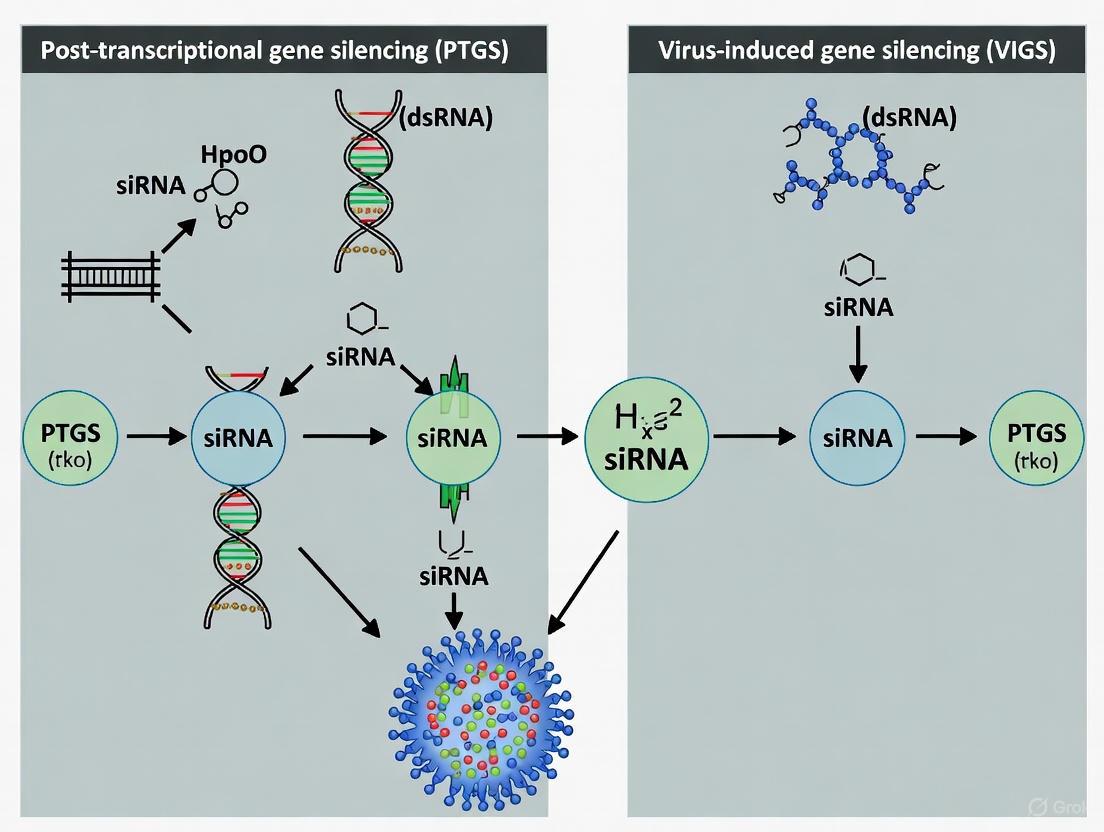
Figure 1: The Core PTGS Pathway. Double-stranded RNA (dsRNA) is processed by DICER enzyme into siRNAs, which are loaded into RISC to guide sequence-specific mRNA degradation.
Initiation Pathways and Biological Contexts of PTGS
PTGS can be initiated through multiple distinct pathways, each with unique triggers and mechanistic nuances while converging on a common silencing outcome.
Antisense-Mediated Gene Silencing
Antisense RNA-mediated silencing represents one of the earliest documented methods for initiating PTGS in plants [1]. This approach involves expressing antisense RNA from integrated transgenes, with successful silencing observed in approximately 5-20% of transformed individuals [1]. The extent of mRNA suppression varies considerably, ranging from no detectable effect to greater than 99% reduction in steady-state RNA levels [1]. The mechanism primarily involves homologous pairing of sense and antisense RNAs to produce dsRNA in vivo, which then serves as the primary initiator of the silencing cascade [1].
RNA Interference (RNAi) and Cosuppression
RNA interference (RNAi) represents a highly efficient induction method for PTGS, where direct introduction of dsRNA triggers near-absolute suppression of targeted mRNA species in over 90% of transformants [1]. This approach typically utilizes inverted-repeat transgenes that produce self-complementary hairpin RNAs upon transcription. Effective induction requires a dsRNA region of at least approximately 100 nucleotides, which explains why the limited secondary structure of endogenous mRNAs does not typically trigger silencing [1].
Cosuppression presents a particularly intriguing initiation pathway, wherein overexpression of an endogenous gene leads to silencing of both the transgene and homologous endogenous sequences [1]. Genetic evidence suggests this pathway is mechanistically distinct from antisense silencing and RNAi, requiring specific catalytic components such as the RNA-dependent RNA polymerase (RdRP) for dsRNA synthesis from abundant sense RNA templates [1]. Cosuppression exhibits the unique property of systemic acquired silencing, where a sequence-specific silencing signal spreads throughout the plant, presumably through plasmodesmata and phloem vasculature [1].
Virus-Induced Gene Silencing (VIGS)
VIGS represents a powerful application of PTGS that exploits natural antiviral defense mechanisms for functional genomics [2] [4]. When plants encounter viruses, their replication intermediates generate dsRNA molecules that trigger PTGS. VIGS technology harnesses this pathway by engineering viral vectors to carry fragments of host genes. When these recombinant viruses infect plants, the RNA silencing machinery processes the viral dsRNA into siRNAs that target both viral RNAs and complementary endogenous mRNAs, leading to systemic silencing of the host gene of interest [2] [5].
Table 1: Comparison of Major PTGS Initiation Pathways
| Initiation Method | Trigger Molecule | Silencing Efficiency | Systemic Spread | Key Applications |
|---|---|---|---|---|
| Antisense Silencing | Antisense RNA | 5-20% of transformants | Limited | Early gene function studies |
| RNA Interference (RNAi) | dsRNA (hairpin) | >90% of transformants | Variable | Targeted gene knockout |
| Cosuppression | Sense RNA (overexpression) | 5-20% of transformants | Strong (graft-transmissible) | Developmental studies |
| VIGS | Viral dsRNA | Variable by system | Strong (viral movement) | High-throughput functional genomics |
Virus-Induced Gene Silencing: Mechanisms and Applications
Molecular Basis of VIGS
VIGS operates by exploiting the plant's innate antiviral RNA silencing machinery [2] [5]. When a recombinant virus carrying a fragment of a host gene infects the plant, the viral replication process generates double-stranded RNA intermediates. The plant recognizes these as foreign and activates its PTGS defense system. Cellular Dicer-like enzymes process the viral dsRNA into 21-24 nucleotide small interfering RNAs (siRNAs) of both sense and antisense polarities [2]. These siRNAs are then incorporated into the RNA-induced silencing complex (RISC), which uses them as guides to identify and cleave complementary viral RNA molecules [2]. Critically, because the viral vector contains sequences from a host gene, the resulting siRNAs also target corresponding endogenous mRNAs for degradation, leading to specific knockdown of the plant gene of interest [5] [6].
A key advantage of VIGS is the systemic nature of the silencing effect. The silencing signal amplifies and spreads throughout the plant via the viral movement machinery, often reaching tissues distant from the initial infection site [2] [1]. This systemic propagation enables analysis of gene functions in multiple organs and developmental stages from a single inoculation event.
Viral Vectors for VIGS
The effectiveness of VIGS depends critically on the choice of viral vector, with different vectors offering distinct advantages for particular host species and experimental applications.
Table 2: Major Viral Vector Systems for VIGS
| Viral Vector | Virus Type | Primary Host Species | Key Features | Applications |
|---|---|---|---|---|
| Tobacco Rattle Virus (TRV) | RNA virus | Solanaceae, Arabidopsis, Soybean | Mild symptoms, efficient systemic movement, meristem targeting [2] [7] | Functional genomics in dicots |
| Barley Stripe Mosaic Virus (BSMV) | RNA virus | Barley, Wheat, Monocots | Effective in cereal crops [6] [4] | Gene function studies in monocots |
| Cotton Leaf Crumple Virus (CLCrV) | DNA virus (Geminivirus) | Cannabis, Cotton | ssDNA genome, nuclear replication [5] | Gene silencing in dicots including medicinal plants |
| Bean Pod Mottle Virus (BPMV) | RNA virus | Soybean | Well-established for legumes [7] [8] | Soybean functional genomics |
The TRV-based system has emerged as one of the most versatile and widely adopted VIGS platforms, particularly for plants in the Solanaceae family [2]. TRV features a bipartite genome requiring two vectors: TRV1 encodes replicase proteins and movement proteins necessary for viral replication and spread, while TRV2 contains the coat protein gene and serves as the insertion site for target gene fragments [2]. This system typically incorporates self-cleaving ribozyme sequences to ensure proper transcript processing and often utilizes Agrobacterium tumefaciens for efficient delivery into plant cells [2] [7].
Experimental Implementation and Methodologies
VIGS Workflow and Protocol
The implementation of VIGS involves a series of carefully optimized steps, from vector construction to phenotypic analysis. Below is a generalized workflow that can be adapted for specific experimental systems.
Figure 2: Generalized VIGS Experimental Workflow. The process begins with target fragment selection and proceeds through vector construction, plant inoculation, and final phenotypic analysis.
Step 1: Target Fragment Selection and Vector Construction
- Select a 200-400 bp gene-specific fragment with 30-70% GC content [5]
- Avoid 5' and 3' UTRs and the first 100 bp of the coding sequence [5]
- Clone the fragment into appropriate viral vector (e.g., pTRV2 for TRV system) [7]
- Verify insert sequence and orientation before proceeding
Step 2: Agrobacterium Transformation and Preparation
- Introduce recombinant vectors into Agrobacterium tumefaciens strains (e.g., GV3101) [7] [5]
- Culture agrobacteria on selective solid media [2]
- Inoculate liquid cultures and grow to optimal density (OD600 typically 0.5-2.0) [2]
- Prepare infiltration buffer containing agrobacteria and induction agents (e.g., acetosyringone)
Step 3: Plant Inoculation
- For difficult-to-infect species like soybean, use optimized methods such as cotyledon node immersion for 20-30 minutes [7] [8]
- For amenable species like Nicotiana benthamiana, use direct leaf infiltration with syringe [2]
- Maintain inoculated plants under appropriate environmental conditions (temperature, humidity, photoperiod) to maximize silencing efficiency [2]
Step 4: Monitoring and Validation
- Observe plants for development of silencing phenotypes (typically 2-4 weeks post-inoculation) [7] [6]
- Validate silencing efficiency using quantitative RT-PCR to measure target transcript reduction [5]
- For fluorescence-based systems, monitor GFP expression to track viral spread [7]
Essential Research Reagents and Materials
Table 3: Essential Research Reagents for VIGS Experiments
| Reagent/Resource | Function/Purpose | Examples/Specifications |
|---|---|---|
| Viral Vectors | Delivery of target gene fragments | TRV1/TRV2 [2], BSMV α, β, γ [6], CLCrV DNA-A/DNA-B [5] |
| Agrobacterium Strains | Biological delivery of viral vectors | GV3101 [7], AGL1 [5] |
| Marker Genes | Visual assessment of silencing efficiency | Phytoene desaturase (PDS) - photobleaching [7] [5], Magnesium chelatase (ChlI) - yellowing [5] |
| Infiltration Buffers | Facilitating Agrobacterium entry into plant cells | MgCl₂, MES, Acetosyringone induction agent [2] |
| Computational Tools | siRNA prediction and off-target analysis | siRNA Scan [3], pssRNAit [5] |
Critical Considerations and Limitations
Off-Target Effects and Specificity Challenges
A significant challenge in PTGS applications, including VIGS, is the potential for off-target effects where unintended genes with sequence similarity to the target are silenced [3]. Computational analyses indicate that approximately 50-70% of gene transcripts in plants have potential off-targets when used for PTGS [3]. Experimental verification has confirmed that up to 50% of predicted off-target genes can be silenced in practice [3]. These off-target effects arise because PTGS functions in a siRNA-specific rather than target-specific manner, with multiple distinct siRNAs derived from a single dsRNA trigger potentially targeting multiple genes sharing contiguous regions of identity (≥21 nucleotides) [3].
To address this challenge, computational tools like siRNA Scan have been developed to identify potential off-targets during PTGS construct design [3]. Careful selection of target fragments with minimal sequence identity to non-target genes is essential for accurate interpretation of PTGS experiments. Additionally, validation of phenotypic outcomes using multiple independent target fragments provides greater confidence in assigning gene function.
Host-Specific Factors Influencing Silencing Efficiency
The efficiency of VIGS is influenced by numerous host-specific factors that must be considered in experimental design. Plant genotype significantly impacts silencing efficiency, with natural variation in virus-host interactions observed across different accessions of the same species [9]. For instance, a screen of 190 Arabidopsis accessions revealed striking diversity in responses to a geminivirus VIGS vector, with only one accession (Pla-1) showing complete immunity [9].
Environmental conditions, including temperature, humidity, and photoperiod, profoundly influence silencing dynamics [2]. Additionally, the plant's developmental stage at inoculation affects both viral spread and silencing establishment, with younger tissues often exhibiting more robust silencing [2]. Viral suppressors of RNA silencing (VSRs) encoded by many plant viruses can also modulate silencing efficiency, with some VSRs like P19 and C2b being exploited to enhance VIGS in certain systems [2].
Post-transcriptional gene silencing represents a fundamental RNA-based regulatory mechanism with profound implications for plant defense and gene regulation. The mechanistic understanding of PTGS has enabled the development of powerful technologies like VIGS, which continues to evolve as an indispensable tool for functional genomics. Current research focuses on expanding the host range of VIGS vectors, improving specificity and efficiency, and integrating VIGS with emerging technologies like CRISPR-Cas systems.
The future of PTGS research will likely see increased emphasis on understanding the intricate relationships between different silencing pathways, including connections to transcriptional gene silencing and epigenetic regulation. As computational tools for predicting siRNA targets and off-effects become more sophisticated, and as viral vectors are further optimized for specific host systems, PTGS-based approaches will continue to provide critical insights into gene function across diverse plant species, accelerating both basic research and crop improvement efforts.
Within the framework of post-transcriptional gene silencing (PTGS), the core molecular machinery comprising Dicer, the RNA-induced silencing complex (RISC), and Argonaute proteins orchestrates the biogenesis and effector functions of small interfering RNAs (siRNAs). This whitepaper provides a comprehensive technical analysis of these components, detailing their conserved domains, mechanistic roles in siRNA pathways, and critical interactions. We further synthesize quantitative data on siRNA design efficacy, outline foundational experimental protocols such as Virus-Induced Gene Silencing (VIGS), and catalog essential research tools. Aimed at researchers and therapeutic developers, this guide serves as a foundational resource for advancing mechanistic studies and applications in RNA interference (RNAi)-based technologies.
Post-transcriptional gene silencing (PTGS), commonly known as RNA interference (RNAi), is a conserved eukaryotic mechanism that degrades target mRNA in a sequence-specific manner, thereby repressing gene expression [10] [11]. This process is central to host defense against transposons and viruses, and it is critical for regulating developmental programs [10] [12]. The initiation of PTGS is triggered by double-stranded RNA (dsRNA), which is processed into the primary effactors of silencing: small interfering RNAs (siRNAs) and microRNAs (miRNAs) [10] [11]. The key molecular players in this pathway include the RNase III enzyme Dicer, which processes dsRNA into small RNAs; the Argonaute protein family, which forms the core of the silencing complex; and the multi-protein RNA-induced silencing complex (RISC), which executes mRNA cleavage or translational repression [10] [13]. Understanding the biogenesis of siRNAs and the assembly of these complexes is fundamental for research in functional genomics and the development of RNAi-based therapeutics.
Molecular Components and Their Functions
Dicer
Dicer is an RNase III-type endonuclease that initiates the RNAi pathway by cleaving long dsRNA precursors into 21-24 nucleotide small RNA duplexes [10] [11]. Its multi-domain structure is conserved across eukaryotes, comprising several key functional regions.
Domain Architecture and Function: A canonical Dicer protein contains the following domains: an N-terminal DExH helicase domain, which aids in ATP-dependent dsRNA binding and processing; a DUF283 domain, a putative RNA-binding platform; a PAZ domain, which recognizes and binds the 3' ends of dsRNA with 2-nucleotide overhangs; and two tandem RNase IIIa and RNase IIIb domains, which form an intramolecular dimer that cleaves dsRNA. The protein is capped by a C-terminal dsRNA-binding domain (dsRBD) [11]. The PAZ and RNase III domains work in concert, with the PAZ domain anchoring one end of the dsRNA and the RNase III domains making sequential cuts approximately 21-24 nucleotides away, determining the length of the resulting siRNAs [11].
Functional Specialization in Different Organisms: The number and specialization of Dicer proteins vary across species, influencing the complexity of their small RNA pathways.
- Humans and other mammals possess a single Dicer enzyme responsible for generating both siRNAs and miRNAs [11] [13].
- The nematode C. elegans encodes two Dicers (DCR-1). DCR-1, in complex with the dsRNA-binding protein RDE-4, is essential for the processing of exogenous dsRNA into primary siRNAs for the RNAi response [14] [15].
- Plants, such as Arabidopsis thaliana, have evolved multiple specialized Dicer-like (DCL) proteins. DCL1 is primarily responsible for miRNA biogenesis, while DCL2, DCL3, and DCL4 generate 22-nt, 24-nt, and 21-nt siRNAs, respectively, involved in antiviral defense and transcriptional silencing [11] [12]. A functional hierarchy exists among these DCLs, where one can partially compensate for the loss of another, providing robustness to the silencing system [11].
Small Interfering RNA (siRNA)
Small interfering RNAs (siRNAs) are the guiding molecules that confer sequence specificity to the RNAi machinery. They are typically 21-24 nucleotides in length and are loaded into the RISC to target complementary mRNAs for destruction [10] [16].
Biogenesis Pathway: The life cycle of an siRNA involves primary and secondary amplification stages.
- Primary siRNA Biogenesis: Long dsRNAs of exogenous (e.g., viral) or endogenous (e.g., transposon) origin are recognized and cleaved by Dicer into short siRNA duplexes with 2-nucleotide 3' overhangs [10] [11].
- Secondary siRNA Amplification: In plants, fungi, and C. elegans, an amplification loop enhances the silencing signal. This process involves RNA-dependent RNA Polymerases (RdRPs), which use the target mRNA as a template to synthesize new dsRNA after it has been primed by a primary siRNA. This secondary dsRNA is subsequently processed by Dicer into a population of secondary siRNAs, which propagates and strengthens the systemic silencing response [10] [17] [12].
Sequence Characteristics for Efficacy: Not all siRNAs are equally effective. High-throughput studies have identified key sequence features that correlate with high knockdown efficiency. Table 1 summarizes these rules, which include nucleotide preferences at specific positions in the siRNA sense strand [16].
Table 1: Sequence Rules for Predicting Functional siRNA Efficacy
| Rule Set | Criteria for Sense Strand | Mean Knockdown Efficacy |
|---|---|---|
| Rule 1 (Best) | A/U at position 10 and 19; G/C at position 1; >3 A/U residues between positions 13-19 | 73% |
| Rule 2 | Matches a specific subset of the above criteria | 60% |
| Rule 3 | Matches a different, less stringent subset of criteria | 68% |
| Rule 4 | Matches another less stringent subset of criteria | 68% |
| Unselected Data | No selection for specific sequence features | 52% |
Applying Rule 1 during siRNA design provides a 99.9% chance of obtaining at least one effective (>50% knockdown) siRNA within a set of three designed sequences [16].
Argonaute and the RISC Complex
The Argonaute (AGO) protein family forms the catalytic heart of the RNA-induced silencing complex (RISC). It is responsible for binding the small RNA guide strand and executing the silencing of the complementary target mRNA [11] [13].
Argonaute Domain Structure and Slicer Activity: Argonaute proteins are characterized by four core domains: the N-terminal, PAZ, MID, and PIWI domains. The PAZ domain anchors the 3' end of the small RNA, while the MID domain binds its 5' phosphate. The PIWI domain adopts an RNase H-like fold and, in "slicer-competent" Argonautes, confers endonuclease activity. This activity cleaves the target mRNA between nucleotides 10 and 11 relative to the 5' end of the siRNA guide strand [11] [13].
RISC Assembly and Loading: RISC assembly is a multi-step process. The siRNA duplex generated by Dicer is first loaded into Argonaute. The passenger strand is then ejected, and the guide strand is retained to form the mature RISC. Recent research in C. elegans has identified that the dsRNA-binding protein RDE-4 not only assists Dicer in processing but also acts as a critical factor for selectively loading siRNAs into the appropriate Argonaute protein, RDE-1. This ensures the initiation of a potent RNAi response and prevents the mis-sorting of siRNAs into non-cognate Argonautes [14] [15].
AGO Protein Diversity and Functional Specialization: Like Dicer, the Argonaute family has expanded and diversified in different lineages. Table 2 classifies AGO proteins based on their phylogenetic relationships and functional specialization in animals and plants [13].
Table 2: Functional Classification of Argonaute Proteins
| Kingdom | Class | Example Proteins | Primary Small RNA Association & Function |
|---|---|---|---|
| Animals | Multifunctional AGOs | Human AGO1-4 | miRNAs & siRNAs; Post-transcriptional gene silencing |
| siRNA-associated AGOs | Drosophila AGO2; C. elegans ERGO-1, RDE-1 | Endo- and exogeneous siRNAs; Antiviral defense, RNAi response | |
| piRNA-associated AGOs | Human PIWIL1-4; Drosophila Piwi, Aub | piRNAs; Transposon silencing, germline genome defense | |
| Plants | Multifunctional AGOs | A. thaliana AGO1, AGO10 | miRNAs & siRNAs; Various developmental processes |
| siRNA-associated AGOs | A. thaliana AGO2, AGO7 | siRNAs (e.g., tasiRNAs); Antiviral defense, leaf development | |
| Complementary functioning AGOs | A. thaliana AGO4, AGO6, AGO9 | siRNAs (24-nt); Transcriptional gene silencing (RdDM) |
Experimental Protocols and Methodologies
Virus-Induced Gene Silencing (VIGS)
Virus-Induced Gene Silencing (VIGS) is a powerful reverse genetics tool that leverages the plant's PTGS/RNAi machinery to silence endogenous genes by introducing a recombinant virus carrying a fragment of the target host gene [17] [18].
Principle: When a recombinant virus infects a plant, its RNA replicates in the host cytoplasm, forming dsRNA intermediates. The host recognizes these as foreign and processes them into vsiRNAs (virus-derived siRNAs). If the viral genome contains a fragment of a host gene, the resulting vsiRNAs will be homologous to the host's mRNA. These vsiRNAs are loaded into RISC and guide the cleavage and degradation of the corresponding host transcript, leading to a loss-of-function phenotype [17].
Protocol: VIGS in Striga hermonthica using Tobacco Rattle Virus (TRV) Vectors
- Vector Construction: Clone a 200-500 bp fragment of the target plant gene (e.g., Phytoene Desaturase (PDS), a visual marker for silencing) into the multiple cloning site of the TRV2 vector. The TRV1 vector contains the viral RNA-dependent RNA polymerase and movement protein genes [18].
- Agrobacterium Transformation: Introduce the engineered TRV2 plasmid and the helper TRV1 plasmid separately into Agrobacterium tumefaciens strain GV3101.
- Plant Inoculation:
- Agro-infiltration: Grow the Agrobacterium cultures to an OD600 of ~1.5. Mix the TRV1 and TRV2 cultures in a 1:1 ratio. Infiltrate the bacterial suspension into the leaves of young plants using a needleless syringe. Silencing phenotypes (e.g., leaf bleaching for PDS) typically appear within 7 days [18].
- Agro-drench: Pour the same bacterial mixture onto the soil around the plant's roots. This method is less efficient, with phenotypes appearing around 14 days post-inoculation [18].
- Validation of Silencing:
- Phenotypic Analysis: Observe and document the expected loss-of-function phenotype.
- Molecular Confirmation: Perform RT-PCR on mRNA extracted from silenced tissue using primers that anneal outside the region targeted by the VIGS construct. A significant reduction in the target gene's mRNA level, compared to control plants, confirms successful silencing [18].
The Scientist's Toolkit: Research Reagent Solutions
Table 3 catalogs essential reagents and their applications for studying siRNA biogenesis and PTGS mechanisms.
Table 3: Key Research Reagents for siRNA/PTGS Studies
| Reagent / Tool | Organism / System | Function & Application in Research |
|---|---|---|
| TRV-VIGS Vectors (pYL156, pYL279) | Plants (N. benthamiana, tomato, Arabidopsis, Striga) | A robust viral vector system for rapid, transient loss-of-function gene silencing studies without stable transformation [17] [18]. |
| Dicer Mutants (e.g., dcl1, dcl2, dcl3, dcl4) | Plants (A. thaliana), Fungi (S. macrospora) | Genetic models to dissect the specific roles of individual Dicer isoforms in various small RNA pathways and their functional redundancy/hierarchy [19] [11]. |
| Argonaute Mutants (e.g., rde-1, sms2, qde-2) | C. elegans, Fungi, Plants | Used to characterize the function of specific Argonaute proteins in RNAi initiation, secondary siRNA amplification, meiotic processes, and transposon silencing [14] [19]. |
| RDE-4 Protein | C. elegans | A dsRNA-binding protein that facilitates Dicer processing and is critical for the specific loading of siRNAs into the Argonaute RDE-1, making it essential for an effective exogenous RNAi response [14] [15]. |
| Chemically Synthesized siRNAs | Mammalian cell culture | 21-mer duplex RNAs with defined sequences, used for direct transfection to induce targeted gene knockdown. Efficacy is maximized by applying rational design rules (see Table 1) [16]. |
The core machinery of Dicer, RISC, and Argonaute proteins provides the cell with a precise and powerful system for sequence-specific gene regulation through siRNA biogenesis and action. While the fundamental steps of this pathway are well-established, recent research continues to uncover new layers of complexity. Discoveries such as the role of RDE-4 in ensuring specific Argonaute loading in C. elegans and the existence of non-canonical RNAi pathways in plants highlight the dynamic nature of this field [14] [12]. Furthermore, the application of this knowledge in tools like VIGS has proven indispensable for functional genomics in a wide range of species.
Future research will likely focus on elucidating the finer details of RISC assembly and trafficking, the interplay between different small RNA pathways, and the development of more sophisticated RNAi-based therapeutics with enhanced specificity and delivery. Integrating the knowledge of these key molecular players with emerging technologies will continue to drive innovation in basic research and drug development.
RNA interference is an evolutionarily conserved mechanism for gene silencing in eukaryotes, serving as a critical defense pathway against viral infections and transposable elements while also playing a fundamental role in regulating endogenous gene expression [20]. The initiation phase of RNAi represents the first critical step in this sophisticated sequence-specific gene silencing pathway, wherein double-stranded RNA is recognized and processed into the effector molecules that direct downstream silencing events. This transformation from long dsRNA to short interfering RNA duplexes establishes the specificity of the entire RNAi process, making the initiation phase fundamental to technologies ranging from functional genomics to therapeutic development [21] [22].
Within the broader context of post-transcriptional gene silencing research, understanding the initiation phase provides crucial insights for developing advanced gene silencing tools. Virus-Induced Gene Silencing leverages this very mechanism, using recombinant viruses to introduce dsRNA into plant cells, which then triggers the endogenous RNAi machinery to silence targeted genes [17] [23]. The efficiency and specificity of this initiation process directly impact the success of both basic research applications and emerging therapeutic strategies, underscoring the importance of elucidating its molecular details.
Molecular Mechanism of Initiation
dsRNA Recognition and Dicer Cleavage
The initiation phase begins when the enzyme Dicer, an RNase III family endonuclease, recognizes and binds to double-stranded RNA molecules in the cytoplasm [24] [21]. Dicer processes long dsRNA precursors into short RNA fragments of defined lengths by cleaving both strands of the duplex. This processing yields siRNA duplexes typically 21-23 nucleotides in length with characteristic 2-nucleotide overhangs on the 3'-ends of each strand [21] [22]. The resulting siRNAs possess 5'-phosphate and 3'-hydroxyl termini, features essential for their subsequent recognition by downstream components of the RNAi machinery [24].
The cleavage mechanism employed by Dicer involves coordinated action of multiple domains. The enzyme measures approximately 21-23 nucleotides from the dsRNA terminus to determine cleavage sites, ensuring production of uniformly sized siRNAs [21]. Structural studies have revealed that Dicer contains a PAZ domain that recognizes the terminus of dsRNA, while its tandem RNase III domains catalyze the cleavage reaction, generating the characteristic 2-nucleotide 3' overhangs [21].
Table 1: Core Components of the RNAi Initiation Machinery
| Component | Structure/Features | Function in Initiation Phase |
|---|---|---|
| Dicer | RNase III family enzyme, PAZ domain, RNase IIIa and IIIb domains, dsRNA-binding domain | Recognizes and cleaves long dsRNA into siRNA duplexes of defined length |
| dsRNA Substrate | Perfect or near-perfect complementarity between strands, variable length | Serves as precursor for siRNA production; source of sequence specificity |
| siRNA Duplex | 21-23 nucleotides, 2-nt 3' overhangs, 5'-phosphate groups | Directing sequence-specific silencing; guides RISC to complementary targets |
Structural Determinants of siRNA Biogenesis
The efficiency of siRNA generation depends on several structural properties of the dsRNA substrate. The A-form helix geometry of dsRNA is critical for recognition and processing by Dicer [24]. Biochemical analyses have demonstrated that modifications altering the major groove structure of the dsRNA helix can abolish processing, indicating that Dicer requires specific structural features for binding and cleavage [24]. Additionally, the thermodynamic stability of the dsRNA ends influences how Dicer interacts with the substrate, with the enzyme typically binding to the thermodynamically less stable end while the more stable end facilitates loading of the antisense strand into RISC [21].
The molecular weight of the dsRNA substrate also impacts siRNA biogenesis and subsequent silencing activity. Research has shown that siRNAs with smaller molecular weights generally enter cells more efficiently, while larger dsRNA substrates that require Dicer processing may have advantages in organismal distribution and persistence [21]. This understanding has led to the development of Dicer Substrate siRNAs – 25-27 nucleotide duplexes designed for improved efficiency through enhanced Dicer processing [21].
Experimental Analysis of Initiation Events
Methodologies for Studying siRNA Biogenesis
Investigating the initiation phase of RNAi requires specialized experimental approaches to detect and quantify the production of siRNAs from dsRNA precursors. The following protocol outlines key methodology for analyzing Dicer processing and siRNA characteristics:
Protocol 1: Analysis of Dicer Processing and siRNA Production
Step 1: In Vitro Dicer Cleavage Assay
- Purify recombinant Dicer enzyme or use cell lysates containing endogenous Dicer activity.
- Incubate with radiolabeled or fluorescently tagged dsRNA substrates under appropriate buffer conditions (including Mg²⁺ as a cofactor).
- Stop reactions at various time points (e.g., 0, 15, 30, 60, 120 minutes) to analyze processing kinetics [24].
Step 2: siRNA Detection and Characterization
Step 3: Functional Validation of Processed siRNAs
Quantitative Analysis of siRNA Features
Systematic analysis of siRNA sequences has identified specific features that impact processing efficiency and silencing activity. Research examining 62 siRNA duplexes targeting morbillivirus genes revealed that specific sequence motifs significantly influence knockdown efficacy [22]. The presence of U13 and A/U19 motifs correlated with improved silencing, while G13 was associated with reduced activity [22]. Additionally, the local secondary structure of the target mRNA influences siRNA efficiency, with accessible regions demonstrating better silencing than highly structured domains [22].
Recent high-throughput studies have further elucidated the relationship between siRNA modification patterns and silencing efficacy. A comprehensive analysis of approximately 1260 differentially modified siRNAs targeting therapeutically relevant mRNAs (APP, BACE1, MAPT, and SNCA) demonstrated that the chemical modification pattern significantly impacts efficacy, while structural features like symmetric versus asymmetric configurations showed less pronounced effects [26].
Table 2: Experimentally Determined Features Impacting siRNA Activity
| Feature Category | Optimal Characteristics | Impact on Silencing Efficacy |
|---|---|---|
| Sequence Motifs | U at position 13, A/U at position 19, G at position 11 | Position-specific nucleotides can increase efficacy by 20-40% [22] |
| Chemical Modifications | Balanced 2'-O-methyl/2'-F patterns (e.g., 18% 2'-F) | Increases nuclease resistance and duration of effect without compromising RISC loading [21] [26] |
| Target Accessibility | Low intramolecular secondary structure at target site | Unstructured regions show 2-3 fold higher silencing than highly structured targets [22] |
| Thermodynamic Asymmetry | Lower stability at 5'-end of antisense strand | Guides proper strand selection; improves RISC loading efficiency by 30-50% [21] |
The Scientist's Toolkit: Essential Research Reagents
Table 3: Key Research Reagents for Studying Initiation Phase
| Reagent/Category | Specific Examples | Research Application |
|---|---|---|
| Dicer Enzymes | Recombinant human Dicer, Drosophila Dicer-2 | In vitro processing assays; biochemical characterization of cleavage parameters [21] |
| dsRNA Substrates | In vitro transcribed dsRNA, Dicer substrate siRNAs (DsiRNAs) | Optimization of processing efficiency; structure-function studies [21] [26] |
| Chemical Modification Tools | 2'-O-methyl, 2'-fluoro, phosphorothioate nucleotides | Enhancing siRNA stability; studying modification tolerance in Dicer processing [24] [21] [26] |
| VIGS Vectors | TRV, TMV, BSMV, PVX-based vectors | Plant functional genomics; high-throughput screening of gene function [17] [18] [23] |
Visualization of the Initiation Pathway
The RNA-induced silencing complex (RISC) serves as the central effector complex in post-transcriptional gene silencing (PTGS), including pathways such as virus-induced gene silencing (VIGS) [2]. This ribonucleoprotein complex utilizes small RNA molecules as guides to identify complementary messenger RNA (mRNA) targets through Watson-Crick base pairing, leading to transcriptional and post-transcriptional repression [27] [28]. The discovery of RISC followed the landmark identification of RNA interference (RNAi) in 1998, with biochemical characterization conducted by Gregory Hannon and colleagues at Cold Spring Harbor Laboratory, who identified RISC as a sequence-specific nuclease activity responsible for targeted mRNA degradation [28]. In PTGS, RISC represents the culmination of the silencing pathway where small interfering RNAs (siRNAs) or microRNAs (miRNAs) direct the inhibition of gene expression through mRNA cleavage, degradation, and translational repression [27] [29].
The fundamental importance of RISC extends across eukaryotic organisms, where it functions as a key defense mechanism against viral infections and transposable elements, while also serving crucial roles in regulating endogenous gene expression [28]. In the context of VIGS, which is grounded in the plant's PTGS machinery, recombinant viral vectors introduce sequences that are processed into siRNAs, which then load into RISC to direct systemic suppression of endogenous plant gene expression [2]. This mechanism has become an powerful tool for functional genomics, allowing researchers to characterize gene function through observable phenotypic changes resulting from targeted gene silencing [2] [7].
RISC Assembly and Architecture
Core Components and Loading Mechanism
RISC assembly is a multi-step process that begins with the processing of double-stranded RNA (dsRNA) precursors into small RNA fragments. The RNase III enzyme Dicer serves as a critical initiation point for RISC formation by cleaving long dsRNA molecules into small interfering RNA (siRNA) duplexes of 21-23 nucleotides with characteristic two-nucleotide 3' overhangs [28]. Similarly, Dicer processes pre-microRNA (pre-miRNA) hairpin structures to generate mature miRNA duplexes [30] [28]. These small RNA duplexes are then transferred to the Argonaute (AGO) protein family, which forms the catalytic heart of RISC [27] [30].
The loading of small RNA duplexes into RISC follows the "asymmetry rule," where thermodynamic stability at the ends of the duplex determines which strand is selected as the guide strand [30] [28]. The strand with less thermodynamically stable 5' pairing is preferentially selected by Argonaute and incorporated into RISC as the guide strand, while the other passenger strand is degraded [28]. This strand selection is critical for determining target specificity, as the retained guide strand will direct RISC to complementary mRNA sequences. The assembly process is facilitated by chaperone complexes in some organisms; for example, in Drosophila, the Dcr2-R2D2 heterodimer helps recognize the siRNA duplex and facilitates its loading into RISC [28].
Structural Organization and Protein Composition
The complete structure of RISC remains an active area of investigation, with studies reporting a range of complex sizes and compositions [28]. The core component across all RISC complexes is an Argonaute protein, which contains two primary domains: the PAZ domain, which binds the 3' end of the small RNA, and the PIWI domain, which shares structural similarity with RNase H and provides the "slicer" activity responsible for mRNA cleavage in some RISC complexes [30] [29].
Table 1: Characterized RISC Complexes and Their Components
| Complex Name | Source | Known/Apparent Components | Estimated Size | Apparent Function in RNAi Pathway |
|---|---|---|---|---|
| Dcr2-R2D2 | D. melanogaster S2 cells | Dcr2, R2D2 | ~250 kDa | dsRNA processing, siRNA binding |
| RLC (A) | D. melanogaster embryos | Dcr2, R2D2 | Not reported | dsRNA processing, siRNA binding, precursor to RISC |
| Holo-RISC | D. melanogaster embryos | Ago2, Dcr1, Dcr2, Fmr1/Fxr, R2D2, Tsn, Vig | ~80S | Target-RNA binding and cleavage |
| Minimal RISC | HeLa cells | eIF2C1 (Ago1) or eIF2C2 (Ago2) | ~160 kDa | Target-RNA binding and cleavage |
| miRNP | HeLa cells | eIF2C2 (Ago2), Gemin3, Gemin4 | ~550 kDa | miRNA association, target-RNA binding and cleavage |
Additional proteins have been identified as RISC components in various organisms, including the Fragile X mental retardation protein (FMRP/FXR), Tudor-staphylococcal nuclease (Tudor-SN), and Vasa intronic gene (VIG) protein [30] [28]. However, the exact composition appears to vary between organisms, cell types, and the class of small RNA involved (siRNA vs. miRNA). In mammalian cells, the minimal RISC capable of target cleavage consists primarily of the Argonaute protein bound to the guide strand [30] [29].
Mechanisms of mRNA Targeting and Degradation
Guide-Target Recognition and Cleavage
The mechanism of mRNA targeting begins when the small RNA guide strand within RISC base-pairs with complementary sequences in target mRNAs. The outcome of this interaction depends significantly on the degree of complementarity between the guide strand and target [28] [29]. For perfect or near-perfect complementarity, particularly common in plant systems and siRNA-mediated silencing, the Argonaute protein catalyzes endonucleolytic cleavage ("slicing") of the target mRNA between nucleotides 10 and 11 relative to the 5' end of the guide strand [30] [29]. This cleavage requires a catalytically active Argonaute protein, with Ago2 serving as the primary slicer in many organisms [29].
The slicing reaction generates two mRNA fragments: a 5' cleavage fragment with a 3' hydroxyl group and a 3' cleavage fragment with a 5' phosphate group [31] [29]. The fate of these fragments is crucial for completing the silencing process and recycling RISC for multiple rounds of target recognition. The 3' cleavage fragment is typically degraded in the 5'-to-3' direction by the conserved exonuclease XRN1 (XRN4 in Arabidopsis) [31] [29]. Degradation of the 5' cleavage fragment is more complex and involves a specialized mechanism discussed in the following section.
Table 2: mRNA Degradation Pathways Following RISC-Mediated Cleavage
| Cleavage Fragment | Degradation Direction | Primary Enzymes | Special Modifications | Conservation |
|---|---|---|---|---|
| 3' Fragment | 5'-to-3' | XRN1/XRN4 (5'-3' exonuclease) | None required | Evolutionarily conserved from plants to mammals |
| 5' Fragment | 3'-to-5' | RICE1/RICE2, exosome complex | Uridylation by HESO1/TUTs | Conserved mechanisms with organism-specific variations |
Specialized Mechanisms for Clearing Cleavage Fragments
Recent research has identified specialized machinery for degrading the 5' cleavage fragments generated by RISC activity. In Arabidopsis, RISC-interacting clearing 3'-5' exoribonucleases (RICEs), specifically RICE1 and RICE2, have been identified as novel cofactors of AGO proteins that specifically degrade uridylated 5' cleavage fragments [31]. These proteins form homohexameric complexes with DnaQ-like exonuclease folds and active sites located at the interfaces between subunits [31].
The degradation of 5' fragments involves a specific marking system. The 5' cleavage products from RISC activity are typically modified through uridylation—the addition of non-templated uridine residues at their 3' ends—by terminal uridylyl transferases (TUTs) such as HESO1 in Arabidopsis [31]. This uridylation serves as a recognition signal for RICE proteins, which then degrade the fragments in the 3'-to-5' direction. When this degradation pathway is disrupted, such as through expression of catalytically inactive RICE1, 5' cleavage fragments accumulate with extended uridine tails, and miRNA levels decrease significantly, indicating that proper clearance of cleavage fragments is essential for maintaining functional RISC [31].
Alternative pathways for 5' fragment degradation also exist, including 5'-to-3' degradation by XRN4 in Arabidopsis and 3'-to-5' degradation by the exosome complex, though the relative contributions of these pathways appear to vary by organism and context [31].
Experimental Analysis of RISC Function
RISC-Sequencing (RISC-Seq) for Target Identification
RISC-sequencing (RISC-Seq) represents a powerful methodological advancement for identifying miRNA and siRNA targets in biological contexts [32]. This technique involves immunoprecipitation of Argonaute-containing complexes followed by high-throughput sequencing of associated RNAs, allowing comprehensive identification of RISC-bound targets without amplification bias [32]. The experimental workflow includes:
- Cross-linking and Cell Lysis: Cells or tissues are cross-linked to preserve RNA-protein interactions, followed by lysis in appropriate buffers.
- Immunoprecipitation: Anti-Argonaute antibodies (e.g., anti-Ago2 monoclonal antibody) bound to protein G-coupled Dynabeads are used to isolate RISC complexes from cell lysates.
- RNA Extraction and Library Preparation: Associated RNAs are extracted, fragmented, and used to construct sequencing libraries without amplification to prevent bias.
- Sequencing and Data Analysis: High-throughput sequencing identifies RISC-associated transcripts, with comparison to transcriptome data revealing enriched targets.
This approach has been successfully applied to identify cardiac-specific miRNA targets in mouse hearts, demonstrating that programming cardiomyocytes with miR-133a or miR-499 overexpression leads to distinct profiles of RISC-targeted mRNAs [32]. The technique is applicable to any tissue or disease state, providing biological context for miRNA target identification that complements bioinformatic predictions.
Assessing Silencing Efficacy: Technical Considerations
Accurate measurement of RISC-mediated silencing requires careful experimental design, particularly when using RT-qPCR to assess mRNA knockdown. A critical technical consideration involves the placement of PCR amplicons relative to the RISC cleavage site [33]. Studies have demonstrated that PCR primers designed to amplify regions 3' of the cleavage site may fail to detect silencing due to incomplete degradation of the 3' fragment, potentially leading to false negative results [33].
To avoid this pitfall, researchers should:
- Design multiple primer sets targeting different regions of the transcript, preferably spanning the expected cleavage site
- Validate silencing with primers located 5' to the cleavage site
- Confirm results with protein-level analysis (western blotting) when possible
- Use both oligo(dT) and random hexamer primers for cDNA synthesis to control for potential artifacts
This phenomenon appears transcript-dependent and may result from RNA secondary structures or RNA-binding proteins that impede complete degradation of cleavage fragments [33].
Applications in Research and Therapeutics
Virus-Induced Gene Silencing (VIGS)
VIGS represents a powerful application of RISC biology that leverages the plant's endogenous PTGS machinery for functional genomics [2]. In this approach, recombinant viral vectors containing fragments of host genes are introduced into plants, where they replicate and generate dsRNA intermediates that are processed by Dicer into siRNAs [2]. These siRNAs are loaded into RISC, which then targets complementary endogenous mRNAs for degradation, effectively silencing the gene of interest [2].
The efficiency of VIGS depends on multiple factors, including:
- Viral vector selection (e.g., Tobacco Rattle Virus, Bean Pod Mottle Virus)
- Insert design and location within the vector
- Agroinfiltration methodology and Agrobacterium strain
- Plant developmental stage and environmental conditions
- Host genotype and RNAi machinery components
TRV-based VIGS has been particularly successful in Solanaceae species like pepper and tomato, enabling functional analysis of genes involved in fruit quality, disease resistance, and stress responses without stable transformation [2]. Recent work has extended TRV-VIGS to soybean, achieving 65-95% silencing efficiency through optimized cotyledon node infiltration [7].
Spray-Induced Gene Silencing (SIGS) and Agricultural Applications
Spray-induced gene silencing (SIGS) represents an emerging agricultural technology that applies dsRNA directly to crops, where it is taken up by pathogens or pests and processed through their RISC machinery to silence essential genes [34]. This approach offers an environmentally sustainable alternative to conventional pesticides, as dsRNA degrades rapidly in the environment and can be designed for high specificity [34].
The process involves:
- dsRNA Design and Production: Target essential pathogen/pest genes and produce dsRNA in vitro or in bacterial systems
- Formulation and Application: Combine dsRNA with carriers or nanomaterial to enhance stability and uptake
- Cellular Processing: Pathogens/pests take up dsRNA, which is processed by Dicer and loaded into RISC
- Gene Silencing: RISC targets complementary mRNAs, impairing pathogen viability
Notably, some pathogens like Botrytis cinerea efficiently take up external dsRNAs, making them particularly susceptible to SIGS approaches [34]. The recent approval of Ledprona as the first sprayable dsRNA biopesticide highlights the translational potential of RISC-based technologies for crop protection [34].
The Scientist's Toolkit: Essential Reagents and Methods
Table 3: Key Research Reagents for RISC Studies
| Reagent/Method | Function/Application | Examples/Specifications |
|---|---|---|
| Anti-Argonaute Antibodies | Immunoprecipitation of RISC complexes | Anti-Ago2 monoclonal antibodies (e.g., Wako Pure, clone #2D4) for RISC-Seq |
| Dicer Enzymes | Generation of siRNAs from dsRNA precursors | Recombinant Dicer for in vitro siRNA production |
| TRV VIGS Vectors | Virus-induced gene silencing in plants | Bipartite TRV system (TRV1 + TRV2 with target insert) |
| Agrobacterium Strains | Delivery of VIGS constructs to plants | GV3101 for agroinfiltration |
| RNAiMAX/siPORT | Transfection of siRNAs into mammalian cells | Lipid-based transfection reagents for siRNA delivery |
| RISC-Seq Protocols | Genome-wide identification of RISC targets | Crosslinking, Ago2-IP, and sequencing of associated RNAs |
| qPCR Primer Design | Assessment of silencing efficacy | Multiple primer sets 5' to expected cleavage site |
Visualizing RISC Mechanisms and Workflows
RISC Assembly and mRNA Degradation Pathway
RISC-Sequencing Experimental Workflow
The effector phase of RISC assembly and sequence-specific mRNA degradation represents the execution stage of post-transcriptional gene silencing pathways. Through precisely orchestrated mechanisms involving small RNA guiding, target recognition, endonucleolytic cleavage, and specialized fragment degradation, RISC achieves potent and specific gene silencing. Ongoing research continues to elucidate the complex regulation of RISC activity, including the role of post-translational modifications, cofactor recruitment, and interactions with other cellular machinery. The experimental approaches and applications discussed here, from basic mechanistic studies to applied VIGS and SIGS technologies, highlight the fundamental importance of RISC in both biological research and translational applications. As our understanding of RISC biology deepens, so too will our ability to harness this powerful machinery for functional genomics, therapeutic development, and sustainable agriculture.
Systemic silencing is a fundamental process in the adaptive immune response of plants, enabling sequence-specific degradation of homologous RNA sequences not only at the site of infection but throughout the organism. This phenomenon represents a sophisticated mechanism for inducible resistance against viral pathogens and has been harnessed for functional genomics through Virus-Induced Gene Silencing (VIGS). Within the context of post-transcriptional gene silencing (PTGS) and VIGS mechanism research, understanding the amplification and spread of silencing signals is paramount for developing efficient tools for gene function characterization, particularly in species recalcitrant to stable genetic transformation [23] [35].
The systemic nature of silencing allows for whole-organism functional analysis without the need for stable transformation, making it an indispensable technique in modern plant genomics [2]. This technical guide examines the molecular mechanisms underlying systemic silencing, explores experimental evidence demonstrating signal amplification and movement, and provides detailed methodologies for studying these processes, with particular emphasis on applications in crop species such as soybean and pepper where VIGS has proven particularly valuable [7] [36].
Molecular Mechanisms of Signal Amplification and Spread
Initiation and Amplification of Silencing Signals
The systemic silencing process initiates when viral vectors introduce double-stranded RNA (dsRNA) replication intermediates or hairpin structures into plant cells. These dsRNA molecules are recognized by the plant's innate antiviral defense system as foreign genetic material. Dicer-like (DCL) enzymes, primarily DCL2 and DCL4, process these dsRNA molecules into 21-24 nucleotide small interfering RNAs (siRNAs), which serve as the primary silencing signals [23].
A critical amplification step occurs through the activity of RNA-dependent RNA polymerases (RDRs). Host RDRs use the cleaved target mRNA as a template to synthesize secondary dsRNA, which is subsequently processed into secondary siRNAs by DCL enzymes [23]. This amplification mechanism dramatically increases the pool of silencing signals and enables the persistence and systemic spread of silencing throughout the plant. The secondary siRNAs exhibit two distinctive properties: they can be transitive, spreading beyond the original trigger sequence, and they can mediate epigenetic modifications through RNA-directed DNA methylation (RdDM) [23].
Systemic Movement of Silencing Signals
The cell-to-cell and long-distance movement of silencing signals occurs through plasmodesmata and the phloem vasculature, respectively. The 21-nucleotide siRNAs primarily facilitate short-range cell-to-cell movement, while the 24-nucleotide species are implicated in long-distance systemic signaling [23]. These mobile siRNAs are incorporated into RNA-induced silencing complexes (RISCs) containing Argonaute (AGO) proteins, which guide the complexes to complementary target RNAs in distant tissues, leading to sequence-specific cleavage and degradation [23].
Viral suppressors of RNA silencing (VSRs) play a crucial role in modulating systemic silencing. Recent research has demonstrated that strategic manipulation of VSRs can enhance systemic silencing efficiency. For instance, truncation of the Cucumber mosaic virus 2b (C2b) silencing suppressor created a mutant (C2bN43) that retained systemic silencing suppression while losing local suppression activity, thereby enhancing VIGS efficacy in pepper by allowing more efficient systemic spread of silencing signals without compromising local silencing induction [36].
Table 1: Key Components in Systemic Silencing Pathways
| Component | Function | Characteristics |
|---|---|---|
| DCL2/DCL4 | Processes dsRNA into siRNAs | Primary siRNA biogenesis (21-24 nt) |
| RDR1/RDR2/RDR6 | Synthesizes secondary dsRNA | Amplifies silencing signal |
| AGO1/AGO2 | RISC catalytic component | mRNA cleavage and degradation |
| Secondary siRNAs | Amplified silencing signals | Transitive silencing beyond target region |
| 24-nt siRNAs | Long-distance signaling | Systemic spread through phloem |
Experimental Evidence for Systemic Silencing
Tracking Silencing Movement
Multiple experimental approaches have demonstrated the systemic nature of silencing signals. Grafting experiments have shown that silencing can be transmitted from rootstock to scion, confirming the long-distance movement of mobile silencing signals [23]. Fluorescence-based assays using GFP or other reporter genes provide visual confirmation of systemic silencing spread, as demonstrated in TRV-based VIGS systems in soybean, where systemic silencing was observed spreading from inoculated cotyledon nodes throughout the plant [7].
Molecular analysis through small RNA sequencing has directly identified mobile siRNA species moving from silenced tissues to distal parts. These mobile siRNAs maintain the same specificity as the primary silencing triggers but can exhibit transitivity, targeting sequences flanking the original trigger region [23]. The efficiency of systemic silencing varies among plant species, with reported silencing efficiencies ranging from 65% to 95% in optimized soybean TRV-VIGS systems [7].
Enhancement Through Viral Suppressor Engineering
Recent advances in understanding VSR function have led to improved systemic silencing efficiency. Structure-guided truncation of the C2b protein in the Cucumber mosaic virus has enabled the separation of its local and systemic silencing suppression activities. The C2bN43 mutant, which lacks local silencing suppression but retains systemic suppression activity, significantly enhances VIGS efficacy in pepper by facilitating viral movement while allowing robust silencing establishment in systemic tissues [36].
This strategic modification demonstrates that selective disruption of local silencing suppression coupled with maintenance of systemic suppression provides a viable strategy for optimizing viral vectors for VIGS. The engineered TRV-C2bN43 system achieved enhanced silencing of endogenous genes, including successful perturbation of CaAN2, an anther-specific MYB transcription factor, resulting in coordinated downregulation of structural genes in the anthocyanin biosynthesis pathway and abolished anthocyanin accumulation in anthers [36].
Table 2: Quantitative Assessment of Silencing Efficiency in Various Systems
| Plant Species | VIGS System | Silencing Efficiency | Key Applications |
|---|---|---|---|
| Soybean (Glycine max) | TRV-based VIGS | 65-95% | Disease resistance genes (GmRpp6907, GmRPT4) [7] |
| Pepper (Capsicum annuum) | TRV-C2bN43 | Significantly enhanced over wild-type TRV | Anthocyanin biosynthesis (CaAN2) [36] |
| Citrus (Citrus reticulata) | TRV-based VIGS | Effective gene knockdown | Organic acid metabolism (CS, ACL) [37] |
| Nicotiana benthamiana | TRV | High efficiency (>80%) | Forward and reverse genetics [2] |
Research Reagent Solutions for Systemic Silencing Studies
Table 3: Essential Research Reagents for Systemic Silencing Studies
| Reagent/Resource | Function/Application | Examples/Specific Use Cases |
|---|---|---|
| TRV Vectors (TRV1, TRV2) | Bipartite RNA virus-based silencing system | pTRV1 (replicase/movement), pTRV2 (capsid/target insert) [7] [2] |
| Agrobacterium tumefaciens GV3101 | Delivery vehicle for VIGS constructs | Mediates plant transformation through cotyledon node infiltration [7] |
| Viral Suppressor Mutants (C2bN43) | Enhanced systemic silencing efficiency | TRV-C2bN43 for improved VIGS in pepper [36] |
| Endogenous Gene Targets (PDS, Rpp6907, RPT4) | Visual markers and functional gene targets | GmPDS for photobleaching phenotype in soybean [7] |
| Fluorescence Reporters (GFP) | Tracking infection and silencing efficiency | Visual assessment of Agrobacterium infection efficiency [7] |
| siRNA Sequencing Kits | Molecular analysis of silencing signals | Identification of 21-24 nt mobile siRNA species [23] |
Detailed Experimental Protocols
TRV-Based VIGS for Systemic Silencing in Soybean
Vector Construction: Amplify target gene fragments (300-500 bp) from cDNA using gene-specific primers containing appropriate restriction sites (e.g., EcoRI and XhoI). Ligate the purified PCR product into the pTRV2-GFP vector digested with corresponding restriction enzymes. Transform the ligation product into E. coli DH5α competent cells, select positive clones, and verify inserts by sequencing. Introduce verified recombinant plasmids into Agrobacterium tumefaciens GV3101 for plant transformation [7].
Plant Inoculation: Sterilize soybean seeds and soak in sterile water until swollen. Prepare half-seed explants by longitudinal bisection. Infect fresh explants by immersion for 20-30 minutes in Agrobacterium suspensions containing either pTRV1 or pTRV2 derivatives (OD600 ≈ 1.0). Co-cultivate infected explants on sterile filter paper for 2-3 days in the dark before transferring to soil [7].
Efficiency Evaluation: Monitor systemic silencing progression by observing phenotypic changes (e.g., photobleaching for GmPDS) beginning at 21 days post-inoculation (dpi). Verify silencing efficiency through quantitative PCR analysis of target gene expression and visualize systemic spread using GFP fluorescence under microscopy [7].
Enhanced VIGS Using Engineered Viral Suppressors in Pepper
Vector Engineering: Generate truncated viral suppressor variants (e.g., C2bN43) through structure-guided mutagenesis. Amplify truncated sequences by PCR and clone into pH7lic4.1 expression vector for initial functional analysis. For VIGS applications, fuse truncated suppressors with the subgenomic RNA promoter from Pea Early Browning Virus (PEBV) and clone into pTRV2-lic vector to generate pTRV2-C2bN43 [36].
Plant Material and Growth Conditions: Grow pepper seedlings (e.g., C. annuum L265) under long-day conditions (16h light/8h dark) at 25°C. For post-inoculation maintenance, transfer plants to 20°C to enhance silencing efficiency [36].
Silencing Assessment and Phenotypic Analysis: For anthocyanin-related genes (e.g., CaAN2), monitor anther color development as a visual marker of silencing efficiency. Quantify gene expression changes through RT-qPCR using appropriate reference genes (e.g., GAPDH). Extract and measure anthocyanin content from silenced tissues to correlate molecular and phenotypic effects [36].
Signaling Pathways and Experimental Workflows
Figure 1: Systemic Silencing Signaling Pathway. This diagram illustrates the molecular pathway from initial viral infection to systemic silencing establishment, highlighting key amplification steps and movement mechanisms.
Figure 2: VIGS Experimental Workflow. This workflow outlines the key steps in implementing VIGS for systemic silencing studies, from target identification to molecular validation.
Systemic silencing represents a sophisticated plant adaptation that has been successfully co-opted as a powerful functional genomics tool. The amplification and spread of silencing signals involve complex molecular mechanisms including primary siRNA production, RDR-mediated amplification, and movement of silencing signals through plasmodesmata and phloem vasculature. Recent advances in VIGS technology, particularly the engineering of viral suppressors of RNA silencing to enhance systemic spread while maintaining local silencing efficiency, have significantly improved the applicability of this technique across diverse plant species.
Future research directions will likely focus on further optimization of viral vectors for enhanced systemic movement, development of tissue-specific silencing systems, and integration of VIGS with emerging genome editing technologies. The continued elucidation of systemic silencing mechanisms will not only advance fundamental understanding of plant defense systems but also provide increasingly sophisticated tools for crop improvement and functional genomics.
Distinguishing PTGS from Transcriptional Gene Silencing (TGS)
Gene silencing represents a fundamental set of mechanisms that regulate gene expression in eukaryotic organisms, playing critical roles in development, genome defense, and response to environmental stimuli. Within this broad field, two distinct mechanisms operate at different levels of gene expression: Transcriptional Gene Silencing (TGS) and Post-Transcriptional Gene Silencing (PTGS). TGS functions at the transcriptional level by preventing mRNA synthesis through epigenetic modifications that render DNA inaccessible to transcription machinery [38]. In contrast, PTGS operates after transcription, allowing mRNA synthesis but targeting specific transcripts for degradation before translation can occur [39] [40]. This distinction is not merely temporal but reflects fundamentally different molecular mechanisms, biological functions, and experimental applications.
The importance of understanding these mechanisms extends beyond basic science into applied biotechnology and therapeutic development. PTGS mechanisms, particularly RNA interference (RNAi) and virus-induced gene silencing (VIGS), have revolutionized functional genomics by enabling rapid characterization of gene function across diverse plant species [2] [41] [42]. For drug development professionals, these pathways offer potential platforms for controlling gene expression therapeutically. Within the broader context of PTGS and VIGS mechanism research, distinguishing these pathways becomes essential for designing precise genetic interventions, interpreting experimental results, and developing novel applications in crop improvement and human medicine [38].
Molecular Mechanisms of TGS and PTGS
Transcriptional Gene Silencing (TGS)
Transcriptional Gene Silencing represents an epigenetic approach to gene regulation that prevents transcription initiation through chromatin modification. The core mechanism involves DNA methylation and histone modifications that create a repressive chromatin environment inaccessible to transcriptional machinery [38]. In TGS, methyl groups are added to cytosine bases in DNA, particularly in promoter regions, which recruits proteins that condense chromatin into a transcriptionally inactive state known as heterochromatin. This process frequently involves RNA-directed DNA methylation (RdDM), where small RNAs guide methyltransferases to specific genomic loci, establishing and maintaining silencing through cell divisions [38].
The consequences of TGS are profound and persistent. Once established, TGS can be maintained throughout development and potentially transmitted to subsequent generations, providing stable, long-term gene repression [38]. This mechanism serves crucial biological functions in regulating transposable elements, maintaining genomic integrity, controlling imprinting, and establishing stable gene expression patterns during cellular differentiation. The heritable nature of TGS distinguishes it fundamentally from PTGS mechanisms and makes it particularly valuable for studies requiring sustained gene repression.
Post-Transcriptional Gene Silencing (PTGS)
Post-Transcriptional Gene Silencing operates through a fundamentally different mechanism that targets already synthesized mRNA molecules for sequence-specific degradation. The process begins with the formation or introduction of double-stranded RNA (dsRNA) molecules, which are recognized and cleaved by the ribonuclease Dicer into small 21-25 nucleotide fragments known as small interfering RNAs (siRNAs) or microRNAs (miRNAs) [2] [42] [38]. These small RNAs are then incorporated into the RNA-induced silencing complex (RISC), which uses them as guides to identify complementary mRNA sequences. The catalytic component of RISC, typically a protein from the Argonaute family, then cleaves the target mRNA, preventing its translation into protein [2] [38].
The PTGS pathway represents a conserved antiviral defense mechanism in plants and other organisms [42]. When viruses infect plants, their replication generates dsRNA intermediates that trigger PTGS, leading to degradation of viral RNAs and limiting infection [39] [40]. This natural defense system has been co-opted for research and application purposes through techniques like VIGS, which introduces modified viruses carrying plant gene fragments to induce silencing of endogenous genes [7] [2] [42]. Unlike TGS, PTGS does not alter the DNA sequence or chromatin state but provides a rapid, sequence-specific response that can be transient or systemic throughout the organism.
Table 1: Key Characteristics of Small RNAs in PTGS
| Feature | siRNA | miRNA |
|---|---|---|
| Origin | Exogenous sources (viruses, transgenes) | Endogenous genome-encoded transcripts |
| Precursor | Long double-stranded RNA | Hairpin-shaped single-stranded RNA |
| Processing | Dicer cleaves dsRNA | Dicer processes pre-miRNAs |
| Length | 21-24 nucleotides | 20-22 nucleotides |
| Complementarity | Fully complementary to target | Partially or fully complementary |
| Primary Function | Defense against transposons and viruses | Regulation of endogenous genes |
| Mode of Action | mRNA degradation, DNA methylation | mRNA degradation, translational repression |
Comparative Molecular Mechanisms
The following diagram illustrates the fundamental differences in the operational levels and mechanisms between TGS and PTGS:
Diagram 1: Fundamental pathways of TGS versus PTGS. TGS prevents transcription through epigenetic modifications, while PTGS targets mRNA after transcription.
Experimental Methodologies and Applications
Virus-Induced Gene Silencing (VIGS) as a Key PTGS Technology
Virus-Induced Gene Silencing represents one of the most powerful applications of PTGS for functional genomics. VIGS harnesses the natural plant defense mechanism against viruses to silence endogenous genes [2] [42]. The methodology involves engineering viral vectors to carry fragments of plant target genes. When these recombinant viruses infect plants, the replication process generates double-stranded RNA intermediates that trigger the plant's RNA silencing machinery, leading to sequence-specific degradation of both viral RNAs and endogenous mRNAs sharing sequence similarity [7] [2].
The development of VIGS has progressed significantly since its initial demonstration in 1995, when Kumagai et al. used a Tobacco mosaic virus vector carrying a phytoene desaturase (PDS) gene fragment to induce photo-bleaching in Nicotiana benthamiana [2]. This pioneering work established VIGS as a rapid alternative to stable transformation for gene function analysis. The technology has since expanded to include vectors based on various viruses including Tobacco rattle virus (TRV), Bean pod mottle virus (BPMV), Pea early browning virus (PEBV), and Cotton leaf crumple virus (CLCrV) [7] [2] [5]. Among these, TRV-based vectors have gained particular prominence due to their broad host range, efficient systemic movement, ability to target meristematic tissues, and mild viral symptoms that don't interfere with phenotypic analysis [7] [2].
Implementation Workflow for VIGS
The following diagram outlines a generalized experimental workflow for implementing VIGS:
Diagram 2: Generalized VIGS experimental workflow from vector construction to phenotypic validation.
Detailed VIGS Protocol: TRV-Based System in Soybean
Recent research has established efficient TRV-based VIGS protocols for functional genomics in soybean, providing an excellent case study for PTGS methodology [7]. The following detailed protocol demonstrates key experimental procedures:
Vector Construction: The target gene fragment (typically 300-500 bp) is amplified from soybean cDNA using gene-specific primers containing appropriate restriction sites (e.g., EcoRI and XhoI) [7]. The fragment is then ligated into the pTRV2 vector digested with the same enzymes. The ligation product is transformed into E. coli DH5α competent cells, and positive clones are verified by sequencing. Verified recombinant plasmids are extracted and introduced into Agrobacterium tumefaciens strain GV3101 through electroporation or freeze-thaw transformation [7].
Plant Material Preparation and Agroinfiltration: Sterilized soybean seeds are soaked in sterile water until swollen, then longitudinally bisected to obtain half-seed explants [7]. Unlike conventional methods (misting, direct injection) that show low efficiency due to soybean's thick cuticle and dense trichomes, the optimized protocol involves infecting fresh explants by immersion for 20-30 minutes in Agrobacterium suspensions containing both pTRV1 and pTRV2 derivatives mixed in 1:1 ratio [7]. The optical density (OD600) of the bacterial suspension is critical, with optimal results typically achieved at OD600 = 1.0 [43].
Evaluation of Silencing Efficiency: Successful infection is initially confirmed by monitoring GFP fluorescence when using pTRV2–GFP vectors, with effective infectivity efficiency exceeding 80% in optimized systems [7]. Silencing efficiency is then quantitatively assessed using real-time RT-PCR with appropriate reference genes (e.g., elongation factor-1α [EF-1] or ubiquitin [ubi3]) [44]. In soybean systems, this approach has demonstrated silencing efficiencies ranging from 65% to 95% for endogenous genes including phytoene desaturase (GmPDS), rust resistance gene (GmRpp6907), and defense-related gene (GmRPT4) [7].
Phenotypic Validation: The photobleaching phenotype in GmPDS-silenced plants typically appears at 21 days post-inoculation (dpi), initially in cluster buds [7]. Beyond visible phenotypes, molecular analyses including quantitative PCR of target transcripts and viral RNA accumulation provide robust validation of silencing efficiency and correlation with observed phenotypes [44].
The Scientist's Toolkit: Essential Reagents for VIGS Research
Table 2: Key Research Reagents for VIGS Experiments
| Reagent/Resource | Function/Purpose | Examples/Specifications |
|---|---|---|
| Viral Vectors | Delivery of target gene fragments to trigger PTGS | TRV, BPMV, CLCrV, ALSV, SYCMV [7] [2] [5] |
| Agrobacterium Strains | Mediate vector transfer into plant cells | GV3101, AGL1 [7] [5] |
| Selection Antibiotics | Maintain plasmid stability in bacterial cultures | Kanamycin, Rifampicin [7] |
| Infiltration Buffers | Facilitate Agrobacterium delivery into plant tissues | Acetosyringone-containing buffers [2] |
| Reference Genes | Normalize gene expression in qRT-PCR | EF-1α, ubiquitin, actin [44] |
| Visual Markers | Monitor infection efficiency and silencing spread | GFP, PDS (photo-bleaching) [7] [43] |
Comparative Analysis of TGS and PTGS
The distinction between TGS and PTGS extends beyond their molecular mechanisms to encompass their experimental applications, persistence, and biological roles. The following table provides a comprehensive comparison:
Table 3: Comprehensive Comparison of TGS and PTGS Characteristics
| Characteristic | Transcriptional Gene Silencing (TGS) | Post-Transcriptional Gene Silencing (PTGS) |
|---|---|---|
| Level of Operation | Transcriptional (before mRNA synthesis) | Post-transcriptional (after mRNA synthesis) |
| Molecular Mechanisms | DNA methylation, histone modifications, heterochromatin formation | RNA interference, mRNA degradation, translational repression |
| Key Effector Molecules | DNA methyltransferases, histone modifiers | Dicer, RISC, Argonaute, siRNAs/miRNAs |
| Silencing Trigger | Chromatin modifications, small RNAs | Double-stranded RNA |
| Inheritance | Meiotically and mitotically stable | Transient, not meiotically inherited |
| Reversibility | Stable, difficult to reverse | Reversible, transient |
| Speed of Onset | Slow (requires chromatin reorganization) | Rapid (direct targeting of existing mRNA) |
| Systemic Spread | Limited to cell lineages | Can spread systemically through plant |
| Primary Biological Role | Genome defense, epigenetic regulation, development | Antiviral defense, regulation of gene expression |
| Experimental Applications | Stable gene repression, epigenetic studies | Rapid functional genomics, VIGS, therapeutic interventions |
| Technology Examples | RdDM, CRISPR-based epigenetic editing | RNAi, VIGS, HIGS, miRNA technologies |
The distinction between Post-Transcriptional Gene Silencing and Transcriptional Gene Silencing represents a fundamental dichotomy in eukaryotic gene regulation. While TGS provides stable, heritable gene repression through epigenetic modifications that prevent transcription, PTGS offers rapid, sequence-specific degradation of target mRNAs without altering the underlying DNA sequence. This mechanistic distinction translates to different biological roles, with TGS serving primarily in genome defense and developmental programming, while PTGS functions as a flexible response system against viruses and for fine-tuning gene expression.
Within the context of PTGS research, VIGS has emerged as a particularly powerful technology for functional genomics, enabling rapid characterization of gene function without stable transformation [7] [2] [42]. The continued refinement of VIGS methodologies, including expanded host ranges, improved vectors, and enhanced silencing efficiency, promises to accelerate gene discovery in both model and crop species. For research professionals, understanding these distinct silencing pathways enables more precise experimental design and interpretation, particularly as emerging technologies like VIGS continue to transform our approach to gene function analysis in the post-genomic era [41]. As these technologies evolve, they will undoubtedly yield new insights into plant biology and provide novel tools for crop improvement and therapeutic development.
Post-transcriptional gene silencing (PTGS), also known as RNA interference (RNAi), represents a conserved eukaryotic mechanism for sequence-specific regulation of gene expression. This in-depth technical review examines the dual natural roles of PTGS in plant systems: as a fundamental antiviral defense mechanism and as a regulator of endogenous gene expression. We explore the molecular machinery underpinning PTGS, detailing the canonical pathway from double-stranded RNA (dsRNA) initiation to target mRNA degradation. The review further analyzes the evolutionary arms race between plant antiviral RNAi and viral suppressors of RNA silencing (VSRs), incorporating recent advances in non-canonical RNAi pathways and their implications. Designed for researchers and drug development professionals, this work synthesizes current understanding of PTGS mechanisms within the broader context of functional genomics and viral pathogenesis research, providing essential foundational knowledge for developing RNAi-based therapeutic and agricultural applications.
Post-transcriptional gene silencing (PTGS) is a sequence-specific gene regulatory mechanism that has emerged as a critical component of the innate immune system in plants, serving as a primary defense against viral pathogens [12] [45]. Simultaneously, PTGS plays indispensable roles in regulating endogenous gene expression during development and in response to abiotic and biotic stresses [46]. The discovery that plants utilize RNA silencing not only for antiviral defense but also for controlling their own transcriptome has revolutionized our understanding of host-pathogen interactions and gene regulatory networks.
The mechanistic foundation of PTGS involves the processing of double-stranded RNA (dsRNA) into small interfering RNAs (siRNAs) that guide the silencing of complementary mRNA sequences [12]. This pathway represents an ancient form of genetic immunity that plants have adapted for diverse regulatory functions. During viral infection, PTGS is triggered by viral dsRNAs, leading to the production of virus-derived small interfering RNAs (vsiRNAs) that direct the cleavage and degradation of viral RNA genomes and transcripts [12] [45]. The evolutionary pressure exerted by this antiviral defense has driven viruses to evolve sophisticated counter-defense strategies, most notably viral suppressors of RNA silencing (VSRs) that target various steps of the PTGS machinery [2] [45].
This technical review provides a comprehensive analysis of the natural roles of PTGS, with emphasis on its dual functions in antiviral immunity and endogenous gene regulation. We examine the core components of the PTGS machinery, detail the canonical and non-canonical pathways, and explore the complex molecular arms race between plant defenses and viral counter-defenses. Additionally, we discuss experimental methodologies for studying PTGS and emerging applications that leverage this knowledge for crop protection and therapeutic development.
Core PTGS Machinery and Molecular Mechanisms
Canonical Antiviral RNAi Pathway
The canonical antiviral PTGS pathway in plants comprises a coordinated sequence of molecular events that recognize, process, and eliminate viral RNA invaders. The pathway initiates with the recognition of double-stranded RNA (dsRNA) molecules, which are common replication intermediates for RNA viruses or can form through intramolecular base-pairing in viral transcripts [12]. These dsRNA structures are recognized as pathogen-associated molecular patterns (PAMPs) by the plant immune system.
The core machinery of canonical PTGS involves several key protein families and enzymatic activities:
Table 1: Core Components of the Canonical PTGS Pathway
| Component | Function | Key Features |
|---|---|---|
| Dicer-like (DCL) enzymes | RNase III endonucleases that process dsRNA into siRNAs | DCL4 produces 21-nt siRNAs; DCL2 produces 22-nt siRNAs; functional specialization and redundancy [12] |
| Argonaute (AGO) proteins | Catalytic components of RISC that slice target mRNAs | AGO1 and AGO2 are primary effectors of antiviral PTGS; contain PIWI domain with RNase H-like activity [12] [47] |
| RNA-dependent RNA Polymerases (RDRs) | Amplify silencing by synthesizing secondary dsRNAs | RDR6 uses aberrant viral RNAs as templates to generate dsRNA for secondary siRNA production [12] |
| Suppressor of Gene Silencing 3 (SGS3) | Stabilizes single-stranded RNA templates for RDRs | Essential for secondary siRNA amplification; contains zinc finger and coiled-coil domains [12] |
The sequential mechanism of canonical PTGS proceeds through the following steps:
Initiation: Viral dsRNAs are recognized and cleaved by DICER-like (DCL) proteins from the RNase III family, generating primary virus-derived small interfering RNAs (vsiRNAs) of 21-24 nucleotides in length [12].
Effector Complex Assembly: These vsiRNAs are loaded onto Argonaute (AGO) proteins to form the RNA-induced silencing complex (RISC). The vsiRNA guides RISC to complementary viral RNA sequences through Watson-Crick base pairing [12] [45].
Target Cleavage: The AGO protein within RISC catalyzes the sequence-specific cleavage (slicing) of target viral RNAs, thereby suppressing viral replication and spread [45].
Amplification: Plant RNA-dependent RNA polymerases (RDRs), particularly RDR6, use the cleavage products as templates to synthesize secondary dsRNAs, which are subsequently processed by DCLs to generate secondary vsiRNAs. This amplification step significantly enhances the robustness and systemic spread of the silencing signal [12].
The systemic nature of PTGS is facilitated by the movement of silencing signals from cell to cell through plasmodesmata and over long distances via the phloem vasculature, enabling the establishment of antiviral immunity throughout the plant [12] [46].
Canonical vs. Non-Canonical RNAi Pathways
Recent research has revealed increasing complexity in plant RNA silencing pathways, with several non-canonical mechanisms supplementing the classical PTGS machinery:
Non-canonical Transcriptional Gene Silencing (TGS):
- RDR6-RdDM pathway: RDR6-mediated dsRNA synthesis followed by DCL3 processing into 24-nt siRNAs that associate with the RdDM pathway, leading to transcriptional repression [12].
- miRNA-directed DNA methylation: RNA polymerase II transcripts directly cleaved by DCL3 into 24-nt sRNAs that participate in RdDM [12].
Non-canonical PTGS pathways:
- Evidence for these pathways emerged from observations that externally applied dsRNA fragments can generate non-canonical pools of small RNAs appearing as ladders of ∼18–30 nt in length, suggesting an unexpected sRNA biogenesis pathway distinct from canonical DCL2/4-AGO1/2-RDR6-mediated cascades [12].
The discovery of these non-canonical pathways expands the functional repertoire of RNA silencing in plants and suggests additional layers of complexity in the regulation of gene expression and antiviral defense.
Antiviral Defense: The Primary Natural Role of PTGS
PTGS as an Antiviral Immune System
PTGS serves as the primary innate immune response against viral pathogens in plants, providing a sequence-specific adaptive defense system without the requirement for prior exposure to pathogens. The antiviral function of PTGS is initiated when viral replication generates dsRNA intermediates, which are recognized as foreign molecular patterns by the plant's silencing machinery [12] [45].
The effectiveness of antiviral PTGS is demonstrated by several key observations:
- Mutations in core PTGS components (DCL, AGO, or RDR genes) typically result in hyper-susceptibility to viral infections [12] [46].
- Successful viruses must evolve strategies to counteract or evade PTGS to establish systemic infections [2] [45].
- Transgenic expression of VSRs in plants often enhances susceptibility to diverse viral pathogens [45].
Antiviral RNAi signaling molecules can move from cell to cell and leaf to leaf, initiating systemic antiviral RNAi in distant, initially uninfected plant tissues [12]. This systemic spread of silencing enables the plant to mount a whole-organism defense response, limiting viral replication and movement beyond the initial infection site.
Viral Counter-Defense Strategies
During long-term co-evolution with their host plants, viruses have developed sophisticated counter-defense strategies to overcome PTGS-mediated immunity. Nearly all known plant viruses encode at least one viral suppressor of RNA silencing (VSR) protein that functions as an immune evasion factor [12] [45].
VSRs employ diverse molecular tactics to inhibit various steps of the antiviral PTGS pathway:
Table 2: Mechanisms of Viral Suppressors of RNA Silencing (VSRs)
| Suppression Mechanism | Representative VSRs | Molecular Function |
|---|---|---|
| dsRNA binding | P19 (Tombusviruses) | Sequesters vsiRNA duplexes to prevent RISC loading [2] [45] |
| AGO protein inhibition | HopT1-1 (Pseudomonas syringae) | Interacts with and suppresses AGO1 function through GW/WG motifs [47] |
| DCL activity interference | P38 (Turnip crinkle virus) | Inhibits DCL4 cleavage activity through direct protein-protein interactions |
| RDR6 function suppression | HC-Pro (Potyviruses) | Prevents secondary siRNA amplification by inhibiting RDR6 activity |
| Signal movement disruption | CMV 2b | Binds to and inhibits the systemic spread of silencing signals |
The ongoing molecular arms race between plant PTGS mechanisms and viral suppressors represents a powerful driving force in the co-evolution of plants and their viral pathogens [45]. This dynamic interaction has shaped both viral genome structure and the plant immune repertoire, resulting in increasingly sophisticated attack and counter-attack strategies.
Endogenous Gene Regulation by PTGS
Developmental and Physiological Roles
Beyond its well-established antiviral functions, PTGS plays crucial roles in regulating endogenous gene expression during plant development and in response to physiological cues. The endogenous PTGS pathways utilize similar molecular machinery as antiviral PTGS but target host-derived transcripts rather than viral RNAs [46].
Key endogenous regulatory functions of PTGS include:
Organ development and morphogenesis: Endogenous siRNAs derived from transposable elements and repetitive genomic regions fine-tune the expression of genes involved in leaf patterning, floral development, and root architecture [46].
Response to abiotic stresses: PTGS pathways modulate gene expression in response to environmental challenges such as drought, salinity, and temperature extremes. Transcriptomic analyses have revealed that systemic PTGS is associated with transcriptional reprogramming of genes involved in abiotic stress responses [46].
Maintenance of genomic stability: By silencing transposable elements and repetitive sequences, PTGS helps maintain chromosomal integrity and prevents deleterious rearrangements [46].
Phase transitions: Endogenous siRNA pathways regulate developmental transitions such as juvenile-to-adult phase change and flowering time through modulation of key regulatory genes.
The importance of PTGS in endogenous regulation is evident from the developmental abnormalities observed in mutants defective in core RNAi components. These phenotypes highlight the essential housekeeping functions of PTGS beyond pathogen defense.
Interactions with Other Defense Pathways
PTGS does not function in isolation but participates in complex networks with other plant defense systems. Emerging evidence indicates significant crosstalk between RNA silencing and conventional immune pathways:
Pattern-Triggered Immunity (PTI): Transcriptomic studies have revealed that systemic PTGS affects the expression of genes involved in PTI, suggesting coordination between these defense systems [46].
Effector-Triggered Immunity (ETI): Components of the PTGS machinery, including AGO4 and RDR6, contribute to ETI responses against viral pathogens, with some NBS-LRR proteins recruiting AGO4 to mediate translational repression of viral mRNAs [46].
Hormonal signaling pathways: Defense hormones such as salicylic acid (SA) and jasmonic acid (JA) influence PTGS efficiency, while RNA silencing components modulate hormone biosynthesis and signaling [46].
Chloroplast-nucleus communication: Chloroplasts play a critical role in orchestrating plant defense responses, and PTGS affects the expression of nuclear-encoded photosynthetic genes, potentially as a defense mechanism to reduce resources available to pathogens [46].
These interactions enable plants to mount coordinated, multi-layered defense responses against invading pathogens while maintaining appropriate developmental programs and resource allocation.
Experimental Analysis of PTGS
Virus-Induced Gene Silencing (VIGS) as a Research Tool
Virus-induced gene silencing (VIGS) has emerged as a powerful technical application of PTGS for functional genomics research. VIGS exploits the natural antiviral RNAi machinery to silence endogenous plant genes by delivering virus vectors carrying sequence fragments of target genes [2] [7].
The molecular basis of VIGS involves:
- Recombinant viral vectors containing fragments of plant genes trigger PTGS through the production of dsRNA replicative intermediates [2].
- The plant's RNAi machinery processes these dsRNAs into siRNAs that target both viral and homologous endogenous mRNAs for degradation [2] [48].
- This leads to systemic suppression of endogenous gene expression and visible phenotypic changes that enable gene function characterization [2].
VIGS has been successfully implemented in diverse plant species, including model organisms (Arabidopsis thaliana, Nicotiana benthamiana) and a wide range of crops (tomato, barley, soybean, cotton, pepper) [2] [7]. The technology is particularly valuable for species that are recalcitrant to stable genetic transformation, as it provides a rapid, transient alternative for gene function analysis [49] [48].
Table 3: Common Viral Vectors Used in VIGS
| Viral Vector | Virus Type | Host Range | Key Features |
|---|---|---|---|
| Tobacco Rattle Virus (TRV) | RNA virus | Broad (Solanaceae, etc.) | Efficient systemic movement; mild symptoms; meristem invasion [2] [7] |
| Bean Pod Mottle Virus (BPMV) | RNA virus | Soybean | High efficiency in legumes; requires particle bombardment [7] |
| Cucumber Mosaic Virus (CMV) | RNA virus | Broad | Useful for monocots and dicots; satellite vectors available [2] |
| Geminiviruses (CLCrV, ACMV) | DNA virus | Specific to host range | DNA-based vectors; different replication mechanism [2] |
Key Methodologies and Protocols
The successful implementation of VIGS requires careful optimization of multiple parameters:
Vector Construction:
- Target gene fragments of 200-500 bp are cloned into viral vectors (e.g., TRV2) using restriction enzymes (EcoRI, XhoI) or recombination-based cloning [7] [48].
- Fragment specificity must be verified through sequence alignment to avoid off-target silencing [48].
- Control constructs containing non-target sequences (e.g., GFP) are essential for distinguishing silencing phenotypes from viral symptoms [7] [46].
Agroinfiltration Protocol:
- Recombinant vectors are transformed into Agrobacterium tumefaciens strains (e.g., GV3101) [7] [48].
- Bacterial cultures are grown to optimal density (OD600 = 0.9-1.5) and induced with acetosyringone [7] [49].
- Inoculation methods vary by plant species and include:
Critical Optimization Parameters:
- Plant developmental stage: Younger tissues generally show higher silencing efficiency [49] [48].
- Environmental conditions: Temperature (20-25°C), humidity, and photoperiod significantly impact silencing efficiency [2].
- Agroinoculum concentration: OD600 typically between 0.5-2.0, optimized for each species [49].
- Silencing duration: Maximum silencing typically occurs 2-4 weeks post-inoculation [7] [48].
The Scientist's Toolkit: Essential Research Reagents
Table 4: Essential Research Reagents for PTGS Studies
| Reagent/Category | Specific Examples | Function/Application |
|---|---|---|
| Viral Vectors | TRV1/TRV2, BPMV, CMV, CLCrV | Delivery of target gene fragments to trigger PTGS [2] [7] |
| Agrobacterium Strains | GV3101, LBA4404 | Delivery of viral vectors into plant tissues [7] [48] |
| Marker Genes | Phytoene desaturase (PDS), GFP | Visual assessment of silencing efficiency [7] [49] |
| Enzymes for Molecular Cloning | Restriction enzymes (EcoRI, XhoI), Ligases | Construction of VIGS vectors [7] [48] |
| siRNA Detection Systems | Northern blot reagents, sRNA sequencing kits | Detection and quantification of vsiRNAs and endogenous siRNAs [12] |
| Antibodies for Protein Analysis | Anti-AGO, Anti-DCL, Anti-RDR | Detection of PTGS machinery components [47] |
| Chemical Inducers/Inhibitors | Acetosyringone, Cycloheximide | Enhancement of transformation efficiency; inhibition of protein synthesis |
Emerging Applications and Future Perspectives
Agricultural and Therapeutic Applications
The understanding of PTGS mechanisms has enabled the development of innovative applications in crop protection and disease management:
Spray-Induced Gene Silencing (SIGS):
- SIGS technology utilizes foliar applications of dsRNA to silence specific genes in pathogens or pests, providing an environmentally sustainable approach to crop protection [34].
- The recent approval of Ledprona as the first sprayable dsRNA biopesticide by the EPA in 2023 marks a significant milestone in the commercial application of RNAi technology [34].
- SIGS offers advantages over transgenic approaches by avoiding genetic modification of crop plants while providing targeted pest and pathogen control [34].
Host-Induced Gene Silencing (HIGS):
- HIGS involves the transgenic expression of dsRNA targeting essential genes in pathogens, providing durable resistance in crop plants [34].
- This approach has been successfully deployed against fungal pathogens, insects, and nematodes, expanding the toolbox for crop protection.
Therapeutic Development:
- Principles of plant PTGS have informed the development of RNAi-based therapeutics in mammals, including treatments for genetic disorders and viral infections.
- The design of synthetic small RNAs with improved stability and targeting specificity represents an active area of research with significant clinical potential.
Future Research Directions
Several emerging areas represent promising frontiers in PTGS research:
Non-canonical RNAi pathways: Further characterization of non-canonical PTGS and TGS mechanisms will likely reveal additional layers of complexity in RNA-mediated gene regulation [12].
Cross-kingdom RNAi: The discovery that sRNAs can move between interacting organisms to induce gene silencing in counterparties opens new possibilities for managing plant diseases [34].
RNAi signaling molecules: Identification of the precise nature of mobile silencing signals and their mechanisms of movement will enhance our understanding of systemic immunity [12] [46].
Nanoparticle-mediated delivery: Integration of SIGS with nanotechnology offers promising approaches to improve the stability and cellular uptake of dsRNA, enhancing the efficacy of RNA-based crop protection strategies [34].
Engineering enhanced VIGS systems: Development of more efficient viral vectors and delivery methods will expand the application of VIGS to recalcitrant plant species and enable high-throughput functional genomics studies [2] [48].
The continued investigation of PTGS mechanisms and their applications promises to yield significant advances in both fundamental knowledge and practical solutions for agricultural and medical challenges.
VIGS in Practice: Vector Systems and Functional Genomics Applications
Principles of Virus-Induced Gene Silencing (VIGS) as a Reverse Genetics Tool
Virus-Induced Gene Silencing (VIGS) is a powerful reverse genetics technique that leverages the plant's innate RNA silencing machinery to achieve targeted knockdown of endogenous genes. As a transient, rapid, and cost-effective alternative to stable transformation, VIGS has become an indispensable tool for functional genomics, particularly in non-model plants and species recalcitrant to genetic transformation. This technical guide details the molecular principles of VIGS, its foundational mechanisms within post-transcriptional gene silencing (PTGS), and provides optimized protocols for its application. By synthesizing recent advances and methodologies, this review serves as a comprehensive resource for researchers aiming to implement VIGS for gene function characterization in diverse plant species.
Virus-Induced Gene Silencing (VIGS) is an RNA-mediated, sequence-specific technique that uses recombinant viral vectors to suppress endogenous gene expression in plants, leading to observable phenotypic changes that enable gene function characterization [2] [17]. As a reverse genetics tool, VIGS operates within the broader framework of Post-Transcriptional Gene Silencing (PTGS), an evolutionarily conserved antiviral defense mechanism in plants [35]. The foundational principle of VIGS involves engineering viral vectors to carry fragments of host plant target genes; upon infection, the plant's PTGS machinery processes these transcripts, generating small interfering RNAs (siRNAs) that guide the sequence-specific degradation of complementary endogenous mRNAs [2] [18].
The significance of VIGS in functional genomics stems from its ability to bypass the need for stable transformation, which remains a major bottleneck for many crop and medicinal plant species [49] [50]. Since its initial development using Tobacco mosaic virus vectors in Nicotiana benthamiana [2], the VIGS toolkit has expanded considerably, with successful applications documented in over 50 plant species including monocots, dicots, and woody perennials [2] [35]. The technique provides a rapid means to link genomic sequences to biological functions, making it particularly valuable for exploiting the wealth of data generated by modern sequencing technologies in species where traditional genetic approaches are impractical [2] [35].
Molecular Mechanisms of VIGS
The molecular mechanism of VIGS is an exploitation of the plant's natural PTGS pathway, which typically serves as a defense against viral pathogens. The process can be divided into distinct stages, as illustrated in the following diagram and detailed in the subsequent sections.
Initiation: Viral Vector Delivery and dsRNA Formation
The VIGS process initiates with the delivery of a recombinant viral vector containing a fragment (typically 200-500 bp) of the plant's target gene [2] [7]. Delivery methods commonly include Agrobacterium tumefaciens-mediated transformation (agroinfiltration) or direct RNA transcript inoculation [17]. Following delivery and viral replication, double-stranded RNA (dsRNA) molecules are formed as replication intermediates of RNA viruses or through host RNA-dependent RNA polymerases (RdRPs) that recognize aberrant viral RNAs [17] [51]. For DNA viruses, dsRNA can form from bidirectional transcription of the viral genome [2]. This dsRNA serves as the primary trigger for the plant's silencing machinery.
Effector Generation: siRNA Biogenesis and RISC Assembly
The central step in VIGS involves the processing of dsRNA into small interfering RNAs (siRNAs) by Dicer-like (DCL) enzymes, primarily DCL2 and DCL4 in Arabidopsis, which cleave dsRNA into 21-24 nucleotide duplexes with 2-nucleotide 3' overhangs [2] [51]. These siRNAs are then loaded into the RNA-induced silencing complex (RISC), where the guide strand (antisense) is selected and the passenger strand (sense) is degraded [51]. The catalytic component of RISC, an Argonaute (AGO) protein (typically AGO1 in Arabidopsis), uses the guide siRNA to identify complementary mRNA sequences through base-pairing interactions [2] [51].
Execution: Target mRNA Cleavage and Systemic Spread
The siRNA-guided RISC complex binds complementary mRNA transcripts and cleaves them between nucleotides 10 and 11 relative to the 5' end of the siRNA guide strand, preventing translation and leading to rapid degradation of the target transcript [17] [51]. The silencing effect is not confined to the initial site of infection due to the systemic nature of PTGS; the silencing signal, likely comprising siRNAs or longer dsRNA precursors, moves cell-to-cell through plasmodesmata and systemically through the phloem vasculature, establishing silencing throughout the plant [2] [52]. This systemic movement is facilitated by viral movement proteins and host RNA trafficking mechanisms [52].
Key Viral Vectors in VIGS Research
The effectiveness of VIGS depends critically on the choice of viral vector, with different vectors offering distinct advantages based on host range, silencing efficiency, and symptom severity. The table below summarizes the characteristics of commonly used VIGS vectors.
Table 1: Key Viral Vectors Used in VIGS Research
| Virus Name | Virus Type | Host Range | Key Features | Applications |
|---|---|---|---|---|
| Tobacco Rattle Virus (TRV) | RNA virus (Tobravirus) | Broad (Solanaceae, Arabidopsis, legumes) | Mild symptoms, efficient systemic movement, meristem penetration [2] [49] | Functional genomics in pepper, tomato, walnut, soybean [2] [49] [7] |
| Tobacco Mosaic Virus (TMV) | RNA virus (Tobamovirus) | Moderate (9 plant families) | First virus developed for VIGS; can cause severe symptoms [17] | Gene silencing in Nicotiana benthamiana [17] |
| Barley Stripe Mosaic Virus (BSMV) | RNA virus (Hordeivirus) | Monocots (barley, wheat) | Efficient in cereal crops [17] [53] | Gene function studies in barley and wheat [17] |
| Bean Pod Mottle Virus (BPMV) | RNA virus (Comovirus) | Legumes (soybean) | High stability and efficiency in legumes [17] [7] | Soybean functional genomics [7] |
| Wheat Dwarf Virus (WDV) | DNA virus (Mastrevirus) | Monocots (wheat, rice) | DNA virus with minimal effects on plant growth [53] | Rice gene function studies, blast resistance research [53] |
Beyond these established vectors, ongoing research continues to develop new VIGS systems, including vectors based on Cotton leaf crumple virus (CLCrV) for cotton [2], Pea early browning virus (PEBV) for legumes [17], and Apple latent spherical virus (ALSV) for broad host range applications [7].
Essential Research Reagents and Materials
Successful implementation of VIGS requires specific biological materials and reagents, each playing a critical role in the experimental workflow.
Table 2: Essential Research Reagents for VIGS Experiments
| Reagent/Material | Function | Examples & Specifications |
|---|---|---|
| Viral Vectors | Delivery of target gene fragments into plant cells | TRV (pTRV1, pTRV2), BSMV, BPMV vectors; contain multiple cloning sites for insert ligation [2] [7] |
| Agrobacterium tumefaciens Strains | T-DNA mediated transfer of viral vectors | GV3101, GV2260; optimized for plant transformation [18] [7] [50] |
| Marker Genes | Visual assessment of silencing efficiency | Phytoene desaturase (PDS): causes photobleaching [2] [49]; Chlorophyll H (ChlH): leads to yellow cotyledons [50] |
| Enzymes for Molecular Cloning | Construction of VIGS vectors | Restriction enzymes (EcoRI, XhoI), ligases, DNA polymerases for PCR amplification [7] [53] |
| Plant Growth Media | Support plant growth and Agrobacterium culture | MS (Murashige and Skoog) medium, LB broth for bacterial culture [53] [50] |
Optimized Experimental Protocols
TRV-Mediated VIGS in Dicotyledonous Plants
The TRV-based system is among the most widely used VIGS protocols, particularly for Solanaceous species. The following workflow details the optimized procedure:
Step 1: Vector Construction - A 200-500 bp fragment of the target gene is amplified and cloned into the multiple cloning site of the TRV2 vector using appropriate restriction enzymes (e.g., EcoRI and XhoI) [7]. The fragment should exhibit minimal self-complementarity to prevent hairpin formation and should be verified by sequencing.
Step 2: Agrobacterium Preparation - The constructed pTRV2-target and the helper pTRV1 plasmids are independently transformed into Agrobacterium tumefaciens strain GV3101. Single colonies are inoculated in liquid LB media with appropriate antibiotics and grown to OD600 = 0.8-1.5 [7] [50]. The bacterial cells are pelleted and resuspended in infiltration buffer (10 mM MES, 10 mM MgCl2, 200 μM acetosyringone) to a final OD600 of 0.3-1.5, depending on plant species sensitivity [49] [7].
Step 3: Plant Inoculation - Equal volumes of pTRV1 and pTRV2-target Agrobacterium suspensions are mixed. For syringe infiltration, the mixture is pressure-infiltrated into fully expanded leaves using a needleless syringe [50]. For vacuum infiltration, whole seedlings or specific tissues are submerged in the bacterial suspension and subjected to vacuum (typically -0.8 to -1.0 bar) for 30 seconds to 5 minutes, followed by rapid release [53] [50]. For cotyledon-VIGS, 5-day-old etiolated seedlings are vacuum-infiltrated, providing a highly efficient and rapid silencing system [50].
Step 4: Silencing Verification - Plants are maintained under standard growth conditions (16h light/8h dark, 22-25°C). Silencing phenotypes typically appear 2-4 weeks post-inoculation. Efficiency is confirmed through: (1) visual assessment of phenotypic changes, (2) quantitative RT-PCR to measure target transcript reduction, and (3) downstream biochemical analyses to validate functional consequences [18] [7].
BSMV-Mediated VIGS in Monocotyledonous Plants
For monocot species like barley and wheat, BSMV-based VIGS offers an effective approach:
Vector Construction - Target gene fragments (150-300 bp) are cloned into the BSMVγ vector using standard molecular techniques [17]. In Vitro Transcription - Linearized plasmid DNA serves as template for in vitro RNA transcription. Plant Inoculation - The RNA transcripts are mixed with inoculation buffer and mechanically applied to carbonundum-dusted leaves through gentle rubbing [17]. Alternatively, for WDV-based systems in rice, vacuum infiltration of germinated seeds or Agrobacterium-mediated delivery can be employed [53].
Factors Influencing Silencing Efficiency
The efficiency of VIGS is influenced by multiple factors that must be optimized for each plant system:
Table 3: Key Factors Affecting VIGS Efficiency and Optimization Strategies
| Factor | Impact on Efficiency | Optimization Strategies |
|---|---|---|
| Insert Design | Determines specificity and potency of silencing | Use 200-500 bp fragments with 60-100% sequence identity to target; avoid complex secondary structures [2] |
| Agroinoculum Concentration | Affects infection efficiency and plant toxicity | Optimize OD600 (typically 0.3-1.5); balance between infection and symptom severity [49] [7] |
| Plant Developmental Stage | Influences systemic spread and phenotype visibility | Younger tissues generally more susceptible; cotyledons ideal for efficient silencing [50] |
| Environmental Conditions | Affects viral replication and movement | Temperature (20-25°C optimal), humidity, and photoperiod control [2] |
| Plant Genotype | Species and cultivar-specific responses | Select susceptible genotypes; some require specific viral suppressors of RNA silencing (VSRs) [2] [7] |
Applications in Functional Genomics
VIGS has been successfully deployed to characterize genes involved in diverse biological processes across numerous plant species. In pepper (Capsicum annuum), VIGS has identified genes governing fruit quality traits including color, biochemical composition, and pungency [2]. For disease resistance, VIGS has facilitated the functional validation of the wheat rust resistance gene Lr21 and the soybean rust resistance gene GmRpp6907 [17] [7]. In soybean, TRV-mediated silencing achieved 65-95% efficiency in knocking down target genes including GmPDS, GmRpp6907, and the defense-related gene GmRPT4 [7]. In rice, WDV-mediated silencing of the blast resistance gene Pi21 significantly increased susceptibility to Magnaporthe oryzae, validating its role in defense [53]. For medicinal plants, cotyledon-VIGS in Catharanthus roseus has elucidated regulatory genes controlling the biosynthesis of valuable terpenoid indole alkaloids [50].
Virus-Induced Gene Silencing represents a sophisticated integration of molecular virology and plant biology that provides researchers with a powerful tool for dissecting gene function. By harnessing the plant's innate PTGS machinery, VIGS enables rapid, specific, and cost-effective functional genomics in a wide range of plant species, including those recalcitrant to stable transformation. As sequencing technologies continue to generate vast genomic resources, VIGS will play an increasingly critical role in bridging the gap between sequence information and biological function, ultimately accelerating crop improvement and fundamental plant research.
Virus-Induced Gene Silencing (VIGS) is a powerful reverse genetics tool that leverages the innate RNA-mediated defense mechanisms of plants for functional characterization of genes. As a form of post-transcriptional gene silencing (PTGS), VIGS uses viral vectors carrying target gene fragments to produce double-stranded RNA (dsRNA), which triggers sequence-specific degradation of complementary endogenous mRNA transcripts [17]. The core mechanism involves processing of dsRNA by the DICER enzyme into 21-25 nucleotide small interfering RNAs (siRNAs) that are incorporated into the RNA-induced silencing complex (RISC), which guides specific cleavage or suppression of target mRNAs [17]. The term VIGS was first coined by A. van Kammen to describe resistance events against viral infection, but its application has since been transformed into a versatile functional genomics tool [17].
The foundational principle of VIGS capitalizes on the natural antiviral defense system of plants. During cytoplasmic replication of positive-sense single-stranded RNA viruses, replicative forms and intermediates create dsRNA structures that represent the initial pool of dsRNAs recognized by the plant's silencing machinery [17]. VIGS vectors exploit this pathway by incorporating fragments of host genes, thereby redirecting the silencing apparatus toward the plant's own transcripts. This technology provides significant advantages compared to other loss-of-function approaches, including rapid phenotype generation, no requirement for plant transformation, relatively low cost, and capacity for large-scale screening studies [17].
Fundamental Mechanisms of PTGS and VIGS
Molecular Machinery of Gene Silencing
The process of PTGS operates through a sophisticated RNA degradation pathway that is widely conserved across kingdoms. In plants, this process can be triggered by transgenes and viruses, functioning as an endogenous defense mechanism against viral invasion by directly targeting viral genome integrity and consequently lowering the titer of invading viruses [54]. The key steps in this pathway include:
- Initiation: DICER-like enzymes process long dsRNA molecules into 21-25 nucleotide small interfering RNAs (siRNAs) [17].
- Effector Phase: siRNAs are incorporated into RISC, where the antisense strand guides the complex to homologous RNA sequences for degradation [17].
- Amplification: Cellular RNA-dependent RNA polymerases (RdRps) can amplify the silencing signal by synthesizing secondary dsRNAs using the target mRNA as a template [17].
The efficiency of VIGS relies primarily on the capacity of the virus to invade the host and replicate to sufficient levels in target tissues. Viral tropism directly reflects the VIGS response, which is typically stronger in tissues where viral replication is most favored [54]. This understanding has guided the development of enhanced viral vectors with improved tissue specificity and silencing efficiency.
Visualization of VIGS Mechanism
Diagram 1: VIGS Mechanism Overview - This diagram illustrates the sequential process from viral entry through target gene silencing, highlighting key molecular steps in the VIGS pathway.
Tobacco Rattle Virus (TRV) Vectors
TRV Vector Development and Features
Tobacco Rattle Virus (TRV) has emerged as one of the most widely used VIGS vectors due to its distinctive biological properties. TRV is a bipartite RNA virus composed of TRV1 and TRV2 components [55]. The TRV2 RNA can be engineered by inserting a cargo expression cassette downstream of the pea early browning virus promoter (pPEBV) [55]. A significant advantage of TRV-based vectors is their vigorous spreading capability throughout the entire plant, including meristem tissues, while causing only mild infection symptoms [17]. Modified TRV vectors such as pYL156 and pYL279 incorporate strong duplicate 35S promoters and a ribozyme at the C-terminus for more efficient and faster spreading [17].
A key advancement in TRV vector technology came with the discovery that retaining the helper protein 2b, which is required for nematode transmission, significantly enhances the vector's capacity to invade roots and meristematic tissues [54]. Compared to the TRV-Δ2b vector, TRV-2b demonstrated substantially improved systemic infection rates (60% vs. 23% in N. benthamiana; 74% vs. 30% in Arabidopsis) and root infection capability (55% vs. 29% in N. benthamiana) [54]. This improved tropism makes TRV-2b particularly valuable for studying genes involved in root development and function.
TRV Applications and Protocols
TRV vectors have been successfully employed for functional genomics in multiple plant species, including N. benthamiana, tomato, Arabidopsis, and other Solanaceous species [17] [54]. The delivery of TRV vectors is typically achieved through agroinfiltration, where the VIGS construct is placed between the Right Border (RB) and Left Border (LB) sites of T-DNA and inserted into Agrobacterium tumefaciens [17].
Experimental Protocol: TRV-Mediated VIGS in Arabidopsis and Tomato
- Vector Construction: Clone 200-300 bp fragment of target gene into TRV RNA2-derived vector (e.g., pYL156) using appropriate restriction sites or recombination cloning.
- Agrobacterium Preparation: Transform constructs into Agrobacterium tumefaciens strain GV3101. Grow overnight cultures in selective media.
- Agroinfiltration: Resuspend bacterial cells in infiltration buffer (10 mM MES, 10 mM MgCl₂, 200 μM acetosyringone) to OD₆₀₀ = 1.0. Incubate for 2-4 hours at room temperature.
- Plant Inoculation: For Arabidopsis, use syringe infiltration on expanded leaves. For tomato, infiltrate cotyledons or first true leaves.
- Plant Growth: Maintain inoculated plants at 19-22°C for optimal viral spread and silencing efficiency.
- Phenotype Monitoring: Assess silencing phenotypes 2-4 weeks post-inoculation, with maximal silencing typically occurring at 3 weeks.
TRV vectors have been particularly valuable for studying root biology, enabling silencing of genes involved in root development (IRT1, TTG1, RHL1, β-tubulin), lateral root-meristem function (RML1), and nematode resistance (Mi) [54]. Recent innovations have further expanded TRV applications to include delivery of genome editing tools, such as the compact RNA-guided TnpB enzyme ISYmu1 and its guide RNA, enabling transgene-free editing of Arabidopsis with inheritance in subsequent generations [55].
Tobacco Mosaic Virus (TMV) Vectors
TMV Vector Evolution and Design
Tobacco Mosaic Virus (TMV) holds historical significance as the first virus to be modified for VIGS applications in plants [17]. TMV is a positive-sense single-stranded RNA virus with a rod-shaped structure containing a 6.3-6.5 kbp genome that encodes four proteins from three open reading frames [56]. Traditional TMV vectors were developed by either replacing a viral gene (typically the coat protein) with a gene of interest or inserting an additional subgenomic promoter to drive expression of foreign genes [57].
A major breakthrough in TMV vector technology came with the development of the TRBO (TMV RNA-based overexpression) vector, which lacks the TMV coat protein (CP) gene coding sequence [57]. This modification resulted in several significant improvements: (1) much higher agroinfection efficiency; (2) higher recombinant protein expression levels; and (3) inability to form virus particles during infection/replication cycle, enhancing biocontainment [57]. The TRBO vector demonstrated remarkable protein expression capabilities, producing some foreign proteins at levels of 3 to 5 mg/g fresh weight of plant tissue, and up to 100 times more recombinant protein than the P19-enhanced agroinfiltration transient expression system [57].
Further refinement led to the creation of TMV-Gate vectors, which combine the favorable attributes of the TRBO vector with Gateway cloning technology [58]. These vectors enable rapid assembly of expression constructs and exploitation of ORFeome collections, facilitating either N- or C-terminal fusions to a broad series of epitope tags and fluorescent proteins [58]. The TMV-Gate vector series includes variants for expression of native proteins (pMW388), N-terminal epitope tags (pSK101-pSK106, pTK251), C-terminal tags (pMW399), and C-terminal fusions to fluorescent proteins (pMW390, pMW391) [58].
TMV Applications and Expression Workflow
TMV-based vectors are particularly valued for high-level transient expression of proteins, with reported yields of recombinant GFP reaching 3.3 to 5.5 g per kilogram of infiltrated N. benthamiana tissue [58]. The applications of TMV vectors extend to affinity purification, immunodetection, subcellular localization studies, protein-protein interaction analysis, and screening of plant pathogen effectors [58].
Diagram 2: TMV Vector Workflow - This diagram outlines the key steps in utilizing TMV-based vectors for high-level protein expression and functional studies in plants.
Bean Pod Mottle Virus (BPMV) Vectors
Bean Pod Mottle Virus (BPMV) is a positive-sense RNA virus that has been developed as an effective VIGS vector for legumes, particularly soybean (Glycine max) [17]. BPMV belongs to the comovirus group and has a bipartite genome consisting of two RNA components: RNA1 and RNA2. RNA1 encodes proteins required for replication and processing, while RNA2 encodes movement and coat proteins. For VIGS applications, modified BPMV vectors typically involve engineering the RNA2 component to accommodate inserts of target gene fragments.
The BPMV-VIGS system has enabled functional genomics studies in soybean, addressing a significant challenge in legume research due to the difficulty of genetic transformation in many soybean cultivars. BPMV vectors have been successfully used to silence various endogenous genes in soybean, including those involved in disease resistance, symbiosis, and metabolic pathways [17].
BPMV Applications and Silencing Protocol
Experimental Protocol: BPMV-Mediated VIGS in Soybean
- Vector Preparation: Clone target gene fragment (200-400 bp) into modified BPMV RNA2 vector using appropriate restriction sites.
- In Vitro Transcription: Generate infectious transcripts from cDNA clones of BPMV RNA1 and modified RNA2 using T7 RNA polymerase.
- Plant Inoculation: Mix RNA1 and RNA2 transcripts with inoculation buffer (0.1M sodium phosphate buffer, pH 7.0) and mechanically inoculate onto carbonundum-dusted primary leaves of young soybean plants.
- Viral Spread: Allow systemic infection to develop over 2-3 weeks post-inoculation.
- Silencing Verification: Monitor visual symptoms (e.g., photobleaching for PDS silencing) and confirm silencing efficiency through RT-PCR or qRT-PCR analysis of target transcripts.
BPMV-mediated silencing typically becomes evident 2-3 weeks post-inoculation and can persist for several weeks, allowing sufficient time for phenotypic characterization. The effectiveness of BPMV VIGS has been demonstrated through silencing of the phytoene desaturase (PDS) gene, resulting in characteristic photobleaching symptoms [17]. This system has been particularly valuable for studying soybean-pathogen interactions and functional analysis of defense-related genes.
Comparative Analysis of RNA Virus Vectors
Performance Metrics and Applications
Table 1: Comparative Analysis of TRV, TMV, and BPMV Vector Systems
| Parameter | TRV | TMV (TRBO) | BPMV |
|---|---|---|---|
| Virus Type | Tobravirus (Bipartite RNA) | Tobamovirus (Single RNA) | Comovirus (Bipartite RNA) |
| Primary Host Species | N. benthamiana, Tomato, Arabidopsis, Potato | N. benthamiana, Tobacco | Soybean, Phaseolus vulgaris |
| Tissue Specificity | Whole plant, including meristems and roots | Primarily leaves and stems | Leaves, stems, limited root silencing |
| Silencing Efficiency | High (especially with TRV-2b variant) | Moderate to High | Moderate to High |
| Protein Expression Level | Not typically used for protein production | Very High (3-5 mg/g FW) | Not typically used for protein production |
| Duration of Silencing | 3-6 weeks | 2-4 weeks | 3-5 weeks |
| Key Advantages | Meristem and root silencing, mild symptoms | Extremely high protein yield, rapid expression | Legume specificity, stable silencing |
| Notable Applications | Root biology, developmental studies, genome editing delivery | Recombinant protein production, protein interaction studies | Soybean functional genomics, disease resistance studies |
| Delivery Methods | Agroinfiltration, in vitro transcripts | Agroinfiltration | Mechanical inoculation, in vitro transcripts |
Quantitative Performance Data
Table 2: Quantitative Performance Metrics of RNA Virus Vectors
| Performance Metric | TRV | TMV (TRBO) | BPMV |
|---|---|---|---|
| Agroinfection Efficiency | 60-74% (with TRV-2b) [54] | Near 100% (confluent expression) [57] | N/A (mechanical inoculation) |
| Time to Maximum Silencing/Expression | 2-3 weeks | 3-5 days [57] | 2-3 weeks |
| Recombinant Protein Yield | Not applicable | 3.3-5.5 g GFP/kg tissue [58] | Not applicable |
| Relative Expression Level | Baseline for VIGS | 100x higher than P19-enhanced expression [57] | Baseline for VIGS |
| Root Silencing Efficiency | 55% (with TRV-2b) [54] | Limited | Limited |
| Germline Editing Efficiency | Demonstrated with TnpB system [55] | Not demonstrated | Not demonstrated |
Advanced Applications and Emerging Technologies
Genome Editing Delivery
RNA virus vectors have recently been harnessed for delivering genome editing reagents, overcoming limitations of traditional plant transformation methods. TRV vectors have been successfully engineered to carry the compact RNA-guided TnpB enzyme ISYmu1 and its guide RNA, enabling transgene-free editing of Arabidopsis with inheritance in subsequent generations [55]. This innovation required designing TRV cargo architectures that accommodate both the TnpB protein and guide RNA within the same mRNA transcript, utilizing HDV ribozyme sequences for proper processing [55].
In parallel, engineered tomato spotted wilt virus (TSWV) vectors have been developed as a broad-spectrum delivery platform for CRISPR/Cas systems across multiple crop species [59]. By eliminating viral elements essential for insect transmission, researchers created non-insect-transmissible viral vectors that effectively deliver Cas12a and Cas9 nucleases as well as adenine and cytosine base editors [59]. This system enables efficient somatic gene mutations and base conversions in multiple crop species with minimal genotype dependency, and heritable mutations can be recovered through tissue culture of infected tissues.
Biocontainment and Regulation Systems
Recent advances address important biosafety considerations through engineered biocontainment systems. The RNA virus-based episomal vector (REVec) platform, derived from Borna disease virus, has been enhanced with artificial aptazymes (GuaM8HDV and P1-F5) that enable fine-tuning of transgene expression and complete elimination of vectors from transduced cells through compound administration [60]. This system allows immediate suppression of gene expression in a guanine or theophylline concentration-dependent manner, addressing a critical safety requirement for viral vector applications [60].
Similarly, the deleted TMV coat protein in TRBO vectors provides natural biocontainment by preventing formation of infectious virus particles [57]. This feature is particularly valuable for large-scale applications where environmental release is a concern.
The Scientist's Toolkit: Essential Research Reagents
Table 3: Key Research Reagents for RNA Virus-Based VIGS Studies
| Reagent/Resource | Function/Application | Specific Examples |
|---|---|---|
| Viral Vectors | Delivery of target gene fragments for silencing | TRV (pYL156, pYL279), TMV (TRBO, TMV-Gate), BPMV vectors |
| Agrobacterium Strains | Delivery of viral vectors to plants | GV3101, LBA4404 |
| Gateway Cloning System | Rapid assembly of expression constructs | pENTR vectors, LR Clonase |
| Silencing Reporters | Visual monitoring of silencing efficiency | PDS (photobleaching), GFP (fluorescence loss) |
| RNAi Suppressors | Enhancement of silencing efficiency | P19 protein from Tomato bushy stunt virus |
| Aptazyme Systems | Regulation of gene expression | GuaM8HDV (guanine-responsive), P1-F5 (theophylline-responsive) |
| Plant Genotypes | Optimized hosts for VIGS | N. benthamiana, rdr6 mutants (reduced silencing suppression) |
RNA virus-based vectors including TRV, TMV, and BPMV have revolutionized plant functional genomics and biotechnology applications. Each system offers distinct advantages: TRV excels in whole-plant silencing including roots and meristems; TMV-based vectors provide unparalleled recombinant protein expression; and BPMV enables legume-specific functional studies. The continued refinement of these platforms through protein engineering, regulatory element incorporation, and host range expansion will further enhance their utility.
The emerging applications in genome editing delivery represent a particularly promising direction, potentially overcoming the transformation bottleneck that limits gene editing in many crop species. As these technologies mature, integration of advanced regulatory systems and improved biocontainment features will be essential for safe implementation in agricultural biotechnology. The ongoing development of RNA virus-based vectors promises to accelerate both basic plant research and crop improvement efforts worldwide.
The Geminiviridae family represents a large group of plant viruses characterized by their unique twinned (geminate) icosahedral capsid structure and circular single-stranded DNA (ssDNA) genomes [61] [62]. Within this family, the genus Begomovirus is the largest and most economically significant, containing over 445 species that cause devastating losses in crops worldwide [62]. Begomoviruses have evolved to effectively hijack host plant cellular machinery for their replication and systemic movement, properties that make them exceptionally suitable for development as viral vectors for plant functional genomics and genome editing [61].
These viruses are exclusively transmitted by the whitefly Bemisia tabaci complex and can be either monopartite (single genome component) or bipartite (two genome components, DNA-A and DNA-B) [61] [62]. The Cotton Leaf Crumple Virus (CLCrV), a begomovirus, has emerged as a particularly valuable vector system for functional genomics in cotton (Gossypium hirsutum), where it has been successfully used for both gain-of-function and loss-of-function analyses [63]. CLCrV vectors consistently advance systemic transgene expression in cotton, outperforming RNA viral vectors like Tobacco Rattle Virus (TRV) for gain-of-function studies in this economically important crop [63].
Many begomoviruses are associated with circular ssDNA satellite molecules that depend on the helper virus for replication, movement, and transmission. These satellites include betasatellites (approximately 1.3 kb), alphasatellites (approximately 1.3 kb), and deltasatellites (approximately 0.7 kb) [62]. They do not share significant sequence similarity with the viral genome but can significantly modulate the pathogenesis and symptom severity of the helper begomovirus, making them additional tools for vector development [62].
Molecular Architecture and Taxonomy
Genomic Organization of Begomoviruses
Begomoviruses possess compact genomes that typically range from 2.5 to 5.5 kb in length and encode proteins essential for replication, encapsidation, movement, and suppression of host defenses [61]. The genomic organization varies between monopartite and bipartite viruses but follows conserved principles:
- Monopartite begomoviruses contain a single DNA-A-like component that encodes all essential viral proteins. The virion-sense strand typically encodes the coat protein (CP/V1) and often a V2 protein involved in movement and suppression of RNA silencing. The complementary-sense strand encodes replication-associated proteins (Rep/C1, C2, C3, C4) that initiate viral replication and interact with host cell cycle machinery [62].
- Bipartite begomoviruses divide their genetic information between two components: DNA-A and DNA-B. The DNA-A component is homologous to the monopartite genome and encodes replication and capsid proteins. The DNA-B component encodes a nuclear shuttle protein (NSP/BV1) and a movement protein (MP/BC1) that coordinate systemic movement throughout the plant [61] [62].
All begomovirus genomes contain an intergenic region (IR) with a conserved nonanucleotide sequence (TAATATTAC) that forms part of the stem-loop structure essential for the initiation of rolling circle replication [62].
Table 1: Major Genera in the Geminiviridae Family
| Genus | Number of Species | Genome Type | Vector | Primary Hosts |
|---|---|---|---|---|
| Begomovirus | 445+ | Mono-/bipartite ssDNA | Whitefly | Dicots, vegetables, fiber crops |
| Mastrevirus | 45 | Monopartite ssDNA | Leafhopper | Cereals, grasses |
| Curtovirus | 3 | Monopartite ssDNA | Leafhopper | Vegetables, dicots |
| Becurtovirus | 3 | Monopartite ssDNA | Leafhopper | Plants |
| Topocuvirus | 1 | Monopartite ssDNA | Treehopper | Plants |
Source: [62]
Satellite Molecules and Their Functions
Satellite molecules associated with begomoviruses play significant roles in pathogenesis and have potential as complementary vector components:
- Betasatellites (≈1.3 kb): These satellites depend completely on the helper begomovirus for replication, movement, and transmission. They typically encode a single protein (βC1) that functions as a pathogenicity determinant, suppressor of RNA silencing, and disruptor of host hormonal signaling pathways. Betasatellites are often essential for causing severe disease symptoms by their helper viruses [62].
- Alphasatellites (≈1.3 kb): These satellites encode a replication-associated protein (Rep) and can autonomously replicate in host cells but still require the helper virus for movement and transmission. They are thought to moderate the virulence of the helper virus-betasatellite complex, potentially by competing for replication resources or suppressing RNA silencing in a modulated manner [62].
- Deltasatellites (≈0.7 kb): These small satellites do not encode known proteins and their function remains poorly understood, though they may influence the accumulation and symptom severity of the helper virus [62].
Table 2: Characteristics of Begomovirus Satellite Molecules
| Satellite Type | Size | Encoded Proteins | Primary Functions | Dependence on Helper Virus |
|---|---|---|---|---|
| Betasatellite | ~1.3 kb | βC1 | Pathogenicity determinant, silencing suppressor | Complete for replication, movement, transmission |
| Alphasatellite | ~1.3 kb | Rep | Autonomous replication, potential virulence modulation | Required for movement and transmission only |
| Deltasatellite | ~0.7 kb | None | Unknown, may affect viral accumulation | Complete for replication, movement, transmission |
Source: [62]
Mechanism of Replication and Viral Lifecycle
The begomovirus lifecycle represents a sophisticated manipulation of host plant cellular processes, particularly those occurring in the nucleus. The following diagram illustrates the key stages of viral replication and the mechanism of Virus-Induced Gene Silencing (VIGS):
Viral Replication Cycle and VIGS Mechanism
The lifecycle begins when the virus enters the plant cell, typically through wounds caused by insect vectors or mechanical damage [64]. Following cellular entry, the viral nucleoprotein complex is transported into the nucleus, where the host's DNA polymerase converts the single-stranded DNA genome into double-stranded DNA (dsDNA) [61]. This dsDNA associates with host histones to form minichromosomes, protecting it from degradation and serving as a template for transcription by host RNA polymerase II [64].
The replication process proceeds via a rolling circle mechanism initiated by the viral replication-associated protein (Rep), which cleaves the conserved nonanucleotide sequence in the intergenic region [64] [61]. Rep recruits host DNA replication machinery to synthesize multiple copies of the viral genome. Newly synthesized single-stranded DNA molecules are either converted to dsDNA for further replication, encapsidated into virions, or move to adjacent cells through plasmodesmata with the assistance of viral movement proteins [64].
The transcription of viral genes occurs bidirectionally from the dsDNA intermediate, producing viral mRNAs that are translated using the host's ribosomes [61]. The systemic movement throughout the plant involves coordination between the coat protein, nuclear shuttle protein (in bipartite viruses), and movement protein, which interacts with plasmodesmata to facilitate cell-to-cell transport [61].
CLCrV and Begomovirus Vectors in PTGS/VIGS Research
Molecular Basis of PTGS and VIGS
Virus-Induced Gene Silencing (VIGS) harnesses the plant's innate post-transcriptional gene silencing (PTGS) antiviral defense mechanism to suppress expression of endogenous plant genes [2] [23]. When plants detect viral infection, they activate an RNA silencing pathway that involves sequence-specific degradation of viral RNA. VIGS technology exploits this pathway by engineering viral vectors to carry fragments of host plant genes, thereby tricking the plant's defense system into targeting its own mRNAs for degradation [2].
The molecular mechanism of VIGS involves several key steps. First, the recombinant viral vector containing a fragment of a plant gene of interest is introduced into the plant cell, where it begins to replicate and transcribe viral RNAs, including the inserted host gene fragment [23]. The plant's RNA-dependent RNA polymerase (RDRP) recognizes these viral RNAs and replicates them, forming double-stranded RNA (dsRNA) molecules [23]. Cellular Dicer-like enzymes (DCL) then process these dsRNAs into small interfering RNAs (siRNAs) 21-24 nucleotides in length [2] [23].
These siRNAs are incorporated into an RNA-induced silencing complex (RISC), where they serve as guides to direct the complex to complementary RNA sequences, including both viral RNAs and endogenous plant mRNAs with sequence homology [23]. The Argonaute (AGO) protein, a core component of RISC, catalyzes the cleavage and degradation of target mRNAs, resulting in silencing of the corresponding gene [23]. The silencing signal amplifies and spreads systemically throughout the plant via secondary siRNAs, enabling whole-plant gene silencing [23].
Application of CLCrV Vectors in Functional Genomics
CLCrV has emerged as a particularly powerful vector for functional genomics in cotton, where it offers significant advantages over other viral vectors. Recent research demonstrates that CLCrV consistently enables systemic gain-of-function analyses in cotton, whereas modified TRV vectors that work effectively in model plants like Nicotiana benthamiana show limited efficacy in this important crop species [63].
The application of CLCrV-based vectors includes both loss-of-function studies through VIGS and gain-of-function studies through ectopic expression of target genes [63]. For loss-of-function approaches, a fragment of the target plant gene is cloned into the CLCrV vector in sense or antisense orientation. When the recombinant virus infects the plant, it triggers silencing of both viral genes and the endogenous plant gene with sequence similarity [63]. For gain-of-function studies, the complete coding sequence of a gene of interest is cloned into the viral vector, leading to overexpression throughout the systemically infected tissues [63].
Comparative studies have shown that CLCrV-based vectors outperform TRV-based systems for gain-of-function analyses in cotton, making them the vector of choice for this economically important crop [63]. The ability of CLCrV to efficiently deliver transgenes to distal tissues and its compatibility with cotton's cellular environment make it particularly valuable for studying traits related to plant architecture, fiber development, and stress responses [63].
Experimental Protocols and Workflows
Vector Construction and Plant Inoculation
The development of effective begomovirus-based vectors requires careful consideration of vector design, inoculation methods, and host-specific factors. The following workflow outlines the key steps in implementing CLCrV-mediated gene silencing:
CLCrV VIGS Experimental Workflow
Vector Construction and Preparation
The first step involves cloning a fragment (typically 200-500 bp) of the target plant gene into the CLCrV vector backbone [2] [63]. For bipartite begomoviruses like some CLCrV isolates, the insert is typically placed in the DNA-A component. The gene fragment should be carefully selected to minimize off-target effects while ensuring high specificity for the intended gene. For genes belonging to multigene families, targeting unique 3' untranslated regions can improve specificity [2].
The recombinant vector is then transformed into Agrobacterium tumefaciens strains such as GV3101 or LBA4404 using standard transformation protocols [65] [63]. Transformed agrobacteria are selected on appropriate antibiotics and single colonies are inoculated into liquid cultures for amplification.
Plant Inoculation Methods
Several inoculation methods can be employed for CLCrV-based vectors, each with specific advantages:
- Agroinfiltration: The most common delivery method, where agrobacteria carrying the viral vector are infiltrated into leaves using a needleless syringe [2] [63]. Bacterial cultures are typically grown to an OD600 of 0.4-1.0, pelleted, and resuspended in infiltration medium (10 mM MES, 10 mM MgCl2, 200 μM acetosyringone) before infiltration.
- Seed Imbibition (Si-VIGS): An emerging technique particularly valuable for studying gene function during early germination stages [65]. Dry seeds are immersed in agrobacterial suspension or virus sap for 12-24 hours, allowing the virus to enter through the emerging radicle during imbibition. This method shows superior silencing in belowground tissues compared to leaf infiltration [65].
- Agrodrench and Vacuum Infiltration: Alternative methods where soil around plants is drenched with agrobacterial suspension or whole plants are subjected to vacuum infiltration to facilitate viral entry [65].
Following inoculation, plants are maintained under controlled environmental conditions (typically 22-25°C with 16-hour light periods) for 2-4 weeks to allow systemic silencing to develop [2].
Validation and Optimization Strategies
Successful implementation of CLCrV-VIGS requires careful validation and optimization:
- Silencing Efficiency Assessment: Include a positive control such as a phytoene desaturase (PDS) gene, which produces a characteristic photobleaching phenotype when silenced [65]. Quantify silencing efficiency through qRT-PCR analysis of target gene transcripts and, when possible, Western blot analysis of protein levels.
- Temporal Monitoring: Silencing typically begins 1-2 weeks post-inoculation and can persist for several weeks to months, depending on the plant species and viral vector [2].
- Tissue-Specific Considerations: CLCrV may show variation in silencing efficiency across different tissue types. Combining different inoculation methods (e.g., leaf infiltration for aerial tissues and seed imbibition for root tissues) can provide comprehensive whole-plant silencing [65].
- Environmental Optimization: Temperature, light intensity, and plant developmental stage significantly impact silencing efficiency. Lower temperatures (18-22°C) often enhance VIGS efficiency by moderating plant defense responses [2].
The Scientist's Toolkit: Essential Research Reagents
Table 3: Key Research Reagents for CLCrV and Begomovirus Vectors
| Reagent/Resource | Specifications | Primary Function | Application Notes |
|---|---|---|---|
| CLCrV Cloning Vectors | Bipartite system (DNA-A, DNA-B); ~2.7 kb each | Delivery of target gene fragments for silencing or overexpression | Available with modified multiple cloning sites; select based on monocot/dicot host range |
| Agrobacterium tumefaciens | GV3101, LBA4404 strains | Biological delivery of viral vectors into plant cells | Use with appropriate antibiotic selection; optimize OD600 for inoculation (0.4-1.0) |
| Infiltration Buffer | 10 mM MES, 10 mM MgCl2, 200 μM acetosyringone, pH 5.6 | Facilitates Agrobacterium delivery into plant tissues | Acetosyringone concentration critical for virulence induction |
| Satellite Molecule Vectors | Betasatellites (≈1.3 kb), Alphasatellites (≈1.3 kb) | Enhance silencing suppression, modulate symptom severity | Select based on compatibility with helper begomovirus |
| Positive Control Constructs | PDS, CLA1, GOLS2 gene fragments | Visual markers for silencing efficiency | PDS/CLA1 cause photobleaching; GOLS2 inhibits germination |
| RNAi Suppressor Proteins | HC-Pro, P19, C2b, V2 | Enhance VIGS efficiency by countering host silencing | Co-express with viral vectors in recalcitrant species |
| PCR Detection Primers | Begomovirus degenerate primers (AV494/CoPR) | Confirm viral infection and systemic spread | Amplify ~570 bp fragment from begomovirus genomes |
Current Research and Emerging Applications
Virus-Induced Genome Editing (VIGE)
Recent advances have extended begomovirus applications beyond gene silencing to Virus-Induced Genome Editing (VIGE), which utilizes viral vectors to deliver CRISPR/Cas components for targeted genome modification [64]. VIGE represents a significant innovation as it potentially allows researchers to obtain transgene-free edited plants in a single generation without the need for in vitro tissue culture [64]. This approach is particularly valuable for perennial crops and plants recalcitrant to transformation.
Geminivirus-based vectors are especially suitable for VIGE because their DNA replicons can serve as both delivery vehicles and potential repair templates for homology-directed repair (HDR) [61]. The high replication efficiency of begomoviruses increases the chance of delivering editing components to meristematic cells, enabling the production of non-chimeric edited plants [64]. Current research focuses on overcoming limitations such as vector capacity constraints, unstable Cas protein expression, and host immune responses through strategies including fusion of mobile elements, RNAi suppressors, and development of novel miniature Cas proteins [64].
Recombinant Viruses and Evolutionary Considerations
Studies of naturally occurring recombinant begomoviruses provide insights for vector engineering. Recent research on the recombinant begomovirus Tomato Yellow Leaf Curl Shuangbai Virus (TYLCSbV) demonstrated that recombination between parental viruses can result in novel pathogens with combined traits, such as the pathogenicity determinants from one parent and vector transmission specificity from another [66]. This understanding of natural recombination events informs the strategic design of synthetic begomovirus vectors with customized properties.
Molecular ecology studies have revealed that shifts in whitefly vector populations can influence the evolution and spread of recombinant begomoviruses [66]. For instance, TYLCSbV outcompeted its parental virus when transmitted by the MED whitefly species but was outcompeted when transmitted by the MEAM1 species, illustrating how vector specificity can provide selective advantages in changing agricultural environments [66]. These findings highlight the importance of considering vector ecology when deploying begomovirus-based technologies in field applications.
CLCrV and begomovirus vectors represent powerful tools for plant functional genomics and genome engineering, particularly within the framework of PTGS/VIGS research. Their ability to efficiently silence or overexpress genes in a wide range of plant species, including agriculturally important crops recalcitrant to stable transformation, makes them invaluable for both basic research and applied crop improvement.
Future developments in begomovirus vector technology will likely focus on several key areas. Capacity expansion through the development of miniature CRISPR/Cas systems and split-vector approaches will enable more sophisticated genome engineering applications [64]. Tissue specificity enhancements through the identification of tissue-specific promoters and mobile RNA elements will improve precision in spatial and temporal control of gene manipulation [65]. Regulatory compliance through the advancement of transgene-free editing strategies will facilitate the commercialization of improved crop varieties [64] [61].
The integration of begomovirus vectors with emerging technologies such as single-cell genomics, advanced imaging, and multi-omics approaches will further enhance our understanding of gene function and regulation in plants. As these vector systems continue to evolve, they will play an increasingly important role in accelerating both fundamental plant biology research and the development of next-generation crops with improved productivity, sustainability, and resilience.
Agroinfiltration is a cornerstone technique in plant biotechnology, enabling the transient delivery of genetic material into plant tissues for functional genomics research. Within the study of post-transcriptional gene silencing (PTGS), this method is paramount for facilitating Virus-Induced Gene Silencing (VIGS), a powerful tool for rapid gene function analysis. VIGS operates by hijacking the plant's innate RNA-based antiviral defense machinery. When a recombinant viral vector carrying a fragment of a host gene is introduced via Agrobacterium, the plant's Dicer-like enzymes process the viral RNA into small interfering RNAs (siRNAs). These siRNAs are then incorporated into the RNA-induced silencing complex (RISC), which guides the sequence-specific degradation of complementary endogenous mRNA transcripts, leading to targeted gene knockdown [2]. The applicability of VIGS has expanded beyond model plants to encompass a wide range of crops and medicinal plants, including soybean, pepper, Catharanthus roseus, and Nepeta cataria [2] [67] [7]. This technique is particularly invaluable for species recalcitrant to stable genetic transformation, as it bypasses the need for tissue culture and plant regeneration, allowing for high-throughput functional screening [2] [67].
Core Agroinfiltration Methodologies
The efficacy of VIGS is highly dependent on the method used to deliver the Agrobacterium strain, carrying viral vectors like Tobacco Rattle Virus (TRV), into the plant. The choice of technique is often determined by the plant species, its developmental stage, and the target tissue.
Established Techniques
- Syringe Infiltration (Leaf Injection): This classical method involves pressing a needleless syringe containing an Agrobacterium suspension against the abaxial (lower) surface of a leaf and applying gentle pressure to infiltrate the intercellular spaces. It is highly effective for soft-leaved model plants like Nicotiana benthamiana but is less suitable for species with thick cuticles or dense trichomes, such as soybean, where penetration is impeded [7].
- Vacuum Infiltration: This method involves submerging entire plant seedlings or explants in an Agrobacterium suspension and applying a vacuum. The sudden pressure release forces the suspension into the plant's intercellular spaces through the stomata. A notable advancement is the cotyledon-based VIGS method, where etiolated, five-day-old seedlings of species like Catharanthus roseus, Glycyrrhiza inflata, and Artemisia annua are vacuum-infiltrated, resulting in highly efficient systemic silencing that manifests in the first true leaves [67]. This approach is faster and more efficient than many traditional methods.
- Sprout Vacuum Infiltration (SVI): Optimized for solanaceous crops like tomato, eggplant, and pepper, SVI is performed on young sprouts and can lead to silencing phenotypes appearing in the first pair of true leaves, offering a rapid alternative to other methods [67].
Advanced and Specialized Techniques
- Cotyledon Node Transformation: Developed for challenging species like soybean, this tissue culture-based protocol uses half-seed explants where the cotyledon node is exposed. The explants are immersed in an Agrobacterium suspension for 20-30 minutes, allowing for highly efficient infection. This method achieves transformation efficiencies of up to 95% in certain soybean cultivars, as confirmed by GFP fluorescence, and results in systemic silencing of endogenous genes [7].
- Pinch Wounding: Previously the only successful method reported for Catharanthus roseus, this technique involves physically wounding the plant tissue at nodal sections before application of the Agrobacterium culture to facilitate entry [67].
- Friction Inoculation: An innovative method developed for cotton uses homogenized leaf tissue from pre-infected N. benthamiana plants, mixed with an abrasive, and gently rubbed onto cotton leaves. This technique enhances and extends VIGS efficiency, enabling functional gene analysis during both vegetative and reproductive growth stages [68].
- Agrodrench: This soil-based method involves drenching the soil around the plant with an Agrobacterium suspension, which is then taken up by the root system to induce silencing, and has been successfully applied in tomato [67].
Quantitative Comparison of Agroinfiltration Techniques
Table 1: Comparison of Key Agroinfiltration Techniques for VIGS
| Technique | Target Species/Stage | Key Advantage | Reported Efficiency | Time to Phenotype |
|---|---|---|---|---|
| Syringe Infiltration | N. benthamiana, soft-leaved plants | Simple, direct application for individual leaves | Varies by species; low in soybean [7] | 1-2 weeks [2] |
| Vacuum Infiltration (Cotyledon-VIGS) | Seedlings of C. roseus, G. inflata, A. annua [67] | High-throughput, systemic silencing, applicable to medicinal plants | High efficiency in optimized species [67] | ~6 days for marker genes [67] |
| Cotyledon Node Transformation | Soybean (half-seed explants) [7] | Bypasses leaf barriers, high infection rate | 65% - 95% silencing efficiency [7] | ~21 days for systemic silencing [7] |
| Sprout Vacuum Infiltration (SVI) | Tomato, eggplant, pepper, N. benthamiana [67] | Faster than other methods for some crops | ~30% in Goji species [67] | In first true leaves [67] |
| Friction Inoculation | Cotton (all growth stages) [68] | Enables gene study in vegetative & reproductive stages | High, allows for constant tracing [68] | 2-3 weeks [68] |
Table 2: Essential Research Reagent Solutions for TRV-based VIGS
| Reagent / Material | Function / Role in Experiment | Example Specifications / Notes |
|---|---|---|
| TRV Vectors (pTRV1, pTRV2) | Bipartite viral vector system; pTRV1 encodes replication/movement proteins, pTRV2 carries the target gene fragment [2] [69]. | Basis for most modern VIGS protocols; broad host range [67]. |
| Agrobacterium tumefaciens | Bacterial delivery vehicle for transferring T-DNA containing the VIGS construct into plant cells. | Strain GV3101 is commonly used [67] [7] [68]. |
| Infiltration Medium (MMA) | Suspension medium for Agrobacterium during infiltration, induces virulence. | 10 mM MES, 10 mM MgCl₂, 200 μM acetosyringone [68]. |
| Visual Marker Genes (ChlH, PDS) | Positive controls to visually confirm silencing efficiency through photobleaching (PDS) or yellowing (ChlH) [67] [7]. | GoPGF is a novel marker for cotton that allows tracing without plant death [68]. |
| Antibiotics | Selection for bacterial and plasmid containment. | Kanamycin (50 μg/mL), Rifampicin (50 μg/mL) [68]. |
Detailed Experimental Protocol: Cotyledon Node Transformation for Soybean
The following protocol, optimized for soybean, demonstrates a highly efficient and reproducible methodology for TRV-based VIGS [7].
Vector Construction andAgrobacteriumPreparation
- Clone Target Fragment: Amplify a 200-400 bp fragment from the coding sequence of the target gene (e.g., GmPDS). Clone this fragment into the multiple cloning site of the pTRV2 vector using restriction enzymes (e.g., EcoRI and XhoI) or homologous recombination.
- Transform Agrobacterium: Introduce the constructed pTRV2 plasmid and the helper pTRV1 plasmid into Agrobacterium tumefaciens strain GV3101 via electroporation or freeze-thaw transformation.
- Culture Preparation: Grow individual Agrobacterium cultures containing pTRV1 or the recombinant pTRV2 overnight at 28°C in LB medium with appropriate antibiotics (e.g., Kanamycin, Rifampicin). Centrifuge the cultures and resuspend the pellets in an infiltration medium (10 mM MgCl₂, 10 mM MES, 200 μM acetosyringone) to a final OD₆₀₀ of 1.5. Incubate the suspensions at room temperature for 3-4 hours.
Plant Material Preparation and Inoculation
- Sterilize and Germinate: Surface-sterilize soybean seeds and soak them in sterile water until they are swollen.
- Prepare Explants: longitudinally bisect the swollen seeds to create half-seed explants, ensuring the cotyledonary node is freshly exposed.
- Inoculate: Immerse the fresh half-seed explants in a 1:1 mixture of the Agrobacterium suspensions (pTRV1 + pTRV2-target gene) for 20-30 minutes with gentle agitation. This duration has been identified as optimal for infection.
Post-Inoculation Culture and Analysis
- Co-cultivation: After inoculation, place the explants on sterile tissue culture media and maintain them under standard growth conditions.
- Efficiency Validation: Around the fourth day post-infection, examine the hypocotyl and cotyledon node under a fluorescence microscope for GFP signals to confirm successful infection. Efficiencies exceeding 80% can be achieved [7].
- Phenotype and Molecular Analysis: Systemic silencing phenotypes, such as photobleaching upon silencing GmPDS, typically appear within 21 days post-inoculation (dpi). silencing efficiency should be quantified using qRT-PCR to measure the reduction in target gene transcript levels.
Critical Factors Influencing Agroinfiltration and VIGS Efficiency
The success of any agroinfiltration-based VIGS experiment is governed by several interconnected biological, environmental, and technical parameters.
Plant Genotype and Developmental Stage: The genetic background of the plant material significantly impacts silencing efficiency. Furthermore, the developmental stage is critical; for cotyledon-VIGS, five-day-old etiolated seedlings are ideal, as younger tissues are more susceptible to Agrobacterium infection and allow for faster systemic movement of the virus [67] [69].
Agroinoculum Parameters: The optical density (OD₆₀₀) of the Agrobacterium suspension is a key determinant. While an OD₆₀₀ of 1.0-1.5 is standard, this may require optimization for specific species. The use of virulence-inducing compounds like acetosyringone is essential for efficient T-DNA transfer [68].
Environmental Conditions: Post-infiltration environmental controls are vital for robust VIGS. Temperature, humidity, and photoperiod must be carefully regulated. For instance, a temperature of 23°C with a 16/8-hour light/dark photoperiod is commonly used for cotton and other species to promote viral replication and spread without triggering severe plant defense responses [68].
Vector and Insert Design: The choice of viral vector is host-dependent, with TRV being dominant for dicots due to its broad host range and mild symptoms. The fragment of the target gene inserted into the vector should be unique, 200-400 bp in length, and designed to minimize off-target effects through a BLAST search against the host genome [2] [69].
Use of Viral Suppressors of RNA silencing (VSRs): Co-expressing well-characterized VSRs like P19 or HC-Pro can enhance silencing efficiency by transiently suppressing the plant's RNAi machinery, allowing for greater viral accumulation and a stronger silencing signal [2].
Visual reporter genes are indispensable tools in plant molecular biology, enabling researchers to directly observe the efficiency of gene silencing techniques such as Post-Transcriptional Gene Silencing (PTGS) and Virus-Induced Gene Silencing (VIGS). This technical guide provides an in-depth examination of two established visual reporter genes—Phytoene Desaturase (PDS) and Magnesium Chelatase subunit I (ChlI)—within the context of PTGS/VIGS mechanism research. We detail their molecular functions, associated visible phenotypes when silenced, quantitative silencing metrics, and comprehensive experimental protocols for their implementation. The whitepaper synthesizes current research data into structured comparisons and provides visual workflows to facilitate their application in functional genomics studies, particularly for plant species lacking efficient stable transformation systems.
Post-Transcriptional Gene Silencing (PTGS) and Virus-Induced Gene Silencing (VIGS) represent powerful reverse genetics approaches that mediate sequence-specific degradation of target mRNA, leading to reduced gene expression. VIGS exploits the plant's innate antiviral RNA interference (RNAi) machinery by introducing recombinant viral vectors carrying host gene sequences, triggering silencing of homologous endogenous genes [23] [70]. The process involves production of double-stranded RNA (dsRNA) from viral replication, which is cleaved by Dicer-like enzymes into 21-24 nucleotide small interfering RNAs (siRNAs). These siRNAs are incorporated into the RNA-induced silencing complex (RISC), which guides sequence-specific cleavage and degradation of complementary target mRNAs [23].
Within this framework, visual reporter genes provide a critical phenotypic readout for silencing efficiency without requiring molecular analysis. These genes encode enzymes involved in pigment biosynthesis pathways, and their silencing produces conspicuous photobleaching or chlorophyll deficiency phenotypes, enabling rapid, visual assessment of VIGS efficacy across different plant species, genetic backgrounds, and experimental conditions [71] [5]. This review focuses on the implementation of PDS and ChlI as the most validated visual reporters in plant VIGS research.
Phytoene Desaturase (PDS) as a Visual Reporter
Molecular Function and Phenotypic Consequences
Phytoene desaturase (PDS) is a core enzyme in the carotenoid biosynthesis pathway that catalyzes the first step in carotene desaturation, converting 15-cis-phytoene to 9,15,9'-tri-cis-ζ-carotene via the intermediate 9,15-di-cis-phytofluene [72] [73]. The enzyme introduces two double bonds into phytoene while simultaneously isomerizing two neighboring double bonds from trans to cis configurations. PDS assembles into homotetramers that interact monotopically with plastid membranes and contains FAD as a prosthetic group that is reoxidized by plastoquinone during catalysis [72] [73].
Silencing of PDS disrupts the carotenoid biosynthesis pathway, leading to:
- Reduced carotenoid production, eliminating photoprotective pigments
- Photo-bleaching of chlorophyll due to oxidative damage from uncontrolled singlet oxygen
- Accumulation of colorless phytoene
- Characteristic white or albino phenotypes in emerging leaves and tissues [71] [5] [74]
The albino phenotype serves as a dominant visual marker that is non-lethal in the short term, allowing researchers to track systemic silencing patterns throughout the plant.
Quantitative Silencing Efficiency Data
Table 1: Quantitative Phenotypic Data for PDS Silencing Across Plant Species
| Plant Species | VIGS Vector | Inoculation Method | Time to Phenotype | Silencing Efficiency | Reference |
|---|---|---|---|---|---|
| Lycoris chinensis | TRV-LcPDS | Leaf tip needle injection | 2 weeks | Significant reduction (qRT-PCR) | [71] |
| Cannabis sativa | CLCrV-CsPDS | Agroinfiltration | 3-4 weeks | 73% transcript reduction | [5] |
| Chinese narcissus | TRV-NtPDS | Syringe infiltration | 4-6 weeks | Successful silencing (qPCR) | [74] |
| Solanum pseudocapsicum | TRV-SpPDS | Leaf infiltration | 3-5 weeks | Obvious albino phenotype | [71] |
Experimental Protocol for PDS VIGS
Vector Construction:
- Identify PDS sequence: Isolate a 300-400 bp fragment from the target plant's PDS coding sequence, avoiding 5'/3' UTRs and the first 100 bp of CDS [5].
- Bioinformatic validation: Use tools like pssRNAit to predict siRNA targets within the selected fragment, optimizing for GC content (30-70%) [5].
- Clone into VIGS vector: Incorporate the PDS fragment into appropriate viral vectors (e.g., TRV-based pTRV2, CLCrV).
Plant Inoculation:
- Agrobacterium preparation: Transform constructs into Agrobacterium strains (e.g., GV3101, AGL1) and culture to OD₆₀₀ = 0.4-0.6 in induction media with appropriate antibiotics [71] [5].
- Infiltration: For plants with waxy leaves (e.g., Lycoris), use leaf tip needle injection (1-2 mL per leaf, 15-20 s infiltration time). For other species, conventional syringe infiltration without needle may suffice [71].
- Post-inoculation conditions: Maintain plants at 22-25°C with reduced light intensity (100-130 µM m⁻² s⁻¹) to enhance phenotype visibility [5].
Phenotypic Validation:
- Visual assessment: Monitor for bleached areas in newly emerging leaves 2-6 weeks post-infiltration.
- Molecular confirmation: Quantify PDS transcript reduction via qRT-PCR using validated reference genes.
- Biochemical analysis: Measure carotenoid content reduction via HPLC to confirm pathway disruption.
Magnesium Chelatase (ChlI) as a Visual Reporter
Molecular Function and Phenotypic Consequences
Magnesium chelatase catalyzes the first committed step in chlorophyll biosynthesis, inserting Mg²⁺ into protoporphyrin IX to produce Mg-protoporphyrin IX [75] [76]. This three-subunit enzyme (ChlH, ChlD, ChlI) operates at the branch point between heme and chlorophyll synthesis, making it a critical regulatory node. The ChlI subunit specifically functions as part of the Mg-chelatase complex that exhibits Mg²⁺-dependent ATPase activity and cooperativity, with hydrolysis of approximately 15 ATP molecules required per chelation event under in vitro conditions [76].
Silencing of ChlI produces distinct phenotypic effects:
- Impaired chlorophyll biosynthesis due to blockage at the Mg-insertion step
- Yellow or chlorotic phenotypes resulting from reduced chlorophyll content
- Potential dwarfism due to disrupted photosynthetic capacity
- "Yellow" phenotype that is visually distinguishable from PDS-mediated bleaching [5]
The ChlI-silenced phenotype typically manifests as uniform yellowing rather than complete bleaching, indicating partial chlorophyll retention despite impaired synthesis.
Quantitative Silencing Efficiency Data
Table 2: Quantitative Phenotypic Data for ChlI Silencing Across Plant Species
| Plant Species | VIGS Vector | Inoculation Method | Time to Phenotype | Silencing Efficiency | Reference |
|---|---|---|---|---|---|
| Cannabis sativa | CLCrV-CsChlI | Agroinfiltration | 3-4 weeks | 70% transcript reduction | [5] |
| Arabidopsis thaliana | TRV-AtChlI | Leaf infiltration | 2-3 weeks | Chlorotic phenotype | [75] |
Experimental Protocol for ChlI VIGS
Vector Construction:
- Sequence identification: Isolate a 300-400 bp fragment from the ChlI coding sequence using similar parameters as for PDS.
- siRNA optimization: Select regions with predicted high siRNA efficiency using bioinformatic tools.
- Vector assembly: Clone into appropriate VIGS vectors (TRV, CLCrV, or others validated for target species).
Plant Inoculation and Validation:
- Agroinfiltration: Follow similar protocols as for PDS VIGS, with optimized bacterial density and infiltration conditions.
- Phenotypic monitoring: Observe emerging leaves for characteristic yellowing 2-4 weeks post-infiltration.
- Validation measures:
- Quantify ChlI transcript reduction via qRT-PCR
- Measure chlorophyll a and b content spectrophotometrically
- Assess photosynthetic parameters if necessary
Comparative Analysis and Applications
Direct Comparison of PDS and ChlI
Table 3: Comparative Analysis of PDS and ChlI as Visual Reporter Genes
| Parameter | Phytoene Desaturase (PDS) | Magnesium Chelatase (ChlI) |
|---|---|---|
| Biosynthetic Pathway | Carotenoid biosynthesis | Chlorophyll biosynthesis |
| Primary Phenotype | White/albino (photobleaching) | Yellow/chlorotic |
| Silencing Onset | Typically 2-3 weeks | Typically 2-4 weeks |
| Phenotype Strength | Strong, easily visible | Moderate, sometimes mosaic |
| Physiological Impact | Loss of photoprotection, oxidative damage | Reduced photosynthesis, potential dwarfism |
| Optimal Use Cases | Species with strong albino response, high-light conditions | Species with uniform chlorosis, lower-light conditions |
| Limitations | Potentially lethal in strong silencing | Phenotype sometimes less distinct |
Research in Lycoris chinensis directly compared both reporters and found that CLA1 (another visual reporter related to chloroplast development) showed stronger silencing than PDS, suggesting that the optimal visual reporter may vary by plant species [71].
Applications in PTGS/VIGS Mechanism Research
Visual reporter genes enable critical advancements in PTGS/VIGS research:
Systemic Silencing Tracking: PDS and ChlI silencing patterns visually map the spread of silencing signals throughout the plant, revealing source-sink relationships and translocation dynamics [23].
VIGS Optimization: Visual reporters facilitate optimization of inoculation methods, viral vectors, and environmental conditions to maximize silencing efficiency across diverse plant species [71] [5].
Epigenetic Studies: VIGS can induce heritable epigenetic modifications when targeting promoter regions. Visual reporters help identify successful epigenetic silencing events that persist across generations [23].
Gene Function Validation: Beyond methodological studies, PDS and ChlI serve as positive controls when establishing VIGS for functional genomics in non-model plant species [71] [74].
The Scientist's Toolkit: Research Reagent Solutions
Table 4: Essential Research Reagents for Visual Reporter Gene Studies
| Reagent/Resource | Function/Application | Examples/Specifications |
|---|---|---|
| VIGS Vectors | Delivery of silencing constructs | TRV (Tobacco Rattle Virus), CLCrV (Cotton Leaf Crumple Virus), ALSV (Apple Latent Spherical Virus) |
| Agrobacterium Strains | Plant transformation | GV3101, AGL1, EHA105 |
| Infiltration Buffers | Facilitate plant transformation | 10 mM MES, 10 mM MgCl₂, 150 μM acetosyringone |
| Reference Genes | qRT-PCR normalization | Actin, tubulin, GAPDH, EF1α, ubiquitin |
| siRNA Prediction Tools | Target sequence optimization | pssRNAit (https://plantgrn.noble.org/pssRNAit/) |
| Phenotype Documentation | Visual phenotype recording | Standardized imaging under consistent lighting |
| Biochemical Assays | Pathway disruption validation | Carotenoid/chlorophyll extraction and quantification |
Visual Workflows and Signaling Pathways
VIGS Mechanism and Reporter Gene Activation
Diagram 1: VIGS Mechanism Activating Visual Reporter Genes
Pigment Biosynthesis Pathways and Silencing Effects
Diagram 2: Pigment Biosynthesis Pathways and Silencing Effects
Phytoene desaturase (PDS) and Magnesium chelatase (ChlI) represent well-established visual reporter genes that continue to enable critical advancements in PTGS and VIGS research. Their distinct molecular functions in separate pigment biosynthesis pathways produce clearly distinguishable phenotypes—albino and chlorotic, respectively—that provide immediate visual feedback on silencing efficiency. The comprehensive protocols, comparative analyses, and reagent resources provided in this technical guide equip researchers with the necessary tools to implement these visual reporters in diverse plant systems. As VIGS technology evolves to include epigenetic applications and high-throughput functional genomics, these visual reporters will remain fundamental components of the plant molecular biologist's toolkit, bridging the gap between genomic data and biochemical function in species resistant to stable transformation.
Virus-Induced Gene Silencing (VIGS) has emerged as a powerful reverse genetics tool for rapidly characterizing gene function in plants. This technical guide explores the application of VIGS technology for validating genes involved in disease resistance and specialized metabolic pathways. We present comprehensive case studies demonstrating how this post-transcriptional gene silencing (PTGS) mechanism enables high-throughput functional screening in various plant species, including crops and non-model medicinal plants. The article details optimized VIGS protocols, experimental design considerations, and data interpretation methodologies, providing researchers with practical frameworks for implementing this technology in functional genomics research.
Virus-Induced Gene Silencing (VIGS) is an RNA-mediated reverse genetics technology that leverages the plant's innate antiviral defense mechanism to suppress endogenous gene expression [23]. As a form of post-transcriptional gene silencing (PTGS), VIGS utilizes recombinant viral vectors to trigger sequence-specific degradation of complementary mRNA targets, enabling functional characterization of genes through transient knockdown phenotypes [2] [77]. Since its initial development using Tobacco mosaic virus (TMV) in Nicotiana benthamiana [23], VIGS has evolved into an indispensable tool for plant functional genomics, particularly valuable for species recalcitrant to stable genetic transformation.
The fundamental principle of VIGS involves engineering viral vectors to carry fragments of host plant genes. When introduced into plants, these recombinant viruses replicate and spread systemically, triggering the plant's RNA silencing machinery. This process generates virus-derived small interfering RNAs (siRNAs) that guide the degradation of complementary endogenous mRNAs, leading to specific gene knockdown [23] [77]. The technology's ability to rapidly link gene sequences to biological functions without requiring stable transformation makes it particularly valuable for studying complex traits such as disease resistance and metabolic pathway regulation.
VIGS has been successfully established in over 50 plant species, including model plants like Arabidopsis thaliana and Nicotiana benthamiana, major crops such as soybean and tomato, and various medicinal plants [2] [23]. The development of vectors based on different viruses, including Tobacco Rattle Virus (TRV), Bean Pod Mottle Virus (BPMV), and Cucumber Mosaic Virus (CMV), has expanded VIGS applications across diverse plant families [2] [7]. Among these, TRV-based vectors have emerged as particularly versatile due to their broad host range, efficient systemic movement, ability to target meristematic tissues, and mild symptom development [2] [49].
Molecular Mechanisms of VIGS
The PTGS Pathway in VIGS
The biological foundation of VIGS lies in the plant's post-transcriptional gene silencing (PTGS) machinery, an evolutionarily conserved antiviral defense system [2]. The mechanism begins with the introduction of recombinant viral vectors containing target gene fragments into plant cells. As the virus replicates, double-stranded RNA (dsRNA) intermediates are formed, which are recognized by the plant's Dicer-like (DCL) enzymes [23]. These RNases cleave the long dsRNA molecules into 21-24 nucleotide small interfering RNA (siRNA) duplexes [23].
The siRNA duplexes are then incorporated into an RNA-induced silencing complex (RISC), where the guide strand directs sequence-specific recognition and cleavage of complementary endogenous mRNA transcripts [2] [23]. This process effectively silences the target gene by preventing its translation into functional protein. Importantly, the silencing signal amplifies and spreads systemically throughout the plant, enabling whole-plant functional analysis [23] [18]. The VIGS process also involves secondary siRNA production through host RNA-dependent RNA polymerase (RDRP) activity, which enhances silencing maintenance and dissemination [23].
Viral Vector Systems for VIGS
Various viral vectors have been engineered for VIGS applications, each with distinct advantages and limitations. The most commonly used systems include:
- Tobacco Rattle Virus (TRV): A bipartite RNA virus requiring two vectors (TRV1 and TRV2) that is widely adopted due to its broad host range, efficient systemic movement, and ability to target meristematic tissues while inducing mild symptoms [2] [49]. TRV-based vectors have been successfully used in model plants, crops, and medicinal species [7] [50].
- Bean Pod Mottle Virus (BPMV): Particularly valuable for legume species, especially soybean, where it has been extensively used to study disease resistance genes [7].
- DNA Virus-Based Vectors: Including geminiviruses like Cotton Leaf Crumple Virus (CLCrV) and African Cassava Mosaic Virus (ACMV), which are useful for certain host species [2].
- Other RNA Viruses: Such as Pea Early Browning Virus (PEBV), Apple Latent Spherical Virus (ALSV), and Cucumber Mosaic Virus (CMV), which offer alternative host range compatibility [7].
Table 1: Commonly Used Viral Vector Systems in VIGS
| Vector System | Virus Type | Primary Host Applications | Key Advantages | Limitations |
|---|---|---|---|---|
| Tobacco Rattle Virus (TRV) | RNA virus (bipartite) | Solanaceae, Arabidopsis, legumes, medicinal plants | Broad host range, meristem infiltration, mild symptoms | Requires two vectors for infection |
| Bean Pod Mottle Virus (BPMV) | RNA virus | Soybean and other legumes | High efficiency in legumes; well-established protocols | Often requires particle bombardment; may cause leaf symptoms |
| Tobacco Mosaic Virus (TMV) | RNA virus | Nicotiana species, some crops | Rapid silencing induction | Limited host range; stronger viral symptoms |
| Geminiviruses (CLCrV, ACMV) | DNA virus | Cotton, cassava, selected crops | Persistent silencing in dividing cells | More complex vector construction |
The following diagram illustrates the core molecular mechanism of the VIGS process:
Figure 1: Molecular Mechanism of Virus-Induced Gene Silencing
VIGS Methodologies and Experimental Design
Vector Construction and Insert Design
The first critical step in VIGS experimental design involves constructing the appropriate silencing vector. For TRV-based systems, this requires two separate vectors: TRV1, encoding replication and movement proteins, and TRV2, containing the coat protein gene and a multiple cloning site for inserting target sequences [2]. A fragment of the target gene (typically 200-500 bp) is amplified and cloned into the TRV2 vector in sense or antisense orientation [7] [49].
Insert design considerations include:
- Sequence specificity: Ensure the fragment is unique to the target gene to avoid off-target silencing
- Fragment length: Optimal ranges between 200-500 bp, though efficient silencing has been achieved with fragments as short as 255 bp [49]
- Sequence position: Target the 3' UTR or coding regions with moderate conservation
- GC content: Maintain 40-60% for optimal silencing efficiency
For species without established VIGS systems, heterologous gene sequences from closely related species can sometimes induce effective silencing, expanding VIGS applicability [77].
Plant Inoculation Methods
Various inoculation methods have been developed for different plant species and experimental requirements:
- Agroinfiltration: Using needleless syringes to infiltrate Agrobacterium suspension into leaves; effective for species with accessible leaf surfaces [50]
- Vacuum Infiltration: Applying vacuum to submerged tissues to facilitate Agrobacterium entry; particularly effective for seedlings and difficult-to-transform species [50]
- Spray Infiltration: Spraying Agrobacterium cultures onto plant surfaces with abrasives; useful for high-throughput applications [49]
- Agrodrench: Applying Agrobacterium suspension to soil around plants; effective for root transformation and certain species [18]
- Cotyledon-Based VIGS: Using young etiolated seedlings for vacuum infiltration; enables rapid screening in medicinal plants [50]
Table 2: Comparison of VIGS Inoculation Methods
| Method | Procedure | Optimal Plant Stage | Efficiency Range | Best For |
|---|---|---|---|---|
| Syringe Agroinfiltration | Force Agrobacterium into leaf intercellular spaces | 2-4 week old plants | 50-80% | Model plants (N. benthamiana), accessible leaves |
| Vacuum Infiltration | Submerge tissue in Agrobacterium suspension under vacuum | 5-10 day old seedlings | 60-95% | High-throughput, medicinal plants, seedlings |
| Spray Infiltration | Spray culture + abrasives onto leaves | 1-2 week old plants | 30-60% | Large-scale applications, species with dense trichomes |
| Agrodrench | Apply to soil around roots | 1-2 week old plants | 10-60% | Root-parasite interactions, soil-based studies |
| Cotyledon-VIGS | Vacuum infiltrate etiolated seedlings | 5-day old etiolated seedlings | Up to 95% | Medicinal plants, rapid screening |
Experimental Optimization Parameters
Multiple factors influence VIGS efficiency and must be optimized for each plant system:
- Agrobacterium strain and density: GV3101 is commonly used at OD600 of 0.5-1.5 [7] [49] [50]
- Plant developmental stage: Younger tissues generally show higher silencing efficiency [50]
- Environmental conditions: Temperature (18-22°C), humidity, and photoperiod significantly affect silencing establishment and persistence [2]
- Plant genotype: Cultivar-specific differences in silencing efficiency necessitate optimization [49]
The following workflow outlines a generalized VIGS experiment from design to analysis:
Figure 2: VIGS Experimental Workflow
Case Study 1: Validating Disease Resistance Genes
Soybean Resistance to Asian Soybean Rust
Background: Asian soybean rust caused by Phakopsora pachyrhizi is a devastating disease threatening global soybean production. Identifying and validating resistance genes is crucial for developing resistant cultivars [7].
Experimental Approach: Researchers employed a TRV-based VIGS system to silence candidate resistance genes in soybean cultivar Tianlong 1. The optimized protocol involved Agrobacterium-mediated infection through cotyledon nodes, with viral transmission initiating from this site to achieve systemic silencing [7].
Methodology Details:
- Target genes: GmRpp6907 (rust resistance gene) and GmRPT4 (defense-related gene)
- Vector: TRV2-GFP derivatives
- Inoculation: Fresh cotyledon explants immersed in Agrobacterium suspension for 20-30 minutes
- Evaluation: Silencing efficiency assessed through phenotypic observation and qPCR at 21 days post-inoculation (dpi)
Results: The TRV-VIGS system achieved 65-95% silencing efficiency. Silencing of GmRpp6907 compromised soybean rust immunity, confirming its role in disease resistance. The photobleaching positive control (GmPDS silencing) appeared at 21 dpi, demonstrating system effectiveness [7].
Significance: This study established a highly efficient TRV-VIGS platform for rapid gene function validation in soybean, providing a valuable tool for genetic and disease resistance research without requiring stable transformation.
Functional Analysis of Defense-Related Genes in Nicotiana benthamiana
Background: Understanding the function of defense-related genes under various stresses is essential for engineering broad-spectrum stress tolerance [78].
Experimental Approach: Researchers developed a high-throughput VIGS methodology combining leaf disk silencing with stress imposition assays. This approach enabled efficient functional characterization of 28 Nicotiana benthamiana genes involved in abiotic and biotic stress responses [78].
Methodology Details:
- Target genes: NtEDS1, NbRBX1, NbCTR1, NtRAR1, NtNPR1, and other defense-related genes
- Vector: TRV-based silencing vectors
- Stress assays: Excised leaf disks from silenced plants subjected to pathogen inoculation and abiotic stress treatments
- Analysis: Phenotypic assessment and molecular analysis of stress response markers
Key Findings:
- NtEDS1 demonstrated functional relevance in abiotic stress and thermotolerance
- NbRBX1 and NbCTR1 were involved in oxidative stress response
- NtRAR1 and NtNPR1 played roles in salinity stress tolerance
- Multiple genes showed involvement in multi-stress tolerance, revealing interconnected defense networks
Advancement: This leaf disk-based VIGS methodology enabled higher throughput functional screening than whole-plant assays, significantly accelerating the characterization of stress-responsive genes [78].
Case Study 2: Validating Metabolic Pathway Genes
Terpenoid Indole Alkaloid Pathway in Catharanthus roseus
Background: Catharanthus roseus produces valuable terpenoid indole alkaloids (TIAs), including anticancer compounds vinblastine and vincristine. Understanding the regulatory mechanisms of TIA biosynthesis is essential for metabolic engineering [50].
Experimental Approach: Researchers developed a cotyledon-based VIGS (cotyledon-VIGS) method for rapid functional analysis of transcriptional regulators in the TIA pathway. Five-day-old etiolated seedlings were vacuum-infiltrated with Agrobacterium carrying TRV vectors targeting key regulatory genes [50].
Methodology Details:
- Target genes: CrGATA1 and CrMYC2 (activators), CrGBF1 and CrGBF2 (repressors) of TIA biosynthesis
- Vector: TRV-based vectors with gene-specific fragments
- Inoculation: Vacuum infiltration of etiolated seedlings (OD600 = 1.0, 30 minutes)
- Analysis: Gene expression (qRT-PCR) and metabolite profiling (HPLC) in silenced tissues
Key Results:
- Silencing CrGATA1 downregulated vindoline pathway genes (T3O, T3R, and DAT) and decreased vindoline content
- Silencing CrMYC2 followed by methyl jasmonate elicitation downregulated ORCA2 and ORCA3 expression
- Co-silencing CrGBF1 and CrGBF2 while overexpressing CrMYC2 significantly upregulated TIA pathway genes
Significance: The cotyledon-VIGS method enabled rapid functional characterization of TIA regulators in a non-model medicinal plant, providing insights for metabolic engineering of valuable compounds [50].
Chlorophyll Biosynthesis in Walnut
Background: Walnut (Juglans regia L.) is a high-value tree species with limited genetic transformation capabilities, hindering gene function analysis [49].
Experimental Approach: Researchers developed the first TRV-mediated VIGS system for walnut seedlings, optimizing key parameters to achieve efficient silencing. The protochlorophyllide reductase (JrPOR) gene, involved in chlorophyll biosynthesis, was targeted to validate the system [49].
Methodology Details:
- Target gene: JrPOR (chlorophyll synthesis-related gene)
- Vector: TRV-based vectors with 255 bp JrPOR fragment
- Infiltration methods: Spray infiltration and leaf injection compared
- Optimization: Agrobacterium density (OD600 = 1.1), fragment length (255 bp), cultivar selection
Results:
- Spray infiltration resulted in whole-plant photobleaching phenotype
- Silencing JrPOR significantly reduced chlorophyll content compared to controls
- Optimal system achieved 48% silencing efficiency in early-fruiting cultivar 'Xiangling'
- System effectiveness confirmed through gene expression analysis and phenotypic assessment
Advancement: This study established the first efficient VIGS system for walnut, providing a valuable tool for reverse genetic studies in this economically important tree species [49].
Research Reagent Solutions Toolkit
Table 3: Essential Research Reagents for VIGS Experiments
| Reagent/Resource | Function/Application | Examples/Specifications | Key Considerations |
|---|---|---|---|
| TRV Vectors | Delivery of target gene fragments for silencing | pTRV1, pTRV2 (with MCS) | Bipartite system requires co-infiltration; broad host range |
| Agrobacterium Strains | Delivery of T-DNA to plant cells | GV3101, GV2260 | Optimization needed for different plant species |
| Marker Gene Vectors | Silencing efficiency validation | PDS (photobleaching), ChlH (yellowing) | Visual confirmation of silencing establishment |
| Enzymes for Cloning | Vector construction and insert cloning | Restriction enzymes, ligases | Ensure compatibility with vector MCS |
| Selection Antibiotics | Bacterial and plasmid selection | Kanamycin, rifampicin | Concentration optimization required for different strains |
| Infiltration Buffers | Agrobacterium suspension for inoculation | 10 mM MgCl₂, 150 μM acetosyringone | Acetosyringone enhances T-DNA transfer |
| qRT-PCR Reagents | Silencing efficiency quantification | SYBR Green, gene-specific primers | Design primers outside VIGS target region |
| RNA Extraction Kits | Quality RNA for downstream analysis | Plant-specific RNA extraction protocols | Ensure RNA integrity for accurate quantification |
Technical Challenges and Optimization Strategies
Despite its widespread application, VIGS implementation faces several technical challenges that require careful optimization:
Silencing Efficiency Variability: Efficiency can vary significantly across plant species, tissues, and genes. Strategies to enhance efficiency include:
- Optimization of Agrobacterium density (typically OD600 0.5-1.5) [7] [49]
- Use of viral suppressors of RNA silencing (VSRs) like P19 to enhance silencing [2]
- Co-silencing of negative regulators to amplify silencing signals [50]
Temporal Limitations: VIGS induces transient silencing that may not persist throughout the experiment. Approaches to address this include:
- Timing experiments to coincide with peak silencing periods (typically 2-4 weeks post-inoculation) [78]
- Using inducible systems for temporal control of silencing initiation
- Multiple inoculations for extended silencing duration
Species-Specific Optimization: VIGS protocols require customization for each plant species. Key optimization parameters include:
- Infiltration method selection based on plant morphology [49] [50]
- Plant developmental stage at inoculation [50]
- Environmental conditions during silencing establishment [2]
Off-Target Effects: Sequence similarity may cause unintended silencing of related genes. Mitigation strategies include:
- Careful design of target fragments with high specificity
- Bioinformatics tools to assess potential off-targets
- Inclusion of multiple independent fragments for validation
Future Perspectives and Emerging Applications
VIGS technology continues to evolve with several promising research directions:
Integration with Multi-Omics Technologies: Combining VIGS with transcriptomic, proteomic, and metabolomic analyses enables comprehensive understanding of gene functions within biological networks [2]. This integrated approach is particularly valuable for studying complex metabolic pathways and defense responses.
VIGS for Epigenetic Studies: Recent research demonstrates that VIGS can induce heritable epigenetic modifications through RNA-directed DNA methylation (RdDM) [23]. This application enables studying transgenerational epigenetic inheritance and developing stable epigenetic variants for breeding.
High-Throughput Functional Screening: Advances in VIGS methodologies, such as leaf disk-based assays and cotyledon-VIGS, enable large-scale functional genomic screening [78] [50]. These approaches accelerate gene discovery in crop improvement programs.
Extension to Non-Model Species: VIGS protocols are increasingly being adapted for non-model plants, including medicinal species and perennial crops [49] [50]. This expansion facilitates gene function studies in economically important species with limited genetic tools.
Spray-Induced Gene Silencing (SIGS): The development of SIGS technology, which applies dsRNA directly to plant surfaces for pathogen and pest control, represents an exciting extension of gene silencing applications [34]. Unlike VIGS, SIGS doesn't involve viral vectors and offers a non-transgenic approach to crop protection.
Virus-Induced Gene Silencing has established itself as an indispensable tool for functional characterization of genes involved in disease resistance and metabolic pathways. The case studies presented in this technical guide demonstrate the versatility and efficiency of VIGS across diverse plant species, from model plants to crops and medicinal species. The technology's ability to provide rapid gene function validation without requiring stable transformation makes it particularly valuable for accelerating crop improvement programs and metabolic engineering efforts.
As VIGS methodologies continue to evolve through integration with multi-omics approaches, expansion to non-model species, and development of high-throughput applications, this technology will play an increasingly important role in plant functional genomics. The ongoing optimization of protocols and vectors will further enhance silencing efficiency and reliability, solidifying VIGS as a cornerstone technology for gene function analysis in plant biology research.
Virus-Induced Gene Silencing (VIGS) is a powerful functional genomics tool that leverages the natural plant defense mechanism of Post-Transcriptional Gene Silencing (PTGS) to silence target genes. As a rapid, transient alternative to stable genetic transformation, VIGS allows researchers to investigate gene function by knocking down expression and observing phenotypic consequences [2]. The core mechanism involves double-stranded RNA (dsRNA) processing by Dicer-like enzymes into 21-24 nucleotide small interfering RNAs (siRNAs) that guide sequence-specific mRNA degradation [79] [80]. This process has been harnessed for high-throughput gene validation across numerous plant species, though its application in certain crops like soybean, cannabis, blueberry, and agapanthus presents unique challenges and opportunities.
The fundamental biology of PTGS begins when double-stranded RNA triggers are processed into small RNAs that direct the RNA-induced silencing complex (RISC) to degrade complementary mRNA sequences, preventing their translation into proteins [79]. VIGS capitalizes on this by using engineered viral vectors to deliver gene fragments that trigger silencing of endogenous plant genes. The effectiveness of this technology depends on multiple factors, including viral vector selection, inoculation methodology, plant developmental stage, and environmental conditions [2]. This technical guide explores recent advances in expanding the host range of VIGS technology, with particular emphasis on applications in soybean, cannabis, blueberry, and agapanthus within the broader context of PTGS mechanism research.
Molecular Mechanisms of PTGS and VIGS
Core PTGS Pathway Components
The PTGS machinery involves several conserved components across plant species. Dicer-like (DCL) enzymes process dsRNA into 21-24 nucleotide small interfering RNAs (siRNAs). These siRNAs are then loaded into Argonaute (AGO) proteins, the catalytic components of the RNA-induced silencing complex (RISC) [79]. The RISC complex guided by siRNAs targets complementary mRNA sequences for degradation. Another critical component is RNA-dependent RNA polymerase (RDR6), which amplifies the silencing signal by converting cleaved transcripts into additional dsRNA molecules [81]. While PTGS was initially thought essential for initiating transcriptional silencing of transposable elements, recent research demonstrates that PTGS is dispensable for the initiation of epigenetic silencing of an active transposon in Arabidopsis, suggesting alternative pathways can compensate [81].
The following diagram illustrates the core PTGS mechanism that forms the foundation for VIGS technology:
VIGS as an Applied PTGS Tool
VIGS technology utilizes engineered viral vectors to deliver gene-specific fragments that trigger the plant's endogenous PTGS machinery. When recombinant viruses carrying host gene sequences replicate in plant tissues, they produce dsRNA intermediates that are recognized by the plant's Dicer-like enzymes. The resulting siRNAs guide the silencing of both viral RNAs and complementary endogenous mRNA targets [2]. The broad host range of certain viral vectors, particularly Tobacco Rattle Virus (TRV), has enabled the application of VIGS across diverse plant species, though optimization is often required for specific crops [7] [2].
A critical advantage of VIGS is its systemic nature - the silencing signal spreads throughout the plant, allowing for whole-plant functional analysis. Additionally, VIGS can silence genes in meristematic tissues, enabling the study of genes involved in early development [2]. The technology is particularly valuable for species resistant to stable transformation, as it requires only transient infection with recombinant viruses. Furthermore, because VIGS operates at the mRNA level, it can induce knockdown phenotypes without permanently altering the plant's genome.
VIGS Applications in Soybean
TRV-Based VIGS System Optimization
Soybean has presented significant challenges for functional genomics due to its difficult genetic transformation and recalcitrance to conventional VIGS approaches. Recent research has established an efficient TRV-VIGS system for soybean using Agrobacterium tumefaciens-mediated infection through the cotyledon node [7]. This optimized method involves soaking sterilized soybeans in sterile water until swollen, longitudinally bisecting them to obtain half-seed explants, then infecting fresh explants by immersion for 20-30 minutes in Agrobacterium tumefaciens GV3101 suspensions containing either pTRV1 or pTRV2-GFP derivatives [7].
This innovative approach addresses the limitation presented by soybean leaves' thick cuticles and dense trichomes, which impede liquid penetration in conventional misting or injection methods. The sterile tissue culture-based procedure achieved transformation efficiencies of 65% to 95%, as evaluated by qPCR-detected GFP expression [7]. Fluorescence microscopy revealed that infection initially infiltrated 2-3 cell layers before gradually spreading to deeper cells, with transverse sections showing more than 80% of cells exhibiting successful infiltration [7].
Key Applications and Validation
The optimized TRV-VIGS system has been successfully used to silence several important soybean genes, demonstrating its utility for functional genomics:
Table: Key Genes Successfully Silenced Using TRV-VIGS in Soybean
| Gene Target | Gene Function | Silencing Phenotype | Silencing Efficiency |
|---|---|---|---|
| GmPDS | Phytoene desaturase in carotenoid biosynthesis | Photobleaching in leaves | 65-95% |
| GmRpp6907 | Rust resistance gene | Compromised rust immunity | 65-95% |
| GmRPT4 | Defense-related gene | Altered disease response | 65-95% |
Notably, photobleaching was observed in leaves inoculated with pTRV:GmPDS at 21 days post-inoculation (dpi), while no such phenotype was detected in pTRV:empty controls [7]. The photobleaching phenotype initially appeared in the cluster buds, demonstrating the systemic spread of the silencing signal. This TRV-VIGS platform enables rapid functional validation of critical soybean genes, expanding genetic resources for disease resistance and stress tolerance research [7].
The following workflow details the optimized protocol for soybean VIGS:
VIGS Challenges and Opportunities in Specialty Crops
Cannabis sativa Functional Genomics
The application of VIGS in cannabis research represents a promising frontier for functional genomics, though significant challenges exist. Cannabis presents unique obstacles including secondary metabolite interference, limited genomic resources, and legal restrictions that have hampered research. However, the development of VIGS protocols for cannabis could revolutionize the study of genes involved in cannabinoid biosynthesis, stress responses, and developmental processes.
While direct references to cannabis VIGS applications were not found in the current literature, principles from successful systems in other species suggest potential strategies. The TRV-based system optimized for soybean [7] and extensively used in pepper [2] provides a foundation for cannabis protocol development. Key considerations would include identifying optimal inoculation sites (potentially young leaf tissue or nodal segments), determining appropriate Agrobacterium strains and optical densities, and optimizing post-inoculation environmental conditions to maximize viral spread while minimizing plant stress.
Blueberry and Agapanthus Applications
Blueberry and agapanthus present similar challenges as perennial species with complex genetics and often extended life cycles. For these ornamental and fruit crops, VIGS technology offers the potential to rapidly characterize genes involved in fruit quality parameters, flower pigmentation, stress tolerance, and disease resistance without the need for stable transformation.
Research in blueberry could benefit from adapting VIGS systems successfully used in other Ericaceae family members or from the well-established TRV systems in solanaceous crops [2]. For agapanthus, systems developed for monocot species may provide better starting points for optimization. Critical factors for success in these species would include identifying susceptible growth stages, optimizing delivery methods to overcome cuticular barriers, and determining the duration of silencing persistence in perennial tissues.
Comparative VIGS Efficiency Across Species
Table: VIGS Efficiency Factors Across Plant Species
| Species/Group | Optimal Vector | Delivery Method | Key Challenges | Silencing Duration |
|---|---|---|---|---|
| Soybean | TRV | Agrobacterium-mediated cotyledon node infection | Thick cuticle, dense trichomes | 3-6 weeks |
| Solanaceous Crops (e.g., pepper, tomato) | TRV, BBWV2, CMV | Leaf infiltration, vacuum infiltration | Genotype-dependent efficiency | 2-8 weeks |
| Cannabis (Potential) | TRV (projected) | Undetermined (likely meristem infection) | Secondary metabolites, legal restrictions | Unknown |
| Blueberry (Potential) | TRV, ALSV (projected) | Stem injection, leaf abrasion (projected) | Perennial nature, waxy cuticle | Unknown |
| Agapanthus (Potential) | CbLCV, BSMV (projected) | Meristem infection (projected) | Monocot defenses, vascular structure | Unknown |
Technical Protocols and Methodologies
TRV Vector Construction and Agrobacterium Preparation
The foundational step in VIGS implementation is the construction of appropriate viral vectors. For TRV-based systems, this involves a bipartite vector system:
TRV Vector Assembly Protocol:
- Amplify target gene fragment: Using cDNA template, amplify 300-500 bp fragment with gene-specific primers containing appropriate restriction sites (e.g., EcoRI and XhoI) [7].
- Digest and purify PCR product: Use restriction enzymes to create compatible ends for cloning.
- Ligate into pTRV2 vector: Insert target fragment into the multiple cloning site of the TRV2 vector.
- Transform into E. coli: Use DH5α competent cells for plasmid propagation and select positive clones using antibiotic resistance.
- Verify constructs by sequencing: Confirm insert sequence and orientation before proceeding.
- Transform into Agrobacterium: Introduce verified plasmids into Agrobacterium tumefaciens GV3101 for plant infection.
Agrobacterium Culture Preparation:
- Inoculate single colonies of both pTRV1 and pTRV2-recombinant Agrobacterium strains in 2-5 mL of LB medium with appropriate antibiotics.
- Grow overnight at 28°C with shaking at 200 rpm until OD600 reaches approximately 1.0.
- Centrifuge cultures at 5000 × g for 10 minutes and resuspend in infiltration medium (10 mM MES, 10 mM MgCl2, 200 μM acetosyringone).
- Adjust OD600 to 1.0-2.0 and incubate at room temperature for 3-4 hours before use.
- Mix pTRV1 and pTRV2-recombinant strains in 1:1 ratio immediately before plant inoculation [7] [2].
Inoculation Methods for Different Species
The optimal inoculation method varies significantly by plant species and must be empirically determined:
Cotyledon Node Method (Soybean Optimization):
- Surface sterilize seeds and imbibe in sterile water for 12-16 hours
- Bisect swollen seeds longitudinally to create half-seed explants
- Immerse explants in Agrobacterium suspension for 20-30 minutes with gentle agitation
- Blot dry and co-culture on medium for 2-3 days in dark conditions
- Transfer to soil and maintain under high humidity for 3-5 days [7]
Leaf Infiltration Method (Standard Protocol):
- Use young but fully expanded leaves from 3-4 week old plants
- Gently abrade leaf surface with carborundum powder if needed
- Press syringe (without needle) against abaxial leaf surface and infiltrate with Agrobacterium suspension
- Mark infiltrated areas and monitor for silencing symptoms [2]
Vacuum Infiltration Method (Seedlings):
- Submerge whole seedlings in Agrobacterium suspension
- Apply vacuum (25-50 mmHg) for 30 seconds to 2 minutes
- Slowly release vacuum to allow bacterial entry through stomata
- Transplant seedlings to soil and maintain under high humidity [2]
Research Reagent Solutions
Table: Essential Reagents for VIGS Implementation
| Reagent/Resource | Function/Purpose | Example Specifications |
|---|---|---|
| TRV Vector System | Bipartite viral vector for VIGS | pTRV1 (RNA1 components) and pTRV2 (RNA2 with MCS) |
| Agrobacterium tumefaciens | Delivery vehicle for TRV vectors | GV3101, GV2260, or LBA4404 strains |
| Acetosyringone | Phenolic inducer of vir genes | 200 μM in infiltration medium |
| Infiltration Medium | Buffer for Agrobacterium suspension | 10 mM MES, 10 mM MgCl₂, pH 5.6 |
| Selection Antibiotics | Maintain plasmid selection | Kanamycin (50 mg/L), Rifampicin (50 mg/L) |
| GmPDS Reference Gene | Positive silencing control | Phytoene desaturase gene fragment (300-500 bp) |
| qPCR Reagents | Silencing efficiency validation | SYBR Green master mix, gene-specific primers |
Future Perspectives and Concluding Remarks
The expansion of VIGS technology to additional host species like cannabis, blueberry, and agapanthus represents an important frontier in plant functional genomics. Future developments will likely focus on vector optimization for specific plant families, improved delivery methods to overcome species-specific barriers, and integration with multi-omics technologies for comprehensive functional analysis [2]. The combination of VIGS with emerging technologies like CRISPR-Cas9 will further enhance our ability to validate gene function across diverse species.
For cannabis specifically, successful VIGS implementation could accelerate the characterization of genes involved in cannabinoid and terpenoid biosynthesis, potentially leading to optimized production of valuable compounds. In blueberry and agapanthus, VIGS could facilitate the rapid breeding of improved cultivars with enhanced ornamental and fruit quality traits. As these technologies develop, researchers must address challenges related to silencing stability in perennial systems, genotype-specific efficiency, and environmental influences on silencing persistence.
The continued refinement of VIGS protocols for challenging species will expand our understanding of plant gene function and accelerate the development of improved crops with enhanced productivity, stress resilience, and nutritional quality. By leveraging the natural PTGS machinery of plants, VIGS provides a powerful, flexible platform for functional genomics across an increasingly broad host range.
Optimizing VIGS Efficiency: Critical Parameters and Troubleshooting Strategies
Virus-induced gene silencing (VIGS) has emerged as a powerful reverse genetics tool for studying gene function in plants, operating through the natural mechanism of post-transcriptional gene silencing (PTGS). This technology exploits the plant's antiviral defense system, where recombinant viral vectors carrying host gene fragments trigger sequence-specific degradation of complementary mRNA transcripts [2] [23]. The efficacy of VIGS critically depends on strategic insert design, which directly influences silencing efficiency, specificity, and experimental outcomes. Proper insert design minimizes off-target effects while maximizing target gene knockdown, making it fundamental to obtaining reliable functional data [82] [3]. This technical guide provides a comprehensive framework for designing optimal VIGS inserts, focusing on the core principles of fragment length, GC content, and specificity within the broader context of PTGS mechanism research.
Core Principles of VIGS Insert Design
Fragment Length Optimization
The length of the inserted gene fragment is a primary determinant of VIGS efficiency. Research systematically testing Tobacco Rattle Virus (TRV)-based silencing of the phytoene desaturase (PDS) gene in Nicotiana benthamiana established clear guidelines for effective silencing [82].
Table 1: Impact of Insert Length on VIGS Efficiency
| Insert Length Range | Silencing Efficiency | Key Observations |
|---|---|---|
| 200 - 1300 bp | High | Sufficient to produce a high number of siRNAs for effective silencing [82]. |
| ~200 - 500 bp | Commonly Used | Frequently employed and effective range in established protocols [83] [48] [84]. |
| < 200 bp | Reduced | May generate an insufficient diversity of siRNAs, leading to inconsistent silencing. A 103 bp fragment showed poor efficiency [82]. |
| > 1300 bp | Potentially Reduced | Larger inserts may impact viral replication or movement, reducing systemic silencing spread [82]. |
Experimental evidence indicates that fragments as short as 192 bp can induce efficient silencing, while a 103 bp fragment performed poorly [82]. Recent studies successfully employed fragments of 210 bp in Chinese jujube and within the 200-300 bp range in Camellia drupifera, confirming the applicability of these guidelines across diverse species [83] [48].
GC Content and Sequence Composition
GC content influences the stability of the double-stranded RNA intermediates crucial for PTGS. While an optimal range is species- and vector-dependent, extreme values should be avoided.
- General Considerations: Very high GC content can create overly stable secondary structures that interfere with efficient processing by Dicer-like enzymes. Very low GC content may result in less stable dsRNA.
- Practical Example: In a successful VIGS system established in Chinese jujube, a 210 bp fragment with 42% GC content was effectively used to silence the ZjCLA1 gene [83].
- Sequence to Avoid: Homopolymeric regions, such as poly(A) or poly(G) tracts of 24 bp, have been shown to reduce silencing efficiency and should be excluded from insert design [82].
Ensuring Specificity and Minimizing Off-Target Effects
A fundamental challenge in PTGS is the potential for off-target silencing, where siRNAs derived from the VIGS construct inadvertently silence non-target genes due to partial sequence complementarity.
Table 2: Strategies to Minimize Off-Target Silencing
| Strategy | Description | Application |
|---|---|---|
| Specificity Screening | Use computational tools (e.g., siRNA Scan, SGN VIGS Tool) to screen the insert sequence against the plant's genome or transcriptome to identify potential off-targets with high sequence similarity [3] [48]. | Pre-design check to select unique target regions. |
| Avoid Highly Conserved Domains | Target gene-specific regions rather than highly conserved functional domains shared among gene family members. | Increases target specificity within gene families. |
| Positioning within cDNA | Select the insert from the middle of the target cDNA. Studies show that fragments from the 5' and 3' ends perform more poorly than those from the central region [82]. | Improves silencing efficiency and can reduce non-specific effects. |
Computational analyses estimate that over 50% of gene transcripts in plants may have potential off-targets during PTGS experiments [3]. Experimental verification has confirmed that up to 50% of computationally predicted off-target genes can be silenced in practice, underscoring the critical importance of this design step [3].
Experimental Workflow for Insert Design and Validation
The following diagram illustrates the integrated workflow for designing, testing, and optimizing a VIGS construct, from initial bioinformatic analysis to final experimental validation.
Detailed Methodologies for Key Experiments
Protocol: Testing Insert Length Efficiency
This protocol is adapted from a systematic study that investigated the effect of host insert length on silencing efficiency [82].
- Fragment Amplification: Amplify fragments of varying lengths (e.g., from 54 bp to 1304 bp) from the target cDNA using specific primer pairs. The study on NbPDS used primers with attB sites for Gateway cloning.
- Cloning into Entry Vector: Clone the PCR products into a Gateway entry vector via BP Clonase reaction.
- LR Recombination: Perform an LR recombination reaction to transfer the insert from the entry clone into the TRV-RNA2 destination vector (e.g., pYL279).
- Plant Inoculation: Transform the resulting plasmids into Agrobacterium tumefaciens. For inoculation, mix cultures carrying TRV1 and the different TRV2-insert constructs. Resuspend bacterial pellets to an OD₆₀₀ of 1.0-1.5 in induction buffer (10 mM MES, 10 mM MgCl₂, 200 µM acetosyringone) and incubate for 3-4 hours at room temperature [82] [85].
- Infiltration: Infiltrate the bacterial mixture into the leaves of 4-week-old plants. A standard method involves puncturing superficial wounds on the abaxial side of cotyledons or true leaves with a needle and flooding the area with the bacterial suspension using a needleless syringe [85] [84].
- Efficiency Quantification: Assess silencing efficiency 2-4 weeks post-inoculation. For a marker gene like PDS, this can involve visual scoring of photobleaching, measuring chlorophyll content via spectrophotometry, or using quantitative RT-qPCR to measure transcript knockdown [82] [83].
Protocol: Verification of Silencing Specificity
This protocol outlines steps to confirm that the observed phenotype is due to specific silencing of the target gene [3] [85].
- Computational Prediction: Before experimentation, use a tool like "siRNA Scan" to input the intended insert sequence and identify all potential off-target genes in the host genome that share contiguous regions of ≥21 nt identity [3].
- Experimental Verification:
- Plant Material: Include control plants infiltrated with empty vector (TRV2-w/o) and a non-silencing control (e.g., TRV2-GFP) [85].
- RT-qPCR Analysis: Design qPCR primers for the primary target gene and for the potential off-target genes identified in step 1.
- RNA Extraction & cDNA Synthesis: Extract total RNA from silenced and control tissues. Use kits such as the Spectrum Plant Total RNA Kit. Synthesize cDNA using a reverse transcription kit.
- Gene Expression Measurement: Perform RT-qPCR using a suitable master mix. Crucially, select stable reference genes (e.g., GhACT7, GhPP2A1 in cotton) validated for VIGS studies to ensure accurate normalization [85].
- Data Interpretation: A successful and specific silencing experiment shows a significant reduction in the target gene's transcript level without significant changes in the expression of potential off-target genes.
The Scientist's Toolkit: Research Reagent Solutions
Table 3: Essential Reagents for VIGS Experiments
| Reagent / Material | Function / Application | Examples / Specifications |
|---|---|---|
| VIGS Vectors | Recombinant viral vectors to deliver the host gene insert and trigger silencing. | TRV (pYL156/TRV2, pYL192/TRV1), CGMMV (pV190), TelMV (pTelMV-GW) [2] [86] [84]. |
| Agrobacterium Strain | Delivery vehicle for transferring the VIGS vector into plant cells. | GV3101 [85] [84]. |
| Induction Buffer | Prepares agrobacteria for efficient plant infiltration. | 10 mM MES, 10 mM MgCl₂, 200 µM acetosyringone, pH 5.6 [85] [84]. |
| Marker Gene Clones | Positive controls to visually confirm the VIGS system is functional. | TRV-PDS, TRV-CLA1, TRV-GFP [83] [71] [85]. |
| Computational Tools | In silico design and specificity checking of VIGS inserts. | siRNA Scan, SGN VIGS Tool (vigs.solgenomics.net) [3] [48]. |
| Stable Reference Genes | Essential for accurate normalization of RT-qPCR data in VIGS studies. | GhACT7/GhPP2A1 (cotton); must be validated for the specific species and condition [85]. |
The precision of VIGS as a functional genomics tool is fundamentally governed by the principles of insert design. Adherence to the optimized parameters for fragment length (200-500 bp), careful consideration of GC content, and rigorous bioinformatic screening for specificity is paramount for generating reliable, interpretable data. By integrating these design principles with robust experimental protocols and appropriate controls, researchers can effectively harness the PTGS mechanism to accelerate gene function discovery and advance molecular breeding programs.
Virus-Induced Gene Silencing (VIGS) has emerged as a powerful reverse genetics tool for rapid functional analysis of plant genes, operating through the natural mechanism of post-transcriptional gene silencing (PTGS) [23] [87]. This technology exploits the plant's antiviral defense system, where recombinant viral vectors carrying host gene fragments trigger sequence-specific mRNA degradation, leading to targeted gene silencing [23] [2]. Despite its widespread application in functional genomics, the efficiency of VIGS is considerably influenced by plant-specific anatomical and immunological factors [7] [2]. The epidermal structures of plants, particularly trichomes and cuticles, constitute the primary physical barriers that significantly impede viral entry and systemic movement [7] [88] [89]. Furthermore, plant defense responses activated upon viral infection can limit the establishment and persistence of silencing [2]. This technical guide examines these host-specific barriers within the context of PTGS-VIGS mechanism research and provides evidence-based strategies to overcome these challenges for efficient gene silencing across diverse plant species.
Trichomes as Physical and Biochemical Barriers
Structure and Function of Trichomes
Trichomes are specialized epidermal structures that appear as hair-like protrusions on most plant organs, including leaves, stems, and fruits [88] [89]. These structures exhibit remarkable diversity in size, shape, and cellular composition across plant species, ranging from unicellular to multicellular forms and classified as either glandular or non-glandular types [89]. From a functional perspective, trichomes serve as a plant's first line of defense against environmental stresses, including herbivory, pathogen attack, and ultraviolet radiation [88] [89]. The protective role of trichomes is achieved through both physical obstruction and the synthesis of specialized secondary metabolites in glandular types [89].
Impact on VIGS Efficiency
The physical density and structural complexity of trichomes significantly hinder VIGS efficiency by preventing effective penetration of viral vectors into epidermal cells. Research in soybean demonstrated that conventional Agrobacterium infiltration methods, including misting and direct injection, showed low infection efficiency due to the "thick cuticle and dense trichomes" on leaves that impeded liquid penetration [7]. Similarly, tomato fruit trichomes create porous surfaces that, when broken, expose pathways for water loss and potentially facilitate pathogen entry, indicating their role as a physical barrier [89]. The challenge is particularly pronounced in species with dense trichome coverage, where viral vectors fail to reach sufficient numbers of epidermal cells to establish systemic silencing.
Strategies to Overcome Trichome Barriers
Recent methodological advancements have developed specific approaches to circumvent trichome-mediated barriers:
- Cotyledon Node Transformation: In soybean, an optimized TRV-VIGS protocol utilizing Agrobacterium-mediated infection through cotyledon nodes achieved systemic silencing with efficiency ranging from 65% to 95%, effectively bypassing the trichome barrier present on true leaves [7].
- Root Wounding-Immersion Method: This innovative approach involves cutting approximately one-third of the root length and immersing the wounded root system in Agrobacterium suspension containing TRV vectors [90]. This method achieved remarkable silencing rates of 95-100% in Nicotiana benthamiana and tomato by completely avoiding the trichome-dense aerial parts during initial infection [90].
- Pericarp Cutting Immersion: For recalcitrant fruits with specialized trichome structures, such as Camellia drupifera capsules, direct pericarp cutting followed by immersion in Agrobacterium suspension successfully achieved silencing efficiencies exceeding 90% [48].
Cuticular Resistance to Viral Entry
Composition and Protective Role of Plant Cuticles
The plant cuticle is a hydrophobic layer covering the aerial surfaces of plants, primarily composed of cutin polyester intertwined with polysaccharides and embedded with waxes [88] [89]. This complex lipid matrix forms a continuous barrier that protects plants against uncontrolled water loss and serves as the primary interface between the plant and its environment [88] [89]. The thickness, composition, and biomechanical properties of the cuticle vary considerably among plant species, developmental stages, and environmental conditions, directly influencing its permeability to external agents, including viral vectors used in VIGS [89].
Cuticle-VIGS Interactions
The impermeable nature of the plant cuticle presents a significant challenge for VIGS applications, as it prevents efficient entry of viral vectors into plant tissues. Research on tomato fruits demonstrated that despite dramatic reductions in cutin biosynthesis (exceeding 90%) in cd2 and cd3 mutants, postharvest water loss was minimally affected, suggesting that cuticle composition alone does not wholly govern permeability [89]. This finding indicates that the relationship between cuticle properties and VIGS efficiency is complex and not easily predicted based on single parameters. The presence of polar pores associated with trichome bases may represent important entry points for viral vectors, highlighting the interconnected nature of different epidermal barriers [89].
Methodological Adaptations for Cuticle Penetration
Successfully overcoming cuticular barriers requires specific infiltration techniques:
- Tissue Culture-Based Sterile Procedures: Utilizing half-seed explants with bisected cotyledons through sterile tissue culture protocols enables direct access to internal tissues, effectively bypassing the cuticular barrier [7].
- Optimized Injection Techniques: Direct injection of Agrobacterium suspensions into specific tissues with thinner cuticles, such as cotyledonary nodes or leaf veins, facilitates viral entry [7] [48].
- Vacuum Infiltration: Applying vacuum pressure to submerged tissues temporarily displaces air from intercellular spaces and creates microscopic fissures in the cuticle, permitting enhanced infiltration of viral vectors [90].
- Peduncle Injection: For fruit tissues with particularly thick cuticles, direct injection through the peduncle (fruit stem) allows the viral vector to bypass the fruit-specific cuticular barrier entirely [48].
Plant Defense Responses and Silencing Suppression
Molecular Mechanisms of Defense Activation
Plants have evolved sophisticated defense mechanisms to recognize and counteract viral pathogens, which can inadvertently limit the effectiveness of VIGS. The core mechanism of VIGS itself exploits the plant's RNA interference pathway, where viral replication generates double-stranded RNA intermediates that are cleaved by Dicer-like enzymes into 21-24 nucleotide small interfering RNAs [23] [87]. These siRNAs are incorporated into the RNA-induced silencing complex, which guides sequence-specific degradation of complementary viral and endogenous mRNAs [23]. However, plants may activate additional defense responses, including pattern-triggered immunity upon recognition of viral components, hypersensitive responses that limit viral spread, and systemic acquired resistance that confers broad-spectrum resistance [2].
Impact on VIGS Efficiency
The interplay between VIGS and plant defense responses creates a paradoxical situation where the very systems being harnessed for gene silencing can also limit its efficiency. If defense responses are triggered too vigorously, they can effectively clear the viral vector before systemic silencing is established, resulting in transient or patchy silencing patterns [2]. Furthermore, some plants possess specialized receptors that recognize specific viral proteins, leading to containment of the infection at the entry site. The efficiency of these defense mechanisms varies significantly among plant genotypes and species, contributing to the observed variability in VIGS success rates across different experimental systems [2].
Strategies to Modulate Plant Defense
Strategic approaches to manage plant defense responses have been developed to enhance VIGS efficiency:
- Vector Selection: Choosing viral vectors that naturally elicit milder defense responses in the target species improves persistence and spread. TRV-based vectors are particularly valued for inducing fewer symptoms compared to other viruses, thereby minimizing harm to plants and preventing masking of silencing phenotypes [7] [90].
- Viral Suppressor Proteins: Co-expressing viral suppressors of RNA silencing, such as P19 or HC-Pro, can temporarily dampen the plant's silencing machinery, allowing more robust establishment of the VIGS vector before host defenses fully activate [2].
- Environmental Optimization: Maintaining plants at lower temperatures (approximately 18-21°C) and high humidity after agroinfiltration can suppress defense response activation and enhance silencing efficiency by reducing the metabolic activity of both plant and Agrobacterium [90].
- Agrobacterium Strain and Concentration: Using attenuated Agrobacterium strains and optimizing bacterial density in the infiltration medium reduces the elicitation of pattern-triggered immunity while maintaining adequate transformation efficiency [7] [90].
Comparative Analysis of Barrier-Specific Methods
Table 1: Quantitative Comparison of VIGS Efficiency Across Different Inoculation Methods
| Inoculation Method | Target Barrier | Plant Species | Silencing Efficiency | Key Advantages |
|---|---|---|---|---|
| Cotyledon Node Transformation [7] | Trichomes, Cuticles | Soybean | 65-95% | Bypasses dense trichomes on true leaves |
| Root Wounding-Immersion [90] | Trichomes, Cuticles | Tomato, Tobacco | 95-100% | Avoids all aerial barriers; suitable for seedlings |
| Pericarp Cutting Immersion [48] | Cuticles, Lignified Tissues | Camellia drupifera | ~90-94% | Effective for recalcitrant woody fruits |
| Leaf Infiltration [7] | Cuticles | Multiple species | Variable (often low) | Simple but limited by cuticular waxes |
| Vacuum Infiltration [90] | Cuticles | Arabidopsis | High | Good for small plants with thin cuticles |
Table 2: Optimized Parameters for Enhanced VIGS Efficiency Against Different Barriers
| Experimental Parameter | Trichome-Rich Species | Thick-Cuticled Species | Defense-Responsive Species |
|---|---|---|---|
| Optimal Developmental Stage | Young cotyledons [7] | Early developmental stages [48] | Pre-flowering stage [2] |
| Agrobacterium OD600 | 0.8-1.0 [90] | 0.8-1.0 [90] | 0.6-0.8 (lower to reduce defense elicitation) [90] |
| Infiltration Duration | 20-30 minutes (immersion) [7] | 30 minutes (immersion) [90] | 15-20 minutes (shorter to minimize stress) [90] |
| Post-Inoculation Conditions | 21°C, high humidity [90] | 21°C, high humidity [90] | 18-20°C, high humidity [90] |
| Ideal Vector System | TRV [7] | TRV or CLCrV [91] | TRV with mild suppressors [2] |
The Scientist's Toolkit: Essential Research Reagents
Table 3: Key Research Reagent Solutions for VIGS Barrier Studies
| Reagent/Vector | Primary Function | Application Context | Considerations |
|---|---|---|---|
| TRV-Based Vectors [7] [90] | Induction of systemic silencing | Broad-host range applications; minimal symptom development | Bipartite system (TRV1 + TRV2) requiring two Agrobacterium strains |
| CLCrV Vectors [91] | DNA virus-based silencing | Cotton and other malvaceous species; potentially different tissue tropism | Geminivirus with nuclear replication; different insertion capacity |
| Marker Genes (PDS, CLA1) [7] [91] | Visual assessment of silencing efficiency | Optimization experiments; system validation | Phenotypes (photobleaching) may affect plant health |
| Agrobacterium GV3101 [7] | Delivery of viral vectors through agroinfiltration | Standard laboratory strains with good transformation efficiency | Requires virulence induction with acetosyringone |
| VSRs (P19, C2b) [2] | Suppression of plant RNA silencing | Enhancing VIGS efficiency in recalcitrant species | May cause unintended effects on endogenous miRNA pathways |
| Acetosyringone [90] [48] | Induction of vir genes in Agrobacterium | Enhancing transformation efficiency during inoculation | Concentration critical (typically 150-200 μM) |
Technical Protocols for Barrier Overcoming
Root Wounding-Immersion Protocol
This protocol, adapted from Li et al. (2024), achieves 95-100% silencing efficiency by circumventing both trichome and cuticular barriers [90]:
- Plant Material: Utilize 3-4 leaf stage seedlings (approximately 3 weeks old) grown under standard conditions.
- Agrobacterium Preparation: Culture TRV1 and TRV2-containing Agrobacterium strains separately in YEB/LB medium with appropriate antibiotics until OD600 reaches 0.8-1.0. Centrifuge and resuspend in infiltration medium (10 mM MgCl₂, 10 mM MES pH 5.6, 150 μM acetosyringone). Incubate in dark for 3 hours [90].
- Root Processing: Carefully remove seedlings from soil, gently wash roots to remove debris, and aseptically remove approximately one-third of the root system lengthwise.
- Inoculation: Immerse wounded roots in the mixed Agrobacterium suspension (TRV1:TRV2 in 1:1 ratio) for 30 minutes with gentle agitation.
- Transplanting: Transfer treated seedlings to fresh soil or growth medium and maintain at 21°C with high humidity for 24-48 hours before returning to standard growth conditions.
- Efficiency Monitoring: Silencing phenotypes typically appear 2-3 weeks post-inoculation, with GFP fluorescence observable in roots, stems, and leaves within 7-10 days [90].
Cotyledon Node Transformation Protocol
This method, optimized for soybean, addresses challenges posed by dense trichomes and thick cuticles [7]:
- Explant Preparation: Surface sterilize soybean seeds and imbibe in sterile water until swollen. Bisect seeds longitudinally to obtain half-seed explants containing the cotyledon node.
- Agrobacterium Preparation: Prepare Agrobacterium GV3101 containing pTRV1 and pTRV2 derivatives as described above, resuspended to OD600 = 0.8.
- Inoculation: Immerse fresh explants in Agrobacterium suspension for 20-30 minutes (optimal duration determined empirically).
- Co-cultivation: Transfer inoculated explants to sterile filter paper for 2-3 days in dark conditions.
- Plant Recovery: Transfer explants to soil or appropriate growth medium for recovery and systemic silencing development.
- Efficiency Assessment: Fluorescence microscopy at 4 days post-infection reveals infection success, with effective infectivity exceeding 80% in optimized systems [7].
Signaling Pathways and Molecular Interactions
Diagram 1: Molecular Interplay Between Host Barriers and VIGS Strategies
This diagram illustrates the complex relationship between physical barriers (trichomes and cuticles), molecular defense pathways, and strategic approaches to enhance VIGS efficiency. The recognition of viral components as PAMPs triggers defense gene activation, while the core RNAi machinery processes viral RNA to initiate silencing. Strategic interventions target specific points in this network to improve VIGS outcomes.
Future Perspectives and Concluding Remarks
The continued refinement of VIGS technology against host-specific barriers promises to expand its applications in plant functional genomics. Emerging approaches include the development of virus-induced gene editing platforms that combine the precision of CRISPR systems with the versatility of viral vectors [92]. Additionally, advances in viral vector engineering, particularly the identification of plant virus variants with reduced elicitation of defense responses, may further enhance VIGS efficiency in recalcitrant species [2]. The integration of VIGS with multi-omics technologies provides a powerful framework for comprehensive functional validation of genes involved in agronomically important traits [87] [92].
Understanding and addressing the challenges posed by trichomes, cuticles, and defense responses is fundamental to advancing PTGS-VIGS mechanism research. The strategic methodologies outlined in this technical guide provide researchers with evidence-based approaches to overcome these barriers, enabling more robust and reproducible gene silencing across a broader range of plant species. As these technologies continue to evolve, VIGS will remain an indispensable tool for accelerating both basic plant biology research and applied crop improvement programs.
In the field of plant functional genomics, Virus-Induced Gene Silencing (VIGS) has emerged as a powerful technique for rapid gene functional analysis. This method leverages the plant's innate post-transcriptional gene silencing (PTGS) machinery, using recombinant viral vectors to trigger sequence-specific degradation of target gene mRNA. The efficiency of VIGS, and by extension the success of PTGS research, is critically dependent on the effective delivery of the viral vector into plant cells. Agroinoculation—the use of Agrobacterium tumefaciens to deliver viral vectors—is a cornerstone of this process. Consequently, the optimization of agroinoculum parameters, including its concentration, infiltration methods, and additive composition, is paramount for achieving high-efficiency gene silencing, especially in recalcitrant plant species. This guide provides a comprehensive technical overview of these critical factors, framing them within the context of advancing PTGS and VIGS research for scientists and drug development professionals.
Agroinoculum Components and Their Functions
The preparation of an effective agroinoculum is a critical first step in VIGS. It involves combining specific bacterial strains, vector constructs, and solution additives to create a mixture capable of efficiently delivering the VIGS construct into plant cells.
Table 1: Essential Research Reagents for Agroinoculum Preparation
| Reagent/Category | Specific Examples | Function & Role in Agroinoculation |
|---|---|---|
| Agrobacterium Strain | GV3101 [7] [48] | Facilitates the transfer of T-DNA containing the viral vector from the plasmid into the plant cell. |
| Viral Vector System | TRV1 & TRV2 [7] [2] [48] | A bipartite system where TRV1 encodes proteins for replication and movement, and TRV2 carries the target gene insert for silencing. |
| Induction Compounds | Acetosyringone (150 μM) [93] [48] | A phenolic compound that activates the Agrobacterium Vir genes, essential for T-DNA transfer. |
| Buffer Components | MES (5-10 mM) [48] | Maintains a stable, slightly acidic pH (around 5.6) optimal for Vir gene induction. |
| Culture Media | YEB Medium [48] | Provides nutrients for Agrobacterium growth prior to resuspension in the inoculation buffer. |
| Selection Antibiotics | Kanamycin, Rifampicin [48] | Maintains selective pressure for the recombinant plasmid within the Agrobacterium culture. |
The core of the VIGS system is the viral vector. The Tobacco Rattle Virus (TRV) is among the most widely used due to its broad host range and ability to spread systemically, including into meristematic tissues [2]. The TRV system is bipartite: the TRV1 plasmid encodes proteins for replication (134K and 194K replicases) and movement, while TRV2 carries the gene for the capsid protein and contains a multiple cloning site for inserting a fragment of the target plant gene [7] [2]. The choice of Agrobacterium strain, typically GV3101, is crucial for its high transformation efficiency and effectiveness in delivering these vectors [7] [48].
The chemical environment of the inoculum is vital for success. Acetosyringone is a key additive that induces the Agrobacterium's virulence (Vir) genes, which are responsible for the excision and transfer of T-DNA into the plant cell. Studies have used concentrations as low as 150 μM in the resuspension medium to significantly enhance transformation efficiency [93] [48]. The buffer is also commonly supplemented with MES to maintain an optimal acidic pH for this induction process [48].
Figure 1: The logical workflow from agroinoculum preparation to the induction of a VIGS phenotype, showing how each component contributes to the activation of the Post-Transcriptional Gene Silencing (PTGS) machinery.
Optimization of Agroinoculum Parameters
Agroinoculum Concentration and Incubation
The concentration of Agrobacterium cells in the inoculum, typically measured by optical density at 600 nm (OD₆₀₀), and the subsequent incubation period are interdependent factors that significantly influence transformation efficiency. Contrary to the assumption that higher concentrations are always better, research indicates that lower titres of agroinoculum combined with prolonged incubation periods can yield superior results.
A key study on rice transformation found that using a low titre of agroinoculum for infection, followed by an extended co-cultivation phase, was a critical factor in achieving a high transformation frequency of 44% [93] [94]. This approach likely reduces the stress and damage to plant tissues that can be caused by dense Agrobacterium cultures, while the prolonged contact ensures adequate time for T-DNA transfer and integration.
Table 2: Optimized Agroinoculum and Incubation Parameters for Different Species
| Plant Species | Optimal OD₆₀₀ | Optimal Incubation/Co-cultivation | Key Finding |
|---|---|---|---|
| Rice (Nagina 22) | Low Titre [93] | Prolonged Period [93] | Low titre with prolonged incubation was key to 44% transformation efficiency [93]. |
| Soybean (Tianlong 1) | Not Specified | 20-30 minute immersion [7] | Soaking half-seed explants for 20-30 min achieved high infection efficiency [7]. |
| Camellia drupifera | 0.9-1.0 [48] | Not Specified (Pericarp cutting immersion) | Culture was grown to OD₆₀₀ of 0.9-1.0 before centrifugation and resuspension for inoculation [48]. |
Infiltration Methodology
The method of delivering the agroinoculum into plant tissues is perhaps the most variable parameter and must be tailored to the specific plant species and tissue type. Leaves with thick cuticles or dense trichomes, like those of soybean, present a significant barrier to conventional methods such as spraying or needleless syringe infiltration [7].
For such recalcitrant species, tissue culture-based methods using explants have proven highly effective. An optimized protocol for soybean involves using longitudinally bisected half-seeds as explants and infecting them by immersion in the Agrobacterium suspension for 20-30 minutes [7]. This method achieved an infection efficiency exceeding 80%, and up to 95% in some cultivars, as validated by GFP fluorescence [7].
Similarly, for firmly lignified capsules of the perennial woody plant Camellia drupifera, the most effective VIGS was achieved through the pericarp cutting immersion method, which resulted in an infiltration efficiency of approximately 94% for target genes [48]. This demonstrates that creating a fresh wound at the site of inoculation is often essential for successful agroinoculation in tough or woody tissues.
Additives and Media Composition
The composition of the inoculation and subsequent regeneration media can dramatically influence the outcome of a VIGS experiment. Additives in the resuspension medium and the hormonal balance in the regeneration media are two critical levers for optimization.
Strikingly, in rice, reducing the basal salt concentration in the resuspension medium significantly enhanced transformation efficiency. The highest efficiency was achieved by using only sterile distilled water supplemented with 150 μM acetosyringone for callus infection [93]. This suggests that simplified, low-ionic-strength environments can be beneficial for certain plant-genotype interactions.
Following transformation, the hormonal regime for regenerating whole plants from transformed tissues is vital. The same rice study found that low auxin concentration in the regeneration media was a key factor for high-frequency transformation and regeneration [93] [94]. Specifically, a combination of 5 mgL⁻¹ of the cytokinin 6-benzylaminopurine (BAP) and a low concentration of the auxin naphthalene acetic acid (0.02 mgL⁻¹ NAA) resulted in a regeneration frequency of approximately 44% [93]. This balance promotes shoot formation and development without promoting excessive callusing.
Figure 2: The cause-and-effect relationships between key agroinoculum optimization parameters and their impact on the desired experimental outcomes in VIGS research.
Detailed Experimental Protocols
optimized TRV-based VIGS in Soybean
This protocol, which achieved high systemic silencing efficiency, uses cotyledonary node immersion [7].
- Vector Construction: Clone a 200-300 bp fragment of the target gene (e.g., GmPDS) into the pTRV2 vector using appropriate restriction enzymes (e.g., EcoRI and XhoI). Transform the construct into Agrobacterium tumefaciens strain GV3101.
- Agroinoculum Preparation:
- Inoculate a single colony of Agrobacterium containing pTRV1 and the recombinant pTRV2 in separate cultures of YEB medium supplemented with appropriate antibiotics (e.g., Kanamycin, Rifampicin) and 10 mM MES.
- Incubate at 28°C with shaking (200-240 rpm) for ~24 hours until the OD₆₀₀ reaches 0.9-1.0.
- Centrifuge the cultures at 5000 rpm for 15 minutes and resuspend the pellets in an induction medium (e.g., containing 10 mM MgCl₂, 10 mM MES, and 150 μM acetosyringone).
- Adjust the final OD₆₀₀ to the desired value and incubate the suspended cultures in the dark for 3-4 hours at room temperature.
- Plant Material Preparation: Surface-sterilize soybean seeds and soak them in sterile water until swollen. longitudinally bisect the seeds to obtain half-seed explants.
- Agroinoculation: Mix the pTRV1 and pTRV2 Agrobacterium suspensions in a 1:1 ratio. Immerse the fresh half-seed explants in the mixed suspension for 20-30 minutes, ensuring full contact.
- Co-cultivation and Regeneration: Blot-dry the explants and transfer them to co-cultivation media. After several days, transfer to regeneration media to induce shoot and root development.
- Phenotyping and Validation: Monitor plants for silencing phenotypes (e.g., photobleaching for GmPDS). Validate silencing efficiency through quantitative PCR (qPCR) on leaf samples, which can range from 65% to 95% [7].
VIGS in Recalcitrant Woody Tissues (Camellia drupifera)
This protocol is optimized for lignified capsules using pericarp cutting immersion [48].
- Vector and Agroinoculum Preparation: Follow steps similar to the soybean protocol for vector construction and Agrobacterium culture preparation (using strains like GV3101), growing the culture to an OD₆₀₀ of 0.9-1.0.
- Plant Material Selection: Collect capsules of C. drupifera at specific developmental stages (e.g., early to mid-stage), as the stage significantly impacts silencing efficiency.
- Inoculation via Pericarp Cutting: Using a sterile blade, make a shallow cut on the pericarp of the capsule. Immerse the wounded capsule into the prepared Agrobacterium suspension (OD₆₀₀ ~0.9-1.0) for the optimized duration.
- Incubation and Observation: Maintain the treated capsules under controlled conditions (e.g., 22°C with a 16/8 h light/dark cycle). Observe for phenotype development (e.g., color fading in the pericarp) and analyze silencing molecularly.
The optimization of agroinoculum is a decisive factor in the successful application of VIGS for PTGS research. As detailed in this guide, there is no universal formula; instead, optimization requires a tailored approach. Key strategies include employing lower bacterial titres with longer incubation times, adopting tissue-specific infiltration methods such as immersion of wounded explants, and fine-tuning the chemical environment with additives like acetosyringone and low-salt buffers. The integration of these optimized parameters enables robust, high-efficiency gene silencing across a wide spectrum of plant species, from model plants to recalcitrant crops and woody perennials. This empowers researchers to accelerate functional genomic studies and the development of novel crop varieties with enhanced traits.
Within the framework of post-transcriptional gene silencing (PTGS) research, Virus-Induced Gene Silencing (VIGS) has emerged as a powerful reverse genetics tool for rapidly characterizing gene function in plants. The efficacy of VIGS is not solely determined by the molecular construct but is profoundly influenced by the plant's physical environment [2]. The PTGS machinery, which VIGS co-opts, is a dynamic cellular process that responds to external cues. This technical guide synthesizes current research on how temperature, humidity, and photoperiod modulate the efficiency of VIGS, providing researchers with evidence-based protocols to optimize these environmental factors for robust and reproducible gene silencing.
The Impact of Temperature on VIGS Efficiency
Temperature is one of the most critical and well-documented environmental factors affecting VIGS efficiency. It influences the complex interplay between viral replication, systemic movement, and the host plant's RNA silencing machinery.
Optimal Temperature Ranges
Research across multiple species indicates that cooler temperatures generally enhance silencing efficiency. A seminal study in petunia demonstrated that a day/night temperature regime of 20°C/18°C induced stronger gene silencing compared to warmer conditions of 23°C/18°C or 26°C/18°C [95]. This phenomenon is attributed to the attenuated plant immune response and reduced viral symptom severity at lower temperatures, which allows for more effective viral spread and siRNA accumulation before the host mounts a full-scale defense.
Temperature-Dependent Viral Spread and Symptomology
The optimal temperature for VIGS often represents a balance that permits sufficient viral activity without triggering extreme host defense responses. In potato, efficient silencing was achieved with growing temperatures of 16–18°C [95]. Conversely, in Nicotiana benthamiana, a standard model for VIGS, effective silencing is typically performed at around 25°C [95]. This species-specific variation underscores the need for empirical optimization, as the temperature optimum depends on the native growing conditions of the host plant and the thermal stability of the viral vector used.
Table 1: Optimal Temperature Ranges for VIGS in Different Plant Species
| Plant Species | Optimal Temperature Regime | Observed Effect on Silencing |
|---|---|---|
| Petunia | 20°C day / 18°C night | 1.8-fold higher silencing efficiency in corollas [95] |
| Potato | 16-18°C | Promotes efficient viral spread and silencing [95] |
| Nicotiana benthamiana | ~25°C | Standard condition for effective agroinfiltration [95] |
| Sunflower | ~22°C (average) | Used in conjunction with optimized vacuum protocol [96] |
The Role of Photoperiod in Regulating VIGS
Photoperiod, the cyclical duration of light and dark exposure, regulates key physiological processes in plants, including the circadian clock and photosynthetic capacity, which indirectly influence the success of VIGS.
Light Duration and Intensity
The systemic movement of the silencing signal and the plant's metabolic state are influenced by light. In optimized VIGS protocols for sunflower and petunia, a long photoperiod of 16-18 hours of light is commonly employed [95] [96]. This extended light period likely supports high metabolic activity and resource allocation, facilitating the production and transport of siRNAs throughout the plant. Light intensity is also a contributing factor; for instance, petunia VIGS experiments were conducted under metal halide lights at approximately 150 μmol m⁻² s⁻¹ [95].
Circadian Integration of Silencing
The plant's circadian clock regulates many aspects of metabolism and defense [97]. While not directly studied in the context of VIGS efficiency, the circadian gating of plant immunity and gene expression suggests that the timing of inoculation and the establishment of silencing could be synchronized with the plant's internal clock for optimal results. Key clock genes like CCA1 and LHY, which are repressed by the evening complex, form part of the intricate network that could potentially interact with the PTGS pathway [97].
Humidity and Hydration Status
While less explicitly quantified than temperature in the available literature, humidity is an integral factor that affects plant cell turgor pressure, stomatal aperture, and overall plant health, thereby influencing the initial Agrobacterium infection process and the subsequent systemic spread of the viral vector.
Maintaining Tissue Hydration for Infection
High humidity levels are crucial immediately after inoculation, particularly for methods like agroinfiltration or vacuum infiltration, to prevent desiccation of the wounded tissues and to facilitate the recovery of Agrobacterium-infected plants. For example, in petunia VIGS protocols, an average relative humidity of 69% was maintained during plant growth [95]. Proper hydration is essential for the cellular activities that underpin PTGS, including the intercellular movement of siRNAs through plasmodesmata.
Optimized Experimental Protocols for Environmental Control
This section provides detailed methodologies that successfully integrated environmental control to achieve high VIGS efficiency.
Protocol for Temperature and Photoperiod Optimization in Petunia
The following protocol, which increased silencing area by up to 69%, highlights the importance of controlled environment growth chambers [95].
- Plant Material & Growth: Sow seeds in a soil-less mix and grow under fluorescent grow lights.
- Pre-Inoculation Environment: Maintain plants in a growth chamber with a 16-hour light/8-hour dark cycle and a day/night temperature of 20°C/18°C.
- Inoculation: Perform Agrobacterium inoculation on plants at 3-4 weeks after sowing, as this stage was more susceptible than older plants.
- Post-Inoculation Environment: Return inoculated plants to the same optimized growth chamber conditions (20°C/18°C, 16-h light).
Integrated Seed-Vacuum Protocol for Sunflower
This protocol for a recalcitrant species demonstrates the combination of environmental control with an optimized inoculation technique [96].
- Plant Material: Use sunflower seeds without surface sterilization, peeling the seed coat only.
- Agroinfiltration: Prepare Agrobacterium strain GV3101 carrying TRV vectors. Use the seed vacuum infiltration technique, followed by 6 hours of co-cultivation.
- Post-Infection Growth: Transfer infected seeds to a greenhouse with a growth medium of 3:1 peat-perlite. Maintain an average temperature of 22°C with an 18-hour light/6-hour dark photoperiod and approximately 45% relative humidity.
Table 2: Key Environmental Parameters from Optimized VIGS Protocols
| Protocol / Plant | Temperature | Photoperiod | Relative Humidity | Primary Inoculation Method |
|---|---|---|---|---|
| Petunia [95] | 20°C day / 18°C night | 16-h light / 8-h dark | ~69% | Mechanical wounding of shoot apical meristems |
| Sunflower [96] | ~22°C (average) | 18-h light / 6-h dark | ~45% | Seed vacuum infiltration |
| Soybean [7] | Information not specified in snippets | Information not specified in snippets | Information not specified in snippets | Agrobacterium-mediated cotyledon node infection |
The Scientist's Toolkit: Essential Research Reagents and Materials
The following table details key reagents and materials essential for conducting VIGS experiments under controlled environmental conditions.
Table 3: Research Reagent Solutions for VIGS Experiments
| Reagent / Material | Function / Application | Example Usage |
|---|---|---|
| TRV Vectors (pTRV1, pTRV2) | Bipartite viral vector system for inducing silencing; pTRV2 contains MCS for gene insert. | Most widely used vector; applied in soybean, pepper, petunia, sunflower [2] [7] [95]. |
| Agrobacterium tumefaciens GV3101 | Bacterial strain for delivering recombinant TRV vectors into plant cells. | Standard strain for agroinfiltration in sunflower, soybean, and Styrax japonicus [7] [96] [98]. |
| Acetosyringone (AS) | Phenolic compound that induces Vir genes in Agrobacterium, enhancing T-DNA transfer. | Critical for efficiency; used at 200 μmol·L⁻¹ in Styrax japonicus VIGS systems [98]. |
| Marker Gene Constructs (PDS, CHS) | Visual indicators of silencing efficiency via photobleaching (PDS) or loss of pigmentation (CHS). | GmPDS in soybean [7], HaPDS in sunflower [96], PhCHS in petunia [95]. |
| Controlled Environment Growth Chambers | Precisely regulate temperature, photoperiod, and often humidity for post-inoculation plant growth. | Essential for maintaining optimized conditions (e.g., 20°C/18°C for petunia) [95]. |
Signaling Pathways and Experimental Workflows
The following diagram illustrates the logical relationship between environmental factors and the key components of the VIGS mechanism within the plant.
Diagram Title: How Environment Influences VIGS Efficiency
The following diagram outlines a generalized workflow for setting up and executing a VIGS experiment, incorporating key environmental control steps.
Diagram Title: VIGS Experimental Workflow
The optimization of temperature, photoperiod, and humidity is not merely a matter of improving plant health but is a fundamental requirement for achieving consistent and high-efficiency VIGS. The evidence indicates that cooler temperatures (e.g., 20°C), long photoperiods (16-18 hours of light), and adequate humidity create an environment that favors viral spread and the PTGS mechanism while tempering the plant's defensive responses. For researchers employing VIGS as a tool for functional genomics, a rigorous, species-specific optimization of these environmental parameters, alongside molecular and inoculation protocols, is indispensable for generating reliable data and advancing our understanding of gene function within the PTGS framework.
In the realm of plant functional genomics, Virus-Induced Gene Silencing (VIGS) has emerged as a powerful reverse genetics tool for elucidating gene function by exploiting the plant's endogenous post-transcriptional gene silencing (PTGS) machinery [2]. This mechanism involves the processing of double-stranded RNA into small interfering RNAs (siRNAs) by Dicer-like enzymes, which then guide sequence-specific mRNA degradation through the RNA-induced silencing complex (RISC) [2]. While vector selection, insert design, and delivery methods receive significant research attention, the developmental stage of the plant at the time of inoculation represents a frequently underestimated yet critical determinant of VIGS success. The efficiency of viral vector movement, replication, and the activation of the host's RNAi pathway are intrinsically linked to the physiological and developmental status of the plant [48] [49]. Selecting an inappropriate growth stage can result in inadequate systemic silencing, weak phenotypic penetration, or even complete experimental failure, particularly in recalcitrant species where VIGS often serves as the primary functional genomics tool [2] [48].
This guide provides a comprehensive framework for researchers to systematically identify and optimize plant developmental timing for VIGS experiments, thereby enhancing the reliability, efficiency, and interpretability of gene functional studies within the broader context of PTGS mechanism research.
Physiological and Molecular Basis for Developmental Optimization
The efficacy of VIGS is contingent upon a sequence of biological events: successful Agrobacterium-mediated delivery (in most protocols), viral replication, systemic movement of the viral vector throughout the plant, and the local and systemic activation of the PTGS machinery targeting the gene of interest [2] [18]. The plant's developmental stage profoundly influences each of these steps.
Young, metabolically active tissues are generally more susceptible to agroinfiltration and viral infection. Their active cell division and less developed cell walls facilitate easier penetration and initial infection [99]. Furthermore, the source-sink relationship of photoassimilates is a primary driver of viral movement. Sink tissues, such as young leaves, meristems, and developing fruits, import sugars and other nutrients, and viral particles harness this phloem-mediated transport for systemic spread [48]. A VIGS experiment targeting a gene expressed in a strong sink tissue is more likely to succeed if the vector is delivered when that tissue is actively importing nutrients. Additionally, the components of the RNAi machinery, including Dicer-like (DCL) and Argonaute (AGO) proteins, may exhibit varying expression levels across different developmental stages, potentially influencing the amplitude and persistence of the silencing signal [2].
Table: Developmental Stage Impact on Key VIGS Processes
| Biological Process | Influence of Young Stage | Influence of Mature Stage |
|---|---|---|
| Agroinfiltration Efficiency | High; thin cuticle and active cell division facilitate uptake [7] | Low; thick cuticle and lignified tissues impede infiltration [48] |
| Viral Replication & Movement | Efficient; aligns with nutrient transport to sink tissues [48] | Reduced; altered source-sink dynamics and possible tissue resistance [49] |
| PTGS Machinery Activity | Typically high; active gene expression and defense responses [2] | May be variable or reduced; potential for developmental regulation |
| Phenotype Observation | Allows monitoring over subsequent development | May be restricted to pre-existing tissues |
Empirical Data: Stage-Dependent Silencing Efficiency Across Species
Empirical studies across a diverse range of plant species consistently demonstrate that developmental timing is a pivotal experimental variable. The following examples and synthesized data highlight its importance:
- Woody Species (Camellia drupifera): A systematic study on tea oil camellia capsules established that the optimal developmental stage for VIGS is gene-dependent. For silencing
CdCRY1, the early capsule stage yielded the highest efficiency (~69.80%), whereas forCdLAC15, the mid-stage of capsule development was superior, achieving a remarkable ~90.91% silencing efficiency [48]. This underscores that the ideal window must be determined empirically for specific target genes and tissues. - Ornamental Species (Iris japonica): Research on the butterfly flower demonstrated a clear effect of seedling age. One-year-old seedlings were identified as the most effective for VIGS, achieving a silencing efficiency of 36.67%, a figure likely optimized for this specific perennial species [99].
- Tree Seedlings (Juglans regia L.): In walnut, the method of infiltration had to be matched to the developmental stage. Spray infiltration was effective on seedlings with 5–10 true leaves, producing a whole-plant photobleaching phenotype, while leaf injection was a more universally effective method for later stages [49].
Table: Optimized Developmental Stages for VIGS in Various Plant Species
| Plant Species | Optimal Stage / Tissue | Key Experimental Findings | Reported Silencing Efficiency |
|---|---|---|---|
| Camellia drupifera (Tea Oil Camellia) | Early-stage capsules (for CdCRY1) / Mid-stage capsules (for CdLAC15) | Silencing efficiency was highly dependent on both the target gene and capsule developmental stage [48]. | ~69.80% (CdCRY1) / ~90.91% (CdLAC15) |
| Iris japonica (Butterfly Flower) | One-year-old seedlings | Older seedlings proved more effective for VIGS compared to younger propagules [99]. | 36.67% |
| Juglans regia L. (Walnut) | Seedlings with 5-10 true leaves (for spray infiltration) | The photobleaching phenotype spread systemically throughout the whole plant when inoculated at this stage [49]. | Up to 48% |
| Glycine max (Soybean) | Cotyledon node / half-seed explants | The use of bisected, swollen seeds as explants overcame barriers posed by thick cuticles and dense trichomes on mature leaves [7]. | 65% to 95% |
The Scientist's Toolkit: Essential Reagents for VIGS Developmental Studies
Table: Key Research Reagent Solutions for VIGS Experiments
| Reagent / Material | Function in VIGS Protocol | Example from Literature |
|---|---|---|
| TRV Vectors (pTRV1, pTRV2) | Bipartite viral vector system; pTRV1 encodes replication and movement proteins, pTRV2 carries the target gene insert [2] [7]. | Standard system used in soybean, cotton, walnut, and Iris japonica [7] [85] [99]. |
| Agrobacterium tumefaciens GV3101 | Standard bacterial strain for delivering T-DNA containing the TRV vectors into plant cells [7] [85]. | Used in the vast majority of cited protocols across species [7] [85] [99]. |
| Acetosyringone | A phenolic compound that induces the Agrobacterium vir genes, essential for T-DNA transfer [85] [68]. | Included in induction and infiltration buffers in all cited Agrobacterium-based protocols. |
| Marker Genes (PDS, CLA1, GoPGF) | Visual reporters for silencing efficiency. PDS/CLA1 silencing causes photobleaching. GoPGF silencing causes gland loss, ideal for long-term studies [49] [68]. | GhCLA1 used in cotton [68]; JrPDS used in walnut [49]; GoPGF proposed as a less detrimental marker [68]. |
| Stable Reference Genes (e.g., GhACT7, GhPP2A1) | Essential for accurate RT-qPCR normalization when validating silencing efficiency, as common reference genes can be unstable under VIGS and stress [85]. | GhACT7 and GhPP2A1 were identified as most stable in cotton under VIGS and herbivory, unlike GhUBQ genes [85]. |
Practical Experimental Protocol for Stage Optimization
To empirically determine the optimal developmental stage for a VIGS experiment in a new plant system or for a new target gene, the following step-by-step protocol is recommended.
Workflow for Developmental Stage Optimization
The following diagram outlines the critical decision points and steps in establishing a robust VIGS protocol centered on developmental timing.
Detailed Methodological Steps
Preliminary Analysis and Vector Construction
- Select a Visual Marker Gene: Begin by cloning a fragment (typically 200-500 bp) of a visual marker gene, such as Phytoene Desaturase (PDS) or Cloroplastos Alterados 1 (CLA1), into the multiple cloning site of the TRV2 vector [49] [68]. The silencing of these genes produces a clear photobleaching phenotype, providing a visual readout of silencing efficiency.
- Transform Agrobacterium: Introduce the constructed pTRV2 vector (e.g., pTRV2-PDS) and the pTRV1 helper vector into Agrobacterium tumefaciens strain GV3101 separately, using electroporation or freeze-thaw methods [7] [85].
Plant Material Preparation and Inoculation
- Define Developmental Cohorts: Establish multiple cohorts of plants representing distinct developmental stages. For annuals, this may range from cotyledon stage, early true leaf (e.g., 2-leaf), vegetative rosette, to pre-bolting stage. For perennial or woody tissues, stages may be defined by days after pollination (e.g., for fruits) or specific morphological changes [48] [49].
- Prepare Agrobacterium Culture: Grow individual Agrobacterium cultures harboring pTRV1 and pTRV2-PDS overnight in LB medium with appropriate antibiotics. Centrifuge and resuspend the bacterial pellets in an induction buffer (10 mM MgCl₂, 10 mM MES, 200 µM acetosyringone) to a final OD₆₀₀ of 0.8-1.5 [85] [49]. Incubate the suspensions at room temperature for 3–4 hours.
- Inoculate: Mix the pTRV1 and pTRV2-PDS suspensions in a 1:1 ratio. Inoculate each developmental cohort using the most appropriate method (e.g., agroinfiltration for tender leaves, vacuum infiltration for germinated seeds, or direct injection for woody tissues) [7] [48] [49].
Phenotypic and Molecular Validation
- Phenotypic Monitoring: Monitor all inoculated plants daily for the appearance of the systemic silencing phenotype (e.g., photobleaching). Record the time of onset, the intensity, and the extent of spread. The cohort showing the earliest, strongest, and most systemic phenotype indicates the optimal developmental stage [49] [68].
- Molecular Confirmation: From tissues showing the phenotype, extract total RNA and synthesize cDNA. Perform reverse-transcription quantitative PCR (RT-qPCR) to quantify the knockdown of the marker gene (e.g., PDS). It is critical to use reference genes that have been validated for stability under VIGS conditions and any concomitant stresses (e.g., GhACT7 and GhPP2A1 in cotton) [85]. Normalization with unstable reference genes (e.g., GhUBQ7) can lead to inaccurate conclusions about silencing efficiency [85].
Developmental timing is not a mere technicality but a fundamental biological parameter that directly interfaces with the molecular mechanics of PTGS. The empirical evidence from diverse species confirms that a "one-size-fits-all" approach is ineffective. A systematic investigation of plant developmental stage, tailored to the specific species, tissue, and gene of interest, is therefore not an optional optimization but a mandatory component of rigorous VIGS experimental design. By adopting the structured framework outlined in this guide—incorporating preliminary marker studies, staged inoculations, and validated molecular confirmation—researchers can significantly enhance the success and reproducibility of their functional genomics studies, thereby accelerating the pace of discovery in plant biology and biotechnology.
Within the framework of post-transcriptional gene silencing (PTGS) and Virus-Induced Gene Silencing (VIGS) mechanism research, a sophisticated molecular arms race continually unfolds between plants and their viral pathogens. RNA silencing represents an evolutionarily conserved, homology-based gene inactivation mechanism that plays a critical role in plant immune responses to viral infections [100]. This defense system is triggered by virus-derived double-stranded RNA (dsRNA) replicative intermediates, which are recognized and processed by the host's enzymatic machinery into 21-24 nucleotide small interfering RNAs (vsiRNAs) [17]. These vsiRNAs are then incorporated into RNA-induced silencing complexes (RISC) to guide the sequence-specific degradation of complementary viral RNA [17], thereby limiting viral replication and spread.
In response to this potent host defense, viruses have evolved diverse countermeasures known as viral suppressors of RNA silencing (VSRs). These proteins are essential for viral pathogenicity and have become indispensable tools for enhancing the stability and expression of viral vectors in molecular farming and biotechnology applications [101]. VSRs display remarkable diversity across virus genera with no obvious sequence similarities, yet they converge functionally to inhibit key steps in the host silencing pathway [101]. The strategic deployment of these suppressors through vector modifications represents a promising approach to overcome host-mediated silencing and achieve high-level recombinant protein expression in plant-based systems.
The Molecular Mechanism of RNA Silencing and Viral Suppression
The Antiviral RNA Silencing Pathway in Plants
The antiviral RNA silencing mechanism in plants operates through a precisely coordinated multi-phase process that can be divided into initiation, effector, and amplification stages [100]:
Initiation Phase: Viral RNA genomes with defective regulatory stem-loop structures are transcribed into complementary dsRNA replication intermediates through virus-encoded RNA-dependent RNA polymerases (RdRps). These dsRNA structures function as virus-associated molecular patterns (VAMPs) and are recognized by host Dicer-like (DCL) enzymes. DCL4 (primarily) and DCL2 (secondarily) process these VAMPs into 21-24 nucleotide vsiRNA duplexes through precise cleavage [100].
Effector Phase: The generated vsiRNA duplexes are stabilized through HUA Enhancer 1 (HEN1)-dependent methylation at their 3' ends [100]. These stabilized duplexes are then unwound by helicase activity, and the guide strand is incorporated into Argonaute (AGO) proteins to form the RNA-induced silencing complex (RISC). The loaded RISC complex targets vsiRNA-complementary viral RNAs for endonucleolytic cleavage or translational inhibition, completing the post-transcriptional gene silencing (PTGS) mechanism [100].
Amplification Phase: To systemically amplify the silencing signal, host RNA-dependent RNA polymerases (RDRs) use the AGO-sliced products as templates to generate secondary vsiRNAs, enabling a robust and systemic antiviral response throughout the plant [17] [100].
The following diagram illustrates this sophisticated host defense mechanism and the corresponding viral countermeasures:
Figure 1: Molecular Arms Race: Host Antiviral RNA Silencing and Viral Suppression Strategies. The diagram illustrates the key stages of the host defense pathway (green) and the corresponding viral countermeasures (red) implemented by various viral suppressors of RNA silencing (VSRs).
Diverse Mechanisms of Viral Suppressors of RNA Silencing
VSRs employ remarkably diverse strategies to inhibit the host RNA silencing machinery at multiple points. Recent research has elucidated several distinct molecular mechanisms:
dsRNA and siRNA Sequestration: Many VSRs function by binding to and sequestering dsRNA precursors or siRNA duplexes, preventing their processing or incorporation into RISC. The P19 protein from Tomato bushy stunt virus (TBSV) forms a head-to-tail homodimer that precisely "measures" and binds siRNA duplexes through tryptophan residues, effectively sequestering them from the silencing machinery [102]. Similarly, the B2 protein from Wuhan nodavirus (WhNV) binds both dsRNA and siRNA, inhibiting their processing by Dicer and incorporation into RISC [103].
AGO Protein Targeting: Some VSRs directly interact with and inhibit the core effector proteins of RNA silencing. The P38 protein from Turnip crinkle virus (TCV) utilizes glycine/tryptophane (GW) motifs as "AGO hooks" to bind and disarm AGO1, the primary effector of antiviral RNA silencing in plants [102]. This GW motif-mediated strategy mimics cellular proteins that normally interact with AGOs, representing a remarkable example of molecular mimicry. Similarly, the 2b protein of cucumber mosaic virus (CMV) interacts with the PAZ domain of AGO1 to inhibit its function [102].
Dicer Enzyme Inhibition: Certain VSRs directly target the initiation phase of RNA silencing by inhibiting Dicer enzymes. The WhNV B2 protein binds to the RNase III and PAZ domains of Dicer-2 (Dcr-2), blocking its ability to process dsRNA into siRNAs [103]. This interaction is enhanced by RNA binding, which promotes B2 homodimerization, creating a more stable inhibitory complex [103].
Component Degradation: Some VSRs promote the degradation of essential components of the silencing machinery. The NSs protein from Tomato zonate spot virus (TZSV) targets SGS3 for degradation via both autophagy and the ubiquitin-proteasome pathway, thereby disrupting the amplification phase of RNA silencing [104]. Similarly, the P0 protein of beet Polerovirus contains an F-box motif that mediates the degradation of AGO1 [102].
Experimental Evidence: Quantitative Analysis of VSR Efficacy
Enhanced Protein Expression via VSR-Equipped PVX Vectors
Recent systematic studies evaluating the efficacy of various VSRs in engineered Potato Virus X (PVX) vectors provide compelling quantitative evidence of their potential to enhance recombinant protein expression. As shown in Table 1, incorporating heterologous VSRs significantly improved target protein accumulation in Nicotiana benthamiana:
Table 1: Enhancement of Recombinant Protein Expression in PVX Vectors with Heterologous VSRs [105] [104]
| Vector Construct | VSR Incorporated | Target Protein | Expression Level (mg/g FW) | Fold Increase vs. Parental PVX |
|---|---|---|---|---|
| pPVX:GFP (Parental) | None (native p25 only) | GFP | 0.13 | 1.0x |
| pP3:GFP:P19 | P19 (Tomato bushy stunt virus) | GFP | ~0.30 | ~2.3x |
| pP3:GFP:P38 | P38 (Turnip crinkle virus) | GFP | ~0.45 | ~3.5x |
| pP3:GFP:NSs | NSs (Tomato zonate spot virus) | GFP | 0.50 | 3.8x |
| pPVX:VP1 (Parental) | None | FMDV VP1 (vaccine antigen) | <0.00015 | 1.0x |
| pP3:VP1:NSs | NSs | FMDV VP1 (vaccine antigen) | 0.016 | >100x |
| pPVX:S2 (Parental) | None | SARS-CoV-2 S2 (vaccine antigen) | <0.00015 | 1.0x |
| pP3:S2:NSs | NSs | SARS-CoV-2 S2 (vaccine antigen) | 0.017 | >100x |
The data reveal striking improvements in protein expression, particularly for structurally complex vaccine antigens, where VSR incorporation boosted yields by over 100-fold compared to parental PVX vectors [105]. Among the tested VSRs, NSs demonstrated the highest efficacy, followed closely by P38, while P19 provided more moderate enhancement [104]. This hierarchy of VSR potency likely reflects differences in their suppression mechanisms and targeting of distinct components within the RNA silencing pathway.
Vector Engineering Strategies to Optimize VSR Performance
The strategic engineering of viral vectors significantly influences VSR efficacy. Critical design parameters include:
Transcriptional Orientation: Reversing the VSR cassette orientation relative to the target gene alleviated transcriptional interference, significantly improving both target protein and VSR expression [104]. This simple but crucial modification demonstrates the importance of vector architecture in synthetic design.
Genomic Position: Positioning VSR expression cassettes downstream of the nopaline synthase terminator (NOS) enabled independent expression under the CaMV 35S promoter without disrupting the PVX coding architecture, resulting in more reliable performance [104].
Native vs. Heterologous VSRs: While PVX encodes its own weak VSR (TGBp1/p25) that degrades AGO1 and AGO2, replacing it with stronger heterologous VSRs like NSs, P38, or P19 dramatically enhanced silencing suppression and protein yields [104].
The following experimental workflow illustrates the systematic approach to vector optimization:
Figure 2: Systematic Workflow for Optimizing VSR-Equipped Expression Vectors. This diagram outlines the key experimental stages in developing and refining viral vectors incorporating viral suppressors of RNA silencing.
Research Reagent Solutions: Essential Tools for VSR Research
Table 2: Key Research Reagents for Investigating VSR Mechanisms and Applications
| Reagent / Tool | Function / Application | Example / Source |
|---|---|---|
| Binary BSMV VIGS Vectors | Gene function analysis in wheat through virus-induced gene silencing; adapted for studying wheat-Zymoseptoria tritici interactions [106]. | Barley stripe mosaic virus (BSMV) based system [106] |
| TRV-Based VIGS Vectors | Vigorous spreading throughout entire plant including meristem; mild infection symptoms; used in N. benthamiana, tomato, and A. thaliana [17]. | Tobacco rattle virus vectors (pYL156, pYL279) [17] |
| PVX-Derived Expression Vectors | Engineered backbones (pP1, pP2, pP3) with deleted movement and coat proteins for enhanced transgene expression capacity [104]. | Deconstructed Potato virus X systems [104] |
| Heterologous VSR Cassettes | Silencing suppression modules (P19, P38, NSs) for co-expression with target genes to enhance protein yields [105] [104]. | Cloned from Tombusvirus, Turnip crinkle virus, Tomato zonate spot virus [104] |
| Agrobacterium tumefaciens Strains | Delivery system for viral vectors through agroinfiltration of plant tissues [17] [104]. | GV3101, LBA4404, AGL1 [17] |
| Nicotiana benthamiana | Model plant host for transient expression assays and VIGS studies due to susceptibility to diverse viruses [105] [17]. | Widely accessible laboratory strains [105] |
Detailed Methodologies: Protocols for Key VSR Experiments
BSMV-Mediated Virus-Induced Gene Silencing in Wheat
The Barley stripe mosaic virus (BSMV) VIGS system provides a powerful reverse genetics tool for functional analysis of wheat genes involved in pathogen interactions. The most up-to-date protocol involves [106]:
Vector Preparation: Utilize binary BSMV vectors (BSMV:α, BSMV:β, and BSMV:γ) modified for Agrobacterium-mediated delivery. The target gene fragment (200-500 bp) is cloned into the BSMV:γ vector in the antisense orientation using appropriate restriction sites or recombination cloning.
Agrobacterium Transformation and Culture: Introduce the recombinant BSMV vectors into appropriate Agrobacterium tumefaciens strains (e.g., GV3101) through electroporation or freeze-thaw transformation. Select positive colonies on appropriate antibiotics and culture in induction media (e.g., YEP with 10 mM MES, 20 μM acetosyringone) to activate virulence genes.
Plant Inoculation: For wheat gene silencing, infiltrate the Agrobacterium cultures (OD₆₀₀ ≈ 0.5-1.0) into the second leaves of 7-10 day old wheat seedlings using a needleless syringe. Maintain high humidity for 24-48 hours post-infiltration, then grow plants under standard conditions (16/8h light/dark, 22°C).
Phenotypic Analysis: Monitor silencing efficiency 10-21 days post-inoculation through visual assessment of photobleaching (if targeting PDS), molecular analysis (qRT-PCR for target transcript reduction), and evaluation of disease susceptibility phenotypes when challenging with pathogens like Zymoseptoria tritici [106].
PVX Vector Engineering with Heterologous VSRs
The engineering of optimized PVX-based expression vectors harboring heterologous VSRs follows a systematic protocol [104]:
Backbone Modification: Start with deconstructed PVX derivatives (pP1 lacking CP; pP2 lacking both TGB and CP). The pP2 backbone typically provides the highest expression enhancement when combined with heterologous VSRs due to removal of native elements that might interfere with heterologous VSR function.
VSR Cassette Integration: Amplify VSR coding sequences (P19, P38, NSs) from viral genomes with appropriate linkers and regulatory elements. Clone the VSR expression cassettes (35S promoter-VSR-NOS terminator) downstream of the PVX genomic elements in both sense and antisense orientations to evaluate transcriptional interference effects.
Agroinfiltration Assay: Transform the recombinant PVX vectors into Agrobacterium tumefaciens strain GV3101. Grow overnight cultures, resuspend in infiltration buffer (10 mM MgCl₂, 10 mM MES, 150 μM acetosyringone) to OD₆₀₀ = 0.5, and infiltrate into leaves of 4-6 week old N. benthamiana plants using needleless syringes.
Expression Quantification: Analyze protein expression 3-7 days post-infiltration through:
- Fluorescence imaging for GFP-targeted constructs
- Western blotting with protein-specific antibodies for quantitative assessment
- qRT-PCR to measure transcript levels of both target gene and VSR
- ELISA for accurate quantification of vaccine antigens [104]
The strategic integration of viral suppressors of RNA silencing into engineered viral vectors represents a powerful approach to enhance stability and expression efficiency in plant biotechnology applications. The quantitative data demonstrate that VSR-equipped vectors can improve recombinant protein yields by over 100-fold for challenging vaccine antigens, highlighting the tremendous potential of this technology for molecular farming [105].
Future developments in this field will likely focus on several key areas:
- Combinatorial VSR Approaches: Utilizing multiple VSRs with complementary mechanisms (e.g., siRNA-binding P19 combined with AGO-hooking P38) may provide synergistic enhancement of silencing suppression while minimizing potential host toxicity.
- Tissue-Specific and Inducible Systems: Developing conditional VSR expression systems could help balance the need for high protein yields with plant viability, particularly for producing proteins that are inherently cytotoxic.
- Vector Stability Optimization: Further engineering of vector architectures to minimize recombination and maintain expression stability over multiple generations will be essential for commercial-scale applications.
As plant molecular farming continues to evolve as a sustainable platform for pharmaceutical production, VSR technology will undoubtedly play an increasingly important role in overcoming host regulatory mechanisms and achieving economically viable protein yields. The ongoing molecular arms race between host silencing mechanisms and viral countermeasures continues to provide valuable insights and tools for biotechnology innovation.
Virus-Induced Gene Silencing (VIGS) has emerged as an indispensable reverse genetics tool for rapid functional analysis of plant genes. As a form of post-transcriptional gene silencing (PTGS), VIGS leverages the plant's innate antiviral RNA interference (RNAi) machinery to achieve targeted downregulation of endogenous genes [42]. However, researchers frequently encounter two significant technical challenges that can compromise experimental results: incomplete or non-uniform silencing of target genes, and interference from viral infection symptoms that confound phenotypic interpretation [95]. This technical guide addresses these challenges within the broader context of PTGS mechanism research, providing evidence-based strategies to enhance data reliability in functional genomics studies.
Molecular Mechanisms of VIGS: Foundation for Optimization
Understanding the underlying PTGS mechanisms is crucial for effectively troubleshooting VIGS experiments. The VIGS process initiates when recombinant viral vectors containing plant gene fragments are introduced into host tissue, typically via Agrobacterium-mediated delivery [23] [42].
The core mechanism involves:
- Viral Replication: The recombinant virus replicates within host cells, generating double-stranded RNA (dsRNA) intermediates [42].
- dicer Processing: Host Dicer-like (DCL) enzymes recognize and cleave these dsRNAs into 21-24 nucleotide small interfering RNAs (siRNAs) [23].
- RISC Assembly: siRNAs are incorporated into the RNA-induced silencing complex (RISC), guiding it to complementary mRNA targets [42].
- Target Degradation: The slicer enzyme Argonaute (AGO) within RISC cleaves homologous endogenous mRNAs, resulting in gene silencing [23].
This process can lead to transcriptional gene silencing (TGS) when siRNAs guide DNA methylation to homologous promoter regions through RNA-directed DNA methylation (RdDM), resulting in more stable, potentially heritable epigenetic modifications [23].
The following diagram illustrates this core VIGS mechanism and the points where common challenges emerge:
Challenge 1: Incomplete or Non-Uniform Silencing
Incomplete silencing occurs when the VIGS response fails to systemically spread throughout the plant or achieves insufficient knockdown of the target gene, resulting in mosaic silencing patterns and ambiguous phenotypes.
Optimization Strategies
Vector Selection and Insert Design
The choice of viral vector and insert design significantly impacts silencing efficiency:
Vector Selection: Tobacco rattle virus (TRV) is widely used due to its broad host range and efficient systemic movement, including into meristematic tissues [2]. For legumes, Bean pod mottle virus (BPMV) has demonstrated high efficiency [7].
Insert Specifications: Inserts of 200-300 bp with high sequence uniqueness minimize off-target effects while maximizing silencing potency [48]. Bioinformatics tools should be used to identify regions with minimal homology to non-target genes [48].
Inoculation Method Optimization
Delivery technique dramatically affects infection efficiency and systemic spread:
Cotyledon Node Immersion: In soybean, this method achieved 65-95% silencing efficiency by directly targeting developing tissues [7].
Pericarp Cutting Immersion: For recalcitrant woody species like Camellia drupifera, this approach reached ~94% infiltration efficiency by bypassing lignified barriers [48].
Mechanical Wounding: In petunia, wounding shoot apical meristems during inoculation significantly improved silencing uniformity compared to simple infiltration [95].
Environmental and Host Factors
Optimizing growth conditions enhances viral spread and silencing:
Temperature Management: Petunia showed stronger silencing at 20°C day/18°C night compared to higher temperatures [95].
Developmental Timing: Inoculating plants at 3-4 weeks post-sowing improved silencing efficiency in petunia compared to later time points [95].
Genotype Selection: Significant variation exists between cultivars, with compact petunia 'Picobella Blue' showing 1.8-fold higher silencing efficiency than other varieties [95].
Table 1: Optimization Parameters for Enhanced Silencing Efficiency
| Parameter | Optimal Condition | Experimental Evidence | Impact |
|---|---|---|---|
| Insert Size | 200-300 bp | 255 bp fragment optimal in walnut [49] | Prevents recombination while maintaining specificity |
| Inoculation Method | Tissue-specific immersion | 93.94% efficiency in camellia capsules [48] | Bypasses structural barriers to viral entry |
| Plant Developmental Stage | 3-4 weeks post-sowing | Significant improvement in petunia [95] | Maximizes viral spread during active growth |
| Temperature Regime | 20°C day/18°C night | Stronger silencing in petunia [95] | Optimizes viral movement without excessive replication |
| Host Genotype | Cultivar-specific selection | 1.8-fold variation in petunia [95] | Capitalizes on natural variation in viral susceptibility |
Challenge 2: Viral Symptom Interference
Viral infection symptoms—including chlorosis, necrosis, and stunting—can mask or confound silencing phenotypes, complicating phenotypic analysis.
Mitigation Approaches
Vector Engineering
Modifying vector systems reduces viral pathogenicity:
Non-Plant Gene Inserts: Using control vectors containing non-plant sequences (e.g., GFP) instead of empty vectors eliminates severe viral symptoms. Petunia plants inoculated with pTRV2-empty showed severe necrosis and stunting, while pTRV2-sGFP controls remained healthy [95].
Suppressor Deletion: Vectors derived from viruses with weak or deleted silencing suppressor proteins minimize host damage while maintaining silencing capacity [42].
Inoculation and Culture Refinements
Protocol adjustments balance viral spread and symptom development:
Strict Temperature Control: Maintaining temperatures between 18-21°C reduces symptom severity while permitting sufficient viral movement for effective silencing [95].
Optical Density Calibration: Balancing Agrobacterium concentration (OD600 = 0.9-1.1) ensures efficient infection without overwhelming host defenses [49].
Appropriate Control Selection: Including both empty vector and non-silencing insert controls enables proper discrimination between viral and silencing phenotypes [95].
Table 2: Troubleshooting Viral Symptom Interference
| Symptom | Potential Causes | Solutions | Validation Approach |
|---|---|---|---|
| Leaf Necrosis/Chlorosis | Overly aggressive viral replication, host defense response | Use pTRV2-GFP instead of empty vector, lower growth temperature [95] | Compare symptom severity between vector controls |
| Plant Stunting | High viral titer, resource competition | Reduce Agrobacterium OD600 (0.9-1.1), optimize inoculation volume [49] | Measure plant height and biomass weekly |
| Patchy/Mosaic Symptoms | Uneven viral distribution, host defense activation | Alternative inoculation methods (e.g., apical meristem wounding) [95] | Monitor GFP fluorescence in pTRV2-GFP controls |
| Premise Senescence | Viral toxicity, photosynthetic disruption | Adjust light intensity (150 μmol m⁻² s⁻¹), ensure balanced nutrition [95] | Assess photosynthetic parameters and chlorophyll content |
Integrated Experimental Workflow
The following diagram outlines a comprehensive workflow that integrates optimization strategies to address both incomplete silencing and viral symptom interference:
The Scientist's Toolkit: Essential Research Reagents
Table 3: Key Reagents for VIGS Experimentation
| Reagent/Vector | Function | Application Notes |
|---|---|---|
| pTRV1 & pTRV2 Vectors | Bipartite TRV system for VIGS | pTRV1 encodes replication proteins; pTRV2 carries target gene insert [2] |
| pTRV2-sGFP Control | Non-plant sequence control | Eliminates severe viral symptoms compared to empty vector [95] |
| Agrobacterium GV3101 | Vector delivery strain | Superior for dicot transformation; use with appropriate antibiotics [7] [48] |
| Acetosyringone | Vir gene inducer | Enhances T-DNA transfer efficiency during agroinfiltration [48] |
| Phytoene Desaturase (PDS) | Visual silencing marker | Photobleaching phenotype confirms successful VIGS [7] [49] |
| Chalcone Synthase (CHS) | Flower pigment marker | White floral sectors indicate silencing in petunia and other species [95] |
Effective implementation of VIGS technology requires careful optimization to overcome the inherent challenges of incomplete silencing and viral symptom interference. By integrating strategic vector selection, method optimization, and environmental control, researchers can significantly enhance silencing efficiency while minimizing confounding viral pathologies. The approaches outlined herein provide a roadmap for maximizing experimental reliability in PTGS mechanism research, enabling more accurate gene function characterization across diverse plant species. As VIGS continues to evolve with advancements in viral vector design and delivery methodologies, its utility as a powerful reverse genetics tool in functional genomics will further expand, particularly for species recalcitrant to stable transformation.
Validation, Epigenetics, and Comparative Analysis with Other Silencing Technologies
Virus-Induced Gene Silencing (VIGS) has emerged as a powerful reverse genetics tool for analyzing gene function by exploiting the plant's endogenous post-transcriptional gene silencing (PTGS) machinery [23] [2]. This RNA-mediated mechanism downregulates endogenous genes by utilizing sequence-specific degradation of target mRNAs, preventing systemic viral infections [23]. The molecular validation of successful VIGS is a critical three-tier process encompassing quantitative PCR (qPCR) for transcript level assessment, phenotypic analysis for visual confirmation of silencing effects, and protein level evaluation to confirm the functional outcome of gene knockdown [107] [44]. This comprehensive technical guide details the methodologies and considerations for robust molecular validation within the context of VIGS-based functional genomics research.
The biological foundation of VIGS lies in the plant's antiviral defense mechanism. When a recombinant viral vector containing a fragment of a host gene is introduced into the plant, the PTGS system processes viral double-stranded RNA replication intermediates into 21-24 nucleotide small interfering RNAs (siRNAs) via Dicer-like enzymes [23] [2]. These siRNAs are incorporated into the RNA-induced silencing complex (RISC), which guides the sequence-specific degradation of complementary viral and endogenous mRNA transcripts, resulting in targeted gene silencing [23]. This process enables researchers to transiently knock down gene expression and observe resulting phenotypes, providing crucial insights into gene function.
Quantitative PCR (qPCR) for Transcript Level Assessment
RNA Extraction and cDNA Synthesis
High-quality RNA extraction forms the foundation of reliable qPCR data. Protocols consistently recommend using Trizol reagent for total RNA extraction from phenotypic leaf tissue collected at appropriate timepoints post-inoculation (typically 5-21 days) [107] [36]. Following extraction, RNA quality should be verified through electrophoresis, demonstrating sharp ribosomal RNA bands, and quantification should confirm optimal 260/280 ratios [108]. For cDNA synthesis, 50-2000 ng of total RNA is reverse transcribed using random primers and high-capacity reverse transcriptase kits [107] [36]. Critical steps include DNase treatment to remove genomic DNA contamination and dilution of RNA to consistent concentrations (e.g., 5 ng/μL) prior to reverse transcription to ensure reproducible results [107] [44].
qPCR Reaction Setup and Conditions
qPCR reactions are typically performed in 10-20 μL volumes containing diluted cDNA template, primers, and SYBR Green or TaqMan Master Mix [107] [36]. The primer-probe mix generally contains 0.5 μM of each primer and 0.25 μM probe when using TaqMan chemistry [107]. Standard cycling conditions employ a two-step cycling protocol: 40 cycles of 95°C for 15 seconds (denaturation) followed by 60°C for 60 seconds (annealing/extension) [107]. Primer validation is essential, with amplification efficiencies between 90-110% and correlation coefficients (R²) >0.9874 considered acceptable [108]. Proper controls must include no-template controls and no-reverse transcription controls to detect contamination or genomic DNA amplification.
Reference Gene Selection and Data Analysis
The selection of appropriate reference genes is particularly crucial in VIGS studies, as viral infections can globally alter host gene expression [108] [44]. Studies in Nicotiana benthamiana have demonstrated that the most stably expressed reference genes differ significantly among viruses, even within the same genus [108]. For accurate normalization, multiple reference genes should be validated using algorithms such as geNorm, NormFinder, BestKeeper, and Delta CT, with RefFinder providing a comprehensive stability ranking [108]. In tomato and N. benthamiana VIGS studies, elongation factor-1 α (EF-1) and ubiquitin (ubi3) have shown minimal variability under experimental conditions [44]. Relative quantification is typically performed using the comparative cycle threshold (Ct) method (2^(-ΔΔCt)), where expression values are calculated as 2-(Ct Target – Ct Reference) [107] [44].
Table 1: Recommended Reference Genes for VIGS Studies in Different Plant Species
| Plant Species | Recommended Reference Genes | Validation Context | Stability Assessment Method |
|---|---|---|---|
| Nicotiana benthamiana | NbeIF4A, NbLip, NbL23 | Infection with 11 ss(+) RNA viruses | RefFinder analysis [108] |
| Tomato | EF-1α, ubi3 | TRV-mediated VIGS | Direct measurement and mathematical assessment [44] |
| Pepper | GAPDH (CA03g24310) | TRV-C2bN43 VIGS system | Internal reference for CaAN2 silencing [36] |
Table 2: qPCR Validation Parameters for VIGS Efficiency Assessment
| Parameter | Optimal Range | Validation Method | Technical Notes |
|---|---|---|---|
| Amplification Efficiency | 90-110% | Standard curve with serial dilutions | Efficiency = (10^(-1/slope)-1)×100% [108] |
| Correlation Coefficient (R²) | >0.985 | Linear regression of standard curve | Indicates precision of serial dilutions [108] |
| Primer Specificity | Single peak in melting curve | Melt curve analysis, gel electrophoresis | Confirms amplification of single product [108] |
| Sampling Timepoint | 5-21 days post-inoculation | Phenotypic observation | Dependent on target gene and plant species [7] [107] |
Phenotypic Analysis in VIGS Validation
Visible Marker Genes and Phenotypic Scoring
Phenotypic analysis provides visual confirmation of successful VIGS and initial insights into gene function. The phytoene desaturase (PDS) gene serves as the most widely adopted visual marker for VIGS efficiency across plant species, with successful silencing resulting in characteristic photobleaching due to chlorophyll degradation in the absence of carotenoid pigments [7] [44] [109]. In soybean TRV-VIGS systems, photobleaching typically appears in leaves inoculated with pTRV:GmPDS at 21 days post-inoculation (dpi), initially manifesting in cluster buds before spreading to newer leaves [7]. This visible phenotype provides a rapid, non-destructive assessment of silencing efficiency before molecular analysis. Other visual markers include anthocyanin pathway genes, where silencing of anther-specific transcription factors like CaAN2 in pepper results in abolished anthocyanin accumulation, producing colorless anthers that serve as excellent phenotypic markers [36].
Documentation and Quantitative Phenotyping
Robust phenotypic documentation requires standardized imaging protocols under consistent lighting conditions. For comprehensive analysis, photographs should be taken using digital cameras with standardized settings, and specific tissues may require specialized imaging approaches [36]. For instance, GFP fluorescence signals can be visualized with a hand-held ultraviolet meter to confirm infection efficiency in TRV-GFP systems [7] [36]. In soybean VIGS systems, fluorescence microscopy reveals infection progression, initially infiltrating 2-3 cell layers before gradually spreading to deeper cells, with transverse sections showing more than 80% of cells exhibiting successful infiltration [7]. Quantitative phenotyping may include disease scoring indices, measurement of lesion sizes, chlorophyll content quantification using SPAD meters, or anthocyanin extraction and spectrophotometric measurement for pigmentation phenotypes [36] [110].
Protein Level Assessment
Protein Extraction and Immunoblot Analysis
Protein-level assessment provides crucial functional validation of VIGS efficacy by confirming reduced accumulation of the target protein. For immunoblot analysis, approximately 200 mg of ground leaf tissue is homogenized in 300 μL of ice-cold extraction buffer containing 20 mM Tris-Cl (pH 8.0), 150 mM NaCl, 0.1% (v/v) Triton X-100, and protease inhibitor cocktail [107]. Following centrifugation to remove insoluble debris, protein concentration is quantified by Bradford assays, and 10 μg of total protein for each sample is separated on 10-12.5% SDS-PAGE gels [107] [36]. Proteins are subsequently transferred to polyvinylidene fluoride (PVDF) membranes, blocked overnight with 5% skim milk in TBST, and probed with target-specific primary antibodies. For GFP-tagged VIGS systems, horseradish peroxidase-conjugated antibodies directed against the full-length GFP protein diluted 1:1,000 in TBST are typically used for detection [107].
Detection, Quantification, and Validation
Following primary antibody incubation, membranes are washed four times with TBST for 10 minutes before chemiluminescent detection using substrates such as ECL plus western substrate [107]. Band intensity quantification is performed using software such as ImageJ, with statistical comparison (e.g., Student's t-test) between samples containing nontargeting siRNA versus targeting siRNA [107]. The efficiency of protein extraction can be verified through Coomassie staining of gels and Ponceau S staining of membranes to ensure equal loading and transfer. For comprehensive validation, protein-level assessment should correlate with both transcript reduction (qPCR) and phenotypic manifestations, confirming the functional consequence of target gene silencing at the molecular, biochemical, and organismal levels.
Integrated Workflow and Technical Considerations
Comprehensive Validation Workflow
A robust molecular validation strategy for VIGS experiments requires an integrated, multi-level approach that systematically correlates data from transcript, protein, and phenotypic analyses. The optimal workflow begins with careful experimental design including appropriate controls and replication, followed by standardized plant growth and VIGS inoculation procedures [7] [2]. Sampling should occur at multiple timepoints based on phenotypic progression rather than fixed schedules, with tissue collection for parallel molecular and histological analyses [44]. Subsequent qPCR analysis with validated reference genes establishes transcript knockdown efficiency, while protein extraction and immunoblotting confirm functional silencing [107]. Finally, comprehensive phenotypic documentation and quantification provide biological context, with data integration across all levels yielding a complete picture of gene silencing efficacy and consequences [7] [110].
Optimization and Troubleshooting
Several technical factors significantly impact VIGS validation outcomes and require careful optimization. Agrobacterium infiltration methodology profoundly affects infection efficiency; conventional methods like misting and direct injection often show low efficiency in species with thick cuticles and dense trichomes like soybean, while optimized protocols involving immersion of longitudinally bisected half-seed explants in Agrobacterium suspensions for 20-30 minutes can achieve up to 95% infection efficiency [7]. Temperature control represents another critical parameter, with post-inoculation temperatures of 20-25°C generally optimal for silencing efficiency and spread [2] [36]. Insert design considerations include fragment lengths of 250-500 bp with minimal self-complementarity to prevent secondary structure formation [36]. For challenging systems, incorporating modified viral suppressors of RNA silencing (VSRs) like truncated C2bN43 can enhance VIGS efficacy by retaining systemic silencing suppression while abolishing local suppression, thereby improving long-distance silencing spread without compromising local efficiency [36].
Table 3: Essential Research Reagent Solutions for VIGS Validation
| Reagent/Category | Specific Examples | Function/Application | Technical Notes |
|---|---|---|---|
| RNA Extraction | Trizol reagent, RNAplant Plus Reagent | Total RNA isolation from silenced tissues | Assess RNA integrity by gel electrophoresis [107] [110] |
| cDNA Synthesis | High-Capacity Reverse Transcription kit, Maxima H Minus First Strand cDNA | Reverse transcription for qPCR template | Use random primers; include no-RT controls [107] [36] |
| qPCR Master Mix | ChamQ SYBR qPCR Master Mix, TaqMan Universal PCR Master Mix | Fluorescence-based detection of amplification | SYBR Green for dye-based; TaqMan for probe-based [107] [36] |
| Protein Extraction | Tris-Cl buffers with protease inhibitors, Triton X-100 | Total protein extraction for immunoblotting | Maintain ice-cold conditions during extraction [107] |
| Immunodetection | Anti-GFP monoclonal antibody, HRP-conjugated secondary antibodies | Target protein detection and visualization | Optimize antibody dilution for specific targets [107] [36] |
| VIGS Vectors | TRV-based vectors (pTRV1, pTRV2), BPMV vectors | Delivery of silencing constructs | TRV has broad host range; BPMV established for legumes [7] [2] |
Comprehensive molecular validation through integrated qPCR, phenotypic analysis, and protein level assessment provides the essential framework for robust VIGS-based functional genomics research. The methodologies outlined in this technical guide enable researchers to confidently establish gene silencing efficacy across molecular, biochemical, and phenotypic levels, ensuring accurate interpretation of gene function data. As VIGS technology continues to evolve with improvements in vector design, silencing enhancement strategies, and multi-omics integration, these validation approaches will remain fundamental to advancing our understanding of plant gene function within the PTGS paradigm.
Virus-induced gene silencing (VIGS) has evolved from a transient gene knockdown tool into a powerful technology for inducing heritable epigenetic modifications in plants. As an RNA-mediated reverse genetics technology, VIGS traditionally operates through post-transcriptional gene silencing (PTGS) to downregulate endogenous genes by utilizing the plant's innate antiviral defense machinery [111]. The groundbreaking advancement, however, lies in its capacity to induce stable epigenetic changes through RNA-directed DNA methylation (RdDM), thereby creating new genotypes with desired traits without altering the underlying DNA sequence [111] [112].
RdDM represents a biological process unique to plants wherein non-coding RNA molecules direct the addition of DNA methylation to specific genomic sequences [113] [114]. This pathway closely resembles the highly conserved RNA interference (RNAi) mechanism but diverges in its ability to establish de novo DNA methylation patterns that can be stably transmitted to progeny [114]. The convergence of VIGS and RdDM technologies has created unprecedented opportunities for functional genomics and crop improvement, particularly for species recalcitrant to stable genetic transformation [7] [2].
Molecular Mechanisms of VIGS-Induced RdDM
From Viral Infection to Epigenetic Modification
The molecular pathway bridging VIGS to heritable RdDM involves a sophisticated interplay between viral vectors and the plant's silencing machinery. The process initiates when recombinant viral vectors carrying target gene fragments infect the plant and begin replication [111]. During replication, the plant's Dicer-like (DCL) enzymes recognize and process viral double-stranded RNA replication intermediates into 21-24 nucleotide small interfering RNAs (siRNAs) [2]. These virus-derived siRNAs are then loaded into Argonaute (AGO) proteins to form RNA-induced silencing complexes (RISC) that traditionally mediate sequence-specific cleavage of complementary viral RNAs [2].
The critical transition from transient PTGS to heritable transcriptional gene silencing (TGS) occurs when virus-derived siRNAs are redirected to target genomic DNA sequences [115] [112]. This redirection involves two distinct RdDM pathways: the RNA Polymerase IV (PolIV)-RdDM pathway that maintains existing TE silencing, and the RNA-dependent RNA Polymerase 6 (RDR6)-RdDM pathway that can establish de novo epigenetic silencing at naïve, non-TE loci [112]. Research has demonstrated that VIGS-mediated establishment of RdDM requires PolV and DRM2 but not Dicer-like 3 and other PolIV pathway components, distinguishing it from the maintenance pathway [112]. DNA methylation in VIGS-RdDM is initially initiated by 21/22-nt siRNAs derived from the viral trigger and subsequently reinforced or maintained by 24-nt siRNAs that ensure long-term stability of the epigenetic marks [112].
Diagram: VIGS-Induced RdDM Pathway
The diagram below illustrates the molecular pathway from viral infection to heritable epigenetic silencing:
Key Protein Components and Their Functions
Table 1: Core Components of VIGS-Induced RdDM Machinery
| Component | Function in VIGS-RdDM | Experimental Evidence |
|---|---|---|
| Dicer-like (DCL) enzymes | Process viral dsRNA into 21-24 nt siRNAs that initiate silencing | Required for processing viral RNA into siRNAs of specific sizes [2] |
| Argonaute (AGO) proteins | Form RISC complexes with siRNAs to guide sequence recognition | Central to all RNA silencing pathways, including RdDM [114] |
| RNA-Dependent RNA Polymerase 6 (RDR6) | Amplifies siRNA signals and is essential for de novo RdDM establishment | Critical for RDR6-RdDM pathway that establishes new silencing [112] |
| DNA Methyltransferase (DRM2) | Catalyzes de novo DNA methylation at target loci | Required for maintenance of RdDM and TGS in subsequent generations [115] [112] |
| RNA Polymerase V (Pol V) | Transcribes scaffold RNAs that recruit silencing complexes to chromatin | Essential for VIGS-mediated RdDM establishment [112] |
| Viral Suppressors of RNA Silencing (VSRs) | Counter host defense to enhance viral spread; can be engineered to optimize VIGS | Truncated C2b mutant enhances systemic VIGS efficiency [36] |
Experimental Evidence for Heritable VIGS-Induced RdDM
Foundational Studies and Inheritance Patterns
Seminal research has demonstrated that RNA-triggered events can lead to heritable changes in gene expression through RdDM. In one cornerstone study, RdDM was initiated in 35S-GFP transgenic plants following infection with plant RNA viruses modified to carry portions of either the 35S promoter or the GFP coding region [115]. When the viral vector targeted promoter sequences, this resulted in both DNA methylation and transcriptional gene silencing (TGS) that was inherited independently of the RNA trigger across generations [115]. In contrast, targeting coding region sequences resulted in methylation that was not heritable, highlighting the critical importance of target site selection for stable epigenetic inheritance [115].
The molecular basis of this heritability was further elucidated through genetic analysis of the RdDM machinery. Research using Arabidopsis thaliana mutants demonstrated that the initial establishment of VIGS-mediated RdDM requires Pol V and DRM2 but not Dicer-like 3 or other Pol IV pathway components [112]. However, the maintenance of methylation and TGS in subsequent generations in the absence of the RNA trigger was found to be dependent on Met1 methyltransferase, revealing a complex handoff between initiation and maintenance mechanisms [115]. This transition from RNA-triggered de novo methylation to Met1-maintained heritable silencing represents the foundation for creating stable epialleles through VIGS technology.
Quantitative Data on Silencing Efficiency and Inheritance
Table 2: Experimental Efficiency of VIGS-Induced RdDM Across Plant Systems
| Plant Species | Target Gene | Silencing Efficiency | Inheritance Pattern | Reference System |
|---|---|---|---|---|
| Arabidopsis thaliana | FWA (Flowering) | ~90% flowering delay | Stable over 2+ generations | VIGS of FWA epiallele [112] |
| Soybean (Glycine max) | GmPDS, GmRpp6907 | 65-95% (varies by method) | Not specifically reported | TRV-VIGS [7] |
| Agapanthus praecox | ApPDS, ApTT8 | Up to 85.09% in tepals | Not specifically reported | TRV-VIGS in floral tissues [43] |
| Nicotiana benthamiana | NbPDS | 95-100% | Not specifically reported | Root wounding-immersion [90] |
| Tomato | SlPDS | 95-100% | Not specifically reported | Root wounding-immersion [90] |
| Pepper | CaPDS, CaAN2 | Significantly enhanced with C2bN43 | Not specifically reported | TRV-C2bN43 [36] |
Technical Implementation and Protocols
VIGS Vector Systems for RdDM
The effectiveness of VIGS-induced RdDM depends critically on the selection of appropriate viral vector systems. The Tobacco Rattle Virus (TRV) has emerged as one of the most versatile and widely adopted vectors due to its broad host range, efficient systemic movement, ability to target meristematic tissues, and induction of relatively mild symptoms that don't interfere with phenotypic analysis [7] [2]. TRV features a bipartite genome organization requiring two vectors: TRV1 encodes replicase proteins and movement proteins that ensure viral replication and systemic spread, while TRV2 contains the capsid protein gene and a multiple cloning site for insertion of target gene fragments [2].
Other viral vectors have been successfully employed for VIGS-induced RdDM, including Bean Pod Mottle Virus (BPMV) for soybean studies [7], DNA viruses like geminiviruses for their capacity to target chromatin [111], and engineered variants incorporating viral suppressors of RNA silencing (VSRs) to enhance efficiency [36]. A recent breakthrough demonstrated that structure-guided truncation of the Cucumber Mosaic Virus 2b (C2b) silencing suppressor created a mutant (C2bN43) that retained systemic silencing suppression while abrogating local suppression activity, significantly enhancing VIGS efficacy in pepper [36]. This strategic separation of VSR functions represents a promising approach for optimizing VIGS vectors across diverse plant species.
Diagram: VIGS Vector Construction and Delivery Workflow
The experimental workflow for implementing VIGS-induced RdDM is illustrated below:
The Scientist's Toolkit: Essential Research Reagents
Table 3: Key Research Reagents for VIGS-Induced RdDM Experiments
| Reagent/Vector | Specific Function | Application Notes |
|---|---|---|
| pTRV1 and pTRV2 Vectors | Bipartite TRV system for VIGS; TRV2 contains MCS for target insertion | Most widely adopted VIGS system; requires both vectors for infection [7] [2] |
| Agrobacterium tumefaciens GV3101 | Delivery vehicle for TRV vectors into plant cells | Preferred strain for VIGS; optimized for virulence [7] [90] |
| pTRV2-GFP Construct | Visual marker for monitoring infection efficiency | GFP fluorescence indicates successful systemic infection [7] [90] |
| pTRV2-PDS Construct | Positive control for silencing experiments | Silencing produces photobleaching phenotype [7] [43] |
| C2bN43 Mutant Vector | Enhanced VIGS efficiency through selective suppression | Retains systemic but not local silencing suppression [36] |
| Acetosyringone Solution | Induces Agrobacterium virulence genes | Critical for successful T-DNA transfer [7] [90] |
Optimization Strategies and Technical Considerations
Methodological Optimization for Enhanced Efficiency
Successful implementation of VIGS-induced RdDM requires careful optimization of multiple parameters. Delivery method selection significantly impacts silencing efficiency, with recent advances including root wounding-immersion techniques that achieve 95-100% silencing efficiency in Nicotiana benthamiana and tomato by cutting one-third of the root length and immersing in Agrobacterium suspension for 30 minutes [90]. For soybean, where conventional methods show low efficiency due to thick cuticles and dense trichomes, optimized protocols involving cotyledon node infection through immersion of longitudinally bisected half-seed explants for 20-30 minutes have proven highly effective [7].
Critical parameters requiring optimization include Agrobacterium concentration (typically OD₆₀₀ = 0.8-1.5), plant developmental stage (typically 3-4 leaf stage seedlings), and environmental conditions post-inoculation [7] [2] [90]. Research has demonstrated that low temperature and low humidity can increase VIGS silencing efficiency, and TRV-VIGS inoculated through appropriate methods can persist for extended periods, in some cases up to two years or more [90]. For epigenetic applications, the size and location of target inserts (300-400bp fragments show optimal efficiency) and the selection of target sequences (promoter regions for heritable TGS versus coding regions for transient PTGS) represent critical design considerations [7] [115].
Advanced Engineering of Viral Suppressors
A cutting-edge approach for enhancing VIGS-induced RdDM involves the strategic engineering of viral suppressors of RNA silencing (VSRs). Traditional VSRs like C2b protein exhibit dual-suppression activity by binding both long and short dsRNAs to inhibit plant RNA silencing, but their local suppression activity can paradoxically reduce the efficacy of gene silencing in initially infected tissues [36]. Through structure-guided mutagenesis, researchers have successfully generated truncated C2b variants (C2bN43 and C2bC79) that display compromised local RNA silencing suppression activity while maintaining systemic suppression function [36].
This functional segregation provides a strategic advantage: the retained systemic suppression promotes long-distance movement of recombinant TRV vectors through phloem-mediated transport, while the abolished local suppression potentiates the efficacy of silencing in systemically infected tissues [36]. The practical implementation of this strategy in pepper has demonstrated significantly enhanced VIGS efficiency, particularly for challenging targets like reproductive organs, establishing a viable approach for optimizing viral vectors across phylogenetically diverse non-model crop species [36].
Applications in Plant Research and Crop Improvement
The integration of VIGS-induced RdDM technology has opened new avenues for functional genomics and crop improvement. In soybean, TRV-based VIGS systems have successfully silenced key genes including phytoene desaturase (GmPDS), the rust resistance gene GmRpp6907, and the defense-related gene GmRPT4, inducing significant phenotypic changes with silencing efficiency ranging from 65% to 95% [7]. This has enabled rapid validation of candidate genes for disease resistance without the need for time-consuming stable transformation.
In ornamental species like Agapanthus praecox, where stable genetic transformation is low efficiency and time-consuming, TRV-mediated VIGS has successfully silenced the ApTT8 transcription factor, reducing anthocyanin content by 12.21-fold compared with empty controls and establishing its essential regulatory role in flower pigmentation [43]. The application of VIGS-RdDM has proven particularly valuable for studying biological processes with long generation times, such as flowering time regulation through FWA silencing in Arabidopsis [114] [112], fruit ripening [114], and complex metabolic pathways [2].
Most significantly, the capacity of VIGS-induced RdDM to create heritable epigenetic modifications enables the development of new stable genotypes with desired traits, potentially accelerating breeding programs for traits controlled by epigenetic mechanisms [111]. This approach is particularly valuable for enhancing biotic and abiotic stress resistance [111] [114], modifying plant architecture and development [2], and manipulating secondary metabolite production [43] in crop species that are recalcitrant to conventional genetic transformation.
Transgenerational epigenetic inheritance represents a paradigm shift in understanding heritability, demonstrating that epigenetic marks can be transmitted across generations to regulate gene expression without altering the underlying DNA sequence. This whitepaper examines the molecular mechanisms governing transgenerational silencing, focusing specifically on its relationship with post-transcriptional gene silencing (PTGS) and virus-induced gene silencing (VIGS) research. We synthesize current understanding of how RNA interference pathways establish stable epigenetic states, analyze experimental models demonstrating multigenerational inheritance, and detail the protein machinery facilitating this process. The findings have profound implications for functional genomics, therapeutic development, and understanding evolutionary biology.
Post-transcriptional gene silencing (PTGS) and virus-induced gene silencing (VIGS) initially emerged as powerful tools for functional genomics, enabling sequence-specific degradation of target mRNAs. VIGS specifically refers to a technique that uses recombinant viruses to trigger the plant's innate RNA silencing mechanisms against endogenous genes [17] [35]. The process involves introducing viral vectors carrying host gene fragments, which produce double-stranded RNA (dsRNA) that the plant's Dicer-like enzymes process into 21-24 nucleotide small interfering RNAs (siRNAs) [39]. These siRNAs guide the RNA-induced silencing complex (RISC) to cleave complementary mRNA sequences [17].
Beyond their utility as experimental tools, these pathways are now recognized as fundamental components of epigenetic regulatory systems capable of mediating transgenerational inheritance. Research in model organisms has demonstrated that RNAi triggers can induce chromatin modifications that persist for multiple generations [116] [117]. This connection establishes PTGS and VIGS not merely as methodological approaches but as foundational biological processes enabling stable epigenetic programming across generations, with particular significance for pathogen defense, environmental adaptation, and developmental regulation.
Molecular Mechanisms of Epigenetic Inheritance
From RNAi to Chromatin Modification
The transition from transient post-transcriptional silencing to stable transcriptional regulation involves a sophisticated molecular pathway that bridges RNA interference and chromatin remodeling. In C. elegans, this process involves a well-defined nuclear RNAi pathway [116]:
- Initial Trigger: Exogenous dsRNA or endogenous small RNAs initiate the silencing cascade.
- Amplification Phase: RNA-dependent RNA polymerases (RdRPs) synthesize secondary siRNAs using targeted mRNAs as templates.
- Nuclear Translocation: Argonaute proteins (e.g., NRDE-3 in soma, HRDE-1 in germline) transport siRNAs to the nucleus.
- Chromatin Modification: siRNA-Argonaute complexes bind nascent transcripts and recruit factors that deposit repressive histone marks, particularly H3K9me3.
This pathway establishes a self-reinforcing epigenetic loop where small RNAs direct histone modifications that consolidate the silenced state, which can then be maintained through cell divisions and across generational boundaries.
Key Epigenetic Marks and Their Functions
Table 1: Major Epigenetic Marks in Transgenerational Silencing
| Epigenetic Mark | Molecular Function | Role in Inheritance | Model Organisms |
|---|---|---|---|
| H3K9me3 (Histone H3 Lysine 9 trimethylation) | Induces heterochromatin formation; inhibits RNA polymerase II elongation | Primary repressive mark; shows transgenerational persistence | C. elegans, S. pombe, Plants |
| DNA Methylation | Direct chemical modification of cytosine bases; typically repressive | Maintains transcriptional silencing across generations | Plants, Mammals |
| H3K27me3 (Histone H3 Lysine 27 trimethylation) | Facultative heterochromatin mark; deposited by Polycomb complexes | Contributes to stable gene silencing | Drosophila, Plants |
| Small RNAs (siRNAs, piRNAs) | Sequence-specific guides for chromatin modifiers; amplifiers of silencing signals | Information carriers between generations | C. elegans, Plants |
The NRDE Pathway for Nuclear RNAi
In C. elegans, the Nuclear RNAi Defective (NRDE) pathway provides a mechanistic link between cytoplasmic RNAi and nuclear epigenetic silencing [116]:
- NRDE-3 (an Argonaute protein) binds secondary siRNAs in the cytoplasm and translocates to the nucleus
- The NRDE-3/siRNA complex associates with nascent pre-mRNA transcripts
- NRDE-2 and NRDE-1 are recruited to the transcription site
- NRDE-1 targets chromatin and facilitates H3K9me3 deposition
- The complex inhibits RNA polymerase II elongation, reinforcing silencing
This pathway demonstrates how the sequence specificity of RNAi can be coupled to transcriptional regulation, establishing a stable epigenetic memory that distinguishes self from non-self sequences [116].
Experimental Models and Methodologies
C. elegans as a Model System
The nematode C. elegans has been instrumental in elucidating mechanisms of transgenerational epigenetic inheritance. Key experimental approaches include:
Genetic Screens: Forward genetic screens identified nuclear RNAi-deficient (nrde) mutants that fail to silence nuclear-localized RNAs but maintain cytoplasmic RNAi capability [116]. These screens revealed critical factors including NRDE-1, NRDE-2, NRDE-3/WAGO-12, and NRDE-4.
Chromatin Immunoprecipitation Sequencing: Genome-wide chromatin profiling demonstrated that RNAi induces H3K9me3 specifically at targeted loci, with spreading up to 9kb from the initiation site [116]. This approach established the direct link between RNAi triggers and chromatin modifications.
Transgenerational Tracking: By monitoring silencing persistence in progeny removed from the original trigger, researchers demonstrated that H3K9me3 levels remain elevated for multiple generations, with specific dependence on RNAi factors and histone methyltransferases like SET-25 and SET-32 [116].
Plant VIGS Systems for Epigenetic Studies
VIGS has evolved beyond a tool for transient gene knockdown to a system for studying epigenetic inheritance. Key methodologies include:
Vector Systems: Modified plant viruses including Tobacco Rattle Virus (TRV), Tobacco Mosaic Virus (TMV), and Barley Stripe Mosaic Virus (BSMV) are engineered to carry host gene fragments [17]. TRV-based vectors are particularly valuable due to their vigorous spreading throughout the plant, including meristematic tissues, and mild infection symptoms [17].
Agro-infiltration and Agro-drench: VIGS vectors in Agrobacterium tumefaciens are introduced into plants through infiltration (direct injection) or soil drenching [18]. Agro-infiltration typically produces faster silencing (7 days vs. 14 days for agro-drench) due to more efficient delivery [18].
Efficiency Assessment: Silencing efficiency is quantified through phenotypic scoring (e.g., photo-bleaching for PDS silencing) and molecular analysis (RT-PCR, small RNA sequencing) [18]. TRV-based VIGS achieves approximately 60% efficiency in systems like Striga hermonthica [18].
Diagram 1: Experimental workflow from VIGS to epigenetic inheritance
Quantitative Assessment of Transgenerational Inheritance
Table 2: Key Metrics in Transgenerational Silencing Experiments
| Parameter | Measurement Approach | Typical Results/Values | Significance |
|---|---|---|---|
| Silencing Duration | Generations tracked after initial trigger | C. elegans: 3-5+ generations; Plants: variable | Determines stability of epigenetic memory |
| H3K9me3 Enrichment | ChIP-seq, ChIP-qPCR | Up to 9kb spreading from target site [116] | Direct measure of heterochromatin formation |
| Small RNA Abundance | Small RNA sequencing | 22G-RNAs in C. elegans; 21-24nt siRNAs in plants | Amplification and maintenance signals |
| Transcriptional Output | RNA-seq, RT-qPCR | 70-95% reduction in target mRNA | Functional consequence of silencing |
| Inheritance Frequency | Phenotypic scoring in progeny | C. elegans: ~30-100% depending on locus [116] | Efficiency of transgenerational transmission |
The Protein Machinery of Transgenerational Silencing
The establishment and maintenance of transgenerational epigenetic states depends on a conserved set of protein factors that recognize RNA triggers and implement chromatin modifications.
Table 3: Core Protein Machinery in Transgenerational Silencing
| Protein/Factor | Organism | Molecular Function | Role in Inheritance |
|---|---|---|---|
| NRDE-3/WAGO-12 | C. elegans | Argonaute protein with nuclear localization signal | Transports siRNAs to nucleus; initiates nuclear RNAi [116] |
| HRDE-1/WAGO-9 | C. elegans | Germline-specific Argonaute | Maintaines transgenerational silencing; loads 22G-RNAs [116] |
| SET-25/SET-32 | C. elegans | Histone H3K9 methyltransferases | Catalyzes H3K9me3 deposition for heritable silencing [116] |
| RRF-1/EGO-1 | C. elegans | RNA-dependent RNA polymerases | Amplifies secondary siRNAs (22G-RNAs) [116] |
| NRDE-1/NRDE-2 | C. elegans | Nuclear RNAi factors | Recruited to nascent transcripts; promote H3K9me3 [116] |
| CGH-1 | C. elegans | P-body component (DDX6 RNA helicase) | Promotes germ granule organization; WAGO-4 stability [118] |
| RNA Pol IV/Pol V | Plants | Plant-specific RNA polymerases | Produce non-coding transcripts for RdDM [117] |
| DRM1/DRM2 | Plants | De novo DNA methyltransferases | Establish RNA-directed DNA methylation [117] |
Advanced Concepts: Biomolecular Condensates in Epigenetic Inheritance
Recent research has revealed that higher-order organization of silencing components in biomolecular condensates plays a critical role in transgenerational inheritance. In C. elegans, germ granules (perinuclear condensates) interact functionally with P bodies (cytoplasmic RNA-protein granules) to establish heritable silencing [118].
Key Findings:
- P bodies form a coating around perinuclear germ granules, creating multi-condensate complexes
- The P body component CGH-1 (DDX6 RNA helicase) promotes segregation of germ granules into sub-compartments
- cgh-1 mutants display normal initiation of silencing but defective transgenerational propagation
- This cooperativity between condensates stabilizes silencing factors like WAGO-4 Argonaute and enables small RNA amplification [118]
This emerging paradigm suggests that spatial organization of silencing machinery in phase-separated compartments enhances the stability and heritability of epigenetic information.
Diagram 2: Condensate cooperativity in transgenerational silencing
The Scientist's Toolkit: Essential Research Reagents
Table 4: Key Research Reagents for Investigating Transgenerational Silencing
| Reagent/Category | Specific Examples | Experimental Function | Applications |
|---|---|---|---|
| VIGS Vectors | TRV (Tobacco Rattle Virus), BSMV (Barley Stripe Mosaic Virus), TMV (Tobacco Mosaic Virus) | Delivery of target gene fragments to trigger RNAi | High-throughput gene silencing in plants [17] [35] |
| Mutant Strains | nrde mutants, set-25, cgh-1 (C. elegans); rdr2, drm1/drm2 (plants) | Genetic disruption of specific pathway components | Functional analysis of silencing factors [116] [118] |
| Antibodies | Anti-H3K9me3, Anti-H3K27me3, Anti-NRDE-3 | Detection and localization of epigenetic marks and proteins | ChIP, immunostaining, Western blot [116] |
| Agrobacterium Strains | GV3101, LBA4404 | Delivery of VIGS vectors into plant tissues | Agro-infiltration, agro-drench [18] |
| Sequencing Tools | Small RNA sequencing, ChIP-seq, BS-seq | Genome-wide profiling of molecular features | Comprehensive epigenetic analysis [116] [119] |
Transgenerational silencing represents a sophisticated biological system that connects the sequence specificity of RNA interference with the stability of chromatin-based epigenetic memory. The mechanistic insights gained from PTGS and VIGS research have been instrumental in revealing how organisms can transmit epigenetic information across generations to establish stable gene expression patterns.
Future research directions include elucidating the precise mechanisms that limit or terminate transgenerational inheritance, understanding the potential adaptive significance of these processes in natural populations, and exploring connections to human biology and medicine. The developing recognition of biomolecular condensates as organizing centers for epigenetic machinery [118] opens new avenues for understanding how cells establish, maintain, and interpret epigenetic memories across generational boundaries.
As technical capabilities in single-cell analysis, genome engineering, and live imaging continue to advance, our understanding of transgenerational epigenetic silencing will undoubtedly reveal additional layers of complexity in this fascinating biological phenomenon.
Within the framework of post-transcriptional gene silencing (PTGS) research, scientists employ a suite of powerful tools to elucidate gene function. PTGS is a conserved biological response to double-stranded RNA that mediates resistance to parasitic nucleic acids and regulates the expression of protein-coding genes [120]. This natural mechanism for sequence-specific gene silencing has been harnessed into technologies that have revolutionized experimental biology. Among these, Virus-Induced Gene Silencing (VIGS), stable transformation, and RNA interference (RNAi) represent cornerstone methodologies, each with distinct advantages and limitations. VIGS is an RNA-mediated reverse genetics technology that has evolved into an indispensable approach for analyzing gene function by downregulating endogenous genes through the PTGS machinery [23]. This review provides a comparative analysis of these techniques, focusing on their mechanistic bases, experimental workflows, and applications in functional genomics and drug development, with particular emphasis on recent advances in the field.
Fundamental Mechanisms: Tracing the PTGS Pathway
All three techniques—VIGS, stable transformation, and RNAi—leverage the core PTGS pathway but differ in their initiation mechanisms and cellular processing. The following diagram illustrates the shared and distinct pathways through which each method engages the cellular gene silencing machinery.
Diagram 1: Comparative Gene Silencing Pathways. This diagram illustrates the mechanistic pathways through which VIGS, stable transformation, and experimental RNAi engage the core PTGS cellular machinery to achieve gene silencing. While initiation methods differ, all three technologies converge on the canonical Dicer-RISC pathway for sequence-specific mRNA targeting.
The molecular mechanism of PTGS begins with the processing of double-stranded RNA (dsRNA) triggers by Dicer ribonucleases into small interfering RNAs (siRNAs) of 21-24 nucleotides [120] [51]. These siRNAs are then loaded into the RNA-induced silencing complex (RISC), where the guide strand binds to complementary mRNA sequences, leading to their cleavage and degradation [120] [51]. In VIGS, this process is initiated when a recombinant viral vector containing a fragment of a host gene is introduced into the plant. The virus replicates and produces dsRNA, which is recognized by the plant's antiviral defense system, leading to the generation of virus-derived siRNAs that target both viral and homologous endogenous plant mRNAs for degradation [23] [84]. This systemic silencing effect enables functional gene analysis without permanent genetic modification.
Technical Comparison: Key Parameters for Method Selection
The choice between VIGS, stable transformation, and RNAi depends on multiple experimental factors, including timeframe, persistence of silencing, specificity, and technical feasibility. The following table summarizes the comparative characteristics of these technologies based on current research and applications.
| Parameter | VIGS | Stable Transformation | Experimental RNAi |
|---|---|---|---|
| Mechanism | Viral vector-delivered PTGS [23] | Genomic integration of transgene [121] | Exogenous dsRNA/siRNA application [120] |
| Development Time | Rapid (2-4 weeks) [84] [2] | Lengthy (months to years) [2] | Variable (days to weeks) [120] |
| Persistence | Transient (weeks to months) [23] | Stable and heritable [121] | Transient to semi-stable [120] |
| Specificity | High, with potential for off-target effects [122] | High, but position effects may occur [121] | High, but requires careful design [123] |
| Systemic Spread | Natural systemic movement in plants [2] | Limited to expressing tissues | Variable (depends on delivery method) |
| Technical Expertise | Moderate (vector handling, agroinfiltration) [84] | High (tissue culture, transformation) [121] | Low to moderate [120] |
| Throughput Capacity | High-throughput screening possible [2] | Low to medium throughput [2] | Medium to high throughput [120] |
| Germline Transmission | Not heritable [23] | Heritable across generations [121] | Not typically heritable [120] |
| Host Range | Species-specific viral vectors required [2] | Limited to transformable species [121] | Broad across eukaryotes [120] |
| Regulatory Considerations | Lower (transgene-free applications possible) [123] | Stringent (GMO regulations) [121] | Variable (depends on application and delivery) [124] |
Table 1: Comparative Analysis of VIGS, Stable Transformation, and RNAi Technologies. This table summarizes the key technical parameters that influence method selection for PTGS research, highlighting the complementary strengths and limitations of each approach.
The comparative data reveals a clear trade-off between speed and persistence in gene silencing technologies. VIGS offers unprecedented speed for functional gene characterization, with silencing phenotypes often observable within 2-3 weeks post-inoculation [84]. For instance, in Luffa acutangula, effective silencing of the Phytoene desaturase (PDS) gene resulted in visible photobleaching symptoms within this timeframe [84]. This rapid turnaround enables high-throughput functional screening of candidate genes, making VIGS particularly valuable in species with long life cycles or difficult transformation systems. However, this speed comes at the cost of persistence, as VIGS-mediated silencing is typically transient and not inherited through meiosis [23]. In contrast, stable transformation, while requiring extensive development time—often months to years for pepper and other recalcitrant species [121]—provides permanent, heritable gene modification. Recent breakthroughs in stable transformation, such as the use of RNAi constructs targeting SUPPRESSOR OF GENE SILENCING 3 (SGS3) in pepper, have improved efficiency but still cannot match the speed of VIGS for initial gene characterization [121].
Experimental Protocols: Methodological Workflows
VIGS Protocol: CGMMV-Based System for Luffa acutangula
The successful implementation of VIGS requires careful attention to vector selection, plant growth conditions, and inoculation parameters. The following workflow details a CGMMV-based VIGS protocol optimized for Luffa acutangula, as demonstrated by recent research [84].
Diagram 2: VIGS Experimental Workflow. This diagram outlines the key steps in implementing a CGMMV-based VIGS system, from vector construction to phenotypic validation, demonstrating the streamlined workflow that enables rapid gene function analysis.
Critical to the success of this protocol is the optimization of plant growth conditions and inoculation parameters. Researchers should maintain plants at 28°C with a 16h light/8h dark photoperiod before and after inoculation [84]. The Agrobacterium suspension should be adjusted to an OD₆₀₀ of 0.8-1.0 immediately before infiltration, and plants should be kept in the dark for 24 hours post-inoculation to enhance infection efficiency [84]. Silencing efficiency can be validated using marker genes like Phytoene desaturase (PDS), which produces a visible photobleaching phenotype when silenced, followed by quantitative RT-PCR to measure target gene expression reduction [84]. Recent advances have demonstrated that VIGS can induce not only transient silencing but also heritable epigenetic modifications when the viral vector insert corresponds to promoter sequences rather than coding regions, leading to DNA methylation and transgenerational gene silencing [23].
Stable Transformation Protocol: Pepper cv Cayenne
Stable transformation represents the gold standard for permanent genetic modification but presents significant technical challenges in many species. The following workflow outlines an optimized protocol for stable transformation and gene editing in the popular pepper cultivar Cayenne, which has historically been recalcitrant to genetic transformation [121].
Visual Marker System Implementation: Employ an anthocyanin-based visual marker system for efficient isolation of transgenic lines with robust transgene expression [121].
RNAi Construct Design: Utilize an RNAi construct targeting the SUPPRESSOR OF GENE SILENCING 3 (SGS3) gene to enhance transformation efficiency [121].
Agrobacterium-Mediated Transformation: Perform standard Agrobacterium-mediated transformation using the GV3101 strain, with optimization of antibiotic selection markers [121].
Transgenic Line Selection: Isolate transgenic lines using the visual marker system and validate through molecular analysis including PCR and Southern blotting [121].
Gene Editing Implementation: For gene editing applications, employ CRISPR-Cas9 systems to disrupt target genes such as ARGONAUTE7 (CaAGO7) or POLYPHENOL OXIDASE (CaPPO), producing wiry-leaf phenotypes or reduced enzymatic activity respectively in edited plants [121].
Inheritance Validation: Confirm heritable transmission of transgenes and gene edits through subsequent generations, assessing segregation ratios and phenotypic stability [121].
This protocol has successfully broken the "glass ceiling" of stable transformation in pepper, enabling functional genomics research and targeted breeding for improved agricultural traits [121]. The critical innovation lies in the combination of visual screening markers with RNAi-mediated suppression of silencing mechanisms, addressing the historical challenges of low transformation efficiency in this economically important crop.
Research Reagent Solutions: Essential Materials for Gene Silencing Studies
The successful implementation of gene silencing technologies requires specific reagents and biological materials optimized for each method. The following table catalogues key research reagents and their applications in VIGS, stable transformation, and RNAi experiments.
| Reagent/Material | Function/Application | Examples/Specifications |
|---|---|---|
| Viral Vectors | Delivery of host gene fragments to trigger PTGS | Tobacco Rattle Virus (TRV), Cucumber Green Mottle Mosaic Virus (CGMMV) [84] [2] |
| Agrobacterium Strains | Delivery of T-DNA for stable transformation or viral vectors for VIGS | GV3101 for pepper transformation and VIGS inoculation [121] [84] |
| RNAi Constructs | Suppression of silencing suppressors to enhance transformation | SGS3 RNAi construct for improving pepper transformation [121] |
| Visual Markers | Identification of successfully transformed tissues | Anthocyanin-based markers for pepper transformation [121] |
| Syn-tasiRNA Systems | High-specificity RNAi with minimal off-target effects | Minimal non-TAS precursors for transgene-free silencing [123] |
| Infiltration Buffers | Preparation of Agrobacterium suspensions for inoculation | 10 mM MgCl₂, 10 mM MES, 200 μM acetosyringone [84] |
| Gene Editing Systems | Permanent gene knockout or modification | CRISPR-Cas9 for AGO7 and PPO disruption in pepper [121] |
Table 2: Essential Research Reagents for Gene Silencing Technologies. This table summarizes key laboratory reagents and materials that enable the successful implementation of VIGS, stable transformation, and RNAi methodologies, providing researchers with a practical resource for experimental planning.
Recent advances in reagent development have significantly enhanced the efficiency and specificity of gene silencing technologies. For VIGS, the development of viral vectors based on diverse viruses such as Tobacco Rattle Virus (TRV), Broad Bean Wilt Virus 2 (BBWV2), Cucumber Mosaic Virus (CMV), and geminiviruses has expanded the host range and applicability of this technique [2]. For RNAi, synthetic trans-acting small interfering RNAs (syn-tasiRNAs) represent a next-generation technology that enables highly specific gene silencing with minimal off-target effects [123]. These 21-nucleotide small RNAs can be produced from minimal, non-TAS precursors and can even be expressed from RNA viruses in a transgene-free manner, creating new opportunities for precision RNA interference and antiviral vaccination in plants [123].
Applications and Future Perspectives: Expanding the Toolbox for PTGS Research
The complementary applications of VIGS, stable transformation, and RNAi technologies continue to expand with ongoing methodological innovations. VIGS has been successfully applied for functional gene analysis in over 50 plant species, enabling the characterization of hundreds of genes involved in disease resistance, abiotic stress responses, and metabolism [2]. In pepper specifically, VIGS has identified genes governing fruit quality (color, biochemical composition, pungency), resistance to biotic and abiotic factors, and genes regulating plant architecture and development [2]. The commercial application of RNAi technologies is evidenced by products such as Ledprona and MON87411 maize, demonstrating the practical potential of RNAi-based pest control strategies [122].
Future developments in gene silencing technologies focus on enhancing specificity, efficiency, and applicability. Machine learning (ML) and genome-wide screening play increasingly critical roles in optimizing siRNA design and reducing ecological risks [122]. The integration of RNAi with CRISPR-Cas9 technologies enhances the precision and efficiency of gene silencing methods, creating powerful combinatorial approaches [124]. Innovations in delivery technologies, including nanoparticle and viral vector-based systems, continue to improve specificity and reduce off-target effects across all gene silencing platforms [124]. For VIGS specifically, recent advances in virus-induced transcriptional gene silencing-mediated DNA methylation (ViTGS-mediated DNA methylation) enable targeted epigenetic modifications, opening new avenues for plant breeding and functional genomics [23].
The global gene silencing market reflects the growing importance of these technologies, estimated to be valued at USD 11.21 billion in 2025 and expected to reach USD 27.88 billion by 2032, exhibiting a compound annual growth rate of 13.9% [124] [125]. This significant growth is driven by advancements in genetic research, increasing prevalence of genetic disorders, and expanding applications of gene silencing technologies in therapeutics and agriculture [124]. The post-transcriptional segment dominates the market with an estimated 62.1% share in 2025, underscoring the continued importance of PTGS technologies in both basic and applied research [124].
The post-genomic era has ushered in a pressing need for robust technologies to characterize the function of thousands of newly discovered genes. While CRISPR/Cas9-mediated genome editing has revolutionized functional genomics by enabling precise DNA modification, Virus-Induced Gene Silencing (VIGS) remains an indispensable tool for transient gene knockdown studies. Both technologies leverage the endogenous cellular machinery of plants but operate through fundamentally distinct mechanisms—CRISPR primarily at the DNA level and VIGS at the RNA level via post-transcriptional gene silencing (PTGS). Within the context of PTGS mechanism research, VIGS represents a powerful application that harnesses the plant's antiviral defense system, wherein double-stranded RNA (dsRNA) triggers sequence-specific degradation of complementary mRNA targets [23] [2]. This technical guide examines the complementary roles of VIGS and CRISPR in modern functional genomics, detailing their molecular mechanisms, experimental applications, and synergistic potential for advancing gene characterization in plants.
Molecular Mechanisms: Contrasting VIGS and CRISPR Pathways
The Virus-Induced Gene Silencing Pathway
VIGS operates as an RNA-mediated reverse genetics technology that utilizes the plant's PTGS machinery to target invasive viral transcripts for degradation, thereby silencing genes with sequence homology to the delivered viral vector [23]. The molecular mechanism unfolds through a well-defined series of cytoplasmic and nuclear events:
- Viral Vector Introduction: Recombinant viral vectors containing target gene sequences are inoculated into the plant host, typically via Agrobacterium-mediated delivery or direct mechanical inoculation [23] [7].
- dsRNA Formation: During viral replication, double-stranded RNA forms either as replication intermediates (for RNA viruses) or through bidirectional transcription of dsDNA (for DNA viruses) [126].
- Dicer Cleavage: Plant Dicer-like (DCL) endoribonucleases recognize and cleave the dsRNA into 21-24 nucleotide small interfering RNAs (siRNAs) [23] [2]. DCL4 typically produces 21-nt siRNAs, while DCL2 and DCL3 generate 22-nt and 24-nt siRNAs, respectively [126].
- RISC Assembly: The siRNA guide strand is loaded into an Argonaute (AGO) protein within the RNA-induced silencing complex (RISC) [23].
- Target mRNA Cleavage: RISC uses the siRNA to identify complementary endogenous mRNA transcripts through base-pairing, leading to their endonucleolytic cleavage and degradation [23] [2].
- Systemic Silencing Spread: The silencing signal amplifies through secondary siRNA production mediated by host RNA-dependent RNA polymerases (RDRs) and moves systemically through the plant via plasmodesmata and phloem [126] [2].
- Epigenetic Modifications: In some cases, AGO-siRNA complexes can enter the nucleus and direct transcriptional gene silencing (TGS) through RNA-directed DNA methylation (RdDM) of homologous promoter sequences, leading to potentially heritable epigenetic modifications [23].
The following diagram illustrates the core VIGS mechanism within the plant cell:
The CRISPR/Cas9 Genome Editing Pathway
In contrast to VIGS, CRISPR/Cas9 functions through a DNA-targeting mechanism that creates permanent genetic modifications. The system involves:
- Guide RNA Design: Synthetic single-guide RNA (sgRNA) is engineered to complement specific DNA target sites.
- Cas9-sgRNA Complex Formation: The Cas9 nuclease complexes with sgRNA.
- Target DNA Recognition: The complex scans DNA for protospacer adjacent motif (PAM) sequences and complementary target sites.
- DNA Cleavage: Cas9 creates double-strand breaks at the target locus.
- DNA Repair: Cellular repair mechanisms (NHEJ or HDR) introduce mutations or insert desired sequences.
While both systems enable functional genomics research, their distinct mechanisms offer complementary advantages for researchers.
Comparative Analysis: Technical Specifications and Applications
Table 1: Comparative Analysis of VIGS and CRISPR/Cas9 Technologies
| Parameter | VIGS | CRISPR/Cas9 |
|---|---|---|
| Molecular Target | mRNA (PTGS) | DNA (permanent mutation) |
| Mechanism of Action | Sequence-specific degradation of transcript | Double-strand break induction and repair |
| Temporal Nature | Transient (days to weeks) | Stable/heritable |
| Experimental Timeline | Rapid (2-4 weeks) | Prolonged (months for stable lines) |
| Technical Efficiency | 65-95% silencing efficiency [7] | Varies by species and transformation method |
| Key Applications | High-throughput screening, functional validation, abiotic/biotic stress studies [2] | Precise gene knockout, allele replacement, trait stacking |
| Cargo Capacity | ~2 kb insert size (TRV vector) [2] | Limited only by delivery vector |
| Tissue Culture Requirement | Not required | Required for stable transformation |
| Multiplexing Capability | Limited | High (multiple gRNAs) |
| Off-Target Effects | Potential for off-target silencing due to sequence similarity | Potential for off-target editing |
| Phenotypic Constraints | May not produce null phenotypes | Can generate complete knockouts |
Table 2: Optimal Vector Systems for VIGS in Different Plant Species
| Vector System | Virus Type | Host Range | Key Features | Example Applications |
|---|---|---|---|---|
| Tobacco Rattle Virus (TRV) | RNA virus | Broad (Solanaceae, Arabidopsis, soybean) [7] [2] | Mild symptoms, efficient systemic movement, meristem invasion [2] | Gene function in pepper, tomato, soybean [7] [2] |
| Bean Pod Mottle Virus (BPMV) | RNA virus | Soybean | Well-established for legumes [7] | Soybean disease resistance genes [7] |
| Apple Latent Spherical Virus (ALSV) | RNA virus | Broad (including legumes) | Mild or no symptoms [7] | Functional studies in soybean and other crops |
| Geminiviruses (CLCrV, ACMV) | DNA virus | Limited to specific hosts | Nuclear replication, potential for VIGS-induced TGS [2] | Epigenetic studies, meristem targeting |
| Turnip Yellow Mosaic Virus (TYMV) | RNA virus | Cruciferous plants | Higher efficiency than TRV in radish [127] | Radish gene function studies [127] |
Experimental Design: Implementing VIGS in the CRISPR Era
VIGS Workflow and Protocol Optimization
A standardized VIGS experimental workflow involves sequential steps from target selection to phenotypic analysis, with multiple optimization points critical for success:
Critical Protocol Steps:
Target Sequence Selection: Identify 150-300 bp gene-specific fragment with minimal off-target potential. Tools like siFi21 or VIGS-Tool can help design optimal fragments [2].
Vector Construction: Clone target fragment into appropriate VIGS vector (e.g., pTRV2 for TRV system) using restriction enzymes (e.g., EcoRI and XhoI) or Gateway recombination [7].
Agrobacterium Transformation: Introduce recombinant vectors into Agrobacterium strains (e.g., GV3101 for TRV). Optimal optical density (OD600 = 0.5-0.8) is critical for infection efficiency [7] [128].
Plant Inoculation: For soybean TRV-VIGS, bisect sterilized seeds and immerse fresh cotyledon node explants in Agrobacterium suspension for 20-30 minutes [7]. For other species, vacuum infiltration or leaf infiltration may be preferred.
Efficiency Validation: Monitor silencing through:
The Researcher's Toolkit: Essential Reagents for VIGS Experiments
Table 3: Essential Research Reagents for VIGS Implementation
| Reagent Category | Specific Examples | Function/Purpose | Optimization Tips |
|---|---|---|---|
| Viral Vectors | pTRV1/pTRV2 (TRV system), BPMV vectors | Deliver target sequence to plant cells | Select based on host compatibility; TRV for broad solanaceous hosts [2] |
| Agrobacterium Strains | GV3101, EHA105, AGL1 | Mediate vector delivery into plant cells | Adjust OD600 (0.5-0.8) and acetosyringone concentration [7] [128] |
| Selection Antibiotics | Kanamycin, Rifampicin | Select for transformed Agrobacterium | Use appropriate concentrations for strain and vector |
| Infiltration Media | MES buffer, Acetosyringone | Enhance Agrobacterium virulence | Include 200μM acetosyringone in inoculation medium [128] |
| Visual Marker Constructs | TRV2-GmPDS, TRV2-NbPDS | Assess silencing efficiency through photobleaching | Include positive control in every experiment [7] [127] |
| RNAi Suppressors | P19, HC-Pro, C2b | Enhance silencing efficiency by countering plant defense | Co-express with VIGS constructs in some systems [2] |
| Detection Reagents | GFP-specific antibodies, qPCR kits | Validate silencing at molecular level | Use multiple detection methods for confirmation |
Synergistic Applications: Integrating VIGS and CRISPR for Enhanced Functional Genomics
Sequential Gene Function Validation
The combination of VIGS and CRISPR enables a powerful two-tiered approach to gene characterization:
Rapid Preliminary Screening: VIGS allows high-throughput functional screening of multiple candidate genes identified through omics studies. For example, in soybean, TRV-VIGS successfully silenced defense-related genes GmRpp6907 and GmRPT4 to rapidly assess their role in disease resistance [7].
Precise Genetic Validation: CRISPR/Cas9 creates stable knockout mutants for genes showing interesting phenotypes in VIGS screens. In radish, both VIGS and CRISPR/Cas9 were employed to validate RsPDS function, with CRISPR generating stable albino lines through base substitutions and indels [127].
Epigenetic Studies and Heritable Modifications
VIGS-induced transcriptional gene silencing (ViTGS) enables exploration of epigenetic mechanisms that can be further investigated with CRISPR-based epigenetic editors:
VIGS for Epigenetic Initiation: VIGS vectors targeting promoter sequences can induce RNA-directed DNA methylation (RdDM). For example, TRV:FWAtr infection in Arabidopsis led to transgenerational epigenetic silencing of the FWA promoter [23].
CRISPR for Epigenetic Reinforcement: CRISPR-dCas9 fused to epigenetic modifiers can stabilize VIGS-induced methylation patterns, creating stable epialleles for breeding [23].
Metabolic Pathway Engineering
The transient nature of VIGS makes it ideal for testing metabolic engineering strategies before committing to stable transformation:
Pathway Branch Analysis: VIGS can knock down competing pathway genes to redirect flux toward desired compounds. In carotenoid biosynthesis, VIGS has been used to manipulate pigment composition in pepper fruits [126] [129].
Combinatorial Screening: Multiple VIGS constructs can be co-infiltrated to assess the effect of simultaneous knockdown of several pathway genes, informing multiplex CRISPR strategies.
Case Studies: Successful Integration of VIGS and CRISPR Technologies
Disease Resistance Gene Validation in Soybean
Researchers established a highly efficient TRV-VIGS system in soybean achieving 65-95% silencing efficiency through cotyledon node agroinfiltration [7]. This system successfully silenced the GmPDS (phytoene desaturase) gene, causing visible photobleaching, and further validated disease resistance genes GmRpp6907 (against rust) and GmRPT4 (defense-related). The rapid validation provided by VIGS (3-4 weeks) enables prioritization of candidate genes for subsequent CRISPR editing to develop stable disease-resistant lines.
Fruit Development Studies in Tomato
Research demonstrates the complementary use of both technologies in studying fruit development. VIGS of SlCMT4 (a DNA methyltransferase) provided initial evidence of its role in fruit development, while CRISPR/Cas9-mediated knockout confirmed severe developmental defects including small fruit size with reduced setting rate and defective seed development [130]. The CRISPR mutants also revealed SlCMT4's role in maintaining genome-wide cytosine methylation patterns during fruit ripening.
Radish Gene Function Verification
A comparative study in radish established both TRV- and TYMV-mediated VIGS alongside CRISPR/Cas9 for verifying RsPDS function [127]. The TYMV system showed higher silencing efficiency than TRV, while CRISPR editing through Agrobacterium rhizogenes and A. tumefaciens created stable edited lines with albino phenotypes. This dual-approach methodology enables rapid gene validation (via VIGS) followed by creation of stable breeding material (via CRISPR).
Future Perspectives and Concluding Remarks
The integration of VIGS and CRISPR technologies represents a powerful paradigm for accelerated functional genomics in the post-genomic era. While CRISPR enables precise, heritable genome modifications, VIGS maintains distinct advantages for rapid, high-throughput gene screening and specialized applications including virus-induced genome editing (VIGE) - a recently developed approach that uses viral vectors to deliver CRISPR components [131].
Emerging innovations such as virus-induced base editing and VIGS-mediated epigenetic modification further expand the synergistic potential of these technologies [23]. For instance, VIGS-induced epigenetic changes can create stable epigenetic variation for breeding without altering DNA sequence, while CRISPR-dCas9 systems can precisely reinforce these modifications [23].
For researchers investigating PTGS mechanisms, VIGS remains not only a practical tool for gene characterization but also a valuable experimental system for studying the fundamental processes of RNA interference, systemic silencing signaling, and RNA-directed DNA methylation. When strategically combined with CRISPR-based approaches, these technologies provide a comprehensive toolkit for dissecting gene function from transcriptional regulation to protein coding sequence, ultimately accelerating both basic plant science and crop improvement programs.
Virus-Induced Gene Silencing (VIGS) is a powerful reverse genetics technique that exploits the plant's natural post-transcriptional gene silencing (PTGS) machinery to target specific endogenous genes for silencing. As a research tool, VIGS has revolutionized functional genomics by allowing rapid characterization of gene functions without the need for stable transformation [132]. The fundamental principle involves using recombinant viral vectors to deliver fragments of host plant genes, triggering sequence-specific mRNA degradation and resulting in knock-down phenotypes that reveal gene function [2] [133].
The PTGS mechanism underlying VIGS begins when double-stranded RNA (dsRNA) replication intermediates from the viral vector are recognized and processed by plant Dicer-like enzymes (DCL) into 21- to 24-nucleotide small interfering RNAs (siRNAs). These siRNAs are incorporated into the RNA-induced silencing complex (RISC), which guides the sequence-specific degradation of complementary viral and endogenous mRNAs [2]. This sophisticated cellular defense mechanism, originally evolved as antiviral protection, has been harnessed by researchers as a potent tool for functional genomics.
Advantages of VIGS Technology
Rapid Implementation and Bypassing of Stable Transformation
The most significant advantage of VIGS is its speed, yielding functional data typically within 2-4 weeks post-inoculation compared to months or years required for stable transformation [132] [109]. This rapid timeline enables researchers to quickly move from gene sequence to functional data, dramatically accelerating research pipelines. VIGS circumvents the need for plant transformation, making it particularly valuable for recalcitrant species like pepper (Capsicum annuum L.) where stable transformation remains difficult and genotype-dependent due to low regeneration efficiency [2]. In such species, VIGS is often the only viable tool for high-throughput functional screening [2].
Applicability Across Diverse Plant Species
VIGS has demonstrated remarkable versatility across a broad phylogenetic range of plant species. The technology has been successfully applied in over 50 plant species, including major crops like tomato, barley, soybean, and cotton [2], as well as challenging systems such as woody plants [48] and quinoa [134]. This wide applicability stems from the development of viral vectors tailored to different plant families, including Tobacco Rattle Virus (TRV), Broad Bean Wilt Virus 2 (BBWV2), Cucumber Mosaic Virus (CMV), and various geminiviruses [2].
Table 1: Selected Viral Vectors and Their Applications in Different Plant Species
| Viral Vector | Virus Type | Example Host Plants | Key Advantages |
|---|---|---|---|
| Tobacco Rattle Virus (TRV) | RNA virus | Nicotiana benthamiana, tomato, pepper [2] [133] | Broad host range, efficient systemic movement, mild symptoms [133] |
| Apple Latent Spherical Virus (ALSV) | RNA virus | Quinoa, tobacco, Arabidopsis [134] | Mild symptoms, transmission to progeny [134] |
| Cotton Leaf Crumple Virus (CLCrV) | DNA virus (geminivirus) | Cotton [133] | Efficient in cotton; duplicates in nucleus [133] |
| Barley Stripe Mosaic Virus (BSMV) | RNA virus | Barley, wheat [133] | Effective in monocot species [133] |
Functional Redundancy and Lethality Studies
VIGS enables researchers to address challenges of functional redundancy in gene families by simultaneously silencing multiple related genes through careful selection of conserved target sequences [132]. This capacity is particularly valuable in polyploid species like cotton [133] and quinoa [134], where gene duplication complicates functional analysis. Additionally, VIGS facilitates the study of essential genes that would cause embryo lethality in stable knock-out lines, as silencing can be induced at specific developmental stages [132] [109].
Limitations and Challenges
Transient Nature and Temporal Limitations
The transient nature of VIGS represents both an advantage and a limitation. While enabling rapid analysis, the non-heritable and often declining silencing effect restricts long-term functional studies [109]. Silencing efficiency typically peaks within 2-3 weeks post-inoculation and gradually diminishes as plants recover, making it challenging to study genes involved in late developmental stages or prolonged physiological processes [109]. However, recent advancements have demonstrated that some VIGS systems, such as ALSV in quinoa, can transmit to progeny plants, extending the silencing duration [134].
Variable Efficiency and Technical Optimization
VIGS efficiency is influenced by numerous factors that require careful optimization for each plant system. Key variables include insert design (typically 200-500 bp fragments), agroinfiltration methodology, plant developmental stage, agroinoculum concentration, plant genotype, and environmental conditions such as temperature, humidity, and photoperiod [2]. For example, in Camellia drupifera capsules, silencing efficiency reached ~93.94% using pericarp cutting immersion, with optimal effects varying by developmental stage (~69.80% at early stage for CdCRY1, ~90.91% at mid stage for CdLAC15) [48].
Off-Target Effects and Phenotype Interpretation
Computational analyses indicate that 50-70% of gene transcripts in plants have potential off-targets during PTGS, with experimental verification showing that up to 50% of predicted off-target genes can be inadvertently silenced [3]. This risk necessitates careful design of silencing constructs using tools like siRNA Scan to minimize unintended silencing [3]. Additionally, viral infection symptoms can sometimes complicate phenotypic interpretation, particularly when using vectors that induce chlorosis, mosaic patterns, or growth alterations [2] [132].
Table 2: Key Limitations of VIGS and Potential Mitigation Strategies
| Limitation | Impact on Research | Mitigation Strategies |
|---|---|---|
| Transient silencing | Restricted to certain developmental windows | Optimize inoculation timing; use seed-transmissible vectors [134] |
| Variable efficiency | Inconsistent results between experiments | Standardize plant growth conditions; optimize inoculation parameters [2] [48] |
| Off-target effects | Misinterpretation of gene function | Careful insert design using computational tools [3] |
| Viral symptoms | Phenotype confusion | Use mild symptom vectors (e.g., TRV); include proper controls [2] [133] |
| Host resistance | Limited applicability in certain genotypes | Explore different viral vectors; use RNAi suppressors [36] |
Tissue-Specific and Developmental Constraints
Not all plant tissues are equally amenable to VIGS, with meristematic tissues and some reproductive organs presenting particular challenges [36]. For instance, silencing genes in pepper reproductive organs has remained difficult, prompting the development of enhanced systems like TRV-C2bN43 that improves efficacy in these tissues [36]. Similarly, recalcitrant tissues in woody plants require specialized infiltration methods such as pericarp cutting immersion or direct injection [48].
Quantitative Assessment of VIGS Efficiency
The efficiency of VIGS systems is routinely quantified using both molecular and phenotypic parameters. Molecular assessment typically involves quantitative RT-PCR to measure transcript abundance reduction, while phenotypic evaluation often employs visible marker genes such as PHYTOENE DESATURASE (PDS) or Chloroplastos alterados 1 (CLA1), whose silencing produces characteristic photobleaching [133]. In cotton, marker genes like PDS and CLA1 have been effectively used across cultivated species (G. hirsutum, G. barbadense, G. arboretum, and G. herbaceum), though silencing efficiency varies by ploidy level, appearing higher in diploids [133].
Recent optimization efforts have demonstrated significant improvements in VIGS efficiency through strategic vector engineering. For example, structure-guided truncation of the Cucumber Mosaic Virus 2b (C2b) silencing suppressor created a mutant (C2bN43) that retained systemic silencing suppression while abolishing local suppression activity, significantly enhancing VIGS efficacy in pepper [36]. Such protein engineering approaches represent promising strategies for overcoming inherent limitations of VIGS technology.
Experimental Protocols and Methodologies
Vector Construction and Agroinfiltration
The fundamental VIGS protocol involves cloning a 200-500 bp fragment of the target gene into an appropriate viral vector, transforming the construct into Agrobacterium tumefaciens, and infiltrating the bacterial suspension into plant tissues. For TRV-based systems, which utilize a bipartite genome, this requires separate TRV1 (encoding replication and movement proteins) and TRV2 (containing the target gene insert) constructs [2] [133].
A standardized protocol for agroinfiltration includes:
- Growing Agrobacterium cultures harboring TRV1 and TRV2 constructs to OD₆₀₀ = 0.9-1.0
- Centrifuging and resuspending cells in infiltration medium (10 mM MES, 10 mM MgCl₂, 200 μM acetosyringone)
- Mixing TRV1 and TRV2 suspensions in 1:1 ratio
- Infiltrating into plant tissues using needleless syringe or vacuum infiltration [36] [48]
Efficiency Optimization Strategies
Advanced optimization approaches include:
- Developmental staging: In Camellia drupifera, optimal silencing was achieved at specific capsule developmental stages [48]
- Infiltration method selection: Different approaches (peduncle injection, direct pericarp injection, pericarp cutting immersion, shoot infusion) yield varying efficiencies [48]
- Environmental control: Post-inoculation plants are typically grown at 20°C under long-day conditions (16h light/8h dark) to enhance silencing [36]
- VSR engineering: Strategic modification of viral suppressors of RNA silencing enhances systemic silencing spread [36]
Efficiency Verification Methods
Confirmation of successful gene silencing requires multiple verification approaches:
- Molecular verification: Quantitative RT-PCR to measure target transcript reduction using reference genes (e.g., GAPDH in pepper) [36]
- Phenotypic markers: Visual assessment of known marker genes (PDS, CLA1) [133]
- Protein-level analysis: Western blotting when suitable antibodies are available [36]
- Histochemical analysis: Microscopic examination of tissue-specific phenotypes [48]
The Scientist's Toolkit: Essential Research Reagents
Table 3: Key Research Reagents for VIGS Experiments
| Reagent/Resource | Function/Purpose | Examples/Specifications |
|---|---|---|
| Viral vectors | Delivery of target gene fragments | TRV, ALSV, CLCrV, BSMV [2] [133] [134] |
| Marker genes | Silencing efficiency validation | PDS, CLA1, GFP, ANS [133] |
| Agrobacterium strains | Plant transformation | GV3101, LBA4404 [36] [48] |
| Infiltration medium | Bacterial delivery | 10 mM MES, 10 mM MgCl₂, 200 μM acetosyringone [48] |
| Silencing suppressors | Enhanced efficiency | C2bN43, P19 [36] |
| Computational tools | Off-target prediction | siRNA Scan [3] |
Future Perspectives and Concluding Remarks
Recent advancements in VIGS technology have significantly expanded its applications in plant functional genomics. The integration of VIGS with multi-omics approaches enables systematic validation of candidate genes identified through transcriptomic and genomic studies [2]. Furthermore, the development of virus-induced genome editing (VIGE) systems represents an innovative convergence of VIGS with CRISPR/Cas technologies, potentially allowing for transient genome editing without stable transformation [64].
Engineering of viral vectors through strategic modification of viral silencing suppressors has demonstrated potential for enhancing VIGS efficiency in challenging tissues [36]. The continued expansion of VIGS to previously recalcitrant species, including woody plants and perennial crops, will further broaden its utility in plant functional genomics [48] [134].
In conclusion, while VIGS presents certain limitations related to its transient nature, efficiency variability, and potential off-target effects, its advantages of speed, versatility, and avoidance of stable transformation make it an indispensable tool for plant functional genomics. Ongoing technical improvements continue to expand its applications, enhancing its value for both basic research and crop improvement programs.
Virus-induced gene silencing (VIGS) has emerged as a powerful reverse genetics technology that enables rapid, large-scale functional analysis of plant genes. As a form of post-transcriptional gene silencing (PTGS), VIGS utilizes the plant's innate antiviral defense mechanism to achieve targeted downregulation of endogenous genes. The foundation of VIGS was established in 1995 when Kumagai et al. used a Tobacco mosaic virus vector carrying a fragment of the phytoene desaturase (PDS) gene from Nicotiana benthamiana to induce silencing, resulting in a characteristic photo-bleaching phenotype [2]. This pioneering work demonstrated the potential of using modified viruses to study gene function, leading to the development of VIGS as a high-throughput tool that bypasses the need for stable transformation [2].
The application of VIGS has expanded dramatically, with successful implementation in over 50 plant species including major crops like soybean, tomato, barley, and cotton [2]. This widespread adoption is largely due to VIGS's significant advantages over traditional stable transformation methods: it's faster, more cost-effective, and enables functional gene analysis in species that are recalcitrant to genetic transformation [7] [84]. For high-throughput screening, VIGS provides an unparalleled platform for systematically characterizing gene functions across entire metabolic pathways and signaling networks, making it indispensable for modern plant functional genomics and drug discovery research.
Molecular Mechanisms of VIGS in Post-Transcriptional Gene Silencing
The Core PTGS Pathway
VIGS operates through the plant's conserved post-transcriptional gene silencing machinery, which naturally defends against viral pathogens. The process begins when a recombinant viral vector containing a fragment of a host target gene is introduced into the plant cell. Once inside, the viral RNA replicates, forming double-stranded RNA (dsRNA) intermediates, a key trigger for the silencing cascade [23].
The core mechanism involves several critical steps:
- Recognition and cleavage: Cellular Dicer-like enzymes (DCL) recognize and cleave the viral dsRNA into small interfering RNAs (siRNAs) 21-24 nucleotides in length [23] [2].
- RISC assembly: These siRNAs are incorporated into an RNA-induced silencing complex (RISC), where the guide strand directs the complex to complementary mRNA sequences [23].
- Target degradation: The catalytic component of RISC, typically an Argonaute (AGO) protein, cleaves the target mRNA, preventing its translation [23].
- Amplification and systemic spread: Cellular RNA-dependent RNA polymerases (RDRPs) amplify the silencing signal by synthesizing secondary dsRNAs from the cleaved targets, while the siRNAs move systemically throughout the plant, spreading the silencing effect [23].
This sophisticated cellular defense mechanism is co-opted in VIGS to target specific endogenous genes for silencing, enabling researchers to study loss-of-function phenotypes without permanent genetic modification.
Schematic Representation of VIGS-Mediated PTGS
The following diagram illustrates the key molecular steps of virus-induced gene silencing:
VIGS Vector Systems for High-Throughput Applications
Comparative Analysis of Viral Vectors
The effectiveness of VIGS for high-throughput screening depends critically on selecting appropriate viral vectors. Different vector systems offer distinct advantages and limitations in terms of host range, silencing efficiency, and symptom severity. The table below summarizes the key characteristics of major VIGS vectors used in large-scale functional genomics:
Table 1: Comparison of Major VIGS Vector Systems for High-Throughput Screening
| Vector Type | Virus Origin | Host Range | Silencing Efficiency | Key Advantages | Primary Limitations |
|---|---|---|---|---|---|
| TRV [7] [2] | Tobacco Rattle Virus | Broad (Solanaceae, Arabidopsis, etc.) | 65-95% [7] | Mild symptoms, strong meristem silencing | Bipartite system requires two vectors |
| BPMV [7] | Bean Pod Mottle Virus | Primarily soybean | High in legumes | Well-established for soybean functional genomics | Often requires particle bombardment |
| CGMMV [84] | Cucumber Green Mottle Mosaic Virus | Cucurbits (cucumber, luffa, etc.) | Effective in multiple cucurbits | Efficient in cucurbit species | Limited host range outside cucurbits |
| CLCrV [5] | Cotton Leaf Crumple Virus | Dicots (cannabis, cotton, etc.) | 70-73% knockdown [5] | DNA virus, suitable for difficult species | Begomovirus with specific host requirements |
| ALSV [7] | Apple Latent Spherical Virus | Very broad (>50 species) | High across species | Extremely wide host range | Less specialized for specific plant families |
Selection Criteria for High-Throughput Studies
Choosing the optimal VIGS vector for large-scale screening involves multiple considerations:
- Host compatibility: The vector must efficiently infect and spread through the target plant species. TRV offers the broadest host range, while specialized vectors like BPMV excel in specific crops [7] [2].
- Insert stability: For genome-wide screens, vectors must maintain inserted sequences through multiple replication cycles without recombination or loss.
- Symptom severity: Vectors like TRV that induce mild viral symptoms are preferred as they minimize phenotyping interference [7].
- Silencing duration: Stable silencing over weeks enables analysis of processes with delayed phenotypes.
- Tissue specificity: Some vectors, including TRV, efficiently target meristematic tissues, expanding the range of studyable developmental processes [2].
Recent advances have also enabled epigenetic applications through virus-induced transcriptional gene silencing (ViTGS), where VIGS triggers RNA-directed DNA methylation (RdDM) at target loci, leading to stable, heritable epigenetic modifications [23]. This expands VIGS beyond traditional PTGS applications into the realm of epigenetic studies.
Optimized Experimental Workflows for Large-Scale VIGS
Integrated High-Throughput VIGS Pipeline
Implementing VIGS for large-scale functional genomics requires standardized, optimized protocols that ensure reproducibility and efficiency. The following workflow diagram illustrates a streamlined pipeline for high-throughput VIGS screening:
Critical Protocol Optimization Strategies
Insert Design and Vector Construction
Effective silencing depends on optimal insert design with several key considerations:
- Fragment size: Typically 200-500 bp sequences showing minimal off-target potential [5]
- Sequence position: Avoid 5' and 3' UTRs; target coding regions with 30-70% GC content [5]
- Specificity verification: Use tools like pssRNAit for siRNA prediction and off-target assessment [5]
- High-throughput cloning: Gateway recombination or Golden Gate cloning systems enable rapid assembly of multiple silencing constructs
For TRV vectors, the bipartite system requires separate transformation: pTRV1 contains the replication machinery, while pTRV2 carries the target gene insert [7] [2]. Equal mixing of both constructs is essential before inoculation.
Plant Material and Inoculation Optimization
Successful high-throughput implementation requires careful optimization of plant/inoculation parameters:
- Developmental stage: Younger plants (2-4 true leaves) generally show higher silencing efficiency [84]
- Growth conditions: Temperature (18-22°C post-inoculation), humidity (>70%), and photoperiod optimization significantly impact silencing efficiency [2]
- Inoculation method: Standard approaches include:
- Agroincubation: Post-inoculation dark period (24-48 hours) enhances infection efficiency [84]
The cotyledon node immersion method has demonstrated particularly high efficiency in soybean, achieving up to 95% infection rates as visualized by GFP fluorescence tracking [7].
Quantitative Assessment of Silencing Efficiency
Efficiency Metrics Across Plant Species
Rigorous quantification of silencing efficiency is essential for validating high-throughput VIGS screens. The table below summarizes efficiency metrics reported for various VIGS systems across different plant species:
Table 2: Quantitative Silencing Efficiency Metrics in Various Plant-VIGS Systems
| Plant Species | VIGS Vector | Target Gene | Silencing Efficiency | Assessment Method | Reference |
|---|---|---|---|---|---|
| Soybean (Glycine max) | TRV | GmPDS | 65-95% | Phenotypic scoring & qPCR | [7] |
| Cannabis (Cannabis sativa) | CLCrV | PDS | 73% | qPCR (transcript reduction) | [5] |
| Cannabis (Cannabis sativa) | CLCrV | ChlI | 70% | qPCR (transcript reduction) | [5] |
| Luffa (L. acutangula) | CGMMV | LaPDS | Significant bleaching | Phenotype & RT-qPCR | [84] |
| Luffa (L. acutangula) | CGMMV | LaTEN | Altered tendril development | Phenotype & RT-qPCR | [84] |
| Pepper (Capsicum annuum) | TRV | Various | Variable by genotype | Phenotypic assessment | [2] |
Molecular Validation Techniques
Confirming successful silencing requires multiple validation approaches:
- Transcript quantification: RT-qPCR is the gold standard for measuring target gene downregulation
- Phenotypic scoring: Visual assessment of known phenotypes (e.g., photobleaching for PDS)
- Protein-level analysis: Western blotting when suitable antibodies are available
- siRNA detection: Northern blotting or sequencing to confirm siRNA production
- Secondary validation: Independent methods like CRISPR/Cas9 or RNAi to confirm phenotypes
For accurate qPCR analysis, stable reference genes must be identified for each species. In cannabis, for example, comprehensive reference gene evaluation identified optimal controls for silencing studies [5].
The Scientist's Toolkit: Essential Research Reagents
Successful implementation of high-throughput VIGS screening requires access to specialized biological reagents and vector systems. The following table catalogs essential research tools:
Table 3: Essential Research Reagents for High-Throughput VIGS Implementation
| Reagent Category | Specific Examples | Function in VIGS Workflow | Key Characteristics |
|---|---|---|---|
| VIGS Vectors | pTRV1, pTRV2 [7] [2] | Viral replication and insert carriage | Bipartite system; mild symptoms; broad host range |
| pV190 (CGMMV-based) [84] | Cucurbit gene silencing | Optimized for cucurbit species | |
| CLCrV vectors [5] | DNA virus-based silencing | Suitable for difficult-to-transform species | |
| Agrobacterium Strains | GV3101 [7] [84] | Vector delivery into plant cells | High transformation efficiency; widely compatible |
| AGL1 [5] | Alternative for difficult species | Different Ti plasmid background | |
| Marker Genes | PDS (phytoene desaturase) [7] [84] [5] | Silencing efficiency visual indicator | Photobleaching phenotype |
| ChlI (magnesium chelatase) [5] | Alternative visual marker | Chlorophyll deficiency phenotype | |
| GFP (green fluorescent protein) [7] | Transformation efficiency tracking | Fluorescence-based monitoring | |
| Selection Agents | Kanamycin [84] | Bacterial plasmid selection | Standard antibiotic resistance |
| Rifampicin [84] | Agrobacterium strain selection | Counterselection for plant contaminants |
Applications in Functional Genomics and Drug Discovery
Large-Scale Gene Function Characterization
VIGS has enabled systematic functional characterization of genes involved in diverse biological processes:
- Disease resistance pathways: Identification of GmRpp6907 and GmRPT4 as mediators of rust resistance in soybean [7]
- Plant architecture development: Silencing of TEN gene in luffa demonstrating role in tendril formation [84]
- Metabolic engineering: Elucidation of specialized metabolite pathways in medicinal plants like cannabis [5]
- Abiotic stress tolerance: Identification of salt, drought, and temperature stress response genes [2]
- Fruit quality traits: Characterization of genes controlling color, biochemical composition, and pungency in pepper [2]
The high-throughput capacity of VIGS is particularly valuable for screening gene families with functional redundancy, where multiple family members must be silenced simultaneously to observe phenotypes.
Integration with Drug Discovery Platforms
VIGS technology has been adapted for pharmaceutical screening applications, particularly in virology research:
- Antiviral target identification: Lentiviral VIGS systems enable genome-wide screens for host factors essential for viral replication [135]
- BSL-1 compliant screening: Engineered systems using ecotropic envelope proteins allow safe high-throughput drug screening under BSL-1 conditions [135]
- Compound validation: VIGS can rapidly validate potential drug targets by confirming that gene knockdown phenocopies compound treatment effects
These applications demonstrate how VIGS technology bridges basic plant science and applied pharmaceutical research, creating opportunities for cross-disciplinary innovation.
Virus-induced gene silencing has matured into an indispensable tool for high-throughput functional genomics, enabling rapid characterization of gene functions across diverse plant species. The continued optimization of VIGS vectors, delivery methods, and validation protocols has significantly enhanced its reliability and scalability for large-scale screening applications.
Future developments in VIGS technology will likely focus on several key areas:
- Integration with multi-omics approaches: Combining VIGS with transcriptomics, proteomics, and metabolomics for comprehensive phenotypic characterization
- CRISPR-VIGS synergy: Using VIGS for rapid validation of CRISPR screen hits
- Single-cell silencing: Developing precision VIGS approaches for cell-type-specific silencing
- Automated phenotyping platforms: Integrating VIGS with high-throughput imaging and analysis systems
- Epigenetic engineering: Expanding ViTGS applications for targeted DNA methylation and epigenetic modification [23]
As these technical advances mature, VIGS will continue to accelerate the pace of gene function discovery, providing critical insights into plant biology and enabling development of improved crop varieties and therapeutic compounds.
Conclusion
Post-Transcriptional Gene Silencing, particularly through the VIGS platform, represents a sophisticated and rapidly evolving toolbox for biomedical research. The integration of mechanistic understanding with optimized methodological applications enables precise gene function analysis across an expanding range of organisms. The discovery of VIGS-induced heritable epigenetic modifications opens new avenues for creating stable phenotypes without altering DNA sequences, offering significant potential for both basic research and therapeutic development. Future directions will likely focus on refining vector specificity, expanding host compatibility, and integrating VIGS with multi-omics technologies and precision gene-editing platforms. For drug development, these advances promise to accelerate target identification and validation, particularly for complex disease pathways, ultimately bridging functional genomics with clinical application. The continued optimization of VIGS will cement its role as an indispensable tool for unraveling gene function and developing novel therapeutic strategies.
References
- 1. Post-Transcriptional Gene Silencing in Plants - NCBI - NIH [https://www.ncbi.nlm.nih.gov/books/NBK6037/]
- 2. Virus-Induced Gene Silencing (VIGS) in Functional Genomics [https://www.mdpi.com/2311-7524/11/11/1297]
- 3. Computational Estimation and Experimental Verification of ... [https://pmc.ncbi.nlm.nih.gov/articles/PMC1586062/]
- 4. Virus-induced gene silencing, a post transcriptional ... [https://db.cngb.org/data_resources/literature/19547658]
- 5. Virus-induced gene silencing (VIGS) in Cannabis sativa L. [https://plantmethods.biomedcentral.com/articles/10.1186/s13007-019-0542-5]
- 6. Virus-induced gene silencing-based functional verification ... [https://www.sciencedirect.com/science/article/abs/pii/S0006291X15002922]
- 7. Efficient Virus-Induced Gene Silencing (VIGS) Method for ... [https://pmc.ncbi.nlm.nih.gov/articles/PMC12115190/]
- 8. Efficient Virus-Induced Gene Silencing (VIGS) Method for ... [https://www.mdpi.com/2223-7747/14/10/1547]
- 9. A VIGS screen identifies immunity in the Arabidopsis Pla‐1 ... [https://pmc.ncbi.nlm.nih.gov/articles/PMC5725698/]
- 10. RNA Interference: Biology, Mechanism, and Applications - PMC [https://pmc.ncbi.nlm.nih.gov/articles/PMC309050/]
- 11. The Fascinating World of RNA Interference [https://www.ijbs.com/v05p0097.htm]
- 12. Antiviral RNA interference in plants: Increasing complexity ... [https://pmc.ncbi.nlm.nih.gov/articles/PMC12546453/]
- 13. Comprehensive Evolutionary Analysis of the Major RNA ... [https://www.nature.com/articles/s41598-018-32635-4]
- 14. Argonaute-siRNA loading via the RNA-binding protein ... [https://www.sciencedirect.com/science/article/abs/pii/S0960982225013855]
- 15. Argonaute-siRNA loading via the RNA-binding protein ... [https://pubmed.ncbi.nlm.nih.gov/41260219/]
- 16. Sequence characteristics of functional siRNAs - PMC [https://pmc.ncbi.nlm.nih.gov/articles/PMC1370771/]
- 17. Virus-Induced Gene Silencing, a Post Transcriptional ... [https://pmc.ncbi.nlm.nih.gov/articles/PMC2699436/]
- 18. A virus-induced gene silencing (VIGS) system for functional ... [https://plantmethods.biomedcentral.com/articles/10.1186/1746-4811-10-16]
- 19. RNAi-Related Dicer and Argonaute Proteins Play Critical ... [https://pmc.ncbi.nlm.nih.gov/articles/PMC8274715/]
- 20. Double-Stranded RNA - an overview [https://www.sciencedirect.com/topics/agricultural-and-biological-sciences/double-stranded-rna]
- 21. Structural Modifications of siRNA Improve Its Performance ... [https://www.mdpi.com/1422-0067/24/2/956]
- 22. Structure and sequence motifs of siRNA linked with in vitro ... [https://www.sciencedirect.com/science/article/abs/pii/S0166354208002003]
- 23. Virus-Induced Gene Silencing (VIGS): A Powerful Tool for ... [https://pmc.ncbi.nlm.nih.gov/articles/PMC10057534/]
- 24. siRNA function in RNAi: A chemical modification analysis [https://pmc.ncbi.nlm.nih.gov/articles/PMC1370469/]
- 25. Posttranscriptional gene silencing in nuclei - PMC [https://pmc.ncbi.nlm.nih.gov/articles/PMC3017132/]
- 26. Systematic analysis of siRNA and mRNA features impacting ... [https://pmc.ncbi.nlm.nih.gov/articles/PMC12205987/]
- 27. Life of RISC: Formation, action, and degradation of RNA ... [https://www.sciencedirect.com/science/article/pii/S1097276521010285]
- 28. RNA-induced silencing complex [https://en.wikipedia.org/wiki/RNA-induced_silencing_complex]
- 29. Control of translation and mRNA degradation by miRNAs and ... [https://genesdev.cshlp.org/content/20/5/515.full]
- 30. RISC-Target Interaction: Cleavage and Translational ... [https://pmc.ncbi.nlm.nih.gov/articles/PMC2646505/]
- 31. RISC-interacting clearing 3'- 5' exoribonucleases (RICEs) ... [https://elifesciences.org/articles/24466]
- 32. RISC RNA sequencing for context-specific identification of in ... [https://pmc.ncbi.nlm.nih.gov/articles/PMC3017647/]
- 33. Detection of siRNA induced mRNA silencing by RT-qPCR [https://bmcresnotes.biomedcentral.com/articles/10.1186/1756-0500-3-53]
- 34. Spray-induced gene silencing for crop protection [https://www.frontiersin.org/journals/plant-science/articles/10.3389/fpls.2025.1527944/full]
- 35. VIGS – genomics goes functional [https://www.sciencedirect.com/science/article/abs/pii/S1360138509002295]
- 36. Truncated CMV2b N43 enhances virus-induced gene ... [https://plantmethods.biomedcentral.com/articles/10.1186/s13007-025-01446-w]
- 37. Transcriptome Analysis and VIGS Identification of Key Genes ... [https://pmc.ncbi.nlm.nih.gov/articles/PMC10297567/]
- 38. Gene silencing and its application in crop improvement | PPTX [https://www.slideshare.net/slideshow/gene-silencing-and-its-application-in-crop-improvement-233413438/233413438]
- 39. Understanding Functional Genomics of PTGS Silencing Mechanisms ... [https://link.springer.com/chapter/10.1007/978-981-10-5813-4_24]
- 40. Understanding Functional Genomics of PTGS Silencing Mechanisms ... [https://www.semanticscholar.org/paper/Understanding-Functional-Genomics-of-PTGS-Silencing-Marwal-Gaur/c743b0aeacbb23d5b11effb5ca4982a87c9d978f]
- 41. Goes Viral: How VIGS Transforms Our Understanding of Plant... VIGS [https://pubmed.ncbi.nlm.nih.gov/35138878/]
- 42. Frontiers | Virus-induced gene silencing is a versatile tool for unraveling... [https://www.frontiersin.org/journals/plant-science/articles/10.3389/fpls.2014.00323/full]
- 43. Development and application of virus-induced gene ... [https://www.sciencedirect.com/science/article/abs/pii/S030442382300763X]
- 44. Methods for effective real-time RT-PCR analysis of virus- ... [https://www.sciencedirect.com/science/article/abs/pii/S0166093406002722]
- 45. Host-virus molecular arms race: RNAi-mediated antiviral ... [https://www.sciencedirect.com/science/article/pii/S2772892725000501]
- 46. Stress-related transcriptomic changes associated with GFP ... [https://www.nature.com/articles/s41598-024-63527-5]
- 47. Identification and characterization of plant factors that ... [https://anr.fr/Project-ANR-15-CE20-0013]
- 48. Development of a robust and efficient virus-induced gene ... [https://plantmethods.biomedcentral.com/articles/10.1186/s13007-024-01320-1]
- 49. Developing and applying a virus-induced gene silencing ... [https://www.sciencedirect.com/science/article/abs/pii/S0378111924009685]
- 50. A Cotyledon-based Virus-Induced Gene Silencing (Cotyledon ... [https://plantmethods.biomedcentral.com/articles/10.1186/s13007-024-01154-x]
- 51. RNA interference [https://en.wikipedia.org/wiki/RNA_interference]
- 52. Graft-Transmitted siRNA Signal from the Root Induces Visual ... [https://journals.plos.org/plosone/article?id=10.1371/journal.pone.0016895]
- 53. An Efficient Rice Virus-Induced Gene Silencing System ... [https://www.mdpi.com/2076-3417/15/11/5818]
- 54. Efficient Virus-Induced Gene Silencing in Roots Using a ... [https://pmc.ncbi.nlm.nih.gov/articles/PMC535832/]
- 55. Viral delivery of an RNA-guided genome editor for ... [https://www.nature.com/articles/s41477-025-01989-9]
- 56. Tobacco mosaic virus [https://en.wikipedia.org/wiki/Tobacco_mosaic_virus]
- 57. TRBO: A High-Efficiency Tobacco Mosaic Virus RNA- ... [https://pmc.ncbi.nlm.nih.gov/articles/PMC2151719/]
- 58. TMV-Gate vectors: Gateway compatible tobacco mosaic ... [https://www.nature.com/articles/srep00874]
- 59. Engineered biocontainable RNA virus vectors for non- ... [https://www.sciencedirect.com/science/article/pii/S1674205223000369]
- 60. Development of an RNA virus-based episomal vector with ... [https://link.springer.com/article/10.1007/s00253-024-13327-8]
- 61. Attaining the Promise of Geminivirus-Based Vectors in ... [https://pmc.ncbi.nlm.nih.gov/articles/PMC12116149/]
- 62. Identification and Genome Characterization of ... [https://www.mdpi.com/2223-7747/14/5/782]
- 63. Viral expression vectors differentially impact gain-of- ... [https://www.sciencedirect.com/science/article/pii/S2214662825001379]
- 64. Virus-Induced Genome Editing (VIGE): One Step Away ... [https://pmc.ncbi.nlm.nih.gov/articles/PMC12111327/]
- 65. A newly established virus-induced gene silencing method ... [https://www.sciencedirect.com/science/article/abs/pii/S0926669021008050]
- 66. Recombinant viruses may gain new evolution potential ... [https://www.bspp.org.uk/recombinant-viruses-may-gain-new-evolution-potential-through-shift-of-vector-specificity-molecular-plant-pathology-highlight/]
- 67. A Cotyledon-based Virus-Induced Gene Silencing (Cotyledon ... [https://pmc.ncbi.nlm.nih.gov/articles/PMC10860238/]
- 68. Visible gland constantly traces virus-induced gene ... [https://www.frontiersin.org/journals/plant-science/articles/10.3389/fpls.2022.1020841/full]
- 69. Integrating cotyledon-based virus-induced gene silencing ... [https://www.frontiersin.org/journals/plant-science/articles/10.3389/fpls.2024.1514614/full]
- 70. Characteristics of post-transcriptional gene silencing - PMC [https://pmc.ncbi.nlm.nih.gov/articles/PMC1084130/]
- 71. Establishing a Virus-Induced Gene Silencing System ... [https://pmc.ncbi.nlm.nih.gov/articles/PMC10346259/]
- 72. Plant-type phytoene desaturase: Functional evaluation of ... [https://pmc.ncbi.nlm.nih.gov/articles/PMC5703498/]
- 73. Structure of Phytoene Desaturase Provides Insights into ... [https://www.sciencedirect.com/science/article/pii/S096921261730182X]
- 74. Virus-induced gene silencing (VIGS) in Chinese narcissus ... [https://www.sciencedirect.com/science/article/pii/S2468014121001151]
- 75. Mechanism and regulation of Mg-chelatase - PMC [https://pmc.ncbi.nlm.nih.gov/articles/PMC1218797/]
- 76. Magnesium-dependent ATPase Activity and Cooperativity ... [https://www.sciencedirect.com/science/article/pii/S0021925820853447]
- 77. Virus-induced gene silencing and its application in ... [https://www.sciencedirect.com/science/article/abs/pii/S0176161708001223]
- 78. A high-throughput virus-induced gene silencing protocol ... [https://pmc.ncbi.nlm.nih.gov/articles/PMC3879149/]
- 79. Comprehensive Mechanism of Gene Silencing and Its Role ... [https://www.frontiersin.org/journals/plant-science/articles/10.3389/fpls.2021.705249/full]
- 80. Posttranscriptional Gene Silencing - an overview [https://www.sciencedirect.com/topics/immunology-and-microbiology/posttranscriptional-gene-silencing]
- 81. PTGS is dispensable for the initiation of epigenetic ... [https://link.springer.com/article/10.1038/s44319-024-00304-5]
- 82. Optimized cDNA libraries for virus-induced gene silencing ... [https://pmc.ncbi.nlm.nih.gov/articles/PMC2254408/]
- 83. Virus-Induced Gene Silencing (VIGS) in Chinese Jujube - PMC [https://pmc.ncbi.nlm.nih.gov/articles/PMC10255484/]
- 84. Establishment of virus-induced gene silencing (VIGS) system ... [https://plantmethods.biomedcentral.com/articles/10.1186/s13007-023-01064-4]
- 85. Selection of stable reference genes for accurate reverse- ... [https://www.nature.com/articles/s41598-025-08940-0]
- 86. One virus, many answers: Toward unlocking passion fruit's ... [https://pmc.ncbi.nlm.nih.gov/articles/PMC12616360/]
- 87. Application and Expansion of Virus-Induced Gene ... [https://www.mdpi.com/2311-7524/9/8/934]
- 88. Subtle interplay between trichome development and cuticle ... [https://pubmed.ncbi.nlm.nih.gov/34704619/]
- 89. The role, regulation and application of plant fruit trichomes [https://molhort.biomedcentral.com/articles/10.1186/s43897-025-00167-x]
- 90. Establishment and application of a root wounding ... [https://www.frontiersin.org/journals/plant-science/articles/10.3389/fpls.2024.1336726/full]
- 91. Applications of Virus-Induced Gene Silencing in Cotton [https://www.mdpi.com/2223-7747/13/2/272]
- 92. Transgene-free genome editing and RNAi ectopic ... [https://www.frontiersin.org/journals/plant-science/articles/10.3389/fpls.2022.979742/full]
- 93. Low titre of agroinoculum with prolonged incubation period ... [https://pubmed.ncbi.nlm.nih.gov/39898234/]
- 94. Low titre of agroinoculum with prolonged incubation period and ... [https://www.semanticscholar.org/paper/Low-titre-of-agroinoculum-with-prolonged-incubation-Kumar-Priyanka/1f8c3d349836ce7a93bd38d7d5fed8aeb272a4fb]
- 95. An Optimized Protocol to Increase Virus-Induced Gene ... [https://pmc.ncbi.nlm.nih.gov/articles/PMC3893464/]
- 96. Advancing virus-induced gene silencing in sunflower [https://plantmethods.biomedcentral.com/articles/10.1186/s13007-024-01241-z]
- 97. Circadian and photoperiodic regulation of the vegetative to ... [https://www.nature.com/articles/s42003-024-06275-6]
- 98. an efficient virus induced gene silencing (VIGS) system [https://www.sciencedirect.com/science/article/pii/S2468014123000833]
- 99. Developing a homology-driven gene silencing system in < ... [https://www.maxapress.com/article/id/68c8b5d4fa6c58419c48f3c2]
- 100. Viral suppressors from members of the family Closteroviridae combating antiviral RNA silencing: a tale of a sophisticated arms race in host-pathogen interactions | Phytopathology Research | Full Text [https://phytopatholres.biomedcentral.com/articles/10.1186/s42483-021-00104-y]
- 101. Viral suppressors of RNA silencing - PubMed [https://pubmed.ncbi.nlm.nih.gov/21439890/]
- 102. A viral suppressor protein inhibits host RNA silencing by hooking up with Argonautes - PMC [https://pmc.ncbi.nlm.nih.gov/articles/PMC2861183/]
- 103. Targeting of dicer-2 and RNA by a viral RNA silencing suppressor in Drosophila cells - PubMed [https://pubmed.ncbi.nlm.nih.gov/22438534/]
- 104. Engineering PVX vectors harboring heterologous VSRs to ... [https://www.nature.com/articles/s41598-025-24470-1]
- 105. Engineering PVX vectors harboring heterologous VSRs to ... [https://pubmed.ncbi.nlm.nih.gov/41254168/]
- 106. Virus induced gene silencing (VIGS) for functional analysis ... [https://www.sciencedirect.com/science/article/pii/S1087184515000729]
- 107. Validation of Gene Silencing [https://bio-protocol.org/exchange/minidetail?id=7453653&type=30]
- 108. Selection and Validation of Reference Genes for RT-qPCR ... [https://pmc.ncbi.nlm.nih.gov/articles/PMC9964245/]
- 109. New dimensions for VIGS in plant functional genomics [https://www.sciencedirect.com/science/article/abs/pii/S1360138511001841]
- 110. Cloning and Functional Validation of the Candidate Gene ... [https://www.mdpi.com/2077-0472/15/14/1561]
- 111. Virus-Induced Gene Silencing (VIGS): A Powerful Tool for ... [https://pubmed.ncbi.nlm.nih.gov/36982682/]
- 112. Epigenetic transitions leading to heritable, RNA-mediated ... [https://pubmed.ncbi.nlm.nih.gov/25561534/]
- 113. RNA-directed DNA methylation [https://en.wikipedia.org/wiki/RNA-directed_DNA_methylation]
- 114. RNA-directed DNA Methylation | PLOS Genetics [https://journals.plos.org/plosgenetics/article?id=10.1371/journal.pgen.1009034]
- 115. RNA-directed transcriptional gene silencing in plants can ... [https://www.sciencedirect.com/science/article/pii/S0960982201002263]
- 116. Transgenerational functions of small RNA pathways in ... [https://pmc.ncbi.nlm.nih.gov/articles/PMC3928184/]
- 117. Transgenerational Epigenetic Inheritance: myths and ... [https://pmc.ncbi.nlm.nih.gov/articles/PMC4020004/]
- 118. Condensate cooperativity underlies transgenerational ... [https://www.sciencedirect.com/science/article/pii/S2211124723008707]
- 119. Molecular mechanisms of transgenerational epigenetic ... [https://www.nature.com/articles/s41576-021-00438-5]
- 120. RNA Interference (RNAi) - NCBI - NIH [https://www.ncbi.nlm.nih.gov/probe/docs/techrnai/]
- 121. Breaking the glass ceiling of stable genetic transformation ... [https://pubmed.ncbi.nlm.nih.gov/40099524/]
- 122. Developing RNAi pesticides in the post genomic era [http://www.sciencedirect.com/science/article/pii/S2773237125000565]
- 123. Syn-tasiR-VIGS: virus-based targeted RNAi in plants by ... [https://pmc.ncbi.nlm.nih.gov/articles/PMC11920798/]
- 124. Gene Silencing Market Size, Share and Forecast, 2025-2032 [https://www.coherentmarketinsights.com/industry-reports/gene-silencing-market]
- 125. Gene Silencing Market Is Booming Worldwide 2025-2032 [https://www.openpr.com/news/4287061/gene-silencing-market-is-booming-worldwide-2025-2032-thermo]
- 126. Horticultural innovation by viral-induced gene regulation of ... [https://pmc.ncbi.nlm.nih.gov/articles/PMC8769041/]
- 127. Establishing VIGS and CRISPR/Cas9 techniques to verify ... [https://www.sciencedirect.com/science/article/pii/S209531192400131X]
- 128. Establishment of a CRISPR/Cas9 gene editing system based ... [https://bmcplantbiol.biomedcentral.com/articles/10.1186/s12870-025-07094-5]
- 129. Update on the genetic and molecular regulation of ... [https://www.sciencedirect.com/science/article/pii/S2214662823000324]
- 130. CRISPR/Cas9-targeted mutagenesis of SlCMT4 causes ... [https://pmc.ncbi.nlm.nih.gov/articles/PMC9233168/]
- 131. Virus-Induced Genome Editing (VIGE): One Step Away ... [https://pubmed.ncbi.nlm.nih.gov/40429744/]
- 132. Virus-induced gene silencing: A versatile tool for discovery ... [https://www.sciencedirect.com/science/article/abs/pii/S0981942809001934]
- 133. Applications of Virus-Induced Gene Silencing in Cotton [https://pmc.ncbi.nlm.nih.gov/articles/PMC10819639/]
- 134. Virus-Mediated Transient Expression Techniques Enable ... [https://www.frontiersin.org/journals/plant-science/articles/10.3389/fpls.2021.643499/full]
- 135. A High-Throughput HIV-1 Drug Screening Platform, Based on ... [https://pmc.ncbi.nlm.nih.gov/articles/PMC7290285/]