Optimizing Co-cultivation Duration in VIGS: A Strategic Guide for Enhanced Gene Silencing Efficiency
Virus-Induced Gene Silencing (VIGS) is a powerful reverse genetics tool, but its efficiency is highly dependent on precise protocol optimization.
Optimizing Co-cultivation Duration in VIGS: A Strategic Guide for Enhanced Gene Silencing Efficiency
Abstract
Virus-Induced Gene Silencing (VIGS) is a powerful reverse genetics tool, but its efficiency is highly dependent on precise protocol optimization. This article provides a comprehensive analysis for researchers and scientists on a critical yet often overlooked parameter: co-cultivation duration. We explore the foundational role of co-cultivation in Agrobacterium-mediated delivery, present methodological insights from successful applications in diverse species like soybean, sunflower, and walnut, and detail a troubleshooting framework for optimizing this step. By synthesizing recent findings and validation strategies, this guide aims to equip professionals with the knowledge to standardize and enhance VIGS protocols for more reliable and high-throughput functional genomics outcomes.
The Science of Co-cultivation: How Duration Influences VIGS Success
Defining Co-cultivation in the Agrobacterium-VIGS Workflow
Frequently Asked Questions (FAQs)
1. What is co-cultivation in the Agrobacterium-VIGS protocol? Co-cultivation is a critical step in the Agrobacterium-mediated Virus-Induced Gene Silencing (VIGS) workflow where plant tissues or whole plants are incubated with live Agrobacterium tumefaciens cells containing the VIGS vector. This contact period allows the bacterium to transfer the T-DNA containing the gene-silencing construct from its plasmid into the plant cells [1]. The process relies on the natural ability of Agrobacterium to act as a genetic engineer, delivering the desired genetic material without integrating it permanently, leading to transient gene silencing [2] [1].
2. Why is co-cultivation duration so critical for VIGS efficiency? The duration of co-cultivation is a primary determinant of VIGS efficiency because it represents the window of opportunity for T-DNA transfer. An insufficient period may result in low transfer rates and poor silencing, while an overly long period can lead to bacterial overgrowth, plant cell damage, and a potentiated plant defense response that reduces transformation success [3]. Research in sunflower, for instance, found that a 6-hour co-cultivation period was optimal for their seed vacuum infiltration protocol, effectively balancing infection efficiency with plant health [4].
3. How do I choose the right co-cultivation time for my plant species? The optimal co-cultivation time is highly species-dependent and often needs empirical determination. The table below summarizes co-cultivation durations successfully used in various plant species, illustrating the need for protocol optimization.
Table 1: Documented Co-cultivation Durations in Different Plant Species
| Plant Species | Co-cultivation Duration | Infiltration Method | Key Factor |
|---|---|---|---|
| Sunflower (Helianthus annuus) [4] | 6 hours | Seed Vacuum Infiltration | Prevents bacterial overgrowth; optimizes silencing spread. |
| Medicago truncatula A17 [5] | 3 days (60-72 hours) | Co-culture of cell suspensions | Simplicity; no vacuum or protoplast preparation required. |
| Soybean (Glycine max) [6] | 20-30 minutes ( immersion) | Cotyledon node immersion | Part of an optimized protocol for systemic silencing. |
| Ridge Gourd (Luffa acutangula) [7] | 1 day (dark condition) | Agroinfiltration of leaves | Performed in dark conditions post-infiltration. |
4. What are the signs of a successful co-cultivation? While molecular confirmation is required for definitive proof, initial signs of a successful co-cultivation and subsequent VIGS process include:
- The emergence of the expected silencing phenotype (e.g., photobleaching for PDS silencing) in new leaves after the incubation period [8] [6] [7].
- For protocols involving fluorescent markers, the presence of fluorescence at the infection site indicates successful T-DNA delivery [6].
- Robust plant health after the co-cultivation period, without signs of bacterial overgrowth or hypersensitive response (e.g., necrosis) [3].
5. My VIGS isn't working. Could the co-cultivation be the problem? Yes, co-cultivation parameters are a common source of failure. Issues can arise from:
- Incorrect Duration: The most common problem, as detailed above.
- Bacterial Concentration (OD600): Using an OD that is too high can cause plant stress, while one that is too low results in insufficient infection [4].
- Plant Defense Responses: Some recalcitrant plant species mount a strong defense, including an oxidative burst that rapidly kills the Agrobacterium cells during co-cultivation, as seen in Hypericum perforatum [3].
- Suboptimal Physical Conditions: Temperature and light during co-cultivation can significantly impact the activity of both the plant cells and the bacterium [4].
Troubleshooting Guide
Table 2: Common Co-cultivation Problems and Solutions
| Problem | Potential Causes | Recommended Solutions |
|---|---|---|
| No Silencing Phenotype | • Co-cultivation too short.• Low bacterial viability or concentration.• Potent plant defense response. | • Increase co-cultivation time in increments (e.g., from 6h to 12h, 24h).• Ensure bacterial culture is in log-phase growth and re-check OD600.• Add antioxidants like ascorbic acid to the co-cultivation medium to suppress plant defense [5]. |
| Plant Tissue Necrosis or Death | • Co-cultivation too long.• Bacterial concentration too high.• Agrobacterium overgrowth suffocating plant tissue. | • Significantly shorten the co-cultivation period.• Dilute the bacterial suspension to a lower OD600 (e.g., 0.5-0.8).• Ensure adequate antibiotics (e.g., Timentin) are used in the post-co-cultivation media to eliminate Agrobacterium [5]. |
| Inconsistent Silencing Between Experiments | • Variations in bacterial growth phase.• Inconsistent plant material age or health.• Fluctuating environmental conditions during co-cultivation. | • Always start bacterial cultures from fresh glycerol stocks and grow under standardized conditions.• Use plant tissues of the same developmental stage and from uniform growth conditions.• Control co-cultivation temperature and light precisely. |
| Silencing Only at Infection Site, Not Systemic | • Co-cultivation may be successful, but virus movement is limited.• Genotype-specific limitation in viral spread. | • Extend co-cultivation slightly to increase initial infection sites.• Test different plant genotypes if available, as susceptibility to TRV and silencing spread can vary significantly [4]. |
Experimental Protocol: Optimizing Co-cultivation Duration
The following is a generalized protocol for determining the optimal co-cultivation time, adaptable to various plant species.
Objective: To identify the co-cultivation period that maximizes VIGS efficiency while maintaining plant health.
Materials & Reagents:
- Plant Material: Uniform plant specimens (e.g., seeds, seedlings, leaf discs).
- Agrobacterium Strain: Commonly GV3101 [6] [7] [4] or EHA105 [3].
- VIGS Vectors: pTRV1 and pTRV2 vectors (e.g., pTRV2-PDS as a visual reporter) [8] [4].
- Infiltration Medium: MS basal salts with sucrose, glucose, and acetosyringone (200 µM) [5].
- Co-cultivation Medium: Solid or liquid medium appropriate for the plant species, often without antibiotics.
- Antibiotics: For selection of transformed plant cells and elimination of Agrobacterium post-co-cultivation (e.g., Kanamycin, Timentin) [5].
Methodology:
- Prepare Agrobacterium: Grow Agrobacterium harboring pTRV1 and pTRV2-PDS to an OD600 of 0.6-1.0. Resuspend the pellet in infiltration medium containing acetosyringone and incubate for 2 hours at room temperature [7] [4].
- Inoculate Plant Material: Infect your plant material using your chosen method (e.g., vacuum infiltration of seeds, leaf injection, immersion of explants) [6] [4].
- Co-cultivation: Divide the infected plant material into several groups. Incubate each group with the Agrobacterium suspension for different time periods (e.g., 0h, 2h, 6h, 12h, 24h, 48h, 72h). Conduct this step in the dark at the appropriate temperature for your plant species.
- Terminate Co-cultivation: After each time point, thoroughly wash the plant material and transfer it to a fresh medium containing antibiotics to kill the Agrobacterium.
- Monitor and Analyze:
- Phenotypic Monitoring: Observe plants for the appearance of the PDS silencing phenotype (photobleaching) and record the time of onset and severity.
- Molecular Verification: Use quantitative RT-PCR to measure the transcript levels of the target gene (PDS) in silenced tissues. A significant reduction indicates successful VIGS.
- Efficiency Calculation: Calculate the VIGS efficiency for each time point as the percentage of plants showing a clear silencing phenotype.
The optimal co-cultivation time is the shortest period that yields the highest silencing efficiency without causing significant tissue damage or bacterial overgrowth.
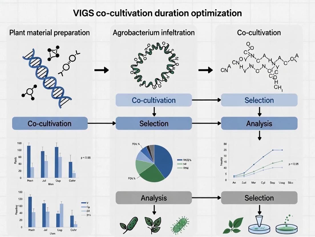
Workflow Visualization
The Scientist's Toolkit: Key Research Reagent Solutions
Table 3: Essential Materials for Agrobacterium-VIGS Co-cultivation Experiments
| Reagent / Material | Function in Co-cultivation | Example & Notes |
|---|---|---|
| Agrobacterium Strain | Engineered to deliver T-DNA; workhorse for gene transfer. | GV3101 (common for VIGS in soy, Luffa) [6] [7], EHA105 [3]. |
| VIGS Vector System | Carries the gene-silencing construct into the plant cell. | TRV-based vectors (pTRV1, pTRV2) are widely used for their mild symptoms and whole-plant spread [8] [4]. |
| Reporter Gene | Visual marker for rapid assessment of VIGS efficiency. | Phytoene Desaturase (PDS): Silencing causes photobleaching [9] [8] [6]. CLA1: Can show a stronger phenotype in some species like Lycoris [8]. |
| Acetosyringone | Phenolic signal molecule that induces the Agrobacterium vir genes, activating the T-DNA transfer machinery. | Critical for efficient transformation; typically used at 100-200 µM in infiltration and co-cultivation media [5] [3]. |
| Antibiotics | Selection for transformed tissues and elimination of Agrobacterium post-co-cultivation. | Kanamycin (for plant selection), Timentin or Carbenicillin (to kill Agrobacterium) [5]. |
| Co-cultivation Medium | Provides nutrients and support for both plant and bacterial cells during the critical T-DNA transfer window. | Often based on MS or Gamborg B5 salts, with sucrose and plant hormones, but without antibacterial agents [5]. |
Biological Mechanism: How T-DNA Transfer Works
The transfer of T-DNA from Agrobacterium tumefaciens into a plant cell is a multistep process, culminating in the integration of foreign genetic material into the plant genome. The following diagram illustrates the key stages of this pathway.
Detailed Mechanism Description
The process begins when acetosyringone, a phenolic compound released by wounded plant tissues, is perceived by the bacterial VirA membrane sensor protein [10] [11]. This activates the VirG regulatory protein, which in turn induces the expression of other virulence (vir) genes located on the Ti plasmid [12] [13].
Key vir gene products then act on the T-DNA region of the Ti plasmid, which is defined by 25-base-pair left and right border repeats [12] [13]. The proteins VirD1 and VirD2 create a single-stranded nick at these borders, liberating a single-stranded T-DNA molecule (the T-strand) with VirD2 covalently attached to its 5' end [13].
The T-strand is coated by numerous molecules of VirE2, a single-stranded DNA-binding protein, forming a protected T-complex [13]. This mature T-complex is then transported through a VirB/VirD4-encoded channel from the bacterial cell into the plant cell cytoplasm [13].
Inside the host cell, the T-complex is directed to the nucleus. Both VirD2 and VirE2 possess nuclear localization signals (NLS) that interact with the host's nuclear import machinery [13]. Once inside the nucleus, the T-DNA is stripped of its protein escort and integrated into the plant genome, a process that is poorly understood but relies on host repair proteins [12] [13].
Troubleshooting Guide & FAQs
Frequently Asked Questions
Q1: What is the primary function of acetosyringone in T-DNA transfer? Acetosyringone is a phenolic signal molecule that activates the bacterial VirA/VirG two-component sensory system. This activation is a crucial first step that triggers the expression of all other
virgenes, initiating the T-DNA processing and transfer process [10] [13] [11].Q2: Why is my transformation efficiency low, and how can acetosyringone help? Low transformation efficiency can stem from inadequate
virgene induction. Many plant species, particularly monocots, do not exude sufficient phenolic signals. Adding acetosyringone (typically at 100-400 µM) to the co-cultivation medium artificially induces thevirgenes, significantly boosting transformation efficiency in a wide range of plants [10] [11].Q3: Can I use Agrobacterium to transform organisms other than plants? Yes. While its natural host range is primarily dicotyledonous plants, Agrobacterium has been successfully used to transform monocots, yeast, fungi, and even human cells under laboratory conditions. The core mechanism of T-DNA transfer is conserved across these diverse hosts [13].
Q4: What is a "disarmed" strain in the context of T-DNA vectors? A disarmed strain is a genetically engineered Agrobacterium strain where the oncogenic (tumor-inducing) genes within the native T-DNA have been removed. This prevents gall formation while retaining the ability to transfer T-DNA. The gene(s) of interest are then cloned into the T-DNA region of a separate binary vector [14].
Troubleshooting Common Experimental Issues
Table 1: Troubleshooting Common T-DNA Transfer and VIGS Experiments
| Problem | Potential Causes | Recommended Solutions |
|---|---|---|
| Low Transformation Efficiency | Inadequate vir gene induction; suboptimal bacterial concentration; incorrect co-cultivation conditions. |
Add 100-400 µM acetosyringone to induction & co-cultivation media [10] [11]; optimize optical density (OD₆₀₀) to 0.4-1.0 for infiltration; ensure optimal co-cultivation duration and temperature [6] [4]. |
| No Silencing Phenotype in VIGS | Inefficient viral vector delivery; poor systemic spread of the virus; target sequence not optimal. | Use vacuum infiltration or direct injection at cotyledon nodes for better delivery [6]; validate viral presence via RT-PCR in new growth; test multiple independent target gene fragments (200-300 bp) for efficient silencing [4]. |
| Plant Cell Death Post-Infiltration | Hypervirulent Agrobacterium strain; too high a bacterial density (OD); overlong co-cultivation. | Titrate bacterial OD₆₀₀ to lower values (e.g., 0.4-0.6); reduce co-cultivation time [4]; use a milder strain (e.g., GV3101) if possible. |
| Unsuccessful T-DNA Integration | Issues with host factors; problems with selection marker; incorrect border sequences. | Confirm T-DNA border sequences are intact and functional [12] [14]; use a robust plant selectable marker (e.g., kanamycin or hygromycin resistance); consider host genotype compatibility and use a super-virulent strain if needed. |
Optimizing Co-cultivation: Key Experimental Data
Optimizing the duration of co-cultivation—the period when Agrobacterium is in intimate contact with plant tissues—is critical for maximizing T-DNA delivery while minimizing tissue damage. The following table summarizes optimal co-cultivation parameters from recent research.
Table 2: Experimentally Determined Optimal Co-cultivation Parameters
| Plant Species / System | Infiltration Method | Optimal Co-cultivation Duration | Key Supporting Findings | Citation |
|---|---|---|---|---|
| Soybean (VIGS) | Cotyledon node immersion | 20-30 minutes (immersion time) | Achieved high infection efficiency, with fluorescence observed in >80% of cells by 4 days post-infection. | [6] |
| Sunflower (VIGS) | Seed vacuum infiltration | 6 hours | This duration produced the most efficient VIGS results, with an infection percentage of up to 77% and significant target gene silencing. | [4] |
| Areca catechu (VIGS) | Callus tissue immersion | 3 days | A 3-day co-cultivation period in the dark was part of the optimized protocol for establishing VIGS in areca palm embryoids. | [9] |
The experimental workflow for determining these parameters typically involves the following steps, which can be visualized in the diagram below.
The Scientist's Toolkit: Essential Research Reagents
Table 3: Key Reagents for T-DNA Transfer and VIGS Experiments
| Reagent / Material | Function / Role in the Experiment |
|---|---|
| Acetosyringone | A phenolic compound used to induce the bacterial vir genes, enhancing T-DNA transfer efficiency, especially in recalcitrant species [10] [11]. |
| T-DNA Binary Vector System | A two-plasmid system where a small binary vector contains the T-DNA with the gene of interest, and a helper Ti plasmid provides the vir genes in trans [14]. |
| Agrobacterium Strains (e.g., GV3101, AGL-1) | Engineered, often disarmed strains used for transformation. Different strains have varying host ranges and transformation efficiencies [14]. |
| TRV-based VIGS Vectors (pTRV1, pTRV2) | Viral vectors derived from Tobacco Rattle Virus used for Virus-Induced Gene Silencing. pTRV2 is modified to carry a fragment of the target plant gene [6] [4]. |
| Plant Selectable Markers (e.g., Kanamycin, Hygromycin Resistance) | Genes incorporated into the T-DNA that allow for the selection of successfully transformed plant cells or tissues on antibiotic-containing media [12] [14]. |
| Reporter Genes (e.g., GFP, GUS) | Genes that encode easily detectable proteins (e.g., Green Fluorescent Protein) used to visually confirm successful transformation or silencing without needing to wait for a phenotypic change [6] [14]. |
Why Co-cultivation Duration is a Critical Determinant of Silencing Efficiency
Co-cultivation duration—the period plant tissues remain in contact with Agrobacterium tumefaciens containing viral vectors—is a pivotal experimental parameter in Virus-Induced Gene Silencing (VIGS) that directly determines the balance between successful gene silencing and plant tissue viability. Within the broader context of VIGS optimization research, co-cultivation represents a critical window where bacterial infection initiates the RNA interference pathway. This technical support center provides evidence-based troubleshooting guidance to help researchers overcome the challenge of identifying species-specific and genotype-dependent co-cultivation windows to maximize silencing efficiency while minimizing cellular damage.
Core Concepts: Co-cultivation and VIGS Efficiency
What is Co-cultivation in VIGS Experiments?
Co-cultivation is the essential step in Agrobacterium-mediated VIGS where explants are incubated with Agrobacterium suspensions containing TRV vectors (pTRV1 and pTRV2 with target gene inserts). During this phase, bacteria attach to plant cells and transfer T-DNA containing viral vectors into the plant genome, initiating the infection process that leads to systemic gene silencing [6] [4].
How Co-cultivation Duration Affects Silencing Outcomes
The relationship between co-cultivation time and VIGS efficiency follows a biphasic pattern. Insufficient duration limits T-DNA transfer and viral establishment, while excessive exposure induces plant stress responses, tissue damage, and reduced viability. Optimal duration ensures adequate bacterial infection without compromising plant health, enabling robust systemic silencing [4].
The diagram below illustrates how co-cultivation duration influences the key stages of the VIGS process:
Experimental Evidence: Co-cultivation Duration Across Plant Systems
Quantitative Data from Published Studies
Table 1: Experimentally Determined Optimal Co-cultivation Durations Across Plant Species
| Plant Species | Optimal Co-cultivation Duration | Silencing Efficiency | Infection Method | Citation |
|---|---|---|---|---|
| Sunflower (Helianthus annuus) | 6 hours | Up to 91% infection rate; 77% silencing efficiency | Seed vacuum infiltration | [4] |
| Soybean (Glycine max) | 20-30 minutes | 65-95% silencing efficiency | Cotyledon node immersion | [6] |
| Tea (Camellia sinensis) | 5 minutes (under vacuum) | 63.34% silencing efficiency | Vacuum infiltration | [15] |
| Taro (Colocasia esculenta) | Not specified (OD₆₀₀=1.0 optimal) | 27.77% silencing plant rate | Bulb vacuum treatment | [16] |
Detailed Experimental Protocols
Sunflower VIGS Protocol with 6-Hour Co-cultivation
Background: Researchers established a highly efficient VIGS protocol for sunflower that identified 6 hours as the optimal co-cultivation period through systematic testing of multiple timepoints [4].
Materials:
- Sunflower seeds (multiple genotypes recommended)
- Agrobacterium tumefaciens strain GV3101
- TRV vectors (pYL192/TRV1 and pYL156/TRV2 with target gene insert)
- Plant growth medium (3:1 peat:perlite ratio)
Methodology:
- Vector Construction: Clone target gene fragment (193bp for HaPDS) into TRV2 vector using appropriate restriction enzymes (XbaI and BamHI sites)
- Bacterial Preparation: Transform recombinant plasmids into Agrobacterium GV3101, culture in LB medium with antibiotics (kanamycin 50μg/mL, gentamicin 10μg/mL, rifampicin 100μg/mL)
- Seed Preparation: Remove seed coats without additional sterilization
- Vacuum Infiltration: Subject seeds to vacuum infiltration with Agrobacterium suspension (OD₆₀₀ = 0.8-1.0)
- Co-cultivation: Incubate infiltrated seeds for 6 hours in co-cultivation medium
- Plant Growth: Transfer to greenhouse conditions (22°C, 18h light/6h dark, 45% RH)
- Efficiency Assessment: Monitor photobleaching symptoms and quantify by qRT-PCR [4]
Key Finding: The 6-hour co-cultivation period significantly increased both infection percentage (up to 91%) and silencing efficiency compared to shorter or longer durations.
Soybean Cotyledon Node Immersion with 20-30 Minute Co-cultivation
Background: This protocol addressed limitations of conventional infiltration methods in soybean with thick cuticles and dense trichomes [6].
Materials:
- Soybean seeds (cv. Tianlong 1)
- Agrobacterium tumefaciens GV3101 with pTRV1 and pTRV2-GFP derivatives
- Sterile tissue culture supplies
Methodology:
- Seed Preparation: Soak sterilized soybeans in sterile water until swollen, longitudinally bisect to obtain half-seed explants
- *Bacterial Preparation": Grow *Agrobacterium to log phase, resuspend in infiltration medium
- *Immersion Co-cultivation": Immerse fresh explants in *Agrobacterium suspensions containing pTRV1 or pTRV2 derivatives for 20-30 minutes
- Tissue Culture": Transfer to sterile tissue culture conditions
- Efficiency Validation": Assess GFP fluorescence at 4 days post-infection, phenotype observation at 21 dpi [6]
Key Finding: The 20-30 minute co-cultivation period achieved 65-95% silencing efficiency with systemic spread throughout the plant, demonstrating that relatively short durations can be effective with proper tissue selection.
Troubleshooting Guide: Co-cultivation Duration Problems
Frequently Asked Questions
Table 2: Troubleshooting Common Co-cultivation Issues
| Problem | Possible Causes | Solutions | Preventive Measures |
|---|---|---|---|
| No silencing phenotype | Insufficient co-cultivation duration; Low bacterial concentration; Incorrect plant developmental stage | Increase co-cultivation time incrementally (1-2 hour steps); Verify OD₆₀₀ = 0.8-1.0; Use younger tissues with higher division rates | Perform time-course pilot experiments; Standardize bacterial growth conditions |
| Tissue browning/death | Excessive co-cultivation duration; Bacterial overgrowth; Plant genotype sensitivity | Reduce co-cultivation time; Add antioxidants to medium; Test different genotypes | Establish species-specific duration windows; Monitor bacterial density carefully |
| Variable silencing between plants | Inconsistent bacterial contact; Genotype-dependent responses; Environmental fluctuations | Ensure uniform immersion/infiltration; Include multiple genotypes in optimization; Control temperature/humidity during co-cultivation | Standardize infection protocols; Use mixed Agrobacterium cultures for consistency |
| Weak transient silencing | Suboptimal T-DNA transfer; Viral movement limitations | Extend co-cultivation within tolerance limits; Include viral suppressors of RNA silencing (VSRs) | Optimize vector design; Use known efficient VIGS vectors (TRV, BPMV) |
Advanced Optimization Strategies
Q: How can I determine the ideal co-cultivation duration for a new plant species?
A: Implement a systematic time-course experiment testing multiple durations (e.g., 0, 15, 30, 60, 120, 180, 360 minutes) while monitoring both efficiency markers (silencing percentage, viral spread) and toxicity indicators (tissue viability, chlorophyll content). The optimal duration balances these competing factors, typically falling where efficiency plateaus before significant toxicity emerges [4] [15].
Q: Does co-cultivation duration requirement vary with infection method?
A: Yes, vacuum infiltration methods often require shorter durations (5 minutes to 1 hour) compared to immersion techniques (20-30 minutes) or simple co-culture (6+ hours), as vacuum forces enhance bacterial penetration. Always optimize duration for your specific infection protocol [4] [15].
Q: How does bacterial density interact with co-cultivation duration?
A: Higher OD₆₀₀ values (0.8-1.0) typically allow shorter co-cultivation periods, while lower densities may require extended contact. However, high densities with long durations often cause tissue damage. For example, in taro, increasing OD₆₀₀ from 0.6 to 1.0 boosted silencing rates from 12.23% to 27.77% without duration adjustment [16].
Essential Research Reagent Solutions
Table 3: Key Reagents for VIGS Co-cultivation Experiments
| Reagent/Equipment | Function/Role | Application Notes | Optimal Specifications |
|---|---|---|---|
| Agrobacterium tumefaciens GV3101 | T-DNA delivery vector | Broad host range, disarmed strain | Contains appropriate virulence genes |
| TRV Vectors (pTRV1/pTRV2) | Viral-induced silencing system | Bipartite system; pTRV2 contains target gene insert | High-copy number plasmids with selection markers |
| Antibiotics (Kanamycin, Gentamicin, Rifampicin) | Selective maintenance of plasmids and strains | Concentration-dependent efficacy | Plant-specific tolerance testing required |
| Infiltration Medium | Bacterial suspension and plant support | Often contains acetosyringone for virulence induction | pH 5.2-5.7 optimal for T-DNA transfer |
| Plant Growth Regulators | Enhance transformation efficiency | Cytokinins/auxins may improve tissue response | Species-specific formulations needed |
Molecular Mechanisms: How Duration Affects Silencing at Cellular Level
The relationship between co-cultivation duration and VIGS efficiency operates through three key molecular mechanisms:
T-DNA Transfer Completion: Adequate time allows full implementation of the Agrobacterium type IV secretion system, mediating transfer of T-DNA complex into plant cells [6] [17].
Viral Establishment and Spread: Longer co-cultivation increases probability of successful TRV replication complex formation, enabling cell-to-cell movement through plasmodesmata and systemic spread via phloem [18] [19].
RNAi Amplification: Extended bacterial contact enhances primary siRNA production, which amplifies through host RNA-dependent RNA polymerase (RDRP) activity, creating secondary siRNAs for sustained silencing [18] [20].
The following diagram illustrates the time-dependent molecular processes activated during co-cultivation:
Co-cultivation duration represents a fundamental determinant in VIGS experimental success that requires species-specific and genotype-dependent optimization. The evidence consistently demonstrates that identifying the precise co-cultivation window balancing efficient T-DNA transfer with plant viability is essential for reproducible, high-efficiency silencing. Future research directions should focus on establishing standardized optimization protocols across taxonomic groups and developing molecular markers to precisely monitor optimal infection windows in real-time.
Frequently Asked Questions (FAQs)
FAQ 1: What is the fundamental relationship between co-cultivation duration and VIGS efficiency? Co-cultivation duration is a critical parameter in Agrobacterium-mediated VIGS that directly influences the balance between successful T-DNA transfer and plant cell survival. Insufficient duration limits bacterial attachment and T-DNA transfer, leading to low infection rates. Excessive duration can cause overgrowth of Agrobacterium (overgrowth), resulting in plant tissue damage or death, which undermines silencing establishment and systemic spread [21]. The optimal window must be determined empirically for each plant species.
FAQ 2: How does co-cultivation time affect systemic silencing in different plant tissues? Adequate co-cultivation ensures sufficient initial viral load in the inoculated tissues, which is a prerequisite for the virus to spread systemically through the vasculature. Research in sunflowers showed that an optimized 6-hour co-cultivation following seed vacuum infiltration allowed the Tobacco Rattle Virus (TRV) to spread effectively, being detected in leaves as high as node 9, indicating extensive systemic movement. Furthermore, silencing spread more actively in young, developing tissues compared to mature ones [21].
FAQ 3: Can the same co-cultivation time be applied across different plant genotypes? No, genotype dependency is a significant factor. While an optimal protocol can be established for a species, the exact infection percentage and the pattern of silencing phenotype spread can vary between genotypes. For instance, in a study with six sunflower genotypes, infection rates varied from 62% to 91% using the same VIGS protocol. One genotype achieved the highest infection rate (91%) but exhibited the lowest spread of the silencing phenotype, highlighting the need for genotype-specific validation [21].
Troubleshooting Guides
Problem: Low Infection Rate and Poor Gene Silencing
Potential Causes and Solutions:
Cause 1: Insufficient Co-cultivation Time
- Solution: Optimize the co-cultivation window. A study in sunflower identified that a 6-hour co-cultivation period after seed vacuum infiltration was key to achieving high infection rates (up to 77%) and strong silencing efficiency [21].
- Actionable Protocol: Test a range of co-cultivation times (e.g., 3, 6, 12, 24 hours) using a marker gene like PDS and quantify silencing through phenotypic observation and qRT-PCR.
Cause 2: Suboptimal Inoculation Method
- Solution: Choose an infiltration method that overcomes the physical barriers of your plant species. For seeds or tough tissues, vacuum infiltration is highly effective. For cotyledonary nodes, as in soybean, a 20-30 minute immersion of bisected seeds in the Agrobacterium suspension proved effective, achieving transformation efficiencies over 80% [22] [6].
- Actionable Protocol:
Cause 3: Incorrect Bacterial Concentration or Plant Growth Conditions
Problem: Silencing is Localized and Does Not Spread Systemically
Potential Causes and Solutions:
Cause 1: Inadequate Viral Establishment from Short Co-cultivation
- Solution: Extend the co-cultivation time within the optimal window to ensure a robust initial infection. The virus requires a strong foothold to replicate and move into the phloem for long-distance travel.
- Actionable Protocol: Refer to the optimized co-cultivation times identified for your plant species. The 6-hour co-cultivation in sunflower facilitated TRV detection in upper nodes, confirming systemic mobility [21].
Cause 2: Plant Genotype with Inherent Limitations in Viral Movement
- Solution: If possible, switch to a genotype known to be more susceptible to VIGS. If not, re-optimize the inoculation protocol, potentially testing higher Agrobacterium densities or different infiltration sites.
- Actionable Protocol: When working with a new genotype, conduct a small pilot study comparing its VIGS efficiency to a known susceptible genotype under your standard protocol.
The following table consolidates key experimental data from recent studies, highlighting the impact of co-cultivation and infiltration methods on key VIGS outcomes.
Table 1: Impact of Infiltration Method and Co-cultivation Duration on VIGS Efficiency
| Plant Species | Infiltration Method | Co-cultivation / Immersion Duration | Key Outcome: Infection Rate | Key Outcome: Systemic Silencing & Efficiency |
|---|---|---|---|---|
| Sunflower [21] | Seed Vacuum Infiltration | 6 hours | Up to 77% infection percentage | TRV detected up to node 9; strong PDS silencing (normalized relative expression = 0.01) |
| Soybean [22] [6] | Cotyledon Node Immersion | 20-30 minutes | Effective infectivity efficiency >80%, up to 95% in some cultivars | Systemic GmPDS silencing observed at 21 days post-inoculation (dpi); silencing efficiency 65-95% |
| Wheat / Maize [23] | Vacuum of (Germinated) Seeds | Co-cultivation duration not specified | Whole-plant level gene silencing achieved | Successful silencing of PDS and MLO homoeoalleles; resistance to powdery mildew demonstrated |
Essential Research Reagent Solutions
Table 2: Key Reagents for VIGS Co-cultivation Optimization Experiments
| Reagent / Material | Function in Experiment | Example from Literature |
|---|---|---|
| TRV Vectors (pTRV1, pTRV2) | Bipartite viral vector system for inducing silencing. pTRV2 carries the target gene fragment. | Used in sunflower, soybean, wheat, and maize studies [21] [22] [23]. |
| Agrobacterium Strain GV3101 | The bacterial vehicle for delivering TRV vectors into plant cells. | The standard strain used across multiple studies in sunflower, soybean, and Luffa [21] [22] [7]. |
| Infiltration Solution (Acetosyringone, Cysteine, Tween 20) | Enhances Agrobacterium virulence and plant cell transformation. | A novel solution containing these components enabled whole-plant VIGS in wheat and maize [23]. |
| Marker Gene (e.g., PDS) | A visual reporter for silencing efficiency. Silencing causes photobleaching. | PDS was used as a marker to optimize protocols in sunflower, soybean, Luffa, and banana [21] [22] [7]. |
Experimental Workflow and Relationship Diagrams
The following diagram illustrates the core experimental workflow for optimizing co-cultivation duration and the logical relationship between duration and outcomes.
Protocols in Practice: Co-cultivation Durations Across Plant Systems
The establishment of a 6-hour co-cultivation period following seed vacuum infiltration represents a significant optimization in sunflower Virus-Induced Gene Silencing (VIGS) protocols. This method specifically addresses the challenges of transforming a recalcitrant species like sunflower, enabling efficient gene silencing for functional genomics studies.
Key Optimized Parameters for Sunflower VIGS:
| Parameter | Optimal Condition | Protocol Impact |
|---|---|---|
| Infiltration Method | Seed Vacuum Infiltration | Enables whole-plant viral spreading without tissue damage from syringe infiltration [4] |
| Co-cultivation Duration | 6 hours | Produced the most efficient VIGS results in terms of infection percentage and target gene silencing [4] |
| Agrobacterium Strain | GV3101 | Standard strain for Agrobacterium-mediated delivery [4] [24] |
| VIGS Vector | Tobacco Rattle Virus (TRV) | Vigorously spreads throughout entire plant with mild infection symptoms [4] [23] |
| Surfactant | Silwet L-77 | Promotes bacterial invasion; significantly higher transformation efficiency vs. Triton X-100 [24] |
Detailed Experimental Methodology
Agrobacterium Culture and Infiltration Suspension Preparation
The success of the protocol begins with proper preparation of the biological materials [4].
- Vector System: Use the TRV-based VIGS vectors pYL192 (TRV1) and pYL156 (TRV2). The target gene fragment (e.g., 193-bp fragment of phytoene desaturase, HaPDS) is cloned into the TRV2 vector.
- Agrobacterium Transformation: Transform the recombinant TRV constructs into Agrobacterium tumefaciens strain GV3101 via electroporation.
- Culture Preparation:
- Streak Agrobacterium glycerol stocks on LB-agar plates with appropriate antibiotics (kanamycin 50 µg/mL, gentamicin 10 µg/mL, rifampicin 100 µg/mL).
- Incubate at 28°C for 1.5 days.
- Pick single colonies to initiate liquid cultures for infiltration suspension.
Seed Vacuum Infiltration Procedure
This optimized protocol eliminates the need for in vitro recovery or surface sterilization [4].
- Plant Material: Use sunflower seeds with seed coats peeled to facilitate infiltration.
- Infiltration Suspension: Prepare Agrobacterium suspension at optimal density in infiltration solution.
- Vacuum Application: Submerge seeds in suspension and apply vacuum. The process removes air from seed coat spaces, allowing the suspension to backfill upon vacuum release.
- Co-cultivation: Transfer infiltrated seeds to co-cultivation medium for the critical 6-hour period.
- Planting: Sow seeds directly in soil mix (peat:perlite, 3:1 ratio) without in vitro recovery steps.
Post-Infiltration Plant Care
- Growth Conditions: Maintain plants at average 22°C with 18-h light/6-h dark photoperiod and approximately 45% relative humidity [4].
- Symptom Monitoring: Silencing symptoms (e.g., photo-bleaching when targeting PDS) typically appear in newly developed tissues after several days.
- Efficiency Assessment: Monitor infection percentages and silencing efficiency through phenotypic observation and molecular analysis (e.g., RT-qPCR).
Troubleshooting Guides & FAQs
Frequently Encountered Experimental Challenges
Q1: What if my sunflower seedlings show poor infection rates after infiltration?
- Potential Cause: Suboptimal Agrobacterium concentration or viability.
- Solution: Ensure bacterial cultures are in log growth phase and adjust OD600 to 0.8-1.0. Confirm viability through plating tests before infiltration [24].
Q2: How can I address excessive tissue damage following infiltration?
- Potential Cause: Prolonged immersion in infiltration solution or incorrect surfactant concentration.
- Solution: Limit immersion time to 2 hours maximum. Use Silwet L-77 at 0.02% concentration for optimal tissue compatibility [24].
Q3: What if silencing symptoms appear inconsistent across plants?
- Potential Cause: Genotype-dependent VIGS efficiency variations.
- Solution: Test multiple sunflower genotypes. Infection percentages vary significantly (62-91%) between genotypes. 'Smart SM-64B' showed highest infection (91%) though with less phenotype spreading [4].
Q4: How can I confirm TRV presence in tissues without visible silencing?
- Potential Cause: TRV mobility not always correlated with observable silencing.
- Solution: Perform RT-PCR analysis on green and bleached tissues from different plant parts. TRV can be present in tissues without observable phenotype [4].
Protocol Optimization FAQs
Q5: Why is the 6-hour co-cultivation period critical?
- Answer: Extensive testing revealed this duration produces the most efficient VIGS results, balancing adequate Agrobacterium interaction with plant viability. Shorter periods reduce transformation efficiency; longer periods increase stress without benefit [4].
Q6: Can this protocol be adapted for other plant species?
- Answer: While optimized for sunflower, the seed vacuum approach has succeeded in wheat, maize, tomato, and other species. However, co-cultivation timing and infiltration parameters require species-specific optimization [23].
Q7: What are the key advantages of seed vacuum over other infiltration methods?
- Answer: This method enables whole-plant level gene silencing, is more rapid and convenient than leaf infiltration, avoids mechanical tissue damage, and allows study of genes involved in early plant development [4] [23].
Research Reagent Solutions
Essential Materials for Sunflower VIGS Protocol:
| Reagent/Equipment | Function/Specification | Application Notes |
|---|---|---|
| Agrobacterium tumefaciens GV3101 | Disarmed strain for DNA delivery | Standard transformation workhorse; compatible with TRV vectors [4] [24] |
| TRV VIGS Vectors | pYL192 (TRV1) & pYL156 (TRV2) | TRV vigorously spreads throughout plant with mild symptoms [4] [23] |
| Silwet L-77 | Surfactant (0.02%) | Critical for reducing surface tension; significantly improves efficiency vs. Triton X-100 [24] |
| Antibiotics | Kanamycin, Gentamicin, Rifampicin | Selection for vector and Agrobacterium strain [4] |
| Vacuum Infiltration System | Chamber and pump | Must achieve sufficient vacuum for solution penetration [4] |
| Co-cultivation Medium | Standard plant growth medium | 6-hour period critical for protocol success [4] |
Experimental Workflow & Visualization
The following diagram illustrates the optimized sunflower VIGS protocol, highlighting the critical 6-hour co-cultivation phase and key decision points:
Sunflower VIGS Workflow with 6-Hour Co-cultivation
The protocol emphasizes the elimination of unnecessary steps like surface sterilization and in vitro recovery, making it more accessible than previous methods. The 6-hour co-cultivation represents the key optimization that balances sufficient bacterial interaction with plant viability.
Technical Applications in Research Context
This optimized protocol enables researchers to effectively apply VIGS to sunflower, a species traditionally considered challenging for transformation. The method has been successfully used to silence phytoene desaturase (PDS) as a visual marker, demonstrating its utility for functional genomics studies [4].
The seed vacuum infiltration approach with defined co-cultivation duration provides a standardized platform for:
- Functional analysis of sunflower genes involved in stress tolerance
- Studies of gene function during early plant development stages
- High-throughput screening of candidate genes
- Investigation of TRV mobility and silencing spread patterns in different tissues
This protocol represents a significant advancement in sunflower biotechnology, providing researchers with a robust tool for reverse genetics in this important oilseed crop.
Virus-Induced Gene Silencing (VIGS) is a powerful reverse genetics tool for rapid functional gene analysis in plants. A critical, yet often variable, factor in its success is the co-cultivation duration—the period plant tissues are exposed to Agrobacterium tumefaciens carrying the viral vector. This technical guide, framed within broader research on optimizing this parameter, addresses key challenges and provides a validated protocol for achieving high-efficiency silencing in soybean using a cotyledon node immersion method.
Troubleshooting Guide: Co-cultivation Duration
Q1: What is the optimal co-cultivation time for soybean VIGS via cotyledon node immersion?
Based on established research, the recommended co-cultivation period is 20-30 minutes of immersion in the Agrobacterium suspension [6]. This duration was identified as optimal for achieving high infection efficiency in soybean cv. Tianlong 1, facilitating effective systemic spread of the Tobacco Rattle Virus (TRV) vector.
Q2: What problems occur if the co-cultivation time is too short or too long?
Deviating from the optimal window can significantly impact experimental outcomes, as summarized in the table below.
| Co-cultivation Time | Potential Consequences & Silencing Outcomes |
|---|---|
| Too Short (< 20 min) | Inadequate bacterial infection; low transduction efficiency; weak or non-systemic silencing [6]. |
| Optimal (20-30 min) | High infection efficiency (65-95% silencing); robust systemic silencing spread [6]. |
| Too Long (> 30 min) | Potential for tissue damage (hyperhydration); increased plant stress; possible overgrowth of Agrobacterium [4]. |
Q3: Why is the cotyledon node a good target for VIGS in soybean?
The cotyledon node is a highly meristematic region, making it an ideal gateway for the TRV vector to establish infection and spread systemically throughout the plant [6]. Research demonstrates that using this method with the 20–30-minute immersion protocol enables the virus to move from the cotyledon node to other tissues, effectively silencing endogenous genes in newer leaves [6].
Q4: Besides time, what other factors are critical for high VIGS efficiency?
Co-cultivation time is just one variable. A successful experiment requires optimizing several key parameters.
| Factor | Optimization Consideration |
|---|---|
| Plant Genotype | Susceptibility to VIGS varies. Efficiency reached up to 95% in 'Tianlong 1' soybean [6], while studies in sunflowers show a range of 62–91% across genotypes [4]. |
| Agrobacterium Concentration | An optical density at 600 nm (OD600) is commonly used. Specific optimal values should be empirically determined for each system. |
| Plant Growth Stage | Younger tissues are generally more susceptible. A cotyledon-based approach using 5-day-old etiolated seedlings has proven highly effective in other species [25] [26]. |
| Environmental Conditions | Post-inoculation, maintaining plants under high humidity for 1-2 days can significantly enhance infection efficiency [4]. |
Validated Experimental Protocol
The following methodology is adapted from a successful TRV-VIGS system established for soybean [6].
Materials
- Plant Material: Soybean seeds (cv. Tianlong 1 used in source study).
- Agrobacterium Strain: GV3101 competent cells.
- VIGS Vectors: Binary TRV vectors (pTRV1 and pTRV2). pTRV2 should be modified to carry a fragment of the target gene (e.g., GmPDS for a visible photobleaching phenotype).
- Growth Media: Luria-Bertani (LB) medium with appropriate antibiotics (Kanamycin, Gentamicin, Rifampicin).
- Induction Medium: LB with antibiotics and 10 mM MES, 20 μM Acetosyringone.
- Infiltration Medium: Liquid plant culture medium (e.g., half-strength Murashige and Skoog basal medium) with 10 mM MES and 200 μM Acetosyringone, pH 5.5.
Step-by-Step Procedure
Vector Construction & Agrobacterium Transformation
Prepare Agrobacterium Culture
- Inoculate single colonies of Agrobacterium containing pTRV1 and pTRV2 into separate flasks of LB medium with antibiotics.
- Incubate at 28°C with shaking (200 rpm) for ~24 hours until the culture is dense.
- Centrifuge the cultures and resuspend the bacterial pellets in induction medium. Adjust the OD600 to between 0.4 and 0.6. Shake for an additional 6-8 hours at 28°C.
Prepare the Agro-infiltration Suspension
- Mix the induced pTRV1 and pTRV2 cultures in a 1:1 ratio.
- Pellet the mixed bacteria and resuspend in infiltration medium to a final OD600 of 0.8-1.5. Let the mixture sit at room temperature for 3-4 hours before use.
Prepare Plant Explants
- Surface-sterilize soybean seeds and soak in sterile water until they are swollen.
- Key Step: Bisect the swollen seeds longitudinally to create half-seed explants, ensuring the cotyledon node is exposed [6].
Co-cultivation: Cotyledon Node Immersion
- Immerse the fresh half-seed explants completely in the agro-infiltration suspension.
- Apply Vacuum Infiltration (if available) for a few minutes to enhance infiltration, then release the vacuum slowly.
- Critical Step: Allow the immersion to continue for the optimized co-cultivation period of 20-30 minutes at room temperature [6].
Post-inoculation Recovery & Cultivation
- After immersion, gently blot the explants to remove excess liquid.
- Transfer the explants to sterile filter paper in Petri dishes or directly to plant culture media.
- Maintain the inoculated explants in the dark at 22-25°C for 2-3 days to facilitate T-DNA transfer and the initiation of viral infection.
- Subsequently, transfer the plants to a growth chamber or greenhouse with a 16/8 hour light/dark cycle and observe for silencing phenotypes, which can appear as early as 10-14 days post-inoculation [6] [25].
Workflow and Parameter Optimization
The diagram below illustrates the experimental workflow and highlights the critical parameters that require optimization for a successful VIGS experiment.
The Scientist's Toolkit: Essential Research Reagents
The following table details key materials required to establish the cotyledon node immersion VIGS protocol for soybean.
| Research Reagent | Function & Application in VIGS |
|---|---|
| pTRV1 & pTRV2 Vectors | Binary plasmid system for TRV-based VIGS. pTRV1 encodes viral replication proteins; pTRV2 carries the coat protein and is modified to include the target gene fragment for silencing [6] [27]. |
| Agrobacterium tumefaciens GV3101 | A disarmed helper strain widely used for delivering TRV vectors into plant cells via a process called agroinfiltration [6] [25]. |
| Acetosyringone | A phenolic compound that induces the Agrobacterium Vir genes, enhancing the efficiency of T-DNA transfer into the plant genome during co-cultivation [4]. |
| Phytoene Desaturase (PDS) Gene Fragment | A common visual marker gene for optimizing VIGS. Silencing PDS disrupts chlorophyll synthesis, causing a distinctive photobleaching (white) phenotype that confirms successful gene silencing [6] [9]. |
| Half-strength MS Medium | A common plant culture medium used as the base for the infiltration suspension, providing osmotic support and nutrients to both the plant tissue and Agrobacterium during the immersion step [6]. |
Key Technical Takeaways
- Adhere to the 20–30-minute window for cotyledon node immersion to balance high infection efficiency with plant health.
- Use the cotyledon node from bisected seeds as it provides direct access to meristematic tissues for efficient viral entry and systemic spread.
- Always include a positive control, such as GmPDS, to visually confirm the system is working in your hands.
- Empirically validate key parameters like Agrobacterium concentration and plant genotype, as optimal conditions can vary.
Technical Support Center
Troubleshooting Guides & FAQs
Question: What is the optimized vacuum infiltration and co-cultivation duration for establishing VIGS in Atriplex canescens, and how was it determined? Answer: For the halophyte Atriplex canescens, the optimized protocol uses vacuum-assisted agroinfiltration for 10 minutes at a pressure of 0.5 kPa, followed by a standard co-cultivation period on vermiculite. This duration was determined through comparative analysis of inoculation materials and methods, achieving an average silencing efficiency of approximately 16.4%. Systemic photobleaching phenotypes from AcPDS silencing appeared in newly emerged leaves at about 15 days post-inoculation [28].
Question: The VIGS protocol for sunflower seeds recommends a 6-hour co-cultivation. What evidence supports this specific duration over shorter or longer times? Answer: Extensive testing of different parameters concluded that a 6-hour co-cultivation period following seed vacuum infiltration produced the most efficient VIGS results. This was quantified by a high infection percentage (up to 77%) and significant silencing efficiency of the targeted gene, with normalized relative expression levels as low as 0.01. This duration likely represents a balance between sufficient Agrobacterium-plant cell interaction and avoidance of overgrowth or hypersensitive defense responses [4].
Question: How does genotype dependency affect VIGS efficiency, and what strategies can mitigate this in challenging species? Answer: Genotype dependency significantly influences VIGS susceptibility and phenotypic spread. In sunflowers, infection percentages varied from 62% to 91% across different genotypes. Furthermore, a genotype with the highest infection rate (91%) showed the lowest spreading of the silencing phenotype. This highlights that infection efficiency and symptom spread are independent variables. Mitigation strategies include [4]:
- Screening Multiple Genotypes: Test several cultivars or lines to identify susceptible genotypes.
- Phenotypic Monitoring: Track both infection rates and the extent of silencing spread in different tissues.
- Protocol Optimization: Adjust factors like Agrobacterium concentration and plant growth conditions for specific genotypes.
Question: For walnut and other highly recalcitrant woody species, what general principles should guide initial optimization of co-cultivation duration? Answer: While specific data for walnut is not available in the provided search results, established principles from other challenging systems guide initial optimization [4] [23] [9]:
- Start with Shorter Durations: Begin with co-cultivation times of 6-24 hours to minimize tissue stress and browning.
- Use a Reporter Gene: Utilize the PDS gene to visually optimize the system through photobleaching.
- Monitor Viral Presence Extensively: Use RT-PCR to track the virus in both symptomatic and non-symptomatic tissues, as TRV presence is not always limited to areas with observable silencing.
- Test at the Seed/Germinated Seed Stage: For species with transformation-resistant mature tissues, seed vacuum infiltration can be a highly effective workaround.
Table 1: Optimized co-cultivation and infiltration parameters across different plant systems.
| Plant Species | Infiltration Method | Vacuum Duration | Vacuum Pressure | Co-cultivation Duration | Key Efficiency Metrics | Reference |
|---|---|---|---|---|---|---|
| Atriplex canescens | Vacuum (germinated seeds) | 10 minutes | 0.5 kPa | Standard duration on vermiculite | ~16.4% avg. silencing efficiency; 40-80% transcript reduction [28]. | |
| Sunflower | Seed Vacuum Infiltration | Information Missing | Information Missing | 6 hours | Up to 77% infection rate; highly efficient silencing [4]. | |
| Wheat & Maize | Vacuum (germinated seeds) | Information Missing | Information Missing | 48-60 hours | Whole-plant level silencing; successful PDS and MLO silencing [23]. | |
| Soybean | Soaking (cotyledon nodes) | 20-30 minutes (soaking) | Not Applied | Standard duration on medium | 65-95% silencing efficiency; high infection rate [22]. |
Detailed Experimental Protocol forAtriplex canescensVIGS
Methodology for TRV-Based VIGS in Atriplex canescens [28]
- Vector Construction: Clone a target fragment (300-400 bp) of the gene of interest (e.g., AcPDS) into the pTRV2 vector using appropriate restriction sites (e.g., EcoRI and BamHI).
- Agrobacterium Preparation:
- Transform recombinant pTRV2 and the helper plasmid pTRV1 into Agrobacterium tumefaciens strain GV3101.
- Grow single colonies in YEP liquid medium with appropriate antibiotics (50 mg/L kanamycin, 50 mg/L rifampicin) at 28°C until OD600 reaches 0.6-0.8.
- Centrifuge bacterial cultures and resuspend the pellet in infiltration buffer (10 mM MES, 200 µM acetosyringone, 10 mM MgCl2, 0.03% Silwet-77) to a final OD600 of 0.8.
- Mix the pTRV1 and pTRV2-derived Agrobacterium suspensions in equal volumes and incubate in the dark at room temperature for 3 hours.
- Plant Material Preparation:
- Treat Atriplex canescens seeds with 50% (v/v) H2SO4 for 8 hours to weaken the seed coat.
- Rinse thoroughly with distilled water and place on moist vermiculite to germinate in darkness at 25°C.
- Select germinated seeds with radicle lengths of 1-3 cm for inoculation.
- Vacuum Infiltration:
- Submerge the germinated seeds in the prepared Agrobacterium suspension.
- Apply a vacuum of 0.5 kPa for 10 minutes.
- Co-cultivation and Plant Growth:
- After infiltration, rinse the materials with sterile distilled water.
- Transfer the inoculated materials to pots filled with vermiculite.
- Co-cultivate and grow plants in a greenhouse under controlled conditions (22°C, 16h light/8h dark cycle).
- Phenotype Observation: Systemic photobleaching in newly emerged leaves is typically observed around 15 days post-inoculation.
Research Reagent Solutions
Table 2: Essential reagents and materials for VIGS establishment in challenging species.
| Item | Function/Application in VIGS |
|---|---|
| pTRV1 & pTRV2 Vectors | Bipartite Tobacco Rattle Virus (TRV)-based vector system; among the most versatile and widely used for VIGS due to broad host range and efficient systemic movement [28] [19]. |
| Agrobacterium tumefaciens GV3101 | Standard bacterial strain for delivering TRV vectors into plant cells via agroinfiltration [4] [28]. |
| Infiltration Buffer | Solution used to suspend Agrobacterium for inoculation; typically contains MES, MgCl2, acetosyringone (an inducer of virulence genes), and a surfactant like Silwet-77 [28]. |
| Phytoene Desaturase (PDS) Gene Fragment | A visual reporter gene; its silencing disrupts chlorophyll biosynthesis, causing a photobleaching phenotype used to optimize and validate VIGS system efficiency [28] [9]. |
| Acetosyringone | A phenolic compound that activates Agrobacterium virulence genes, crucial for enhancing the efficiency of T-DNA transfer into the plant genome [28] [23]. |
Visualized Experimental Workflows
VIGS Optimization Workflow
Mechanism of VIGS
This guide details the protocols for preparing essential reagents for Agrobacterium tumefaciens-mediated plant transformation, with specific optimization for Virus-Induced Gene Silencing (VIGS) experiments.
Agrobacterium Growth Media and Culture Preparation
Standard YEP Medium (for A. tumefaciens strain GV3101)
A complex medium for robust growth of Agrobacterium [21] [29] [7].
- Composition:
- YEP Solid Medium (per Liter): 10 g Peptone, 10 g Yeast Extract, 5 g NaCl, 15 g Bacto-Agar.
- YEP Liquid Medium (per Liter): 10 g Peptone, 10 g Yeast Extract, 5 g NaCl.
- Antibiotics: Add appropriate antibiotics based on the resistance markers of your binary vector (e.g., 50 mg/L Kanamycin, 50 mg/L Rifampicin) [29].
- Protocol:
- Adjust pH to 7.0 with NaOH.
- Autoclave at 121°C for 20 minutes.
- For solid media, cool to approximately 55°C before adding antibiotics and pouring plates.
- Inoculate a single colony of Agrobacterium harboring your plasmid (e.g., TRV1 or TRV2 derivatives) into liquid YEP with antibiotics.
- Incubate at 28°C with shaking at 200 rpm for 1-2 days until the culture reaches the mid-logarithmic growth phase (OD600 = 0.6-0.8) [29] [7].
AT Minimal Medium
A defined medium that limits the development of auxotrophic mutants and is useful for specific experimental needs [30].
- Composition (1X):
- 20X AT Buffer: 0.079 M KH2PO4, 0.044 M NaOH (pH to 7.0).
- 20X AT Salts: 0.015 M (NH4)2SO4, 0.6 mM MgSO4·7H2O, 0.06 mM CaCl2·2H2O, 0.0071 mM MnSO4·H2O.
- 50X Iron Stock: 0.125 M FeSO4·7H2O.
- Carbon Source: 0.5% (28 mM) glucose or other suitable carbon sources.
- Protocol: Prepare sterile stocks, mix components aseptically, and add a carbon source [30].
Infiltration Buffer Formulations
The infiltration buffer is critical for inducing Agrobacterium virulence and facilitating plant infection. Key components include MgCl2 for osmotic balance, MES as a pH buffer, and acetosyringone (AS) as a virulence inducer.
Table 1: Common Infiltration Buffer Compositions
| Component | Final Concentration (Example 1) [29] | Final Concentration (Example 2) [31] | Function |
|---|---|---|---|
| MgCl2 | 10 mM | - | Osmotic regulation |
| MES | 10 mM | - | pH Buffer |
| Acetosyringone (AS) | 200 µM | 200 µM | Induces Vir genes |
| Silwet L-77 | 0.03% | - | Surfactant |
| Sucrose | - | 50 g/L | Osmoticum/Energy source |
| Glucose | - | 2 g/L | Energy source |
Infiltration Buffer Preparation Workflow
The following diagram illustrates the preparation of the infiltration buffer and the subsequent steps for resuspending the bacterial culture.
Troubleshooting Guide & FAQ
Q1: What is the optimal optical density (OD600) for the final Agrobacterium suspension?
The optimal OD600 varies by plant species and infiltration method, typically ranging from 0.5 to 1.0 [31] [29] [32]. Using an OD that is too high can reduce transformation efficiency [31].
Table 2: Optimal OD600 and Acetosyringone (AS) Concentrations for Different Species
| Plant Species | Recommended OD₆₀₀ | Recommended AS Concentration | Primary Inoculation Method |
|---|---|---|---|
| Sunflower | Information missing | 200 µM | Seed Vacuum Infiltration [21] |
| Atriplex canescens | 0.8 - 1.0 | 200 µM | Vacuum Infiltration [29] |
| Luffa acutangula | 0.8 - 1.0 | 200 µM | Leaf Infiltration [7] |
| Styrax japonicus | 0.5 - 1.0 | 200 µM | Vacuum or Friction-osmosis [32] |
| Medicago truncatula | 0.5, 0.6, or 1.0 | 200 µM | Co-cultivation with cells [31] |
Q2: Why is acetosyringone (AS) critical, and how should it be prepared?
Acetosyringone is a phenolic compound that activates the Agrobacterium Virulence (Vir) genes, which are essential for T-DNA transfer [31] [29] [23].
- Stock Solution: Prepare a 200 mM stock in 100% ethanol or DMSO.
- Storage: Aliquot and store at -20°C protected from light.
- Working Concentration: Add to the infiltration buffer at a final concentration of 200 µM immediately before use.
Q3: How long should the Agrobacterium suspension be incubated in the infiltration buffer before use?
A pre-infiltration incubation of 2 to 3 hours at room temperature is commonly used to fully induce the virulence machinery [31] [7].
Q4: What are common reasons for low transformation efficiency?
- Bacterial Overgrowth: Cultures grown beyond the mid-log phase can have reduced virulence [31].
- Incorrect OD600: Suspensions that are too dense or too dilute.
- Old Acetosyringone Stock: AS is light-sensitive and can degrade. Always use a fresh stock.
- Inadequate Wounding: For some methods, proper wounding is necessary for infection.
The Scientist's Toolkit: Key Research Reagent Solutions
Table 3: Essential Materials and Their Functions
| Reagent/Item | Function/Application in VIGS |
|---|---|
| pTRV1 & pTRV2 Vectors | The bipartite Tobacco Rattle Virus (TRV) system used for VIGS; pTRV2 carries the target gene insert [21] [29] [23]. |
| A. tumefaciens GV3101 | A widely used disarmed strain for plant transformation due to its high virulence [31] [21] [29]. |
| Acetosyringone (AS) | A phenolic signal molecule that induces the expression of bacterial vir genes, essential for T-DNA transfer [31] [29] [23]. |
| Silwet L-77 | A surfactant that reduces surface tension and improves the wetting and penetration of the bacterial suspension into plant tissues [29]. |
| MES Buffer | A buffering agent used to maintain a stable and slightly acidic pH (around 5.5) in the infiltration buffer, which is favorable for vir gene induction. |
| Antibiotics (e.g., Kanamycin, Rifampicin) | Selective agents to maintain the binary vector in Agrobacterium and ensure axenic culture conditions [31] [29]. |
Fine-Tuning Co-cultivation: A Troubleshooting Framework for Maximum Efficiency
FAQs and Troubleshooting Guides
FAQ 1: How do I select the optimal Agrobacterium strain for my VIGS experiment?
Answer: The choice of Agrobacterium strain is critical and depends on your plant species and the virulence properties of the strain. Hypervirulent strains are often preferred for recalcitrant species.
- Hypervirulent Strains: For challenging plant species like wheat, hypervirulent strains such as AGL0 and AGL1 are highly effective. These strains contain the pTiBo542 plasmid, which carries additional copies of key
virgenes (vir B, C, and G), enhancing the efficiency of T-DNA delivery [33]. - Standard Strains: Common laboratory strains like GV3101 are successfully used in established VIGS protocols for plants such as sunflower and pepper [34] [4].
- Strain LBA4404: This strain can be effective when augmented with a "superbinary" plasmid containing extra
virgenes, as demonstrated in wheat transformation [33].
FAQ 2: What is the recommended OD600 for the Agrobacterium inoculation suspension?
Answer: The optimal optical density (OD600) is not universal and must be balanced with co-cultivation time to ensure efficient infection without overgrowth.
- Common Range: A standard starting point is an OD600 of 0.5, as used in white clover transformation with a 4-day co-cultivation period [35].
- Higher OD for Specific Protocols: Some protocols for pepper transformation use a higher OD600 of 0.6 [34].
- Empirical Optimization is Key: It is crucial to empirically test a range of OD600 values (e.g., 0.4 to 1.6) in your specific system, as the optimal density can vary with the Agrobacterium strain, plant genotype, and explant type [36].
FAQ 3: My experiment is plagued by Agrobacterium overgrowth after co-cultivation. How can I mitigate this?
Answer: Overgrowth is a common issue that can be managed by optimizing co-cultivation conditions and employing effective washing steps.
- Limit Co-cultivation Time: Reduce the co-cultivation period to the minimum required for efficient T-DNA delivery. For instance, a 6-hour co-cultivation was optimal in a sunflower VIGS protocol [4].
- Use Bacteriostats: After co-cultivation, thoroughly wash explants and transfer them to a medium containing antibiotics like Timentin (360 mg L⁻¹) or Carbenicillin (500 mg L⁻¹) to suppress further bacterial growth [34] [36].
- Avoid Excessive Bacterial Load: Ensure you remove excess Agrobacterium suspension after inoculation and do not use an excessively high OD600, as this directly contributes to overgrowth.
FAQ 4: How do Agrobacterium strain and OD600 interact with co-cultivation time?
Answer: These three variables form a tightly linked optimization triangle. A more virulent strain or a higher bacterial density may require a shorter co-cultivation time to achieve the same transformation efficiency while avoiding overgrowth. Conversely, a less virulent strain might need a longer co-cultivation period for sufficient T-DNA delivery. The optimal combination must be determined empirically for each experimental system.
The following table consolidates optimized parameters from various successful Agrobacterium-mediated transformation studies.
Table 1: Optimized Parameters for Agrobacterium-mediated Transformation in Different Plant Species
| Plant Species | Agrobacterium Strain | OD600 | Co-cultivation Time | Key Supporting Factors | Primary Source |
|---|---|---|---|---|---|
| White Clover | EHA105 | 0.5 | 4 days | 20 mg L⁻¹ Acetosyringone [35] | [35] |
| Sunflower | GV3101 | Information Missing | 6 hours | Seed vacuum infiltration [4] | [4] |
| Pepper | Specific strain not stated | 0.6 | 2 days | Vacuum infiltration, no pre-culture [34] | [34] |
| Wheat | AGL0, AGL1 | Information Missing | 2-3 days | 200 µM Acetosyringone, 0.01% Silwet L-77 [33] | [33] |
| Rose (Hairy Root) | MSU440, Ar Qual | 0.8 - 1.0 | 2-3 days (Aseptic) | Acetosyringone, specific media [36] | [36] |
Detailed Experimental Protocols
Protocol 1: Agrobacterium-mediated VIGS in Sunflower (Optimized Seed-Vacuum Method)
This protocol highlights the critical interaction between a short co-cultivation time and a highly efficient infiltration method [4].
Vector and Agrobacterium Preparation:
- Use the Tobacco Rattle Virus (TRV)-based vectors pYL192 (TRV1) and pYL156 (TRV2) carrying the target gene fragment.
- Transform the constructs into Agrobacterium tumefaciens strain GV3101.
- Grow single colonies in LB medium with appropriate antibiotics until the logarithmic growth phase.
Plant Material Preparation:
- Partially peel the coats of sunflower seeds to facilitate infiltration.
- Note: No surface sterilization or in vitro recovery steps are required.
Inoculation and Co-cultivation:
- Suspend the Agrobacterium culture in an induction medium.
- Vacuum Infiltration: Subject the peeled seeds to the bacterial suspension under a vacuum pressure of -0.06 MPa.
- Co-cultivation: After infiltration, co-cultivate the seeds for 6 hours. This short duration is key to achieving high efficiency while controlling bacterial overgrowth.
Post Co-cultivation:
- Sow the seeds directly in soil or an appropriate growing medium.
- Cultivate plants under controlled greenhouse conditions (e.g., 22°C, 18-h light/6-h dark photoperiod).
Protocol 2: Stable Transformation of Pepper Cotyledon Explants
This protocol demonstrates the use of vacuum treatment and the avoidance of pre-culture to enhance transformation efficiency with a 2-day co-cultivation [34].
Plant Material:
- Use 12-day-old seedlings of a transformable genotype (e.g., PC69).
- Prepare explants from cotyledon and hypocotyl segments.
Agrobacterium Inoculation:
- Prepare an Agrobacterium suspension at an OD600 of 0.6.
- Inoculation: Directly inoculate explants with the suspension under a mild vacuum (-0.06 MPa). Avoid a pre-culture step for optimal results.
- Co-cultivation: Co-culture the explants for 2 days on a suitable medium.
Selection and Regeneration:
- Transfer explants to a callus-inducing medium (CIM) containing 75 mg L⁻¹ kanamycin for selection and 4 mg L⁻¹ AgNO₃ (an ethylene inhibitor).
- Upon the appearance of green bud primordia, transfer to a shoot-inducing medium (SIM) with a reduced cytokinin (ZR) concentration and 0.17 mg L⁻¹ GA₃.
- Elongated shoots are excised and cultured on a root-inducing medium (RIM) containing 2 mg L⁻¹ IBA.
The Scientist's Toolkit
Table 2: Essential Research Reagents and Materials
| Reagent/Material | Function in Experiment | Example Usage |
|---|---|---|
| Acetosyringone | A phenolic compound that induces the expression of bacterial vir genes, enhancing T-DNA transfer. |
Added to the Agrobacterium inoculation and co-cultivation media at 100-400 µM [33]. |
| Silwet L-77 | A surfactant that reduces surface tension, improving the wettability and penetration of the bacterial suspension into plant tissues. | Used at 0.01-0.05% in the inoculation medium for wheat transformation [33]. |
| Timentin / Carbenicillin | Broad-spectrum antibiotics that inhibit Agrobacterium overgrowth after co-cultivation, without affecting plant regeneration. | Added to post-co-cultivation media at 150-500 mg L⁻¹ to control bacterial growth [34] [36]. |
| Silver Nitrate (AgNO₃) | An ethylene action inhibitor. Ethylene can accumulate under stress and inhibit organogenesis. | Supplemented at 4 mg L⁻¹ in the callus-inducing medium for pepper to facilitate regeneration [34]. |
| TRV Vectors (pYL192/156) | A bipartite virus-based system for Virus-Induced Gene Silencing (VIGS). TRV1 encodes replication proteins, and TRV2 carries the target gene fragment for silencing. | The most widely used VIGS vector for functional genomics in Solanaceae and other plant families [4] [19]. |
Logical Workflow for Parameter Optimization
The diagram below outlines a logical, iterative process for optimizing Agrobacterium strain, OD600, and co-cultivation time in your experiments.
Addressing Species and Genotype-Specific Requirements
Frequently Asked Questions (FAQs) on VIGS Co-cultivation
1. What is the primary challenge of applying VIGS across different plant species? The main challenge is the variation in physiological and genetic traits between species and genotypes. For instance, soybean leaves have a thick cuticle and dense trichomes that can impede conventional agroinfiltration methods like misting or direct injection, requiring optimized protocols for efficient infection [6].
2. How does the optimal co-cultivation duration vary? The ideal co-cultivation duration is not universal and must be determined empirically for each species-genotype combination. Research on Fraxinus mandshurica involved testing various Agrobacterium tumefaciens infection durations, while a soybean protocol identified a 20-30 minute immersion as optimal for their specific system [6] [37].
3. What plant material is best for optimizing co-cultivation? The choice of explant is critical. Successful systems have used:
- Soybean: Cotyledon node explants [6].
- Areca catechu (Betel nut): Embryogenic callus tissue [9].
- Fraxinus mandshurica: Embryos and plant growth points [37]. Using explants that are actively dividing and susceptible to Agrobacterium infection significantly increases transformation efficiency.
4. Which bacterial strain and density are recommended? The engineered Agrobacterium strain GV3101 is commonly used with TRV vectors [6]. The optical density (OD600) of the bacterial suspension is a key parameter. Studies optimize this value, with one protocol for Fraxinus mandshurica testing OD600 values of 0.5, 0.6, 0.7, and 0.8 to find the most effective concentration for gene editing [37].
Troubleshooting Guide for VIGS Co-cultivation
Table 1: Common Co-cultivation Issues and Solutions
| Problem Phenotype | Potential Cause | Recommended Solution |
|---|---|---|
| Low transformation efficiency | Incorrect Agrobacterium density (OD600) | Empirically test a range of OD600 values (e.g., 0.5-0.8); use high-quality, freshly prepared suspensions [37]. |
| Low transformation efficiency | Suboptimal explant type or physiological state | Use actively dividing tissues like cotyledon nodes or embryogenic callus; avoid old or dormant tissues [6] [9]. |
| No systemic silencing | Co-cultivation duration too short or long | Test immersion or infection times; for soybean cotyledon nodes, 20-30 minutes was effective [6]. |
| Excessive bacterial overgrowth | Co-cultivation duration too long; inadequate washing | Optimize the duration and ensure thorough washing after co-cultivation to remove excess Agrobacterium [9]. |
| Plant tissue necrosis/death | Agrobacterium concentration too high; toxic response | Titrate the OD600 to a lower concentration; ensure the bacterial suspension is prepared in an appropriate induction medium [37]. |
Table 2: Species-Specific Co-cultivation Parameters from Recent Studies
| Plant Species | Genotype/Cultivar | Optimal Explant | Optimal Co-cultivation Method | Key Parameter (e.g., OD600, Duration) | Reported Efficiency |
|---|---|---|---|---|---|
| Soybean (Glycine max) | Tianlong 1 | Cotyledon node | Immersion in Agrobacterium suspension | 20-30 minutes | 65% - 95% silencing efficiency [6] |
| Areca catechu (Betel nut) | Not specified | Embryogenic callus | Co-culture on solid medium | 30 days (full process) | Effective AcPDS silencing observed [9] |
| Fraxinus mandshurica (Manchurian ash) | Wild-type | Plant growth points | Agrobacterium-mediated infection | OD600 tested (0.5-0.8) | 18% gene editing in induced buds [37] |
Essential Research Reagent Solutions
Table 3: Key Reagents for VIGS Co-cultivation Experiments
| Reagent / Material | Function in Co-cultivation | Example & Notes |
|---|---|---|
| TRV Vectors (pTRV1, pTRV2) | Viral vectors for delivering silencing constructs. pTRV1 encodes replication proteins, pTRV2 carries the target gene insert [6] [19]. | |
| Agrobacterium tumefaciens | Engineered bacterium to deliver TRV vectors into plant cells. | Common strains: GV3101 [6] and EHA105 [37]. |
| Antibiotics | Select for transformed Agrobacterium and prevent bacterial contamination after co-cultivation. | Kanamycin is commonly used for vectors with nptII resistance [9] [37]. |
| Acetosyringone | A phenolic compound that induces the Vir genes of Agrobacterium, enhancing its ability to transfer T-DNA into plant cells. | Added to the co-cultivation medium [9]. |
| MS or WPM Medium | Provides essential nutrients and a supportive environment for plant tissues during and after co-cultivation. | WPM (Woody Plant Medium) is often used for woody species like Fraxinus [37]. |
Visualizing the VIGS Pathway and Optimization Workflow
VIGS Mechanism and Co-cultivation
Co-cultivation Parameter Optimization
Detailed Experimental Protocol: TRV-VIGS in Soybean Cotyledon Nodes
This protocol, adapted from a 2025 study, outlines the steps for establishing a highly efficient VIGS system in soybean, achieving up to 95% silencing efficiency [6].
Principle: The method uses Agrobacterium tumefaciens (strain GV3101) carrying Tobacco Rattle Virus (TRV) vectors to deliver gene-specific fragments into soybean cells via the cotyledon node, leading to systemic silencing of the target gene.
Materials:
- Plant Material: Sterilized soybean seeds (e.g., cultivar 'Tianlong 1').
- Bacterial Strains: Agrobacterium tumefaciens GV3101 harboring pTRV1 and pTRV2-derived plasmids (e.g., pTRV2-GmPDS for a positive control).
- Growth Media: LB broth with appropriate antibiotics (e.g., kanamycin), induction medium (LB with antibiotics and 200 µM acetosyringone).
- Equipment: Sterile culture facilities, fluorescence microscope (for GFP-tagged vectors), shaker incubator.
Procedure:
- Seed Preparation: Soak sterilized soybean seeds in sterile water until swollen. Bisect the seeds longitudinally to obtain half-seed explants containing the cotyledon node.
- Agrobacterium Preparation: Grow recombinant Agrobacterium strains overnight in LB with antibiotics. Resuspend the bacterial pellet in induction medium containing acetosyringone and grow for another 4-6 hours to an OD600 of approximately 0.8 [6] [37].
- Co-cultivation (Infection): Immerse the fresh cotyledon node explants in the Agrobacterium suspension for 20-30 minutes with gentle agitation. This immersion method was critical for overcoming the barriers posed by soybean's thick cuticle and dense trichomes [6].
- Draining and Co-culture: Remove the explants from the suspension and blot off excess liquid. Transfer the explants to a co-cultivation medium and incubate in the dark at a controlled temperature (e.g., 22-25°C) for 2-3 days.
- Recovery and Selection: Transfer the explants to a recovery medium containing antibiotics to suppress Agrobacterium overgrowth.
- Efficiency Evaluation: Around 4 days post-infection, examine the hypocotyl sections under a fluorescence microscope for GFP signals to confirm successful infection. Silencing phenotypes, such as photobleaching for GmPDS, can be observed in leaves approximately 21 days post-inoculation [6].
Key Optimization Notes:
- Explant Vitality: Using fresh, actively growing explants is crucial for high transformation efficiency.
- Bacterial Density: The OD600 of the suspension is a critical factor. While an OD600 of ~0.8 was successful in one study, this parameter should be optimized for your specific system [6] [37].
- Co-cultivation Duration: The 20-30 minute immersion time was identified as optimal in the cited soybean study. Shorter or longer durations may reduce efficiency [6].
Frequently Asked Questions (FAQs)
FAQ 1: What are the most critical environmental factors to control for efficient VIGS co-cultivation? The success of VIGS co-cultivation is highly dependent on three critical environmental factors: temperature, light, and humidity. Temperature directly influences Agrobacterium virulence and plant cell metabolic activity, with an optimal range typically between 19-22°C. Light intensity and photoperiod regulate plant physiology and defense responses, while high humidity (≥70%) is crucial for preventing plant desiccation stress during the co-cultivation period [4] [38].
FAQ 2: How does temperature during co-cultivation affect TRV viral spread and silencing efficiency? Temperature significantly impacts TRV replication and systemic movement. Lower temperatures (19-21°C) generally enhance silencing efficiency by moderating plant defense responses and promoting viral replication. Studies in tomato and Nicotiana benthamiana demonstrate that temperatures above 25°C can accelerate plant growth but simultaneously reduce VIGS efficiency by activating host RNA silencing mechanisms more rapidly [38].
FAQ 3: What is the optimal co-cultivation duration for different plant species? Co-cultivation duration varies by species and inoculation method, typically ranging from 6 hours to 2 days. Research in sunflowers established that 6 hours of co-cultivation produced the most efficient VIGS results [4]. For the root wounding-immersion method in solanaceous species, a 30-minute immersion period proved optimal [38]. Longer durations risk bacterial overgrowth, while shorter periods may not allow sufficient T-DNA transfer.
FAQ 4: How do different plant genotypes respond to standardized VIGS protocols? Genotype dependency significantly affects VIGS efficiency. Research in sunflowers revealed infection percentages varying from 62% to 91% across different genotypes when using the same protocol [4]. Similarly, soybean studies showed silencing efficiency ranging from 65% to 95% across cultivars [6] [22]. This underscores the necessity of protocol optimization for specific plant genotypes.
Troubleshooting Guides
Table 1: Troubleshooting Common VIGS Co-cultivation Issues
| Problem | Possible Causes | Solutions | Preventive Measures |
|---|---|---|---|
| Low silencing efficiency | Incorrect temperature, insufficient co-cultivation time, improper light conditions | Adjust temperature to 19-22°C; extend co-cultivation to 6-48 hours; ensure appropriate light intensity | Optimize all three parameters simultaneously; conduct pilot studies [4] [38] |
| Plant tissue damage/death | Bacterial overgrowth, excessive wounding, temperature stress | Reduce co-cultivation time; optimize Agrobacterium density (OD600=0.5-1.0); minimize tissue damage | Include surfactant (e.g., Silwet L-77) in infiltration medium; control bacterial concentration [23] |
| Inconsistent silencing patterns | Variable environmental conditions, uneven inoculation | Standardize growth conditions; ensure uniform inoculation technique | Implement controlled environment chambers; train personnel on consistent technique [4] [19] |
| No silencing observed | Incorrect vector construction, poor viral spread, incompatible genotype | Verify insert orientation and sequence; confirm TRV presence via PCR; try different genotype | Use positive control (PDS/CLA1); include GFP-tagged vectors to monitor infection [6] [8] |
Table 2: Optimized Co-cultivation Parameters for Different Plant Species
| Plant Species | Optimal Temperature (°C) | Co-cultivation Duration | Light Conditions | Efficiency | Citation |
|---|---|---|---|---|---|
| Sunflower | 22 | 6 hours | 18-h light/6-h dark photoperiod | Up to 91% infection | [4] |
| Soybean | 22-25 | 20-30 min (immersion) | Standard growth conditions | 65-95% silencing | [6] [22] |
| Tomato | 19-21 | 30 min (root immersion) | 16-h light (28°C)/8-h dark (20°C) | 95-100% silencing | [38] |
| Nicotiana benthamiana | 19-22 | 30 min (root immersion) | 16-h light/8-h dark | ~100% silencing | [38] |
| Wheat & Maize | 21 (light)/19 (dark) | 2 days (seed vacuum) | 16-h light/8-h dark | Whole-plant level silencing | [23] |
| Lycoris | 25 | 15-20 s (leaf tip injection) | Controlled environment | High silencing efficiency | [8] |
Experimental Protocols
Protocol 1: Seed Vacuum Infiltration and Co-cultivation for Sunflowers
This protocol achieved up to 91% infection efficiency in sunflowers [4].
Key Materials:
- Agrobacterium strain GV3101 containing pTRV1 and pTRV2 derivatives
- Sunflower seeds with seed coats partially removed
- Infiltration solution (10 mM MgCl₂, 10 mM MES, 150 μM acetosyringone, pH 5.6)
Methodology:
- Prepare Agrobacterium cultures to OD600 = 0.8 in infiltration solution
- Mix pTRV1 and pTRV2 cultures in 1:1 ratio
- Place sunflower seeds in bacterial suspension
- Apply vacuum (0.5-1 bar) for 5-10 minutes
- Release vacuum gradually to ensure proper infiltration
- Co-cultivate seeds for 6 hours in the dark at 22°C
- Transfer seeds to soil or growth medium for further development
Critical Step: The 6-hour co-cultivation period was identified as optimal through systematic testing. Longer durations promoted bacterial overgrowth, while shorter periods reduced infection rates.
Protocol 2: Root Wounding-Immersion Method for Solanaceous Species
This method achieved 95-100% silencing efficiency in tomato and N. benthamiana [38].
Key Materials:
- 3-4 week old seedlings with 3-4 true leaves
- Agrobacterium strain GV1301 with TRV vectors
- Sterile scissors or blade for root wounding
Methodology:
- Prepare Agrobacterium cultures to OD600 = 0.8 in infiltration solution
- Carefully remove seedlings from soil, preserving root integrity
- Wash roots with pure water to remove soil contaminants
- Remove approximately one-third of the root system lengthwise
- Immerse wounded roots in TRV1:TRV2 mixed solution for 30 minutes
- Maintain temperature at 19-22°C during immersion
- Replant seedlings in fresh soil or growth medium
Critical Step: The root wounding must be sufficient to allow bacterial entry without compromising plant viability. The 30-minute immersion at controlled temperature ensures optimal T-DNA transfer.
Signaling Pathways and Experimental Workflows
Diagram 1: Environmental Factor Interplay in VIGS Efficiency
Diagram 2: Root Wounding-Immersion Workflow
Research Reagent Solutions
Table 3: Essential Materials for VIGS Co-cultivation Optimization
| Reagent/Material | Function | Application Notes | Citation |
|---|---|---|---|
| TRV Vectors (pTRV1/pTRV2) | Viral delivery system for gene silencing | Bipartite system; TRV1 encodes replication proteins, TRV2 carries target gene fragment | [6] [19] |
| Agrobacterium GV3101 | T-DNA delivery vehicle | Preferred for VIGS; high transformation efficiency; requires rifampicin, gentamicin, kanamycin selection | [4] [22] |
| Acetosyringone | Inducer of vir genes | Critical for T-DNA transfer; typically used at 150-200 μM concentration | [23] [38] |
| MES Buffer | pH stabilization | Maintains optimal pH (5.6) for Agrobacterium virulence gene induction | [23] [38] |
| Silwet L-77 Surfactant | Enhances tissue penetration | Reduces surface tension; improves infiltration efficiency; use at 0.01-0.05% | [23] |
| Phytoene Desaturase (PDS) | Positive control reporter gene | Silencing produces photobleaching phenotype; validates system functionality | [6] [8] |
| CLA1 Gene | Alternative reporter gene | Produces albino phenotype; may yield stronger silencing than PDS in some species | [8] |
Frequently Asked Questions
Q1: My VIGS experiments are resulting in very low infection rates. What are the key factors I should optimize?
Low infection rates are often due to suboptimal inoculation methods, plant material selection, or Agrobacterium preparation. The table below summarizes critical factors to check.
| Factor | Issue | Solution & Reference |
|---|---|---|
| Inoculation Method | Unsuitable for plant species; fails to deliver Agrobacterium effectively. | Use vacuum infiltration for germinated seeds or cuttings [28] [15]; use apical meristem inoculation for certain species like petunia [39]. |
| Plant Material | Dense trichomes or thick cuticles block infiltration; wrong developmental stage. | Use germinated seeds or young seedlings with cotyledons [6] [4]; for soybeans, use bisected cotyledon nodes [6]. |
| Agrobacterium Concentration (OD600) | Concentration is too low or too high. | Standardize OD600 to 0.5-1.0 for infiltration [6] [28]. |
| Co-cultivation Duration | Insufficient time for T-DNA transfer. | Optimize duration; a 6-hour co-cultivation proved effective in sunflower [4]. |
Q2: I get infection, but the gene silencing is weak or inconsistent between experiments. How can I improve this?
Inconsistent silencing can be attributed to environmental conditions, genetic factors, and vector design.
| Factor | Issue | Solution & Reference |
|---|---|---|
| Temperature | Non-optimal temperature affects viral replication and spread. | Maintain lower temperatures (e.g., 20°C day/18°C night for petunia) [39]; test for your species. |
| Plant Genotype | The plant cultivar has natural resistance to the virus or poor silencing response. | Screen different genotypes/cultivars; efficiency can vary from 62% to 91% [4]. |
| Target Gene Fragment | The fragment is too short/long, has high complexity, or lacks specificity. | Use fragments 300-500 bp from specific regions (5', middle, 3') of the gene ORF; use online tools (e.g., SGN-VIGS) for design [28]. |
Q3: My control plants are showing severe viral symptoms (stunting, necrosis), which confounds the experiment. What can I do?
This is a common problem when using an empty pTRV2 vector as a control. The solution is to use a non-plant insert control.
- Problem: The empty vector can cause severe viral symptoms like necrosis and plant death, making it hard to distinguish from true silencing phenotypes [39].
- Solution: Replace the empty pTRV2 vector (pTRV2:00) with a vector containing a fragment of a non-plant gene, such as green fluorescent protein (pTRV2-sGFP). This nearly eliminates severe viral symptoms while maintaining the viral load in control plants [39].
Troubleshooting Guide: A Step-by-Step Workflow
Follow this decision-making workflow to diagnose and resolve the most common VIGS issues.
Essential Experimental Protocols
Protocol 1: Vacuum Infiltration for Germinated Seeds (from Sunflower & Atriplex) [4] [28] This protocol is highly effective for difficult-to-transform species.
- Germinate Seeds: Surface sterilize seeds and allow them to germinate on moist vermiculite in the dark until radicles are 1-3 cm long.
- Prepare Agrobacterium: Resuspend Agrobacterium (carrying pTRV1 and pTRV2-target gene) in infiltration buffer (10 mM MES, 10 mM MgCl₂, 200 µM Acetosyringone) to a final OD600 of 0.8.
- Mix and Induce: Combine equal volumes of the pTRV1 and pTRV2 cultures and incubate in the dark for 3 hours.
- Infiltrate: Submerge the germinated seeds in the Agrobacterium suspension. Apply a vacuum of 0.5 kPa to 0.8 kPa for 5-10 minutes.
- Co-cultivate and Plant: Gently blot the seeds dry and co-cultivate on moist vermiculite for ~6 hours. Finally, transfer to soil or growth medium [4].
Protocol 2: Cotyledon Node Method for Soybean [6] This method overcomes the challenge of dense trichomes on soybean leaves.
- Imbibe and Bisect: Soak sterilized soybean seeds in water until swollen. Carefully bisect the seeds longitudinally to create half-seed explants.
- Prepare Agrobacterium: Grow Agrobacterium strain GV3101 to mid-log phase and resuspend in infiltration buffer.
- Inoculate: Immerse the fresh half-seed explants in the Agrobacterium suspension for 20-30 minutes with gentle shaking.
- Co-cultivate and Grow: After inoculation, transfer the explants to a sterile tissue culture setup to recover and grow.
The Scientist's Toolkit: Key Research Reagent Solutions
| Reagent / Material | Function in VIGS Experiment | Key Considerations |
|---|---|---|
| TRV Vectors (pTRV1, pTRV2) | pTRV1 encodes viral replication and movement proteins. pTRV2 carries the target gene fragment for silencing. | The pTRV2 vector is modified to include multiple cloning sites (MCS) or Gateway attR sites for insert cloning [27]. |
| Agrobacterium tumefaciens (GV3101) | Delivers the T-DNA containing the TRV vectors into plant cells. | The strain should be disarmed and contain the appropriate antibiotics resistance for the plasmids used [6] [28]. |
| Infiltration Buffer | The solution used to deliver Agrobacterium into plant tissues. | Must contain Acetosyringone, a phenolic compound that induces the Agrobacterium virulence genes, crucial for high transformation efficiency [28]. |
| Phytoene Desaturase (PDS) Gene | A visual marker gene for silencing; its knockdown causes photobleaching (white leaves). | Used to rapidly optimize and validate any new VIGS protocol before targeting genes of unknown function [6] [28] [15]. |
| pTRV2-sGFP Control Vector | A superior control vector containing a non-plant gene insert. | Prevents severe viral symptoms (necrosis, stunting) common with empty pTRV2 vector, providing a healthier baseline for comparison [39]. |
Measuring Success: Validating and Comparing Co-cultivation Protocols
Frequently Asked Questions (FAQs)
1. Why is qRT-PCR the recommended method for assessing gene knockdown in VIGS experiments? qRT-PCR is the method of choice because it provides a highly sensitive, quantitative, and reliable measure of transcript abundance. It allows researchers to directly quantify the remaining mRNA levels of the target gene after VIGS treatment, providing a numerical value for the silencing efficiency. This is crucial for correlating any observed phenotypic changes with the molecular-level knockdown of the gene [40] [41].
2. My VIGS-treated plants show a strong phenotypic change (e.g., photobleaching). Do I still need to perform qRT-PCR? Yes, it is highly recommended. Visible phenotypes, such as photobleaching from silencing a gene like PDS, are excellent initial indicators of successful VIGS. However, they are often qualitative. qRT-PCR provides quantitative data that confirms the phenotype is indeed due to a specific reduction in your target gene's mRNA and not an off-target effect or general stress response. Most publishers require this molecular validation to support phenotypic observations [41].
3. What is an acceptable level of gene knockdown as measured by qRT-PCR? The acceptable level can vary by experiment, but effective VIGS protocols often report significant knockdown. For instance, optimized TRV-VIGS systems in soybean have demonstrated silencing efficiencies ranging from 65% to 95% reduction in target gene expression [6]. In sunflower, a robust seed-vacuum protocol achieved a normalized relative expression as low as 0.01 for the targeted HaPDS gene [4].
4. Where should I design my qPCR assays on the target transcript? IDT recommends using at least two qPCR assays designed to target different regions of the same transcript [40]. This controls for potential artifacts. For example, if the VIGS construct targets a specific region of the mRNA, having a second assay in a different location verifies that the entire transcript is degraded and not just the fragment complementary to the VIGS insert.
5. What are the key steps to ensure high-quality RNA for qRT-PCR? The quality of your RNA is paramount. Key rules to follow include [42]:
- Harvest material from at least three biological replicates, freeze immediately in liquid nitrogen, and store at -80°C.
- Use an RNA isolation procedure that yields high-quality RNA, with an A260/A280 ratio > 1.8 and an A260/A230 ratio > 2.0.
- Digest purified RNA with DNase I to remove contaminating genomic DNA that can lead to false-positive signals.
Troubleshooting Guide: Common Issues with qRT-PCR Validation of VIGS
| Problem | Potential Cause | Recommended Solution |
|---|---|---|
| No knockdown observed (normal mRNA levels) | VIGS system did not work; ineffective vector delivery or poor viral spread. | Optimize Agrobacterium infiltration method (e.g., try vacuum infiltration) [4]. Confirm viral presence with PCR. Use a positive control (e.g., PDS). |
| High variability between biological replicates | Inconsistent VIGS infection or plant material collection. | Standardize plant growth conditions and infiltration protocols. Ensure material is harvested from the same tissue type and developmental stage. Increase sample size (n≥5) [4]. |
| Inconsistent results between multiple qPCR assays for the same gene | The VIGS construct may only be effective against a specific isoform, or one assay may be inefficient. | Design qPCR assays in the 3'-untranslated region (3'-UTR), which is often more unique [42]. Check primer specificity and PCR efficiency for each assay. |
| No signal or poor cDNA quality | RNA degradation or inefficient reverse transcription. | Check RNA integrity (RIN >7). Use a robust reverse transcriptase with no RNase H activity [42]. Test cDNA quality with a stable reference gene. |
| False positive from genomic DNA contamination | DNA contamination in RNA sample gives spurious PCR amplification. | Treat RNA samples with DNase I during purification. Include a "no-RT" control in your qPCR setup [42]. |
Experimental Protocol: Validating VIGS Efficiency via qRT-PCR
This protocol outlines the key steps for quantifying target gene knockdown following a VIGS experiment, adapted from established methods [6] [42] [4].
I. Sample Collection
- Biological Replicates: Harvest tissue from at least three to five independent VIGS-treated plants per construct to account for biological variation [42] [4].
- Appropriate Tissue: Collect tissue where the silencing phenotype is observed or where the virus is known to spread. For systemic silencing, young leaves are often optimal.
- Controls: Include tissue from:
- Plants infiltrated with an empty vector (e.g., pTRV2-empty) as a negative control.
- Untreated wild-type plants.
- Plants infiltrated with a positive control (e.g., pTRV2-PDS).
- Preservation: Flash-freeze the tissue immediately in liquid nitrogen and store at -80°C until RNA extraction.
II. RNA Extraction and cDNA Synthesis
- Extraction: Isolate total RNA using a reliable kit or method. Assess RNA quality and quantity using spectrophotometry (A260/A280 ~1.8-2.0) and/or a Bioanalyzer (RIN >7) [42].
- DNA Digestion: Treat the purified RNA with DNase I to eliminate genomic DNA contamination [42].
- Reverse Transcription: Perform first-strand cDNA synthesis using a robust reverse transcriptase (e.g., SuperScript III). Use consistent priming, such as oligo(dT) or random hexamers, and the same amount of total RNA for all samples in the experiment [42].
III. Quantitative PCR (qPCR) Setup
- Primer Design:
- Reference Gene Selection:
- Select and validate at least three stable reference genes (e.g., Actin, EF1α, UBQ) from the literature for your plant species and treatment conditions.
- Use software like geNorm or BestKeeper to identify the most stable genes for your dataset [42].
- qPCR Reaction:
- Use a SYBR Green master mix for detection.
- Set up reactions in a clean environment to prevent contamination and use a "no template control" (water).
- Run reactions in technical duplicates or triplicates.
IV. Data Analysis
- Calculate the PCR efficiency for each primer pair using LinRegPCR or similar software [42].
- Calculate the relative transcript levels using a method that incorporates PCR efficiency, such as the Pfaffl method [42].
- Normalize the expression of the target gene in the VIGS sample to the expression in the empty-vector control plants, using the validated reference genes for normalization.
- Perform statistical analysis (e.g., Student's t-test) to determine if the knockdown is significant.
Research Reagent Solutions
Table: Essential reagents and their functions for qRT-PCR validation of VIGS.
| Reagent / Tool | Function / Application |
|---|---|
| TRV VIGS Vectors (pTRV1 & pTRV2) | Bipartite viral vector system for delivering the gene-silencing construct into plant cells [6] [4]. |
| Agrobacterium tumefaciens GV3101 | Standard bacterial strain used to deliver the TRV vectors into plant tissues via agroinfiltration [6] [4]. |
| SYBR Green Master Mix | Fluorescent dye that binds double-stranded DNA during qPCR, allowing for quantification of amplified products [42] [41]. |
| Stable Reference Genes | Genes with constant expression used to normalize qRT-PCR data and account for variations in RNA input and cDNA synthesis (e.g., Actin, GAPDH) [42]. |
| Sequence-Specific qPCR Assays | Primers (and probes) designed to uniquely amplify the target gene. Using multiple assays per gene increases result confidence [40]. |
qRT-PCR Validation Workflow
The following diagram illustrates the logical workflow for using qRT-PCR to validate gene knockdown in a VIGS experiment, from initial phenotypic observation to final data interpretation.
Quantitative Data from Recent VIGS Studies
Table: Silencing efficiencies and key parameters from optimized VIGS protocols in various crops.
| Plant Species | VIGS Delivery Method | Target Gene | Silencing Efficiency / Key Parameter | Reference |
|---|---|---|---|---|
| Soybean (Glycine max) | Agrobacterium-mediated (cotyledon node) | GmPDS | Systemic silencing with photobleaching at 21 dpi; efficiency range: 65% - 95% [6]. | [6] |
| Sunflower (Helianthus annuus) | Seed vacuum infiltration | HaPDS | Normalized relative expression as low as 0.01; infection rate up to 91% (genotype-dependent) [4]. | [4] |
| Areca catechu | Agrobacterium-mediated (embryogenic callus) | AcPDS | Significant photobleaching observed in callus tissues, confirming system establishment [9]. | [9] |
The Scientist's Toolkit: Essential Research Reagents and Materials
The following table details key reagents and materials essential for establishing PDS photobleaching as a visual reporter in VIGS experiments.
Table 1: Key Research Reagent Solutions for VIGS-based PDS Silencing
| Reagent/Material | Function/Application | Example Specifications & Notes |
|---|---|---|
| pTRV1 & pTRV2 Vectors | Binary vectors for the Tobacco Rattle Virus (TRV)-based VIGS system; pTRV2 carries the inserted target gene fragment [43] [29]. | Often kanamycin-resistant; pTRV2-empty vector or pTRV2-GFP can serve as controls to minimize viral symptoms [39] [6]. |
| Agrobacterium tumefaciens Strain GV3101 | Bacterial strain used to deliver the TRV vectors into plant cells [43] [44]. | Culture typically supplemented with antibiotics (e.g., 50 mg/L kanamycin, 50 mg/L rifampicin) [29]. |
| Infiltration Buffer | Solution for suspending Agrobacterium before inoculation, enhancing infection efficiency [43] [29]. | Common composition: 10 mM MgCl₂, 10 mM MES, and 200 µM acetosyringone (AS) [43] [32]. |
| Silwet-77 | A surfactant added to the infiltration buffer to reduce surface tension and improve tissue penetration [29]. | Typically used at low concentrations, such as 0.03% [29]. |
| Acetosyringone (AS) | A phenolic compound that induces the virulence genes of Agrobacterium, facilitating T-DNA transfer into the plant genome [29] [32]. | Commonly used at 200 µM; concentration is a key optimization parameter [29] [32]. |
Understanding PDS Photobleaching in Functional Genomics
What is the molecular function of PDS, and why is it a favored visual reporter?
Answer: Phytoene desaturase (PDS) is a key enzyme in the carotenoid biosynthesis pathway. It catalyzes the conversion of phytoene to ζ-carotene, a critical early step in producing colored carotenoids. Carotenoids play a dual role: they are precursors for important signaling molecules and, crucially, they protect chlorophyll from photo-oxidation by dissipating excess light energy. When the PDS gene is silenced, the carotenoid pathway is disrupted, leading to a depletion of protective carotenoids. Consequently, chlorophyll is degraded upon exposure to light, resulting in a characteristic photobleaching phenotype—white or yellow leaves and tissues [39] [9] [29]. This non-lethal, easily scorable visual marker makes PDS an ideal reporter for assessing the success and spatial distribution of VIGS in various plant species, from model organisms to crops [45] [44] [6].
How can researchers confirm that photobleaching is due to PDS silencing and not other factors?
Answer: A proper experimental design includes multiple controls and confirmatory assays. The photobleaching phenotype should be validated through the following steps:
- Control Groups: Always include plants inoculated with a mixture of Agrobacterium containing pTRV1 and the empty pTRV2 vector (TRV2:00). These plants should not exhibit photobleaching, confirming that the phenotype is due to the PDS insert and not the viral infection itself [43] [29].
- Molecular Validation: Use quantitative real-time PCR (qRT-PCR) to measure the transcript abundance of the PDS gene in photobleached tissues and compare it to control tissues. A significant reduction (e.g., 40-80%) in PDS mRNA levels confirms successful silencing at the molecular level [29] [44].
- Downstream Gene Expression: In some systems, such as tomato, PDS silencing also leads to the transcriptional downregulation of other carotenoid biosynthesis genes like ZDS, CrtISO, and CrtR-b2. Analyzing these can provide additional confirmation [43].
Troubleshooting Common Issues in PDS Silencing
What should I do if my experiment shows no photobleaching phenotype?
Answer: A lack of photobleaching indicates that gene silencing has not occurred efficiently. This can be due to several factors, which should be systematically optimized.
Table 2: Troubleshooting Guide for Absent or Weak Photobleaching
| Problem | Potential Causes | Recommended Solutions & Optimization Strategies |
|---|---|---|
| No Silencing | Suboptimal plant growth stage [39] [45]. | Inoculate younger, meristematic tissues. For petunia, 3-4 weeks after sowing is optimal; for Centaurea cyanus, seedlings with four true leaves showed best results [39] [45]. |
| Low silencing efficiency of the chosen PDS gene fragment [29]. | Use bioinformatics tools (e.g., SGN-VIGS) to design and test multiple fragments (300-400 bp) from different regions (5', middle, 3') of the PDS gene [29]. | |
| Inefficient inoculation method [29] [44]. | For difficult-to-transform species, switch from simple agroinfiltration to more efficient methods like vacuum infiltration (e.g., 0.5 kPa for 10 min) or apical meristem inoculation [45] [29] [6]. | |
| Weak or Patchy Silencing | Suboptimal Agrobacterium culture density [45] [32]. | Adjust the OD600 of the infiltration suspension. Test a range from 0.5 to 1.0; for Centaurea cyanus, OD600 of 0.5 was optimal [45] [32]. |
| Incorrect environmental conditions [39]. | Adjust growth temperature post-inoculation. In petunia, 20 °C day/18 °C night induced stronger silencing than higher temperatures [39]. | |
| Natural variability in viral spread. | Increase your sample size and ensure consistent handling. Silencing may not be uniform across all tissues [39]. |
The negative control (empty vector) is causing severe viral symptoms like stunting or necrosis. How can this be mitigated?
Answer: Severe viral symptoms in control plants can mask genuine silencing phenotypes and complicate analysis. This issue has been noted in several species, including petunia [39]. The solution is to avoid using a completely "empty" pTRV2 vector as a control. Instead, use a modified control vector that contains a non-plant insert, such as a fragment of the green fluorescent protein (GFP) gene (pTRV2-sGFP or pTRV2-GFP) [39] [6]. The presence of an insert in the viral backbone appears to attenuate viral replication and movement, thereby minimizing severe symptom development while still serving as an effective negative control for comparison with plants silenced for your gene of interest [39].
Optimized Experimental Protocols
What is a generalized, optimized protocol for establishing VIGS using PDS in a new plant species?
Answer: Based on successful implementations in multiple species, the following workflow provides a robust starting point. The diagram below outlines the key experimental steps.
Detailed Protocol Steps:
- Vector Construction: Clone a species-specific ~300-400 bp fragment of the PDS gene into the multiple cloning site of the pTRV2 vector using appropriate restriction enzymes (e.g., EcoRI and BamHI) or homologous recombination [29] [6].
- Agrobacterium Preparation: Introduce the recombinant pTRV2-PDS and the helper plasmid pTRV1 into Agrobacterium tumefaciens GV3101. Culture single colonies in YEP medium with appropriate antibiotics until the OD600 reaches 0.6-0.8 [43] [29].
- Inoculum Preparation: Pellet the bacteria and resuspend in an infiltration buffer containing 10 mM MgCl₂, 10 mM MES, and 200 µM acetosyringone. Adjust the final OD600 to a value between 0.5 and 1.0. Mix the pTRV1 and pTRV2-PDS suspensions in a 1:1 ratio and incubate in the dark for 3-6 hours before inoculation [43] [29] [44].
- Plant Inoculation: Select plants at an optimal growth stage (often with 2-4 true leaves). Use an efficient inoculation method such as:
- Vacuum Infiltration: Submerge plant tissues (e.g., germinated seeds, detached leaves) in the Agrobacterium suspension and apply a vacuum (e.g., 0.5 kPa for 5-10 minutes) [29].
- Apical Meristem Inoculation: Mechanically wound the shoot apical meristem and apply the bacterial suspension [39] [45].
- Agroinjection: Use a needleless syringe to infiltrate the suspension directly into tissues, such as tomato fruit through the carpopodium [43].
- Post-Inoculation Care: Maintain inoculated plants under controlled environmental conditions. Research indicates that lower temperatures (e.g., 20 °C day/18 °C night) can significantly enhance silencing efficiency in some species [39].
- Phenotypic Monitoring: Systematically observe new growth for the development of photobleaching, which typically begins to appear 14-21 days post-inoculation [45] [29].
- Molecular Confirmation: Sample photobleached tissues and perform RNA extraction followed by qRT-PCR to quantify the reduction in endogenous PDS transcript levels compared to control plants [43] [44].
What specific parameters are critical for optimizing co-cultivation duration and efficiency?
Answer: While the "ideal" co-cultivation duration can be species-specific, the parameters listed in the table below are universally critical and should be optimized in pilot studies. The interplay between these factors determines the success of VIGS and the manifestation of PDS photobleaching.
Table 3: Key Parameters for Optimizing VIGS Co-Cultivation and Efficiency
| Parameter | Optimal Range / Condition | Impact on Silencing Efficiency |
|---|---|---|
| Plant Age/Growth Stage | Seedlings with 2-4 true leaves; 3-4 weeks after sowing [39] [44]. | Younger, actively growing tissues with active meristems are more susceptible to infection and support better systemic viral movement. |
| Agrobacterium OD₆₀₀ | 0.5 - 1.0 [45] [32]. | Lower densities may not cause sufficient infection; higher densities can induce plant stress or hypersensitive responses, reducing efficiency. |
| Acetosyringone Concentration | ~200 µM [29] [32]. | This phenolic compound is crucial for inducing Agrobacterium's virulence (vir) genes, which is essential for T-DNA transfer. |
| Co-cultivation Temperature | 20-23°C [39]. | Lower temperatures post-inoculation have been shown to enhance the systemic spread of the virus and the strength of silencing, likely by slowing plant defense responses. |
| Inoculation Method | Vacuum infiltration, Apical meristem inoculation [45] [29]. | These methods ensure deep and widespread delivery of Agrobacterium into plant tissues, overcoming barriers like thick cuticles or dense trichomes. |
Advanced Considerations for Experimental Design
Beyond a marker, does PDS silencing itself affect plant physiology?
Answer: Yes, and this is a critical consideration for experimental design. While PDS is an excellent visual marker, it is not a phenotypically neutral gene. Silencing PDS disrupts carotenoid biosynthesis, which has downstream consequences. A key study in tomato fruit demonstrated that PDS silencing not only reduced carotenoids but also affected the expression of key fruit-ripening genes, including RIN, TAGL1, FUL1/FUL2, and ethylene biosynthesis/response genes (ACO1, ACO3, E4, E8) [43]. This finding positions PDS as a potential positive regulator of ripening in tomato. Therefore, in studies investigating ripening, development, or stress responses, researchers should be cautious about using PDS as a mere marker and should design controls to account for these potential side effects [43]. For other types of studies, it remains a highly reliable visual tool.
How is PDS photobleaching distinguished from other bleaching phenomena?
Answer: It is essential to distinguish genetically induced photobleaching (VIGS-PDS) from other causes. The table below outlines key differentiators.
Table 4: Differentiating PDS Photobleaching from Other Bleaching Phenomena
| Feature | VIGS-induced PDS Silencing | Nutrient Deficiency | Pathogenic Infection | Chemical or Physical Damage |
|---|---|---|---|---|
| Spatial Pattern | Often systemic, appearing on new growth after inoculation; can be patchy or uniform [29]. | Often specific patterns related to nutrient mobility (e.g., interveinal chlorosis). | Can be localized lesions, mosaics, or ringspots. | Typically localized to area of contact. |
| Timing | Onset is predictable, ~14-21 days post-inoculation [45]. | Develops gradually over time as deficiency worsens. | Progresses with disease development. | Immediate or shortly after exposure. |
| Molecular Marker | Confirmed by qRT-PCR showing specific reduction in PDS transcripts [44]. | No specific reduction in PDS mRNA. | No specific reduction in PDS mRNA. | No specific reduction in PDS mRNA. |
| Control Plants | Plants inoculated with empty/GFP control vector show no bleaching [29]. | All plants in same growth medium may show symptoms. | May spread to non-inoculated controls. | Only affected plants show symptoms. |
This technical support guide provides detailed protocols and troubleshooting advice for researchers tracking the mobility of Tobacco rattle virus (TRV) in Virus-Induced Gene Silencing (VIGS) experiments. Effective monitoring of TRV systemic movement is crucial for optimizing co-cultivation duration and ensuring successful gene silencing. This resource addresses common experimental challenges in using GFP fluorescence and Reverse Transcription-Quantitative Polymerase Chain Reaction (RT-qPCR) to visualize and quantify viral spread, enabling more reliable functional genomics studies in plant systems.
Frequently Asked Questions (FAQs)
Q1: What time frame should I expect for initial GFP fluorescence detection after TRV inoculation? GFP fluorescence typically first appears at the inoculation site within 2-4 days post-infection (dpi). For systemic spread, initial detection in newly emerged leaves generally occurs between 14-21 dpi, with peak intensity around 21-28 dpi [6]. The specific timing can vary based on plant species, inoculation method, and environmental conditions.
Q2: How can I distinguish between weak true-positive signals and autofluorescence? Autofluorescence typically appears uniform across tissues and maintains consistent intensity when you switch filter sets. True GFP fluorescence is localized to specific cell types or tissues and will diminish significantly when examined with non-GFP filter sets. Including proper controls (e.g., empty vector-inoculated plants) is essential for accurate interpretation [46] [47].
Q3: My RT-qPCR results show amplification in no-template controls. What could be causing this? Amplification in no-template controls typically indicates contamination. Common sources include: (1) carryover contamination from previous PCR products, (2) contaminated reagents, or (3) cross-contamination during sample setup. To resolve this, prepare fresh reagents, use dedicated equipment and workspace for pre- and post-PCR steps, and include uracil-N-glycosylase (UNG) in your reactions to degrade previous amplicons [48] [49].
Q4: What specific plant factors affect TRV mobility and detection timing? Plant species, developmental stage, and physiological condition significantly impact TRV mobility. Plants with thick cuticles or dense trichomes may show delayed or reduced viral spread. Environmental factors such as temperature, humidity, and light intensity also influence viral replication and movement [6] [19]. Optimizing these parameters for your specific plant system is essential for reproducible results.
Troubleshooting Guides
Issue 1: Weak or No GFP Fluorescence
Possible Causes and Solutions:
| Possible Cause | Recommended Solution | Additional Notes |
|---|---|---|
| Suboptimal expression levels | Use strong promoters, optimize codon usage, or consider tandem fluorescent protein sequences to enhance brightness [46]. | Transient transfections often yield higher expression than stable lines [46]. |
| Photobleaching | Limit exposure time, use oxygen scavengers (e.g., ascorbic acid), and select more photostable fluorescent proteins [46] [47]. | Photobleaching appears as exponential decay of signal over time [46]. |
| Low pH environment | Use pH-insensitive variants (e.g., ECFP, DsRed) when targeting acidic compartments like lysosomes [46]. | Enhanced green and yellow FPs are quenched at acidic pH [46]. |
| Slow chromophore maturation | Allow sufficient time between protein expression and imaging (often several hours) [46]. | Distinguishing between expression delays and maturation artifacts is challenging [46]. |
Issue 2: Inconsistent RT-qPCR Results
Possible Causes and Solutions:
| Possible Cause | Recommended Solution | Principle |
|---|---|---|
| Poor RNA quality | Assess RNA integrity via gel electrophoresis; use isolation methods that minimize shearing and nicking [48]. | Degraded template leads to reduced amplification efficiency and false negatives [48]. |
| PCR inhibitors in sample | Re-purify RNA; precipitate with 70% ethanol to remove residual salts or inhibitors [48]. | Residual phenol, EDTA, or proteins can inhibit polymerase activity [48]. |
| Suboptimal primer design | Verify specificity; ensure primers do not form dimers; optimize concentration (typically 0.1-1 μM) [48]. | Problematic primer design is a major cause of nonspecific amplification [48]. |
| Insufficient cDNA synthesis | Include RNA quality controls; verify reverse transcriptase activity; use appropriate RNA input [48]. | Incomplete reverse transcription reduces target availability for amplification [49]. |
Issue 3: Low TRV Infection Efficiency
Possible Causes and Solutions:
- Inefficient delivery method: For plants with thick cuticles or dense trichomes (like soybean), conventional spraying or injection may prove ineffective. Optimize by using Agrobacterium-mediated infection through cotyledon nodes with immersion for 20-30 minutes [6].
- Suboptimal Agrobacterium concentration: Use OD₆₀₀ = 0.4-0.6 for infiltration; higher densities can cause tissue damage while lower densities reduce infection rates [6] [9].
- Incorrect plant developmental stage: Younger plants (e.g., 2-week-old seedlings) generally show higher transformation efficiency compared to older plants with more developed cuticles [6] [19].
Experimental Protocols
Protocol 1: GFP Fluorescence Monitoring of TRV Spread
Workflow Overview:
Materials and Reagents:
- TRV-GFP constructs: pTRV1 and pTRV2-GFP viral vectors for VIGS [6]
- Agrobacterium tumefaciens GV3101: Delivery strain for plant transformation [6]
- Infiltration buffer: 10 mM MES, 10 mM MgCl₂, 200 μM acetosyringone, pH 5.6 [6]
- Antibiotics: Kanamycin for bacterial selection [6]
- Microscope equipped with GFP filter set: Excitation 450-490 nm, emission 500-550 nm [46]
Step-by-Step Procedure:
- Prepare Agrobacterium culture: Transform A. tumefaciens with pTRV1 and pTRV2-GFP plasmids. Grow overnight in LB medium with appropriate antibiotics at 28°C with shaking [6].
- Induce bacterial cells: Harvest cells by centrifugation and resuspend in infiltration buffer to OD₆₀₀ = 0.4-0.6. Incubate for 3-4 hours at room temperature [6].
- Infiltrate plants: Use syringe infiltration or immersion (20-30 minutes) depending on plant species. For cotyledon node infection, bisect seeds and immerse fresh explants in Agrobacterium suspension [6].
- Incubate plants: Maintain inoculated plants under standard growth conditions (typically 22-25°C, 16h light/8h dark photoperiod) for 14-21 days to allow systemic spread [6].
- Monitor fluorescence: Beginning at 14 dpi, regularly examine newly emerged leaves using a fluorescence microscope with appropriate GFP filter sets [6] [46].
- Document results: Capture images of fluorescence distribution at different time points to track viral movement patterns.
Technical Notes:
- Include control plants inoculated with empty TRV vectors to distinguish specific GFP signals from autofluorescence.
- For quantitative measurements, maintain consistent microscope settings across all imaging sessions.
- GFP fluorescence is typically first observed in vascular tissues before spreading to mesophyll cells [6].
Protocol 2: RT-qPCR Quantification of TRV Accumulation
Workflow Overview:
Materials and Reagents:
- RNA extraction kit: Suitable for plant tissues (e.g., silica-based columns)
- DNase I: For removing genomic DNA contamination [48]
- Reverse transcriptase: For cDNA synthesis [49]
- qPCR master mix: Contains DNA polymerase, dNTPs, and buffer [48] [49]
- TRV-specific primers: Target TRV coat protein or replicase genes
- Reference gene primers: Housekeeping genes (e.g., EF1α, GAPDH, Ubiquitin) for normalization
Step-by-Step Procedure:
- RNA extraction: Harvest 100 mg of plant tissue from different zones (inoculation site, adjacent leaves, systemic leaves). Extract total RNA using appropriate methods, treating with DNase I to remove genomic DNA [48].
- RNA quality control: Assess RNA integrity by gel electrophoresis (clear 18S and 28S rRNA bands) and measure concentration using a spectrophotometer (A₂₆₀/A₂₈₀ ratio ~2.0) [48].
- cDNA synthesis: Use 1 μg of total RNA for reverse transcription with random hexamers or gene-specific primers following manufacturer's protocols.
- qPCR reaction setup: Prepare reactions containing 2× qPCR master mix, TRV-specific primers (optimized concentration, typically 0.1-1 μM), and cDNA template. Include no-template controls and standard curves with known copy numbers [48] [49].
- qPCR cycling: Use the following typical conditions: initial denaturation at 95°C for 3 min; 40 cycles of 95°C for 15 sec, 58-62°C for 30 sec, 72°C for 30 sec; followed by melt curve analysis [49].
- Data analysis: Calculate TRV copy numbers using standard curves. Normalize values to reference genes. Compare viral load across different tissue zones and time points.
Technical Notes:
- Design primers that span exon-exon junctions to minimize amplification of genomic DNA.
- Optimize Mg²⁺ concentration (typically 1.5-4 mM) for maximum PCR efficiency [48].
- Include at least three biological replicates and two technical replicates for each sample.
- The dynamic range of optimized RT-qPCR assays typically spans 7-8 orders of magnitude, with detection limits of 1-10 target molecules per reaction [49].
Research Reagent Solutions
| Reagent/Category | Specific Examples | Function in Experiment |
|---|---|---|
| Viral Vectors | pTRV1, pTRV2-GFP | TRV genome components for VIGS and fluorescent tracking [6] [19] |
| Agrobacterium Strains | GV3101 | Delivery system for plant transformation [6] |
| Selection Antibiotics | Kanamycin, Rifampicin | Selection of transformed bacteria [6] |
| Fluorescent Proteins | eGFP, mCherry | Visual markers for viral movement [46] [47] |
| PCR Enzymes | Hot-start DNA polymerases | High-specificity amplification for qPCR [48] [49] |
| Reverse Transcriptase | M-MLV, AMV | cDNA synthesis from RNA templates [49] |
| RNA Extraction Kits | Silica-column based | High-quality RNA isolation [48] |
Virus-Induced Gene Silencing (VIGS) has emerged as a powerful reverse genetics tool for rapid functional analysis of plant genes. Within VIGS methodology, the co-cultivation duration—the period during which plant tissues remain in contact with Agrobacterium carrying viral vectors—represents a critical optimization point that dramatically influences silencing efficiency. Research demonstrates that improper co-cultivation can yield silencing rates as low as 16%, while optimized protocols consistently achieve efficiencies exceeding 80%. This technical support center addresses the key factors influencing this variability and provides evidence-based troubleshooting guidance for researchers aiming to maximize VIGS efficiency in their experimental systems.
Technical FAQs: Resolving Common VIGS Co-cultivation Challenges
Q1: What is the optimal co-cultivation time for seed vacuum infiltration in sunflower VIGS? A: In sunflower, the most efficient VIGS results were obtained using the seed vacuum technique followed by 6 hours of co-cultivation. This specific duration produced an infection percentage of up to 77% and significantly reduced normalized relative expression of the target gene to 0.01. The protocol requires no in vitro recovery or surface sterilization steps, significantly simplifying the process compared to previous methods [4].
Q2: How does plant genotype affect VIGS efficiency and how can I optimize for it? A: Genotype dependency significantly impacts VIGS outcomes. Research in sunflowers revealed susceptibility variations ranging from 62% to 91% across different genotypes. Interestingly, the genotype 'Smart SM-64B' showed the highest infection percentage (91%) but exhibited the lowest silencing phenotype spread. This indicates that both infection rate and systemic movement must be evaluated when selecting genotypes for VIGS experiments [4].
Q3: What environmental conditions significantly impact VIGS efficiency? A: Temperature during and after inoculation proves critical. In sorghum, incubating plants at 18°C dramatically increased BMV infection rates compared to standard 22°C conditions. At 18°C, 100% of inoculated plants developed disease symptoms, whereas only ~10% showed limited symptoms at 22°C. Maintaining consistent low temperatures post-inoculation is essential for efficient viral spread and silencing establishment [50].
Q4: Which marker gene provides the most reliable visual indication of silencing efficiency? A: While Phytoene Desaturase (PDS) is widely used, research in sorghum indicates Ubiquitin (Ubiq) serves as a superior visual marker for VIGS efficiency. Additionally, in Lycoris, Cloroplastos Alterados 1 (CLA1) produced a more pronounced and extensive chlorosis phenotype compared to PDS, with higher silencing efficiency based on qRT-PCR validation [50] [51].
Q5: How can I silence genes that don't produce visible phenotypes? A: Implement a co-silencing system with a visual marker gene. In tomato, constructing a TRV vector containing both the target gene (SIARG2) and a reporter gene (SIPDS) enabled researchers to track silencing efficiency spatially and temporally. This approach confirmed that silencing initiated early in development persisted through to fruit ripening stages, allowing for functional analysis of genes without visible phenotypes [52].
Troubleshooting Guide: Addressing Low Silencing Efficiency
Table: Common VIGS Problems and Evidence-Based Solutions
| Problem | Potential Causes | Verified Solutions |
|---|---|---|
| Low infection rate | Suboptimal inoculation method; thick plant cuticles | Use cotyledon node immersion (20-30 min) in soybean; Apply leaf tip needle injection for waxy leaves [6] [22] [51] |
| Partial or uneven silencing | Limited viral spread; improper incubation temperature | Maintain consistent low temperature (18°C); Use antisense strand of target gene fragment [50] |
| No visible phenotype | Incorrect marker gene selection; insufficient silencing duration | Use Ubiquitin or CLA1 as alternative markers; Extend observation period to 8 weeks [50] [51] |
| Genotype-dependent efficiency | Natural variation in viral susceptibility | Screen multiple genotypes; Optimize protocol for specific cultivars [4] |
| Inefficient fruit silencing | Limited viral movement into reproductive tissues | Use sprout vacuum-infiltration to establish early systemic infection [52] |
Table: Comparative Silencing Efficiencies Across Plant Species and Methods
| Plant Species | VIGS Vector | Delivery Method | Co-cultivation/Duration | Silencing Efficiency | Reference |
|---|---|---|---|---|---|
| Sunflower | TRV | Seed vacuum infiltration | 6 hours | Up to 77% infection; 91% in optimal genotypes | [4] |
| Soybean | TRV | Cotyledon node immersion | 20-30 minutes | 65-95% | [6] [22] |
| Wheat | TRV | Seed vacuum infiltration | Specific duration not stated | Whole-plant level silencing achieved | [23] |
| Tomato | TRV | Sprout vacuum-infiltration | Specific duration not stated | Persistent silencing to fruit stage | [52] |
| Sorghum | BMV | Rub inoculation + 18°C incubation | N/A | 100% infection rate | [50] |
| Lycoris | TRV | Leaf tip needle injection | 15-20 seconds per leaf | Effective silencing established | [51] |
Optimized Experimental Protocols
Sunflower Seed Vacuum Infiltration Protocol
This protocol achieved up to 91% infection efficiency in susceptible sunflower genotypes [4]:
- Vector Construction: Clone target gene fragment (193bp for HaPDS) into TRV2 vector using appropriate restriction sites (XbaI and BamHI)
- Agrobacterium Preparation: Transform recombinant plasmids into A. tumefaciens strain GV3101
- Seed Preparation: Peel seed coats without additional sterilization
- Infiltration: Submerge seeds in Agrobacterium suspension and apply vacuum infiltration
- Co-cultivation: Maintain seeds in infiltration solution for 6 hours
- Planting: Sow directly in soil without in vitro recovery step
- Growth Conditions: Maintain at 22°C with 18-h light/6-h dark photoperiod
Soybean Cotyledon Node Immersion Method
This optimized protocol achieved up to 95% efficiency in the Tianlong 1 cultivar [6] [22]:
- Seed Preparation: Soak sterilized soybeans in sterile water until swollen
- Explant Preparation: Bisect seeds longitudinally to obtain half-seed explants
- Infiltration: Immerse fresh explants in Agrobacterium suspension (GV3101 strain) for 20-30 minutes
- Co-cultivation: Maintain explants in sterile tissue culture conditions
- Evaluation: Assess infection efficiency via GFP fluorescence at 4 days post-infection
Essential Research Reagent Solutions
Table: Key Reagents for Optimized VIGS Protocols
| Reagent/Vector | Specifications | Function in VIGS | Application Examples |
|---|---|---|---|
| TRV Vectors | pYL192 (TRV1), pYL156 (TRV2) | Bipartite RNA virus system for gene silencing | Sunflower, tomato, soybean, Lycoris [4] [52] [51] |
| Agrobacterium tumefaciens | Strain GV3101 | Delivery of TRV vectors to plant cells | Most dicot species; some monocots [4] [6] [22] |
| Infiltration Solution | Acetosyringone, cysteine, Tween 20 | Enhances Agrobacterium infection efficiency | Wheat, maize seed vacuum infiltration [23] |
| Marker Genes | PDS, CLA1, Ubiquitin | Visual indicators of silencing efficiency | Species-specific optimization recommended [50] [51] |
| BMV Vectors | RNA1, RNA2, RNA3 components | VIGS vector for monocot species | Sorghum, barley, maize [50] |
Achieving consistent, high-efficiency VIGS requires systematic optimization of co-cultivation conditions tailored to specific plant species and genotypes. The evidence demonstrates that method selection alone can elevate silencing rates from minimal (16%) to highly efficient (>80%). Successful implementation depends on: (1) matching delivery methods to plant morphology; (2) optimizing co-cultivation duration and conditions; (3) selecting appropriate visual markers for the target species; and (4) maintaining optimal environmental conditions throughout the experiment. By applying these troubleshooting principles and validated protocols, researchers can significantly enhance VIGS efficiency for reliable functional gene analysis.
Conclusion
Optimizing co-cultivation duration is not a one-size-fits-all endeavor but a strategic variable that can significantly elevate VIGS from a finicky technique to a robust, high-efficiency tool. Evidence from diverse species confirms that precise calibration of this step—ranging from 20-minute immersions to 6-hour co-cultures—is fundamental to achieving high infection rates and strong, systemic silencing. The interplay between co-cultivation time and other factors like plant genotype, Agrobacterium concentration, and environmental conditions underscores the need for a systematic optimization approach. Moving forward, the standardization of co-cultivation protocols will be crucial for accelerating functional genomics, enabling more reliable high-throughput screens, and facilitating the study of gene function in non-model organisms. By adopting these optimized strategies, researchers can unlock the full potential of VIGS to drive discoveries in plant biology and beyond.
References
- 1. Transient plant transformation mediated by Agrobacterium ... [https://www.sciencedirect.com/science/article/abs/pii/S0734975015000634]
- 2. Agrobacterium-mediated plant transformation: biology and ... [https://pmc.ncbi.nlm.nih.gov/articles/PMC6501860/]
- 3. Hypericum perforatum plant cells reduce Agrobacterium ... [https://pmc.ncbi.nlm.nih.gov/articles/PMC2756370/]
- 4. Advancing virus-induced gene silencing in sunflower [https://plantmethods.biomedcentral.com/articles/10.1186/s13007-024-01241-z]
- 5. A simplified protocol for Agrobacterium-mediated ... [https://link.springer.com/article/10.1007/s11240-023-02495-6]
- 6. Efficient Virus-Induced Gene Silencing (VIGS) Method for ... [https://pmc.ncbi.nlm.nih.gov/articles/PMC12115190/]
- 7. Establishment of virus-induced gene silencing (VIGS) system ... [https://plantmethods.biomedcentral.com/articles/10.1186/s13007-023-01064-4]
- 8. Establishing a Virus-Induced Gene Silencing System in ... [https://pmc.ncbi.nlm.nih.gov/articles/PMC10346259/]
- 9. Construction and optimization of the TRV-mediated VIGS ... [https://www.sciencedirect.com/science/article/abs/pii/S030442382400774X]
- 10. Acetosyringone [https://en.wikipedia.org/wiki/Acetosyringone]
- 11. Acetosyringone - an overview [https://www.sciencedirect.com/topics/biochemistry-genetics-and-molecular-biology/acetosyringone]
- 12. Transfer DNA [https://en.wikipedia.org/wiki/Transfer_DNA]
- 13. Nuclear Import of Agrobacterium T-DNA - NCBI - NIH [https://www.ncbi.nlm.nih.gov/books/NBK6493/]
- 14. A Guide to T-DNA Binary Vectors in Plant Transformation [https://www.goldbio.com/blogs/articles/a-guide-to-agrobacterium-t-dna-binary-vectors?srsltid=AfmBOopdN6hXVhM6NP7olcOblffsz__o8Ou688YzfC230dV0sbacwZ1w]
- 15. Enhancing virus-induced gene silencing efficiency in tea ... [https://www.sciencedirect.com/science/article/abs/pii/S0304423824007398]
- 16. Construction and application of a virus-induced gene ... [https://www.maxapress.com/article/doi/10.48130/tp-0024-0025]
- 17. Plant biomacromolecule delivery methods in the 21st century [https://www.frontiersin.org/journals/genome-editing/articles/10.3389/fgeed.2022.1011934/full]
- 18. Virus-Induced Gene Silencing (VIGS): A Powerful Tool for ... [https://pmc.ncbi.nlm.nih.gov/articles/PMC10057534/]
- 19. Virus-Induced Gene Silencing (VIGS) in Functional Genomics [https://www.mdpi.com/2311-7524/11/11/1297]
- 20. Application and Expansion of Virus-Induced Gene ... [https://www.mdpi.com/2311-7524/9/8/934]
- 21. Advancing virus-induced gene silencing in sunflower [https://pmc.ncbi.nlm.nih.gov/articles/PMC11318282/]
- 22. Efficient Virus-Induced Gene Silencing (VIGS) Method for ... [https://www.mdpi.com/2223-7747/14/10/1547]
- 23. Vacuum and Co-cultivation Agroinfiltration of (Germinated) ... [https://www.frontiersin.org/journals/plant-science/articles/10.3389/fpls.2017.00393/full]
- 24. Establishment of Agrobacterium-Mediated Transient ... [https://www.mdpi.com/2223-7747/14/15/2412]
- 25. A Cotyledon-based Virus-Induced Gene Silencing (Cotyledon ... [https://plantmethods.biomedcentral.com/articles/10.1186/s13007-024-01154-x]
- 26. Integrating cotyledon-based virus-induced gene silencing ... [https://www.frontiersin.org/journals/plant-science/articles/10.3389/fpls.2024.1514614/full]
- 27. A Methodological Advance of Tobacco Rattle Virus ... [https://www.frontiersin.org/journals/plant-science/articles/10.3389/fpls.2021.671091/full]
- 28. based virus-induced gene Silencing in Atriplex canescens [https://pmc.ncbi.nlm.nih.gov/articles/PMC12330136/]
- 29. based virus-induced gene Silencing in Atriplex canescens [https://plantmethods.biomedcentral.com/articles/10.1186/s13007-025-01427-z]
- 30. UNIT 3D.1 Laboratory Maintenance of Agrobacterium - PMC [https://pmc.ncbi.nlm.nih.gov/articles/PMC3350319/]
- 31. A simplified protocol for Agrobacterium-mediated ... [https://pmc.ncbi.nlm.nih.gov/articles/PMC10034231/]
- 32. an efficient virus induced gene silencing (VIGS) system [https://www.sciencedirect.com/science/article/pii/S2468014123000833]
- 33. Review of methodologies and a protocol for the ... [https://plantmethods.biomedcentral.com/articles/10.1186/1746-4811-1-5]
- 34. Establishment of an efficient Agrobacterium‐mediated ... [https://pmc.ncbi.nlm.nih.gov/articles/PMC12576473/]
- 35. Development of two protocols for Agrobacterium-mediated ... [https://pmc.ncbi.nlm.nih.gov/articles/PMC10148932/]
- 36. Agrobacterium rhizogenes-Mediated Hairy Root ... [https://www.mdpi.com/2311-7524/11/1/49]
- 37. Establishment of a CRISPR/Cas9 gene editing system ... [https://pmc.ncbi.nlm.nih.gov/articles/PMC12379479/]
- 38. Establishment and application of a root wounding ... [https://www.frontiersin.org/journals/plant-science/articles/10.3389/fpls.2024.1336726/full]
- 39. An Optimized Protocol to Increase Virus-Induced Gene ... [https://pmc.ncbi.nlm.nih.gov/articles/PMC3893464/]
- 40. How can I assess knockdown of my target gene? | IDT [https://www.idtdna.com/pages/support/faqs/how-can-i-assess-knockdown-of-my-target-gene]
- 41. qRT-PCR to verify RNAi knockdown – Bytes of the Apple [https://blogs.reed.edu/bytes-of-the-apple/2018/10/23/84/]
- 42. Eleven Golden Rules of Quantitative RT-PCR - PMC [https://pmc.ncbi.nlm.nih.gov/articles/PMC2518243/]
- 43. Silencing of the phytoene desaturase (PDS) gene affects ... [https://pmc.ncbi.nlm.nih.gov/articles/PMC6777038/]
- 44. Establishment of virus-induced gene silencing (VIGS) ... [https://pmc.ncbi.nlm.nih.gov/articles/PMC10470258/]
- 45. Tobacco Rattle Virus-induced Phytoene Desaturase (PDS) ... [https://www.sciencedirect.com/science/article/pii/S2468014120300820]
- 46. Imaging Fluorescent Proteins [https://www.microscopyu.com/techniques/fluorescence/fluorescent-protein-imaging-parameters]
- 47. Choosing a Fluorescent Protein: 6 Critical Considerations [https://bitesizebio.com/54287/choosing-a-fluorescent-protein/]
- 48. PCR Troubleshooting Guide | Thermo Fisher Scientific - US [https://www.thermofisher.com/us/en/home/life-science/cloning/cloning-learning-center/invitrogen-school-of-molecular-biology/pcr-education/pcr-reagents-enzymes/pcr-troubleshooting.html]
- 49. Detection and monitoring of virus infections by real-time PCR [https://pmc.ncbi.nlm.nih.gov/articles/PMC7112306/]
- 50. An efficient and improved method for virus-induced gene ... [https://pmc.ncbi.nlm.nih.gov/articles/PMC6006947/]
- 51. Establishing a Virus-Induced Gene Silencing System in ... [https://www.mdpi.com/2223-7747/12/13/2458]
- 52. A co-silencing system for functional analysis of genes ... [https://www.sciencedirect.com/science/article/abs/pii/S0304423818304746]