Harnessing Virus-Induced Gene Silencing: A Technical Guide to Studying Rose Petal Abscission
This article provides a comprehensive resource for researchers employing Virus-Induced Gene Silencing (VIGS) to investigate the molecular genetics of petal abscission in roses.
Harnessing Virus-Induced Gene Silencing: A Technical Guide to Studying Rose Petal Abscission
Abstract
This article provides a comprehensive resource for researchers employing Virus-Induced Gene Silencing (VIGS) to investigate the molecular genetics of petal abscission in roses. It covers foundational principles, detailing how VIGS exploits the plant's post-transcriptional gene silencing machinery to knock down target genes. A detailed methodological protocol for implementing the Tobacco Rattle Virus (TRV) system in rose is presented, followed by critical troubleshooting and optimization strategies to overcome common challenges. The guide concludes with rigorous validation techniques and comparative analyses, ensuring accurate interpretation of phenotypic and molecular data to advance our understanding of abscission and contribute to the development of longer-lasting ornamental varieties.
Understanding the Basics: How VIGS Unlocks Rose Petal Abscission Mechanisms
Core Principles of Virus-Induced Gene Silencing (PTGS)
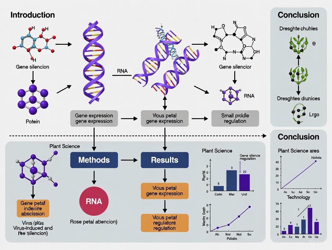
Virus-Induced Gene Silencing (VIGS) is an RNA-mediated reverse genetics technology that has evolved into an indispensable approach for analyzing gene function in plants. It is a powerful form of post-transcriptional gene silencing (PTGS) that enables researchers to transiently knock down targeted gene expression without the need for stable transformation [1]. VIGS utilizes the plant's innate antiviral defense mechanism, where recombinant viral vectors trigger sequence-specific suppression of endogenous plant gene expression, leading to visible phenotypic changes that enable functional characterization of the targeted genes [2]. This technique is particularly valuable for studying non-model organisms and species that are recalcitrant to genetic transformation, including ornamental plants like rose (Rosa hybrida), where it has been successfully applied to study genes involved in critical processes such as petal abscission and coloration [3] [4].
Molecular Mechanism of PTGS in VIGS
The biological foundation of VIGS lies in the plant's post-transcriptional gene silencing machinery, an evolutionarily conserved antiviral defense system [2]. The process initiates when a recombinant viral vector, carrying a fragment of a plant gene of interest, is introduced into the plant tissue. The molecular mechanism unfolds through a highly coordinated sequence of events:
Viral Replication and dsRNA Formation: Following introduction, the viral vector replicates within plant cells. During replication, double-stranded RNA (dsRNA) molecules are formed as replication intermediates or through the activity of host RNA-directed RNA polymerases (RDRPs) that use viral single-stranded RNA as templates [1].
dicer-like Enzyme Cleavage: Cellular Dicer-like enzymes (DCL) recognize and cleave these long dsRNA molecules into small interfering RNA (siRNA) duplexes of 21-24 nucleotides in length [2] [1].
RISC Complex Assembly: These siRNAs are incorporated into an RNA-induced silencing complex (RISC), where the guide strand binds to Argonaute (AGO) proteins, the catalytic core of the complex [1].
Target mRNA Degradation: The RISC complex uses the siRNA as a guide to identify complementary endogenous mRNA sequences. Once bound, the AGO protein catalyzes the cleavage and degradation of the target mRNA, preventing its translation into protein [2] [1].
Systemic Silencing Spread: The silencing signal amplifies and moves systemically throughout the plant, leading to widespread knockdown of the target gene. Secondary siRNAs are produced by host RDRPs using the cleaved mRNA as templates, enhancing silencing maintenance and dissemination [1].
The entire process occurs in the cytoplasm and represents an epigenetic phenomenon that results in sequence-specific degradation of endogenous mRNAs [1]. The diagram below illustrates this coordinated molecular process:
Essential Research Reagents and Materials
Successful implementation of VIGS requires specific biological materials and reagents carefully optimized for the target plant species. The table below details the essential components of a VIGS toolkit, with particular emphasis on rose functional genomics studies:
Table 1: Essential Research Reagent Solutions for VIGS in Rose Functional Genomics
| Reagent/Material | Function/Purpose | Specifications & Examples |
|---|---|---|
| Viral Vector System | Delivers target gene fragment into plant cells to initiate silencing | Tobacco Rattle Virus (TRV) is most common; TRV1 encodes replication proteins, TRV2 contains target gene insert [2] [3] |
| Agrobacterium Strain | Bacterial host for viral vector delivery | GV3101 is widely used for rose and other ornamentals [4] |
| Plant Selection | Experimental material with known growth characteristics | Uniform, healthy rose stems ('Pink Floyd' cultivar used in petal studies) [4] |
| Target Gene Fragment | Specific sequence for silencing | 300-500 bp fragment cloned from gene of interest (e.g., RhILL1 for auxin metabolism) [4] |
| Infiltration Buffer | Medium for Agrobacterium delivery during inoculation | Contains 10 mM MgCl₂, 10 mM MES, and 200 μM acetosyringone [5] |
| Antibiotics | Selective maintenance of bacterial strains | Kanamycin (50 μg/mL) and gentamicin (25 μg/mL) for TRV vectors in Agrobacterium [5] |
| Reference Genes | RT-qPCR normalization for silencing validation | Species-specific stable genes (e.g., RhUBI2 for rose) [4] |
VIGS Experimental Protocol for Rose Petal Abscission Studies
The following section provides a detailed, step-by-step methodology for implementing VIGS in rose petal abscission research, compiled from established protocols in model plants and optimized for rose systems [3] [4].
Vector Construction and Agrobacterium Preparation
Target Sequence Selection and Amplification:
- Identify a 300-500 bp fragment from the coding sequence of the target gene (e.g., genes involved in abscission zone development or hormone response) [4].
- Design gene-specific primers with appropriate restriction sites (e.g., EcoRI and XhoI) for directional cloning [6].
- Amplify the target fragment using PCR with high-fidelity DNA polymerase under the following conditions:
- Initial denaturation: 94°C for 3 minutes
- 35 cycles of: 94°C for 30 seconds, 55-60°C for 30 seconds, 72°C for 45 seconds
- Final extension: 72°C for 5 minutes [6]
Vector Ligation and Transformation:
- Digest both the PCR product and TRV2 vector with appropriate restriction enzymes [6].
- Purify fragments using gel extraction kits and ligate using T4 DNA ligase at 16°C for 16 hours [4].
- Transform ligation product into E. coli competent cells (e.g., DH5α) and select on LB agar with kanamycin (50 μg/mL) [6].
- Verify positive clones by colony PCR and sequencing.
Agrobacterium Preparation:
- Transform verified plasmid into Agrobacterium tumefaciens strain GV3101 [5].
- Plate on LB agar with kanamycin (50 μg/mL) and gentamicin (25 μg/mL), incubate at 28°C for 2 days [5].
- Inoculate single colonies into 5 mL liquid LB with antibiotics, shake overnight at 28°C, 50 rpm.
- Dilute 1:10 in 50 mL fresh LB with antibiotics, 10 mM MES, and 20 μM acetosyringone, grow to OD₆₀₀ = 0.8-1.2 [5].
- Pellet bacteria by centrifugation (3000 × g, 10 minutes) and resuspend in induction buffer (10 mM MES, 10 mM MgCl₂, 200 μM acetosyringone) to OD₆₀₀ = 1.5 [5].
- Incubate at room temperature for 3-4 hours before plant inoculation [5].
Plant Inoculation and Incubation
Plant Material Preparation:
Inoculation Methods:
- Mix TRV1 and TRV2 (with target insert) Agrobacterium cultures in 1:1 ratio [5].
- For vacuum infiltration: Submerge stem ends in Agrobacterium suspension (OD₆₀₀ = 0.5) with 200 μM acetosyringone, apply vacuum (25-30 in Hg) for 30 seconds, slowly release, repeat once [7].
- For friction-osmosis infiltration: Gently abrade cotyledons or stem surfaces with carborundum powder, apply Agrobacterium suspension (OD₆₀₀ = 1.0) with 200 μM acetosyringone using needleless syringe [7].
Post-Inoculation Care:
The experimental workflow from vector construction to phenotypic analysis is summarized in the following diagram:
Efficiency Optimization and Validation
Critical parameters that significantly influence VIGS efficiency must be carefully controlled and optimized for successful gene silencing:
Table 2: Key Optimization Parameters for Efficient VIGS in Rose
| Parameter | Optimal Conditions | Impact on Silencing Efficiency |
|---|---|---|
| Agroinoculum Density | OD₆₀₀ = 0.5-1.0 | Higher OD (0.8-1.2 during growth) improves infection but excessive density causes plant stress [5] [7] |
| Acetosyringone Concentration | 200 μM | Enhances T-DNA transfer; essential for efficient transformation [7] |
| Plant Developmental Stage | Young, actively growing tissue | Meristematic activity promotes systemic spread; 7-10 day seedlings optimal [5] |
| Inoculation Method | Vacuum or friction-osmosis | Rose cuticle presents barrier; vacuum achieves 83% efficiency [7] |
| Environmental Conditions | 22±1°C, 16h light/8h dark | Temperature affects viral replication; 22°C optimal for TRV [4] |
| Incubation Time | 14-21 days | Required for systemic spread and phenotypic development [4] |
Molecular Validation and Phenotypic Analysis
Rigorous validation of gene silencing and comprehensive phenotypic assessment are crucial for reliable data interpretation in VIGS experiments.
Molecular Validation of Silencing:
- Extract total RNA from silenced and control tissues using commercial kits optimized for plant tissues with high polysaccharide and polyphenol content [4].
- Treat with DNase I to remove genomic DNA contamination.
- Synthesize cDNA using reverse transcriptase with oligo(dT) or random hexamer primers.
- Perform quantitative RT-PCR with gene-specific primers and reference genes (e.g., RhUBI2 for rose) [4].
- Calculate relative expression using the 2^(-ΔΔCT) method [4].
- Confirm successful silencing when target gene expression is reduced by ≥70% compared to empty vector controls [7].
Phenotypic Assessment of Petal Abscission:
- Document abscission zone development using stereomicroscopy at regular intervals.
- Quantify petal abscission rates by applying standardized gentle force and recording detachment percentages.
- Measure endogenous phytohormone levels (IAA, ethylene) in abscission zones using HPLC or ELISA.
- Analyze cellular changes in abscission zones using histochemical staining (e.g., toluidine blue for cell wall modifications).
- Assess expression of abscission-related genes (e.g., polygalacturonases, expansins, cellulases) in silenced versus control tissues.
Statistical Analysis:
- Perform experiments with at least three biological replicates, each containing multiple technical replicates [4].
- Apply appropriate statistical tests (t-test, ANOVA) with post-hoc analysis to determine significance (p < 0.05).
- Correlative gene expression and phenotypic data to establish functional relationships.
Applications in Rose Petal Abscission Research
The application of VIGS technology has enabled significant advances in understanding the molecular mechanisms governing rose petal abscission. By selectively silencing genes putatively involved in abscission zone formation, hormone signaling pathways, and cell wall degradation, researchers can directly assess their functional roles in this economically important process. The transient nature of VIGS allows for rapid screening of candidate genes identified through transcriptomic studies of abscission zones, significantly accelerating the functional validation pipeline. Furthermore, the ability to silence genes in specific rose cultivars with varying abscission characteristics facilitates comparative studies to identify key genetic determinants of petal retention. This approach has been successfully implemented in rose to silence the auxin amide hydrolase gene RhILL1, resulting in altered petal coloration through modulation of auxin homeostasis, demonstrating the potential for functional studies in rose petals [4]. When integrated with complementary approaches such as hormone profiling, histological analysis, and advanced imaging techniques, VIGS provides a powerful platform for elucidating the complex regulatory networks controlling rose petal abscission, ultimately contributing to the development of rose varieties with enhanced postharvest characteristics.
The Biological and Commercial Imperative for Studying Rose Petal Abscission
Rose (Rosa hybrida) is one of the most economically important ornamental crops worldwide, with petal abscission significantly determining its postharvest quality and commercial value [8] [9]. The abscission process occurs through a highly regulated sequence of events in a specialized region of cells known as the abscission zone (AZ) [10]. Ethylene-sensitive rose varieties, such as Rosa bourboniana, undergo rapid petal abscission, resulting in a short vase life of just 1-2 days, which drastically reduces their commercial appeal [8]. In contrast, hybrid roses exhibit reduced ethylene sensitivity and delayed abscission [8]. Understanding the molecular mechanisms governing petal abscission is therefore crucial for developing strategies to improve rose longevity. Virus-induced gene silencing (VIGS) has emerged as a powerful functional genomics tool to validate gene functions in this process [3], enabling researchers to dissect the complex hormonal crosstalk and regulatory networks that control petal shedding.
Hormonal Regulation of Petal Abscission
Ethylene and Auxin: A Balancing Act
The initiation and progression of petal abscission are primarily regulated by a delicate balance between ethylene and auxin. Ethylene serves as a potent promoter of abscission, while auxin acts as a key inhibitor of the process [8].
Table 1: Key Hormonal Regulators of Rose Petal Abscission
| Hormone/Regulator | Effect on Abscission | Key Genes/Proteins | Mechanism of Action |
|---|---|---|---|
| Ethylene | Promoter | RhERF1, RhERF4, ACS, ACO | Upregulates cell wall-modifying enzymes; suppresses auxin pathways [8] [10] |
| Auxin | Inhibitor | RhARF7, RhIAA16, RhILL1 | Maintains auxin gradient across AZ; represses abscission-related genes [11] [10] [12] |
| Jasmonic Acid (JA) | Suppressed during abscission | LOX, AOS | Pathway suppression associated with abscission initiation [8] |
| Silver Thiosulfate (STS) | Inhibitor (ethylene blocker) | - | Blocks ethylene perception; delays abscission [10] [13] |
Transcriptomic studies reveal that ethylene-induced abscission is associated with large-scale transcriptional reprogramming, with 8.5% of the AZ transcriptome (3,700 genes) undergoing differential regulation [8]. Ethylene promotes the upregulation of its own biosynthesis and signaling pathway components while simultaneously suppressing auxin, jasmonic acid, and light-regulated pathways [8]. The ethylene-induced abscission process involves upregulation of 1,496 genes and downregulation of 2,199 genes in the petal AZ [9].
Auxin plays a critical protective role against abscission through several mechanisms. The changing auxin gradient across the AZ is a primary determinant of abscission initiation [14]. Research demonstrates that silencing the auxin-related gene RhIAA16 promotes petal abscission, while the auxin response factor RhARF7, in synergy with the sucrose transporter RhSUC2, inhibits ethylene-induced petal abscission [10]. Additionally, the auxin amide hydrolase RhILL1 contributes to petal coloration and may indirectly influence abscission by modulating auxin homeostasis [12].
Reactive Oxygen Species (ROS) in Abscission Signaling
Recent evidence indicates that reactive oxygen species (ROS) function as important signaling molecules in ethylene-mediated petal abscission [13]. Ethylene treatment induces ROS accumulation in both AZ cells and petals by upregulating genes associated with ROS production (RhRHS17, RhRBOHD, RhRBOHC) and suppressing genes involved in ROS scavenging (RhSOD1, RhAPX6.1, RhCATA) [13]. This redox imbalance creates a permissive environment for the activation of cell separation processes in the AZ.
Diagram 1: Ethylene-ROS Signaling Pathway in Petal Abscission. Ethylene promotes ROS accumulation by upregulating production genes and suppressing scavenging genes, leading to cell wall modification and AZ cell separation.
Multi-Omics Insights into Abscission Mechanisms
Integrative multi-omics approaches have provided comprehensive insights into the complex regulatory networks governing petal abscission.
Table 2: Multi-Omics Changes During Rose Petal Abscission
| Analysis Level | Regulated Elements | Key Pathways/Processes Affected | Reference |
|---|---|---|---|
| Transcriptome | 3,695 DEGs (1,496 up, 2,199 down) | Starch/sucrose metabolism, plant hormone signal transduction, phenylpropanoid biosynthesis | [9] |
| Proteome | 715 DEPs (271 up, 444 down) | Extracellular components upregulated; intracellular components downregulated | [9] |
| Ubiquitome | 148 differentially ubiquitinated proteins | Protein degradation, metabolic regulation | [9] |
| Metabolome | 5 key metabolites affected by STS | Shikonin, JA, gluconolactone, stachyose, D-Erythrose 4-phosphate | [10] |
Proteomic and ubiquitomic analyses reveal that during petal abscission, extracellular proteins (including those involved in cell wall modification) are significantly upregulated, while intracellular proteins (particularly those in membrane-bounded organelles) are downregulated [9]. This pattern reflects the active cell wall remodeling occurring in the AZ during the separation process. Additionally, ubiquitination plays a crucial role in targeted protein degradation during abscission, with 139 ubiquitination sites in 100 proteins being upregulated and 55 sites in 48 proteins downregulated [9].
Application Notes: VIGS for Functional Analysis of Abscission Genes
VIGS Protocol for Rose Petal Abscission Studies
Virus-induced gene silencing (VIGS) provides an efficient approach for functional characterization of genes involved in rose petal abscission [3]. The following protocol outlines the key steps for implementing VIGS in rose abscission research:
Materials Required:
- Tobacco rattle virus (TRV)-based vectors (TRV1 and TRV2)
- Agrobacterium tumefaciens strain GV3101
- Rose plants at appropriate developmental stage
- Antibiotics for selection (kanamycin, rifampicin)
- Acetosyringone solution
- Infiltration buffer (10 mM MES, 10 mM MgCl₂, 200 μM acetosyringone)
Methodology:
- Gene Fragment Selection and Cloning: Amplify a 300-500 bp fragment of the target gene and clone it into the TRV2 vector using appropriate restriction enzymes or recombination cloning.
- Agrobacterium Preparation: Transform the recombinant TRV2 construct and the TRV1 helper plasmid into Agrobacterium tumefaciens strain GV3101. Select positive colonies on LB plates containing appropriate antibiotics.
- Agroinfiltration: Grow bacterial cultures to OD₆₀₀ = 1.0-1.5. Harvest cells by centrifugation and resuspend in infiltration buffer. Mix TRV1 and TRV2 cultures in 1:1 ratio and incubate at room temperature for 3-4 hours.
- Plant Inoculation: Infiltrate the bacterial suspension into rose petals or flower buds using a needleless syringe. Alternatively, use the agro-injection method for rose flower buds [15].
- Phenotypic Analysis: Monitor plants for abscission phenotypes after ethylene treatment. Assess silencing efficiency through qRT-PCR and document abscission timing and rate.
Troubleshooting Tips:
- Optimize fragment length and position to maximize silencing efficiency
- Include empty vector (TRV:00) and non-silenced controls
- Validate target gene silencing through qRT-PCR before phenotypic assessment
- For abscission studies, apply ethylene treatment after confirming gene silencing
Auxin Immunolocalization Protocol
Precise localization of auxin in the rose petal AZ provides critical insights into its role in abscission regulation [14]. The following protocol enables spatial visualization of auxin distribution:
Materials:
- Rose petal AZ tissues at different developmental stages
- Anti-IAA antibodies
- Fixation solution (4% paraformaldehyde in PBS)
- Permeabilization buffer (PBS with 0.1% Triton X-100)
- Blocking solution (1% BSA in PBS)
- Secondary antibodies conjugated to fluorescent dyes
- Mounting medium with DAPI
Methodology:
- Tissue Fixation: Collect AZ tissues and fix immediately in 4% paraformaldehyde for 4-6 hours at 4°C.
- Tissue Sectioning: Embed fixed tissues in paraffin and section to 8-10 μm thickness using a microtome.
- Immunostaining:
- Deparaffinize and rehydrate sections through graded ethanol series
- Perform antigen retrieval using citrate buffer (pH 6.0)
- Permeabilize with 0.1% Triton X-100 for 15 minutes
- Block with 1% BSA for 1 hour at room temperature
- Incubate with primary anti-IAA antibodies overnight at 4°C
- Incubate with fluorescent-conjugated secondary antibodies for 1-2 hours
- Visualization: Mount sections with anti-fade mounting medium and image using confocal microscopy.
This protocol can be combined with plant hormone metabolomics to comprehensively reflect auxin changes during abscission [14].
Abscission Zone Isolation Technique
Isolation of high-quality AZ tissue is challenging due to its small size and susceptibility to stress artifacts. The following optimized protocol facilitates AZ sampling for molecular analyses [15]:
Key Considerations:
- Perform dissections rapidly to minimize wound and dehydration stress
- Use sharp, fine forceps and scalpels for precise excision
- Immediately freeze excised AZ tissues in liquid nitrogen
- Pool multiple AZs to obtain sufficient material for RNA/protein extraction
Diagram 2: Experimental Workflow for Rose Petal Abscission Studies. Integrated approach combining tissue isolation, VIGS, and physiological analysis.
Research Reagent Solutions
Table 3: Essential Research Reagents for Rose Petal Abscission Studies
| Reagent/Tool | Function/Application | Example Use in Abscission Research |
|---|---|---|
| TRV VIGS Vectors | Gene functional analysis | Silencing candidate abscission genes to assess function [3] |
| Anti-IAA Antibodies | Auxin immunolocalization | Visualizing auxin distribution in AZ tissues [14] |
| Silver Thiosulfate (STS) | Ethylene action inhibitor | Blocking ethylene responses to delay abscission [10] [13] |
| Ethephon | Ethylene-releasing compound | Inducing synchronous abscission for experimental studies [10] [13] |
| AZ-Specific Promoters | Tissue-specific expression | Driving gene expression specifically in abscission zones [15] |
The study of rose petal abscission represents both a biological imperative for understanding fundamental plant developmental processes and a commercial necessity for improving the postharvest quality of ornamental crops. The integration of VIGS-based functional genomics with multi-omics approaches has significantly advanced our understanding of the complex regulatory networks governing abscission. Key challenges remain in translating this knowledge into practical applications for the floriculture industry, particularly in developing non-transgenic strategies to modulate abscission. Future research should focus on identifying master regulatory genes that could serve as targets for breeding programs aimed at extending rose vase life without compromising other ornamental qualities. The methodological approaches outlined in this article provide a framework for systematic investigation of abscission processes not only in roses but also in other economically important plant species.
Virus-Induced Gene Silencing (VIGS) is a powerful reverse genetics technique that leverages the plant's innate RNA-based antiviral defense system to down-regulate endogenous genes. The application of VIGS in roses provides a rapid, high-throughput method for functional genomics, enabling researchers to elucidate gene functions without the need for stable transformation. This is particularly valuable for studying complex processes such as petal abscission, a critical area of research for improving the postharvest quality and ornamental value of roses. The Tobacco Rattle Virus (TRV) has emerged as a particularly versatile and efficient vector for VIGS in roses, capable of systemic movement and inducing effective silencing in various tissues. This Application Note details the methodologies and protocols for implementing TRV-VIGS in rose, with a specific focus on its application in studying genes involved in petal abscission.
The TRV-VIGS Mechanism and System Components
The fundamental mechanism of TRV-VIGS begins with the delivery of a modified TRV vector containing a fragment of the target plant gene into plant cells, often via Agrobacterium-mediated transformation (agroinfiltration). Inside the plant cell, the viral RNA is replicated, leading to the formation of double-stranded RNA (dsRNA), a key trigger in the silencing pathway. This dsRNA is recognized and cleaved by the plant's Dicer-like (DCL) enzymes into small interfering RNAs (siRNAs). These siRNAs are then incorporated into the RNA-induced silencing complex (RISC), which uses them as a guide to identify and cleave complementary endogenous mRNA transcripts, such as those involved in petal abscission, thereby preventing their translation into protein and resulting in a silenced phenotype [16] [17].
The standard TRV-VIGS system is a two-component system, with each component housed in a separate binary vector [16]:
- pTRV1 (RNA1): Encodes genes for viral replication and movement (e.g., 134K/194K replicases, movement protein, and a 16K cysteine-rich protein).
- pTRV2 (RNA2): Typically modified to carry the coat protein (CP) and a multiple cloning site (MCS) for inserting a fragment of the target plant gene. The Rose AGAMOUS homolog (RhAG) or a petal abscission-related gene fragment would be cloned into this site [3] [18].
The following diagram illustrates the molecular workflow of the TRV-VIGS mechanism and the structure of the essential vectors.
Detailed Protocol for TRV-VIGS in Rose
Vector Construction and Preparation
- Target Gene Fragment Selection: Identify and select a 300-500 base pair (bp) cDNA fragment unique to the target gene (e.g., an abscission-related gene or RhAG). Avoid regions of high homology with other genes to ensure silencing specificity and homopolymeric regions [16] [17].
- Cloning into pTRV2: Insert the selected fragment into the Multiple Cloning Site (MCS) of the pTRV2 vector. The use of GATEWAY recombination cloning (e.g., pTRV2 vectors with attR1 and attR2 sites) can significantly streamline this process [16].
- Control Vectors:
- Positive Control: pTRV2-PDS (Phytoene desaturase) to induce photobleaching, validating the system's efficiency [17].
- Empty Vector Control: A pTRV2 with no insert. However, this can sometimes cause severe viral symptoms like necrosis and stunting. A superior alternative is pTRV2-sGFP, containing a fragment of the green fluorescent protein gene, which minimizes viral symptoms while serving as an effective negative control [17].
- Agrobacterium Preparation: Transform the recombinant pTRV2 and the helper pTRV1 vectors into separate Agrobacterium tumefaciens strains (e.g., GV3101). Grow individual colonies in LB broth with appropriate antibiotics. Resuspend the bacterial pellets in an induction medium (e.g., containing 10 mM MES, 10 mM MgCl₂, and 200 μM acetosyringone) and incubate for 3-4 hours at room temperature before mixing the pTRV1 and pTRV2 cultures in a 1:1 ratio for inoculation [17].
Plant Inoculation
The inoculation method is critical for success. For roses, mechanical inoculation of wounded shoot apical meristems has been demonstrated to induce the most effective and consistent silencing compared to other methods like leaf agroinfiltration [17].
Protocol: Apical Meristem Inoculation
- Plant Material: Use rose plants at 3-4 weeks after sowing. Younger plants generally show higher silencing efficiency [17].
- Wounding: Gently wound the shoot apical meristem and the youngest visible leaves using a sterile needle or scalpel.
- Inoculation: Apply approximately 10-20 μL of the prepared Agrobacterium mixture directly onto the wounded meristem.
- Post-Inoculation Care: Maintain inoculated plants in high humidity conditions (e.g., under a transparent cover) for 2-3 days to facilitate infection.
Post-Inoculation Conditions and Phenotyping
- Growing Temperature: Maintain plants at optimized temperatures. Studies in related species like petunia have shown that 20°C day/18°C night temperatures induce stronger gene silencing compared to higher temperatures [17]. Furthermore, temperature is a key environmental factor influencing flower development; low temperatures (15/5 °C) have been shown to increase petal number in rose by suppressing RhAG expression, highlighting the importance of temperature control in experiments studying floral organ identity and abscission [18].
- Phenotype Monitoring: Silencing phenotypes, such as altered petal abscission rates or changes in floral organ identity, typically appear 3-4 weeks post-inoculation.
- Validation: Use molecular techniques like quantitative RT-PCR to confirm the down-regulation of the target gene transcript levels in treated tissues compared to controls.
Key Experimental Parameters for Optimization
Successful application of TRV-VIGS depends on several factors. The following table summarizes key parameters that require optimization for efficient gene silencing in rose, drawing from optimizations in rose and related species.
Table 1: Key Experimental Parameters for Optimizing TRV-VIGS in Rose
| Parameter | Recommendation & Impact | Biological Rationale & Evidence |
|---|---|---|
| Inoculation Method | Mechanical wounding of apical meristem [17] | Ensures direct viral entry into the actively dividing cells, promoting rapid and systemic spread throughout the plant. |
| Plant Age | 3-4 weeks after sowing [17] | Younger plants may have less developed defense responses and more active meristems, facilitating higher viral replication and movement. |
| Growing Temperature | 20°C day / 18°C night (based on Petunia optimization) [17] | Cooler temperatures may slow plant defense responses, thereby promoting viral spread and enhancing silencing efficiency. |
| Cultivar Selection | Varies; testing of multiple rose cultivars is recommended. | Silencing efficiency is genotype-dependent. In petunia, 'Picobella Blue' showed 1.8-fold higher silencing efficiency than other cultivars [17]. |
| Control Construct | pTRV2-sGFP (superior to empty pTRV2) [17] | The presence of a non-plant insert like GFP in the viral vector minimizes severe viral symptoms (necrosis, stunting) often caused by the empty vector, leading to healthier control plants. |
Application in Rose Petal Abscission and Flower Development
The TRV-VIGS system has been successfully applied to study genes controlling important traits in rose. A prime example is the functional analysis of RhAG, a C-class floral organ identity gene homologous to AGAMOUS from Arabidopsis.
- Role of RhAG: RhAG is a key regulator of stamen and carpel development. Its suppression leads to the conversion of stamens into petals, resulting in flowers with increased petal number (double flowers) [18].
- VIGS Validation: Silencing RhAG in rose using TRV-VIGS significantly increased petal number by promoting stamen petaloidy, effectively mimicking the natural double-flower phenotype and confirming the gene's function [18].
- Link to Abscission & Environment: Research has shown that low ambient temperatures can increase petal number by attenuating RhAG expression. This suppression is linked to DNA hypermethylation of the RhAG promoter, revealing an epigenetic mechanism for environmental regulation of flower development [18]. This makes TRV-VIGS an indispensable tool for dissecting the genetic and environmental pathways, including those involving DNA methylation, that control floral organ identity and the subsequent processes of petal abscission.
The experimental workflow for conducting such a study, from target selection to analysis, is outlined below.
The Scientist's Toolkit: Research Reagent Solutions
Table 2: Essential Research Reagents for TRV-VIGS in Rose
| Reagent / Material | Function & Application in Protocol |
|---|---|
| pTRV1 & pTRV2 Vectors | The core binary vector system for viral RNA replication (pTRV1) and delivery of the target gene insert (pTRV2) [16]. |
| pTRV2-sGFP Control Vector | A critical control vector containing a non-plant insert to minimize confounding viral symptoms in control plants [17]. |
| Agrobacterium tumefaciens (GV3101) | Bacterial strain used for the delivery of TRV vectors into plant cells via agroinfiltration or meristem inoculation. |
| Acetosyringone | A phenolic compound added to the Agrobacterium induction medium to activate the Vir genes, essential for efficient T-DNA transfer. |
| Shoot Apical Meristem | The primary site of inoculation for achieving high-efficiency, systemic silencing in rose and other plants [17]. |
The TRV-based VIGS system provides a robust and versatile platform for functional genomics in rose. Its ability to rapidly silence target genes, such as those involved in petal abscission and floral organ development like RhAG, offers unparalleled insights into gene function. By adhering to the optimized protocols detailed herein—including apical meristem inoculation, use of appropriate controls like pTRV2-sGFP, and maintenance of specific growth conditions—researchers can reliably employ this technology to accelerate the identification and characterization of genes underlying economically important traits in rose.
Plant organ abscission is a highly regulated developmental process essential for plant survival and reproductive success. It facilitates the detachment of organs such as petals, leaves, and fruits through the formation and activation of a specialized abscission zone (AZ) [19]. The timing of abscission significantly impacts the ornamental and economic value of horticultural species, particularly in cut flowers like roses and peonies [20] [19]. The initiation and progression of abscission are coordinated by complex interactions between phytohormones, primarily ethylene, auxin, and cytokinin [20] [21]. While auxin acts as a potent suppressor of abscission, ethylene is a key promoter of the process [19] [21]. Cytokinins also contribute, though their role appears more context-dependent [21] [22]. Understanding the crosstalk between these signaling pathways provides crucial insights for manipulating abscission in agricultural and horticultural contexts, particularly when employing reverse-genetic tools like Virus-Induced Gene Silencing (VIGS) for functional characterization of abscission-related genes.
Quantitative Data on Hormonal Regulation
Research across species has quantified the specific effects of hormonal perturbations on abscission-related phenotypes. The following tables consolidate key experimental findings.
Table 1: Effect of Modifying Hormone-Related Gene Expression on Abscission and Longevity
| Plant Species | Gene Manipulated | Experimental Approach | Effect on Longevity/Abscission | Key Hormonal Changes |
|---|---|---|---|---|
| Petunia [23] | PhHD-Zip (HD-Zip I TF) |
VIGS | Flower longevity increased by ~20% (9.7 days vs. 8.2-8.4 days in controls) | Ethylene production reduced; ACS, ACO, NCED transcripts decreased |
| Petunia [23] | PhHD-Zip (HD-Zip I TF) |
Over-expression | Accelerated flower senescence | N/D |
| Rose [20] | RhIAA16 (Aux/IAA) |
VIGS | Promoted petal abscission | N/D |
| Itoh Peony [19] | IpAUX1 (Auxin Influx Carrier) |
VIGS | Accelerated petal abscission | IAA content in AZ decreased; ACS, ACO expression increased |
| Itoh Peony [19] | IpAUX1 (Auxin Influx Carrier) |
Transient Overexpression | Delayed petal abscission | IAA content in AZ increased; ACS, ACO expression suppressed |
Table 2: Hormone and Stress-Induced Changes in Gene Expression
| Inducing Signal | Plant Species | Target Gene/Pathway | Observed Effect | Citation |
|---|---|---|---|---|
| Ethylene | Petunia | PhHD-Zip |
Transcript abundance induced | [23] |
| Abscisic Acid (ABA) | Petunia | PhHD-Zip |
Transcript abundance induced | [23] |
| Abiotic Stress (Drought, NaCl, Cold) | Petunia | PhHD-Zip |
Transcript abundance induced | [23] |
| Auxin Deficiency / Ethylene | Peony | ACS, ACO (Ethylene biosynthesis) |
Gene expression increased | [19] |
| Auxin (via IpAUX1) | Peony | ACS, ACO (Ethylene biosynthesis) |
Gene expression suppressed | [19] |
Core Signaling Pathways and Molecular Mechanisms
The Ethylene-Promoted Abscission Pathway
Ethylene serves as a primary promoter of abscission across diverse species, including rose, tomato, Arabidopsis, and peony [19] [21]. Its role is intrinsically linked to a decline in auxin levels within the abscission zone. The regulatory cascade involves:
- Ethylene Biosynthesis Genes: The expression of 1-aminocyclopropane-1-carboxylate (ACC) synthase (ACS) and ACC oxidase (ACO) genes is upregulated, leading to a climacteric rise in ethylene production that triggers the abscission process [19].
- Transcriptional Reprogramming: Ethylene signaling activates transcription factors that execute the senescence program. In petunia, the homeodomain-leucine zipper transcription factor PhHD-Zip is upregulated by ethylene and is necessary for senescence; its silencing extends flower longevity by 20% and drastically reduces the expression of ethylene (
ACS,ACO) and ABA (NCED) biosynthesis genes, as well as senescence-associated genes (SAG12,SAG29) [23]. - Cell Wall Degradation: Ethylene promotes the weakening of cell walls within the AZ, a prerequisite for organ separation [22].
The Auxin-Inhibited Abscission Pathway
Auxin functions as a powerful negative regulator of abscission. A continuous, stable flow of auxin through the abscission zone is required to maintain its insensitivity to ethylene [19] [21]. The molecular mechanism involves:
- Auxin Transport: Polar auxin transport, mediated by influx (AUX/LAX) and efflux (PIN) carriers, is critical for maintaining auxin flux. In Itoh peony, the auxin influx carrier IpAUX1 plays a vital role; its silencing accelerates abscission, while its overexpression delays it [19].
- Auxin Signaling and Gene Repression: High auxin levels promote the degradation of Aux/IAA transcriptional repressors (e.g., RhIAA16 in rose), freeing Auxin Response Factors (ARFs) to activate genes that suppress the abscission program [20] [19]. Silencing
RhIAA16leads to premature petal abscission, demonstrating its role as a repressor of the process [20]. - Suppression of Ethylene Biosynthesis: Auxin signaling directly or indirectly suppresses the expression of key ethylene biosynthesis genes (
ACS,ACO), thereby inhibiting the ethylene-triggered pathway [19].
Cytokinin and Hormonal Crosstalk
The role of cytokinin in abscission is complex and appears to be integrated with other hormonal pathways rather than acting as a primary regulator.
- Interaction with Auxin and Ethylene: Cytokinin can regulate the expression of auxin transporters like
AUX1, suggesting a point of crosstalk between the two hormones [19]. Cytokinin accumulation is also influenced by auxin and strigolactones [24]. - Delay of Senescence: Cytokinins are known to delay leaf senescence (the Richmond-Lang effect) and can promote nutrient mobilization [22]. This antagonistic relationship with senescence-promoting hormones like ethylene may indirectly influence abscission.
- Integration via Brassinosteroid Signaling: Recent research in tomato shoot branching reveals that cytokinin signaling can promote brassinosteroid (BR) synthesis, which in turn suppresses the expression of
BRANCHED1 (BRC1), a central integrator that inhibits bud outgrowth [24]. This illustrates how cytokinin signals can be relayed through other hormone pathways to influence developmental decisions.
Experimental Protocols for VIGS-Based Abscission Research
Protocol 1: VIGS in Rose or Peony for Petal Abscission Studies
VIGS is a powerful reverse-genetics tool for rapid functional analysis of genes in non-model plants like roses and peonies [20] [19].
Materials:
- Plant Material: Rooted plantlets of a VIGS-responsive cultivar (e.g., Rosa hybrida 'Samantha' for rose, Itoh peony 'Bartzella' for peony) [20] [19].
- Agrobacterium Strain: GV3101.
- VIGS Vectors: TRV-based vectors (e.g., pTRV1, pTRV2).
- Cloning Reagents: Restriction enzymes, T4 DNA ligase, or gateway BP/LR clonase.
- Growth Media: LB broth and agar with appropriate antibiotics.
- Infiltration Buffer: 10 mM MES, 10 mM MgCl₂, 200 µM acetosyringone, pH 5.6.
Method:
- Gene Fragment Cloning: Clone a ~200-300 bp gene-specific fragment of the target gene (e.g.,
RhIAA16,IpAUX1) into the pTRV2 silencing vector [20] [19]. - Agrobacterium Preparation:
- Transform the constructed pTRV2 and the helper plasmid pTRV1 into Agrobacterium strain GV3101.
- Inoculate single colonies in 5 mL LB with antibiotics and incubate at 28°C for 24 hours.
- Subculture 1 mL into 50 mL of fresh medium and grow to an OD₆₀₀ of 1.0-1.5.
- Pellet cells and resuspend in infiltration buffer to a final OD₆₀₀ of 2.0. Incubate at room temperature for 3-4 hours.
- Plant Infiltration:
- Mix the pTRV1 and pTRV2 (with insert) agrobacterium suspensions in a 1:1 ratio.
- Using a syringe without a needle, infiltrate the mixture into the abaxial side of young leaves or petals of the target plant [20].
- Post-Infiltration Care:
- Maintain infiltrated plants in a growth chamber at 18-22°C with high humidity for 2-3 days to facilitate viral propagation and silencing.
- Subsequently, transfer plants to standard growth conditions.
- Phenotypic Analysis:
- Silencing Efficiency: After 3-4 weeks, assess silencing by quantifying target gene transcript levels in the petal AZ using RT-qPCR.
- Abscission Monitoring: Record the timing of petal abscission in silenced plants compared to empty vector (TRV2) and wild-type controls. Document abscission rates over time.
Protocol 2: Quantifying Hormonal and Molecular Markers
To correlate phenotypic changes with molecular events, quantify key hormonal and gene expression markers.
Materials:
- Tissue from the Abscission Zone (AZ)
- Liquid Nitrogen
- RNA extraction kit (e.g., TRIzol)
- cDNA synthesis kit
- Real-Time PCR system and reagents
- ELISA kits or LC-MS equipment for hormone quantification
Method:
- Tissue Sampling: Precisely dissect the AZ (base of petal and adjacent receptacle tissue, <1 mm) from experimental and control plants at defined developmental stages [20]. Flash-freeze in liquid nitrogen.
- RNA Extraction and Gene Expression:
- Extract total RNA from the AZ tissue.
- Synthesize cDNA and perform RT-qPCR using gene-specific primers.
- Analyze the expression of:
- Normalize data using reference genes (e.g.,
Actin,Ubiquitin).
- Hormone Quantification:
- Ethylene Production: Place detached flowers or AZ tissues in a sealed container. Withdraw a gas sample and measure ethylene concentration using Gas Chromatography (GC) [23].
- Auxin (IAA) Levels: Grind AZ tissue to a fine powder. Extract IAA and quantify using Enzyme-Linked Immunosorbent Assay (ELISA) or the more sensitive Liquid Chromatography-Mass Spectrometry (LC-MS) [19].
The Scientist's Toolkit: Key Research Reagents
Table 3: Essential Reagents for Abscission Research
| Reagent / Material | Function / Application | Example Use |
|---|---|---|
| TRV VIGS Vectors (pTRV1, pTRV2) | RNA virus-based system for transient gene silencing in plants. | Functional analysis of RhIAA16 in rose and IpAUX1 in peony petal abscission [20] [19]. |
| Agrobacterium tumefaciens (GV3101) | Delivery vehicle for introducing TRV vectors into plant cells. | Used in the infiltration step of the VIGS protocol [20] [19]. |
| Ethylene Precursor (e.g., Ethephon) | Ethylene-releasing compound used to induce or accelerate abscission. | Experimental treatment to study ethylene-responsive genes and phenotypes [22]. |
| Ethylene Inhibitors (1-MCP, STS) | Compounds that block ethylene perception or action. | Used to test ethylene dependency of abscission processes [23] [21]. |
| Auxin Transport Inhibitors (NPA, TIBA) | Inhibitors of polar auxin transport. | Used to disrupt auxin flow through the AZ, sensitizing it to abscission signals [19]. |
| Gene-Specific Primers for RT-qPCR | Oligonucleotides for quantifying transcript abundance of target genes. | Assessing silencing efficiency and expression of pathway genes like ACS, ACO, SAGs [23] [19]. |
A Step-by-Step VIGS Protocol for Targeted Gene Silencing in Rose
Within the framework of a thesis investigating rose petal abscission, the construction of a Tobacco Rattle Virus (TRV)-based vector for Virus-Induced Gene Silencing (VIGS) is a critical first step. This transient reverse genetics tool allows for the rapid functional validation of genes hypothesized to regulate abscission, such as those involved in auxin and ethylene signaling pathways [3] [19]. By cloning a fragment of a target rose gene into the TRV2 vector, researchers can silence the gene in planta and observe the subsequent effect on petal abscission, thereby elucidating its function. This application note details the protocols for inserting target sequences into the TRV2 backbone, a prerequisite for initiating VIGS studies.
The TRV system is bipartite, requiring two plasmid vectors: TRV1, which contains the genes for viral replication and movement, and TRV2, which is modified to carry the target gene fragment [2] [16]. The process of cloning into TRV2 involves selecting a unique fragment of the target gene and inserting it into the multiple cloning site (MCS) of the TRV2 plasmid using molecular cloning techniques. The resulting recombinant TRV2 plasmid is then transformed into Agrobacterium tumefaciens, which serves as the delivery vehicle for infecting plant tissues [6] [25].
The following diagram illustrates the core workflow for constructing the VIGS vector and its mechanism of action in a rose plant.
Cloning Methodologies
Several established methods can be used to clone a target fragment into the TRV2 vector. The choice of method depends on available resources, desired throughput, and laboratory preference. The table below summarizes the key characteristics of three common techniques.
Table 1: Comparison of TRV2 Cloning Methods
| Cloning Method | Principle | Key Features | Silencing Efficiency | Primary Application |
|---|---|---|---|---|
| Restriction Enzyme (RE) & Ligation [16] | Uses restriction enzymes to open the vector and ligase to insert the fragment. | - Requires specific restriction sites.- Can be time-consuming.- Lower efficiency for high-throughput work. | ~65-95% (system-dependent) [6] | Low-throughput, single-gene studies. |
| Gateway Recombination [26] [16] | Uses site-specific recombination (attB/attP sites) to transfer the fragment. | - High cloning efficiency.- Requires proprietary enzymes (costly).- Ideal for high-throughput screening. | ~65-95% (system-dependent) [6] | High-throughput functional genomics. |
| Ligation-Independent Cloning (LIC) [26] | Uses T4 DNA polymerase to create complementary overhangs on the vector and insert. | - No requirement for restriction enzymes or ligase.- 100% efficiency in obtaining correct clones reported [26].- Cost-effective for high-throughput. | ~65-95% (system-dependent) [6] | High-throughput, cost-sensitive projects. |
Protocol: Ligation-Independent Cloning (LIC) into TRV2
The LIC method is highly efficient and avoids the cost of proprietary enzymes [26]. The following protocol is adapted from Dong et al. (2007) and can be applied to clone fragments of genes involved in rose petal abscission.
Reagents and Materials:
- pTRV2-LIC vector (e.g., pYY13 [26])
- Proof-reading DNA polymerase
- T4 DNA polymerase
- dATP and dTTP nucleotides
- PCR purification kit
- Chemically competent E. coli cells
Procedure:
- Vector Preparation:
- Linearize the pTRV2-LIC vector by digesting with PstI restriction enzyme.
- Purify the linearized vector.
- Treat the linearized vector with T4 DNA polymerase in the presence of dTTP only. This creates vector with 5' single-stranded overhangs.
Insert Preparation:
- Design gene-specific primers with added 5' extensions that are complementary to the LIC adaptor sequences in the vector.
- Amplify the target ~200-500 bp fragment from rose cDNA using a proof-reading polymerase.
- Purify the PCR product.
- Treat the PCR product with T4 DNA polymerase in the presence of dATP only. This creates inserts with complementary 5' single-stranded overhangs.
Annealing and Transformation:
- Mix the treated vector and insert fragments and incubate to allow annealing via the complementary overhangs.
- Transform the annealed product into competent E. coli cells and plate on selective media.
- Screen colonies by colony PCR or sequencing to confirm the presence of the insert.
Protocol: Restriction Enzyme/Enzymatic Assembly Cloning
This is a common method suitable for labs without specialized LIC vectors. The example below is based on a recent protocol developed for soybean [6].
Reagents and Materials:
- pTRV2 vector (e.g., pTRV2-GFP [6])
- Restriction Enzymes (e.g., EcoRI and XhoI [6])
- T4 DNA Ligase or a DNA Assembly Master Mix
- Chemically competent E. coli cells
Procedure:
- Vector Digestion:
- Digest the pTRV2 vector with the selected restriction enzymes (e.g., EcoRI and XhoI) to create a linearized, cohesive-ended vector.
- Dephosphorylate the vector to prevent self-ligation.
- Gel-purify the digested vector fragment.
Insert Preparation:
- Design primers to amplify the target gene fragment. Add the appropriate restriction enzyme sites (e.g., EcoRI and XhoI) to the 5' ends of the forward and reverse primers, respectively.
- Amplify the fragment from rose cDNA using a high-fidelity PCR mix.
- Digest the PCR product with the same restriction enzymes to create compatible ends.
- Gel-purify the digested insert.
Ligation/Assembly:
- Option A (Ligation): Mix the digested vector and insert in a molar ratio (e.g., 1:3) with T4 DNA Ligase and buffer. Incubate.
- Option B (Enzymatic Assembly): Use a commercial DNA assembly master mix according to the manufacturer's instructions, which can offer higher efficiency and is more tolerant of complex cloning strategies.
- Transform the ligation/assembly reaction into competent E. coli cells. Select positive clones on kanamycin-containing media and verify by colony PCR and sequencing.
Experimental Workflow for Rose Petal Abscission Studies
After successful cloning and Agrobacterium transformation, the following integrated workflow is implemented to study gene function in rose petal abscission.
The Scientist's Toolkit: Research Reagent Solutions
Table 2: Essential Reagents for TRV-VIGS Vector Construction
| Reagent / Material | Function / Description | Example Use Case |
|---|---|---|
| pTRV1 Vector [16] | Encodes viral proteins for replication and movement. Essential component of the bipartite TRV system. | Co-delivered with pTRV2-Target during Agrobacterium inoculation to enable viral infection and spread. |
| pTRV2 Empty Backbone [16] | The recipient plasmid for the target gene fragment. Contains viral elements and a multiple cloning site (MCS). | Used as the starting material for all cloning procedures described in Section 3. |
| Phytoene Desaturase (PDS) | A visual marker gene for VIGS. Silencing causes photobleaching, confirming system functionality. | Cloned into TRV2 (TRV2-PDS) as a positive control to optimize inoculation and monitor silencing spread [6] [27]. |
| Agrobacterium tumefaciens GV3101 | A disarmed strain used for plant transformation. Delivers the T-DNA containing TRV vectors into plant cells. | The standard host for mobilizing pTRV1 and recombinant pTRV2 plasmids for plant infection [6] [27]. |
| Gateway BP Clonase | Enzyme mix for in vitro recombination, facilitating rapid transfer of a PCR product into a Gateway-compatible TRV2 vector. | Used in Gateway cloning for high-throughput construction of silencing vectors [26] [16]. |
Agrobacterium Preparation and Strain Selection for Efficient Transformation
Within the context of studying rose petal abscission using Virus-Induced Gene Silencing (VIGS), the selection and preparation of optimal Agrobacterium tumefaciens strains are critical foundational steps. Efficient genetic transformation enables researchers to unravel molecular mechanisms, such as the roles of reactive oxygen species and ethylene signaling in petal abscission [13]. The tobacco rattle virus (TRV)-based VIGS system, delivered via Agrobacterium, provides a powerful reverse genetics tool for functional gene validation in roses [3]. This protocol details evidence-based methodologies for strain selection and preparation to achieve high-efficiency transformation, specifically framed within VIGS applications for rose petal abscission research.
Strain Selection Criteria and Comparison
Selecting an appropriate Agrobacterium strain significantly impacts transformation efficiency. Different strains exhibit varying levels of virulence, host range compatibility, and suitability for plant species such as roses.
Table 1: Agrobacterium tumefaciens Strains for Plant Transformation
| Strain | Genetic Background | Key Features | Optimal Applications | Transformation Efficiency |
|---|---|---|---|---|
| AGL1 [28] | C58 derivative | Hypervirient strain, recA mutant, carbenicillin resistant | High-efficiency transformation of suspension cells [28] | Near 100% in Arabidopsis suspension cells [28] |
| GV3101 [29] [30] | C58 derivative | Disarmed strain, widely used for transient transformation, good for Arabidopsis and sunflower | Agroinfiltration, transient expression, VIGS [29] [30] | >90% in sunflower transient transformation [29] |
| EHA105 [31] | A281 derivative | Disarmed version of hypervirulent strain EHA101, kanamycin resistant | Monocot and dicot transformation, compatible with ternary systems [31] | High in maize with ternary vectors [31] |
| LBA4404 [31] | Ach5 derivative | Disarmed strain, spectinomycin resistant, classic for monocots | Stable transformation, especially in cereals [31] | Moderate, improved with ternary helpers [31] |
| AGL1 Thy- [31] | C58 derivative | Thymidine auxotroph, reduces overgrowth post-co-cultivation | Species with overgrowth issues, enhances biosafety [31] | Comparable to parental strain with cleaner recovery [31] |
For VIGS studies in roses, research indicates that the tobacco rattle virus (TRV) system is effective for validating gene functions in petal abscission [3]. Strain GV3101 has demonstrated high efficiency in transient transformation and is widely used for VIGS approaches [30].
Agrobacterium Transformation Protocols
Freeze-Thaw Transformation (High-Throughput Method)
This miniaturized protocol enables efficient transformation suitable for automated, high-throughput workflows [30].
Materials:
- Agrobacterium strain (e.g., GV3101)
- LB medium and agar plates with appropriate antibiotics
- Plasmid DNA (200 ng/μL concentration)
- 200 μL PCR tube strips or 96-well plates
- Liquid nitrogen
- Thermal cycler
Procedure:
- Prepare Competent Cells: Pick a single colony and inoculate 10 mL LB medium. Grow overnight at 28°C with shaking.
- Harvest Cells: Centrifuge at 3,500 × g for 10 minutes. Resuspend pellet in 1 mL of cold 20 mM CaCl₂ solution (10x concentration).
- Aliquot: Dispense 50 μL aliquots into PCR tubes or 96-well plates. Store at -80°C for up to several months.
- Transform: Add 2 μL plasmid DNA (~200 ng) to thawed competent cells. Flash-freeze in liquid nitrogen for 10 seconds.
- Heat Shock: Transfer to thermal cycler programmed for 5 minutes at 37°C, then 60 minutes at 28°C.
- Plate: Spread transformed cells directly onto six-well plates containing LB agar with appropriate antibiotics.
- Incubate: Grow at 28°C for 2-3 days until colonies appear [30].
Transformation efficiency averages 8 × 10³ CFU/μg DNA, sufficient for most experimental applications [30]. This method reduces reagent volumes and enables automation using platforms like Opentrons OT-2.
Electroporation Method
Electroporation typically yields higher transformation efficiency than chemical methods.
Materials:
- Electrocompetent Agrobacterium cells
- Electroporator and cuvettes (2 mm gap)
- SOC recovery medium
- Plasmid DNA (100-500 ng)
Procedure:
- Prepare Cells: Thaw electrocompetent cells on ice.
- Electroporate: Mix 50 μL cells with 1 μL plasmid DNA. Transfer to pre-chilled cuvette. Apply pulse (2.5 kV, 25 μF, 400-600 Ω).
- Recover: Immediately add 1 mL SOC medium. Transfer to tube and incubate at 28°C for 2-4 hours with shaking.
- Plate: Spread 100-200 μL on selective plates. Incubate at 28°C for 2-3 days [28].
Culture Preparation for Plant Transformation
Proper culture preparation ensures optimal bacterial viability and T-DNA transfer efficiency.
Pre-culture and Main Culture Preparation:
- Inoculation: Pick a verified colony from freshly transformed plates. Inoculate 5-10 mL of YEB or LB medium with appropriate antibiotics.
- Incubation: Grow at 28°C with shaking at 160-200 rpm for 20-24 hours until OD600 reaches 1.0-2.0 [28].
- Main Culture: Dilute pre-culture to OD600 = 0.2 in AB-MES medium (for virulence induction) containing antibiotics and 200 μM acetosyringone.
- Induction: Grow main culture at 28°C with shaking for 16-20 hours until OD600 reaches 0.3-0.5 [28].
Induction Medium Composition:
- AB-MES medium: 17.2 mM K₂HPO₄, 8.3 mM NaH₂PO₄, 18.7 mM NH₄Cl, 2 mM KCl, 1.25 mM MgSO₄, 100 μM CaCl₂, 10 μM FeSO₄, 50 mM MES, 20 g/L glucose, pH 5.5 [28]
Harvest and Resuspension:
- Pellet: Centrifuge bacterial culture at 6,800 × g for 10 minutes.
- Resuspend: Resuspend pellet in infiltration medium (ABM-MS: 50% AB-MES, 1.1 g/L MS basal salts, 0.25% sucrose, pH 5.5) to final OD600 = 0.8 [28].
- Additives: Include 200 μM acetosyringone and 0.05% Pluronic F68 or 0.02% Silwet L-77 surfactant for enhanced efficiency [28] [29].
Table 2: Key Additives for Enhanced Transformation Efficiency
| Additive | Concentration | Function | Application |
|---|---|---|---|
| Acetosyringone [28] | 200 μM | Vir gene inducer, enhances T-DNA transfer | Co-cultivation medium, bacterial resuspension |
| Silwet L-77 [29] | 0.02% | Surfactant, improves tissue penetration | Agroinfiltration, injection, vacuum infiltration |
| Pluronic F68 [28] | 0.05% | Surfactant, reduces shear stress | Suspension cell transformation |
| AgNO₃ [32] | 5-20 μM | Ethylene action inhibitor, reduces explant senescence | Co-cultivation for stable transformation |
Ternary Vector Systems for Enhanced Efficiency
Ternary vector systems incorporate an additional helper plasmid containing virulence (vir) genes to significantly boost transformation frequency.
Components:
- T-DNA Binary Vector: Contains gene of interest between border sequences
- Ternary Helper Plasmid: Carries additional vir genes (e.g., virG, virB, virC, virD, virE, virJ, virA) [31]
Implementation:
- Strain Selection: Use compatible strains (EHA105, LBA4404).
- Transformation: Introduce both T-DNA vector and ternary helper (pKL2299A) into Agrobacterium.
- Evaluation: Assess transformation efficiency compared to binary system alone [31].
The ternary helper pKL2299A, which carries virA from pTiBo542 in addition to other vir genes, demonstrated 33.3% maize transformation frequency compared to 25.6% with the original version [31].
Integration with VIGS for Rose Petal Abscission Studies
For VIGS studies targeting rose petal abscission genes, the optimized Agrobacterium preparation enables efficient delivery of TRV-based silencing constructs.
VIGS-Specific Modifications:
- Use GV3101 or AGL1 strains with TRV1 and TRV2 vectors [3] [33]
- Resuspend bacteria to OD600 = 1.5 in infiltration medium for VIGS [33]
- Include 200 μM acetosyringone and 0.02% Silwet L-77 in infiltration medium
- Infiltrate rose petals at optimal developmental stage [3]
Experimental Workflow:
- Clone target gene fragment into TRV2 vector
- Transform into optimized Agrobacterium strain
- Prepare cultures using protocols above
- Infiltrate rose petals or whole flowers
- Analyze silencing efficiency and abscission phenotypes [3]
The Scientist's Toolkit
Table 3: Essential Research Reagent Solutions
| Reagent/Kit | Composition/Type | Function in Protocol |
|---|---|---|
| AB-MES Medium [28] | Specific salt formulation with MES buffer, pH 5.5 | Virulence induction medium for Agrobacterium |
| Acetosyringone [28] | 200 μM in resuspension medium | Phenolic compound that induces vir gene expression |
| Silwet L-77 [29] | 0.02% in infiltration medium | Surfactant that enhances tissue penetration |
| Pluronic F68 [28] | 0.05% in co-cultivation medium | Non-ionic surfactant that reduces shear stress |
| AgNO₃ [32] | 5-20 μM in co-cultivation medium | Ethylene action inhibitor, reduces explant senescence |
| Ternary Helper Plasmids [31] | pKL2299A with additional vir genes | Enhances T-DNA delivery efficiency |
Troubleshooting and Optimization
Common Issues and Solutions:
- Low Transformation Efficiency: Verify plasmid quality, use fresh competent cells, optimize electroporation parameters
- Bacterial Overgrowth: Use thymidine auxotrophic strains (e.g., EHA105Thy-), increase antibiotics, reduce co-cultivation time [31]
- Poor VIGS Efficiency: Optimize plant growth stage, ensure correct OD600, verify surfactant concentration [33]
- Hypersensitive Response: Pre-culture explants before inoculation, adjust bacterial density [32]
Optimal Parameters for Rose VIGS:
- Bacterial OD600: 0.8-1.5 [29] [33]
- Acetosyringone: 200 μM [28]
- Surfactant: 0.02% Silwet L-77 [29]
- Infiltration pressure: 0.05 kPa (vacuum) [29]
- Co-cultivation: 2 days in continuous light [28]
Optimized Agrobacterium preparation and strain selection are fundamental to successful plant transformation, particularly for VIGS-based functional studies of rose petal abscission. Implementation of hypervirulent strains, ternary vector systems, and optimized culture conditions significantly enhances transformation efficiency. These protocols provide a robust foundation for investigating gene function in rose petal abscission through VIGS, enabling researchers to dissect the roles of ethylene signaling, reactive oxygen species, and related regulatory factors in this economically important physiological process [13].
Agro-infiltration is a cornerstone technique in modern plant biotechnology, enabling the transient expression of foreign genes for rapid functional analysis. Within the specific research context of a thesis investigating Virus-Induced Gene Silencing (VIGS) for rose petal abscission study, this method provides a powerful means to quickly assess gene function in planta [3]. Unlike stable transformation, which is notoriously lengthy and recalcitrant in roses, agro-infiltration offers a rapid alternative to silence target genes and study their effects on physiological processes like abscission [34] [35]. This protocol details the application of agro-infiltration for rose flower buds, framing it as a critical precursor or complementary technique to VIGS studies aimed at understanding the genetic regulation of petal drop.
Key Findings and Data
The efficacy of agro-infiltration is highly dependent on the choice of plant material and genetic construct. Systematic evaluation has identified optimal parameters for achieving high transient expression in rose tissues.
Table 1: Quantitative Analysis of Agro-infiltration Efficiency in Selected Rose Cultivars [34]
| Rose Cultivar | Flower Color | Expression Vector | Delphinidin Content (µg g⁻¹ FW) | Relative Gene Expression (qPCR) |
|---|---|---|---|---|
| 'Purple Power' | Dark Pink | pBIH-35S-Del2 | 4.67 ± 0.45 | High |
| 'High & Mora' | Dark Pink | pBIH-35S-Del2 | Data Shown | Data Shown |
| 'Marina' | Dark Pink | pBIH-35S-Del2 | Data Shown | Data Shown |
| 'Gulmira' | Not Specified | pBIH-35S-Del2 | Not Detected | Not Tested |
Table 2: Impact of Experimental Conditions on Transient Expression Efficiency [35]
| Experimental Factor | Tested Conditions | Optimal Condition | Impact on Expression |
|---|---|---|---|
| Seedling Age | 20, 40, 60, 80 days | 20-28 days (3/4 weeks) | Strong inverse correlation with age |
| Infiltration Buffer | Standard, MS medium, Sterilized H₂O | Standard, MS medium, or H₂O | No significant difference |
| Acetosyringone (AS) | 100 µM AS, No AS | No AS | No significant benefit |
| Co-infiltration (p19) | With p19, Without p19 | Without p19 | No significant improvement |
Material and Methods
Research Reagent Solutions
The following table catalogues the essential materials required for the successful agro-infiltration of rose flower buds.
Table 3: Essential Research Reagents and Materials
| Item Name | Function/Description | Example/Specification |
|---|---|---|
| Agrobacterium tumefaciens | Vector for gene delivery. | Strain EHA101 [34]. |
| Expression Vectors | Carries gene(s) of interest for transient expression. | pBIH-based vectors with CaMV35S promoter [34]. |
| VIGS Vector | For subsequent gene silencing studies. | Tobacco Rattle Virus (TRV)-based vectors [3]. |
| Infiltration Buffer | Medium for preparing bacterial suspension for infiltration. | 5 g/L D-glucose, 500 mM MES, 50 mM Na₂HPO₄, pH 5.0 [34]. |
| YEP Liquid Medium | For Agrobacterium culture growth. | 10 g/L Bacto Tryptone, 10 g/L Yeast Extract, 5 g/L NaCl, pH 7.2 [34]. |
| Antibiotics | Selection pressure for maintaining plasmids in Agrobacterium. | Hygromycin (50 mg/L), Chloramphenicol (25 mg/L) [34]. |
| Plant Material | Target for infiltration. | Cultivars 'Purple Power', 'High & Mora'; 1-day-old blossoms or 3/4-week-old seedlings [34] [35]. |
Detailed Agro-infiltration Protocol
Day 1: Agrobacterium Culture Initiation
- Pick a single colony of Agrobacterium tumefaciens harboring the desired expression vector (e.g., pBIH-35S-Del2 for functional analysis or a TRV-VIGS vector for silencing) from a freshly streaked plate.
- Inoculate into 5-10 ml of YEP liquid medium supplemented with the appropriate antibiotics (e.g., 50 mg/L hygromycin).
- Incubate the culture overnight at 28°C with continuous shaking at 180 rpm [34].
Day 2: Agrobacterium Preparation and Infiltration
- Harvest Bacteria: Pellet the bacterial cells by centrifugation at 3,000 rpm for 10 minutes at room temperature.
- Wash and Resuspend: Gently resuspend the pellet in infiltration buffer to wash, then centrifuge again. Finally, resuspend the pellet in infiltration buffer to a final optical density at 600 nm (OD₆₀₀) of 0.5 [34].
- Infiltrate Plant Material:
- For Detached Flowers: Use newly blossomed roses. Hold the flower petal and place the tip of a needleless 2 ml syringe containing the bacterial suspension against the adaxial (upper) surface. Gently apply pressure to infiltrate the tissue, causing a water-soaked appearance. Maintain infiltrated flowers in a solution of distilled water with 2.5 g/L sucrose and a small amount of bleach (5% sodium hypochlorite) [34].
- For Young Seedlings (Alternative): Use 3/4-week-old in vitro rose shoots. Submerge the seedlings in the bacterial suspension and apply a vacuum for a few minutes, then release the vacuum to allow the bacteria to be drawn into the intercellular spaces [35].
- Incubate: Store the infiltrated plants or petals in a growth chamber at 22 ± 1°C in dark conditions for 2-3 days before analysis to allow for gene expression or silencing to occur [34].
Downstream Analysis
- Molecular Validation: Confirm transient gene expression via quantitative Real-Time PCR (qPCR). For VIGS experiments, assess the silencing efficiency of the target gene using qPCR [34] [3].
- Biochemical Analysis: Analyze biochemical outcomes, such as changes in anthocyanin composition, using High-Performance Liquid Chromatography (HPLC). This is crucial for studies on flower color but can be adapted for other metabolites relevant to abscission [34].
- Phenotypic Assessment: For petal abscission studies, monitor and quantify the timing and rate of petal drop in control versus VIGS-infiltrated flowers, following established protocols for abscission research [3].
Visualized Workflows
The following diagrams, created with Graphviz using the specified color palette and contrast rules, illustrate the experimental and biological pathways.
Experimental Workflow for Rose Agro-infiltration
VIGS Mechanism for Abscission Study
Isolation of Abscission Zone (AZ) Tissue for Downstream Molecular Analysis
The study of organ abscission in plants, such as the shedding of rose petals, requires a precise understanding of the cellular and molecular events within the specialized abscission zone (AZ). A major technical challenge in this field is the effective isolation of high-quality AZ tissue for downstream molecular analyses, as the AZ is a small structure that is difficult to separate cleanly from surrounding non-AZ cells [15]. Furthermore, the excised tissue is immediately subjected to wound and dehydration stresses, which can rapidly induce stress-related artifacts that compromise the integrity of molecular data, such as transcriptome studies [15]. This protocol details a refined methodological approach for the isolation of petal AZ tissues from rose, optimized to minimize contamination and prevent stress-induced alterations. The application of this protocol is presented within the broader context of utilizing Virus-Induced Gene Silencing (VIGS) for functional gene studies during rose petal abscission, providing a comprehensive workflow from tissue preparation to functional analysis.
A Methodological Approach to AZ Isolation in Rose
The following section provides a detailed, step-by-step protocol for the isolation of rose petal abscission zones.
Key Challenges in AZ Isolation
- Small Size: The AZ is a minuscule and defined tissue region, making clean dissection difficult [15].
- Rapid Stress Response: Upon excision, the tissue is quickly exposed to wounding and dehydration, leading to rapid changes in gene expression that are not related to the abscission process itself [15].
- Contamination: The primary risk is contamination from adjacent, non-AZ petal and receptacle tissues, which can severely confound molecular analyses [15].
Materials and Reagents
Research Reagent Solutions
| Item | Function/Application in Protocol |
|---|---|
| Fine Forceps & Scalpels | Precise dissection and separation of the AZ from surrounding tissues. |
| Liquid Nitrogen | Immediate flash-freezing of excised tissue to halt enzymatic activity and preserve RNA integrity. |
| Agrobacterium tumefaciens | Bacterial strain used as a vector for VIGS construct delivery via agro-injection. |
| TRV-based VIGS Vector | Viral vector for inducing gene silencing; carries a fragment of the target rose gene [33]. |
| RNA Isolation Kit | For extracting high-quality RNA from the isolated frozen AZ tissue for transcriptomic studies. |
Step-by-Step Protocol
- Plant Material Preparation: Use rose flowers at a developmental stage where the petal AZ is competent to respond to abscission signals.
- Tissue Dissection:
- Under a dissection microscope, carefully remove the petals.
- Identify the narrow abscission zone at the base of the petal.
- Using fine forceps and a sharp scalpel, make precise incisions to cleanly separate the AZ tissue (approximately 0.5-1.0 mm in width) from the adjacent non-AZ petal and receptacle tissues. Exercise extreme care to minimize mechanical damage during this step.
- Immediate Freezing:
- Immediately upon excision, transfer the AZ tissue to a pre-chilled microfuge tube.
- Plunge the tube into liquid nitrogen to instantly freeze the tissue. This step is critical to "fix" the molecular profile and prevent degradation or stress-induced changes in RNA.
- Storage: Store the frozen tissue at -80°C until used for RNA or protein extraction.
The entire workflow, from intact flower to molecular analysis, is summarized in the diagram below.
Integration with VIGS for Functional Gene Analysis
The isolation of high-quality AZ tissue is foundational for downstream functional studies. Virus-Induced Gene Silencing (VIGS) is a powerful reverse genetics technique used to investigate gene function by knocking down target gene expression [33]. This section outlines how to employ VIGS to study gene function during rose petal abscission.
VIGS Workflow for Rose Abscission Studies
- Candidate Gene Identification: Use transcriptome data from your isolated AZ tissue (e.g., comparing samples from different stages or treatments) to identify genes that are differentially expressed during abscission [36].
- VIGS Vector Construction: Clone a unique fragment (typically 200-500 bp) of the candidate rose gene into a Tobacco Rattle Virus (TRV)-based VIGS vector, such as TRV2 [33].
- Agro-inoculation:
- Introduce the recombinant TRV vector into an Agrobacterium tumefaciens strain.
- Infiltrate the bacteria into young rose flower buds using a needleless syringe (agro-infiltration) or the agro-injection method specifically mentioned for rose [15]. The optimal stage for inoculation is when the plant is young and most susceptible to the virus [33].
- Phenotypic Analysis: After a suitable period for silencing and abscission induction, observe and quantify the abscission phenotype. This is complemented by molecular analysis, including assessing the transcript levels of the target gene in the isolated AZ tissue.
Key Considerations for Effective VIGS
- Plant Growth Conditions: Silencing efficiency is highly dependent on plant age and growth conditions. Younger plants (e.g., at the two-to-three-leaf stage in Arabidopsis) are significantly more responsive [33].
- Agrobacterium Culture Density: The concentration of the Agrobacterium culture used for infiltration can impact silencing efficiency. An optical density at 600 nm (OD₆₀₀) of 1.5 is recommended for Arabidopsis, which may require optimization for rose [33].
Molecular Signature of the Abscission Zone
Transcriptomic analyses of AZ tissues from diverse species like soybean, tomato, and Arabidopsis have revealed a conserved set of molecular players involved in the abscission process [36]. The following table summarizes the key gene families upregulated during the activation of abscission (Stage 3), many of which are prime candidates for functional analysis via VIGS.
Key Gene Families in Abscission
| Gene Family | Function in Abscission | Example Genes |
|---|---|---|
| Cell Wall Degradation | Loosening and disassembly of the cell wall and middle lamella. | Cellulases (CELs), Polygalacturonases (PGs e.g., ADPG1, ADPG2), Pectate Lyases [36] [37] |
| Cell Wall Modifiers | Mediate breakage and reconnection of xyloglucan cross-links. | Xyloglucan Endotransglucosylase/Hydrolases (XTHs) [36] |
| Cell Wall Loosening | Disrupt hydrogen bonds between cellulose microfibrils. | Expansins (EXPs) [36] |
| Signaling Peptides | Key signaling ligands that activate the abscission pathway. | IDA (INFLORESCENCE DEFICIENT IN ABSCISSION) [37] |
| Signaling Receptors | Receptor kinases that perceive the IDA signal. | HAE (HAESA), HSL2 (HAESA-LIKE2) [37] |
| Pathogenesis-Related (PR) | Proposed to restructure the extracellular matrix or form a protective boundary layer. | Small, secreted PR proteins [36] |
| Cuticle Synthesis | Biosynthesis of a waxy boundary layer on separating cell surfaces. | Genes for wax biosynthesis [36] |
The core molecular pathway regulating abscission, from signal perception to cell separation, is illustrated in the following diagram.
The meticulous isolation of abscission zone tissue is a critical first step in generating reliable molecular data for the study of rose petal abscission. The protocol outlined herein, emphasizing precise dissection and immediate freezing, directly addresses the primary challenges of tissue contamination and stress artifacts. When this high-quality tissue analysis is integrated with the powerful functional tool of VIGS, researchers are equipped with a robust pipeline to not only identify key transcriptional changes but also to validate the functional role of candidate genes. This combined approach significantly advances our ability to dissect the complex molecular network that controls organ separation in roses, with potential applications for improving horticultural traits and reducing yield loss in crops.
Maximizing Silencing Efficiency: Critical Factors and Troubleshooting in Rose VIGS
Optimizing Agrobacterium Concentration and Plant Developmental Stage
Within the context of a broader thesis on the use of Virus-Induced Gene Silencing (VIGS) for studying rose petal abscission, the optimization of Agrobacterium concentration and plant developmental stage is a critical foundational step. VIGS, using the Tobacco Rattle Virus (TRV), has been established as a key reverse genetics tool for validating the functional roles of genes involved in petal abscission in rose [3] [38]. The efficiency of this technique is highly dependent on the successful delivery of the viral vector into plant cells, a process often facilitated by Agrobacterium-mediated transformation. The precise tuning of bacterial concentration and the selection of physiologically receptive plant tissues are therefore paramount to achieving high silencing efficiency, which in turn enables the functional analysis of genes regulating complex processes like abscission [39] [40]. This protocol provides detailed guidance on optimizing these key parameters to ensure reliable and reproducible results in VIGS experiments focused on rose petal abscission.
Background
The Role of VIGS in Rose Petal Abscission Research
Petal abscission is a hormonally regulated process in rose, orchestrated by a complex interplay between auxin and ethylene [39] [40]. Auxin acts as a suppressor of abscission, while ethylene promotes it. Research has shown that a RhARF7-RhSUC2 module in the abscission zone (AZ) plays a critical role, where the auxin response factor RhARF7 modulates sucrose transport by directly binding to the promoter of the sucrose transporter RhSUC2 [39]. Disruption of this module reduces petal sucrose content and accelerates abscission. VIGS serves as a powerful method to functionally characterize such candidate genes identified from transcriptome studies [40] [38] by enabling targeted gene silencing without the need for stable transformation. This allows for rapid assessment of gene function in the context of petal shedding.
Key Factors Influencing VIGS Efficiency
The efficacy of VIGS is contingent upon several factors, with Agrobacterium concentration and plant developmental stage being two of the most crucial. An optimal Agrobacterium concentration ensures sufficient T-DNA delivery for robust viral replication and systemic spread without triggering a severe plant defense response that could contain the infection. Similarly, the plant's developmental stage influences its physiological receptivity to Agrobacterium infection and the capacity for systemic viral movement. Young, actively growing tissues are generally more amenable to transformation and silencing.
Application Notes: Optimization Data
Based on generalized data from Agrobacterium-mediated transformation protocols in other dicot species, the following tables provide a framework for designing optimization experiments for VIGS in rose. These parameters should be empirically tested and refined for specific rose cultivars and experimental conditions.
Table 1: Evaluation of Agrobacterium Concentration Effects
| Agrobacterium Strain | OD₆₀₀ Range | Infection Efficiency | Silencing Efficiency | Phytotoxicity Observations |
|---|---|---|---|---|
| GV3101 | 0.5 – 1.0 | Moderate to High | Moderate to High (≈70%) | Low at OD<0.8; Moderate at higher ODs |
| EHA105 | 0.4 – 0.8 | High | High (≈70-80%) | Low at OD<0.6; can be significant at OD>1.0 |
| LBA4404 | 0.8 – 1.2 | Moderate | Moderate | Generally Low |
| K599 (A. rhizogenes) | 0.6 – 1.0 | High (root transformation) | N/A for aerial VIGS | Altered root morphology [41] |
Note: Infection efficiency is assessed by the presence of a reporter gene (e.g., GFP) shortly after infiltration, while silencing efficiency is measured by phenotypic assessment or molecular analysis of target gene expression in the abscission zone weeks later. Phytotoxicity includes symptoms like yellowing or necrosis at the infection site.
Table 2: Influence of Plant Developmental Stage on VIGS Outcome
| Rose Developmental Stage | Description | Suitability for VIGS | Rationale |
|---|---|---|---|
| Young Seedling (4-6 weeks) | 2-4 true leaves, actively growing | High | High cell division activity and metabolic rate facilitate Agrobacterium infection and viral movement. |
| Pre-Bud Stage | Vegetative, robust growth | High | Strong sink strength and active vasculature development support systemic spread of the silencing signal. |
| Early Bud Stage | Buds visible but closed | Moderate | Effective but may be slower; plant resources begin partitioning to reproductive structures. |
| Flowering Stage (Stage 1-5) [40] | From partially opened bud to fully opened flower | Low | Resource allocation is prioritized for flowering; older tissues are less receptive to transformation and systemic silencing. |
Experimental Protocols
Protocol 1: Optimizing Agrobacterium Concentration
This protocol outlines the steps to determine the optimal Agrobacterium concentration for VIGS infiltration in rose.
Materials:
- Agrobacterium tumefaciens strain GV3101 or EHA105 harboring the TRV VIGS vector [38].
- Luria-Bertani (LB) medium with appropriate antibiotics (e.g., kanamycin, rifampicin).
- Infiltration buffer (10 mM MgCl₂, 10 mM MES, pH 5.6, 200 µM acetosyringone).
- Young rose plants (e.g., 4-6 weeks old, pre-bud stage).
- 1-mL needleless syringe.
Method:
- Agrobacterium Culture: Inoculate a single colony of Agrobacterium into 5 mL of LB medium with antibiotics. Incubate at 28°C with shaking (200 rpm) for 24 hours.
- Secondary Culture: Dilute the primary culture 1:50 into fresh LB medium with antibiotics and acetosyringone (200 µM). Grow again at 28°C until the OD₆₀₀ reaches approximately 0.5.
- Preparation for Infiltration: Pellet the bacterial cells by centrifugation at 5000 rpm for 10 minutes. Resuspend the pellet in infiltration buffer to final OD₆₀₀ values of 0.3, 0.6, and 0.9. Allow the suspensions to incubate at room temperature for 3-4 hours without shaking.
- Plant Infiltration: Select fully expanded young leaves. Gently press the open end of a needleless syringe filled with the bacterial suspension against the underside of the leaf while supporting the top side with a finger. Apply gentle pressure to infiltrate the solution, creating a water-soaked area. Infiltrate multiple leaves per plant for each OD concentration.
- Post-Infiltration Care: Maintain the infiltrated plants in a greenhouse or growth chamber at 22-24°C with a 16-hour light/8-hour dark cycle. High humidity should be maintained for the first 24-48 hours to facilitate infection.
- Analysis: Monitor plants for phytotoxicity. After 2-3 weeks, assess silencing efficiency for a visible marker gene (e.g., PDS) or quantify target gene expression in the AZ via RT-qPCR.
Protocol 2: Determining the Optimal Plant Developmental Stage
This protocol describes how to identify the most receptive developmental stage in rose for efficient VIGS.
Materials:
- Standardized Agrobacterium suspension (e.g., OD₆₀₀ = 0.6, as determined in Protocol 1).
- Rose plants at different developmental stages: young seedlings (4-week), pre-bud stage (6-week), early bud stage, and flowering stage.
- Materials for syringe infiltration as in Protocol 1.
Method:
- Plant Preparation: Cultivate a cohort of rose plants under consistent environmental conditions to ensure uniform development. Tag and group plants according to the defined developmental stages.
- Standardized Infiltration: On the same day, infiltrate plants from each developmental group using the standardized Agrobacterium suspension. Ensure the number of infiltrated leaves and the volume of suspension are consistent across all plants.
- Monitoring and Sampling: Maintain all plants under identical conditions post-infiltration. Monitor the onset and spread of silencing (e.g., photobleaching for PDS). At a set time point (e.g., 3 weeks post-infiltration), collect tissue samples from the petal abscission zone [40].
- Efficiency Quantification: Extract total RNA from the AZ samples and synthesize cDNA. Use RT-qPCR to measure the transcript levels of the silenced target gene. Compare the degree of silencing across the different developmental stages.
- Data Interpretation: The developmental stage that yields the highest level of target gene silencing with the least phenotypic abnormality is considered optimal for future VIGS experiments.
The Scientist's Toolkit
Table 3: Essential Research Reagent Solutions for VIGS in Rose
| Reagent/Material | Function/Application | Key Considerations |
|---|---|---|
| TRV-based VIGS Vectors | RNA virus vector for delivering target gene segments to induce silencing [38]. | Vectors often contain a multiple cloning site (MCS) for inserting gene fragments and may include a visual marker like GFP. |
| Agrobacterium tumefaciens | Bacterial vehicle for delivering the TRV VIGS vector DNA (T-DNA) into plant cells. | Strains GV3101 and EHA105 are commonly used for their high transformation efficiency in dicots [42]. |
| Acetosyringone | A phenolic compound that induces the Agrobacterium virulence (vir) genes, enhancing T-DNA transfer. | Typically used in infiltration buffers at 100-200 µM. Critical for efficient transformation of many plant species. |
| Silencing Suppressor (e.g., p19) | Enhances VIGS efficiency by suppressing the plant's RNA silencing defense mechanism, allowing stronger viral replication. | May be co-expressed from the vector or a separate plasmid in Agrobacterium. |
| Abscission Zone (AZ) Dissection Tools | For precise collection of the 1-2 mm region at the petal base where separation occurs [39] [40]. | Fine forceps and scalpel are essential. Sampling accuracy is critical for downstream transcriptomic or qPCR analysis. |
Signaling Pathways and Workflows
The following diagrams illustrate the molecular context of the study and the optimized experimental workflow.
Regulatory Network in Rose Petal Abscission
Figure 1: Molecular interplay of hormones and genes in rose petal abscission. This network, based on findings from [39] and [40], shows key regulatory nodes (RhARF7, RhIAA16) that are prime targets for functional study using the VIGS protocol outlined in this document.
VIGS Workflow for Functional Gene Analysis
Figure 2: A streamlined workflow for VIGS-based functional gene analysis in rose, highlighting the critical optimization steps for Agrobacterium concentration and plant developmental stage.
Application Notes
The Role of Environmental Conditions in VIGS Efficiency
Virus-Induced Gene Silencing (VIGS) is a powerful functional genomics tool in plants, but its efficacy is profoundly influenced by environmental factors. For recalcitrant species like rose (Rosa hybrida), used in petal abscission and senescence studies, controlling these conditions is not merely beneficial—it is critical for achieving reproducible and high-efficiency silencing. Environmental parameters such as temperature, humidity, and photoperiod directly impact Agrobacterium virulence, viral replication, the plant's immune response, and the systemic movement of the silencing signal. Optimizing these conditions is therefore a prerequisite for successful functional validation of genes involved in processes like rose petal abscission [27] [43].
The table below synthesizes key environmental factors that significantly influence the success of Agrobacterium-mediated VIGS protocols, particularly for non-model plants.
Table 1: Optimal Environmental Conditions for VIGS in Plant Studies
| Environmental Factor | Optimal Range for VIGS | Impact on VIGS Efficiency | Supporting Evidence |
|---|---|---|---|
| Temperature | 22 ± 1 °C [43] | Influences Agrobacterium growth, plant cell metabolism, and viral replication speed. Sub-optimal temperatures can reduce infection rates. | Standardized growth condition for rose plantlets in VIGS studies [43]. |
| Photoperiod | 16-h light / 8-h dark cycle [43]18-h light / 6-h dark cycle [27] | Affects plant physiology and source-sink relationships, potentially influencing the mobility of the silencing signal. | Used in established protocols for rose and sunflower VIGS [43] [27]. |
| Relative Humidity | ~60% [43]Approx. 45% [27] | Post-infection humidity can affect plant stress and recovery. Lower humidity may be used to manage microbial contamination in some setups. | Applied in rose senescence studies and sunflower VIGS optimization [43] [27]. |
| Light Intensity & Quality | Not specified in protocols; use of LED systems noted [27] | Essential for plant health. Specific quality may influence gene expression related to stress and defense responses. | LED light systems used in controlled greenhouse VIGS experiments [27]. |
Impact on Physiological Processes in Rose Petal Studies
In the specific context of rose petal abscission and senescence, environmental conditions serve a dual purpose. They are not only technical requirements for VIGS but also key modulators of the biological process under investigation.
- Temperature as a Senescence Signal: Temperature stress can accelerate senescence, potentially confounding experiments designed to study gene function. Maintaining a constant optimal temperature, such as 22°C, helps isolate the effect of gene silencing from environmental stressors [43].
- Photoperiod and Hormonal Crosstalk: The light cycle regulates plant physiology and interacts with hormone signaling pathways central to petal senescence, such as ethylene and jasmonic acid (JA) [43]. A controlled photoperiod ensures consistent hormonal rhythms across experimental replicates.
- Humidity and Dehydration Tolerance: Petal senescence is closely linked to dehydration tolerance. Regulating humidity is crucial for studying genes like RhNAP, which modulates cytokinin catabolism to enhance dehydration tolerance in young rose petals and accelerate senescence in mature ones [44].
Experimental Protocols
A Workflow for VIGS in Rose Petal Abscission Studies
The following protocol leverages the seed vacuum infiltration method, adapted from successful applications in sunflowers, which offers a robust approach for challenging species like rose [27]. This method is particularly suited for high-throughput functional screening of genes involved in petal abscission and senescence.
Diagram 1: VIGS workflow for rose petal abscission study.
Protocol Steps
- VIGS Construct Preparation: Design an insert targeting your gene of interest (e.g., RhMYB108 or RhNAP involved in rose senescence) [43] [44]. For greater precision, consider using virus-delivered short RNA inserts (vsRNAi) of ~32 nt, which can trigger robust silencing [45]. Clone the insert into a Tobacco Rattle Virus (TRV)-based vector, such as pTRV2 (e.g., pYL156) [27].
- Agrobacterium Preparation: Transform the recombinant pTRV1 and pTRV2 constructs into Agrobacterium tumefaciens strain GV3101. Grow single colonies on LB-agar plates with appropriate antibiotics (e.g., kanamycin, gentamicin, rifampicin) at 28°C for 1.5 days [27]. Inoculate a liquid culture and resuspend the bacterial pellet in an induction medium (e.g., with 10 mM MES, 200 μM acetosyringone) to an final OD600 of ~1.0 for infiltration [43] [27].
- Plant Material and Vacuum Infiltration: Use rose seeds (e.g., R. hybrida 'Samantha') [43]. Peel the seed coats to enhance infection. Subject the seeds to vacuum infiltration in the prepared Agrobacterium suspension. This method has proven highly effective for VIGS in non-model species [27].
- Co-cultivation and Growth: After infiltration, co-cultivate the seeds for 6 hours in the dark to facilitate T-DNA transfer [27]. Then, transplant the seeds to soil and grow them in a controlled environment chamber set to the standardized conditions: 22°C, 60% relative humidity, and a 16-h/8-h light/dark photoperiod [43].
- Phenotypic and Molecular Validation: Monitor plants for visible silencing phenotypes, such as delayed petal abscission or senescence [43]. Validate silencing efficiency by quantifying the expression of target genes (e.g., RhMYB108, RhNAP) and senescence-associated genes (SAGs) like RhSAG113 using qRT-PCR. Assess physiological changes like ion leakage rates, which increase during senescence [43] [44].
Hormonal Treatment to Accelerate Senescence
To study genes involved in hormone-induced petal abscission, this protocol can be combined with controlled hormone treatments post-silencing.
Table 2: Protocol for Hormonal Acceleration of Petal Senescence
| Treatment | Concentration & Method | Purpose in Petal Abscission Study | Expected Outcome in Control Plants |
|---|---|---|---|
| Ethylene | 10 μL/L gaseous, 24 h exposure [43] | To activate ethylene-signaling pathways that accelerate senescence and abscission. | Marked acceleration of petal senescence [43]. |
| Methyl Jasmonate (MeJA) | 100 μM, spray application, 24 h exposure [43] | To probe Jasmonic Acid (JA) signaling and its crosstalk with ethylene in promoting senescence. | Acceleration of petal senescence [43]. |
| 1-MCP (1-methylcyclopropene) | 2 μL/L, 24 h exposure [43] | An ethylene action inhibitor used to block ethylene responses; tests specificity of ethylene-dependent senescence. | Delay of ethylene-induced petal senescence [43]. |
Signaling Pathways in Petal Senescence
The following diagram integrates the key molecular players in rose petal senescence, illustrating how environmental and hormonal signals converge to regulate this process.
Diagram 2: Signaling network regulating rose petal senescence.
This pathway shows that:
- Ethylene and JA act as upstream hormonal signals that induce the expression of the transcription factor RhMYB108 [43].
- RhMYB108 directly promotes the expression of key Senescence-Associated Genes (SAGs), driving the senescence program [43].
- In a parallel pathway, the transcription factor RhNAP is induced by dehydration and aging. It directly activates RhCKX6, a cytokinin oxidase that degrades cytokinins (hormones that delay senescence), thereby promoting the process [44].
The Scientist's Toolkit: Research Reagent Solutions
Table 3: Essential Reagents for VIGS-based Rose Petal Abscission Research
| Reagent / Material | Function and Application in VIGS Studies |
|---|---|
| Tobacco Rattle Virus (TRV) Vectors (pTRV1, pTRV2) | The most widely used VIGS system; pTRV1 contains replicase and movement proteins, while pTRV2 carries the insert from the target gene. Provides systemic silencing [3] [27]. |
| Agrobacterium tumefaciens (strain GV3101) | A disarmed Ti-plasmid strain used as a vehicle to deliver the TRV vectors into plant cells through agro-infiltration [27]. |
| Gene-Specific Inserts (200-400 nt or 32-nt vsRNAi) | A fragment of the target gene (e.g., RhMYB108, RhNAP) cloned into TRV2. The virus triggers silencing of the endogenous gene based on this sequence. Short 32-nt vsRNAi designs can improve specificity and efficiency [43] [45] [44]. |
| Acetosyringone | A phenolic compound that induces the Agrobacterium Vir genes, enhancing the efficiency of T-DNA transfer into the plant genome during infiltration [43]. |
| Hormone Solutions (Ethylene, MeJA, 1-MCP) | Used as experimental treatments to manipulate senescence pathways. They allow researchers to study gene function under controlled, accelerated senescence conditions [43]. |
| qRT-PCR Reagents | Essential for molecular validation. Used to quantify the silencing efficiency of the target gene and subsequent changes in the expression of downstream genes (e.g., RhSAG12, RhCKX6) [43] [44]. |
Addressing Genotype-Dependency and Variable Silencing Penetrance
Virus-Induced Gene Silencing (VIGS) has emerged as a powerful reverse-genetics tool for functional gene characterization in plants, enabling rapid transient knockdown of host gene expression without the need for stable transformation [46]. Within the context of rose petal abscission research, VIGS presents unparalleled opportunities for deciphering the molecular mechanisms governing this economically significant process [3]. However, the efficacy of VIGS is fundamentally challenged by two interconnected phenomena: genotype-dependent variation in silencing efficiency and inconsistent penetrance of silencing phenotypes [27]. These technical constraints can substantially impact experimental reproducibility and data interpretation in rose functional genomics. This application note synthesizes current methodological advances to address these challenges, providing optimized protocols and analytical frameworks specifically contextualized for rose petal abscission studies. By implementing these standardized approaches, researchers can enhance silencing reliability and generate more robust functional data on genes regulating petal abscission.
Key Challenges in VIGS Application
Genotype-Dependent Silencing Efficiency
The efficacy of VIGS varies considerably across plant genotypes, influencing both infection success and systemic spreading of silencing signals. Recent investigations in sunflower demonstrate striking genotype-dependent susceptibility to tobacco rattle virus (TRV) infection, with infection percentages ranging from 62% to 91% across different cultivars [27]. This genetic background effect significantly impacts experimental design and requires careful genotype selection or protocol optimization. The "Smart SM-64B" genotype exhibited the highest infection rate (91%), while showing the most restricted spreading of the visible silencing phenotype [27], indicating that infection efficiency and phenotypic manifestation are independently influenced by genetic factors.
Variable Penetrance and Expressivity
Variable penetrance (whether silencing occurs) and expressivity (the degree of silencing) present substantial challenges in VIGS experiments. Incomplete penetrance results in only a subset of infected plants exhibiting the target gene silencing phenotype, while variable expressivity manifests as a continuum of silencing strengths among individuals [47]. These phenomena are influenced by multiple factors, including viral tropism, efficiency of viral movement, and host regulatory mechanisms [46]. Studies in zebrafish provide mechanistic insights, demonstrating that variable expression of gene paralogs can underlie phenotypic variation in mutant backgrounds [48], offering a potential explanatory model for variable silencing penetrance in VIGS experiments.
Table 1: Factors Contributing to Variable Silencing Outcomes in VIGS Experiments
| Factor Category | Specific Factors | Impact on Silencing |
|---|---|---|
| Host Plant Characteristics | Genotype/cultivar, Developmental stage, Tissue type | Determines viral susceptibility, movement, and silencing response [27] |
| Viral Vector System | Vector type (TRV, CymMV, etc.), Insert size, Target gene sequence | Affects silencing initiation, systemic spread, and specificity [46] [49] |
| Environmental Conditions | Temperature, Light intensity, Photoperiod, Humidity | Influences viral replication and plant defense responses [27] |
| Inoculation Method | Delivery technique, Bacterial density, Co-cultivation duration | Determines initial infection efficiency and distribution [27] |
Optimized VIGS Protocols for Enhanced Reliability
TRV-Mediated VIGS for Rose Petal Abscission Studies
The tobacco rattle virus (TRV) vector system has been successfully implemented for studying gene function in rose petal abscission [3]. The following protocol represents an optimized framework for enhancing silencing consistency:
Vector Construction:
- Utilize TRV-based RNA virus vectors containing target gene segments from rose abscission zone [3]
- For improved root and meristem invasion (relevant for abscission zone studies), employ TRV vectors retaining the helper protein 2b, which enhances viral tropism [46]
- Clone 100-300 bp target gene fragments using appropriate restriction sites or Gateway recombination [27]
Plant Material Preparation:
- Use uniform rose plantlets at consistent developmental stages
- Maintain plants under controlled conditions: 22±1°C temperature, 16h/8h photoperiod, ~60% relative humidity [50]
- For abscission studies, select flower buds at specific developmental stages (e.g., soft bud stage) [19]
Agroinfiltration Procedure:
- Transform TRV constructs into Agrobacterium tumefaciens strain GV3101 or EHA105 [27]
- Culture Agrobacterium in LB medium with appropriate antibiotics to OD₆₀₀ = 0.8-1.0 [49]
- Resuspend bacterial pellets in Murashige and Skoog (MS) medium containing 100 μM acetosyringone [49]
- Allow bacterial suspension to stand at room temperature for 30 minutes before infiltration
- For rose petals, employ needleless syringe infiltration of abaxial epidermis or cotton swab application on scratched tissue [27]
Post-Inoculation Management:
- Maintain inoculated plants under high humidity conditions for 24-48 hours
- Monitor silencing progression regularly through phenotypic assessment and molecular validation
- For petal abscission studies, document developmental stages according to established categories: soft bud (SB), begin opening (BO), middle opening (MO), full blooming (FB), and initial senescence (IS) [19]
Seed-Vacuum Infiltration Method for Enhanced Consistency
Recent advances in sunflower VIGS demonstrate that seed vacuum infiltration significantly improves silencing efficiency and reproducibility [27]. While optimized for sunflower, this approach offers valuable insights for rose transformation:
- Partially remove seed coats to enhance Agrobacterium access
- Apply vacuum infiltration to seeds or early sprouts rather than mature plants
- Utilize optimal bacterial concentration (OD₆₀₀ = 0.8-1.0) in infiltration medium
- Implement 6 hours co-cultivation period for enhanced T-DNA transfer [27]
- Directly transfer to growth medium without in vitro recovery step
This method achieves up to 77% infection efficiency with significantly improved consistency across individuals [27], addressing key challenges of genotype-dependency and variable penetrance.
Quantitative Assessment of Silencing Efficiency
Table 2: Quantifiable Parameters for Assessing VIGS Efficiency and Penetrance
| Assessment Method | Specific Measurement | Interpretation Guidelines |
|---|---|---|
| Molecular Validation | qRT-PCR of target gene mRNA levels | >70% reduction indicates strong silencing [50] |
| Infection Percentage | PCR detection of viral vector in tissues | >75% indicates high infection efficiency [27] |
| Phenotypic Penetrance | Percentage of infected plants showing expected phenotype | <60% indicates incomplete penetrance issues |
| Silencing Expressivity | Degree of phenotypic severity (e.g., photo-bleaching area) | High variation suggests expressivity problems [27] |
| Spatial Distribution | Presence of TRV in different plant tissues (RT-PCR) | Determines systemic spreading efficiency [27] |
The Scientist's Toolkit: Essential Research Reagents
Table 3: Key Research Reagent Solutions for VIGS in Rose Petal Abscission Studies
| Reagent/Resource | Specifications | Application Function |
|---|---|---|
| Viral Vectors | TRV (Tobacco Rattle Virus) with 2b protein [46] | Systemic silencing including roots and meristems |
| Agrobacterium Strains | GV3101, EHA105 [49] [27] | T-DNA delivery for viral vector inoculation |
| Selection Antibiotics | Kanamycin (50 μg/mL), Gentamicin (10 μg/mL), Rifampicin (100 μg/mL) [27] | Selective maintenance of plasmid-containing Agrobacterium |
| Induction Compounds | Acetosyringone (100-200 μM) [49] [27] | Vir gene induction for enhanced T-DNA transfer |
| Plant Growth Media | Murashige and Skoog (MS) salts [49] | Basal medium for Agrobacterium resuspension |
| Target Gene Fragments | 100-300 bp with high siRNA score [27] | Sequence-specific silencing with minimized off-target effects |
Experimental Workflow and Signaling Pathways
The following diagram illustrates the optimized experimental workflow for implementing reliable VIGS in rose petal abscission studies:
The signaling pathways involved in petal abscission regulation represent key targets for VIGS functional studies. The following diagram summarizes the core hormonal interactions:
Concluding Remarks and Future Perspectives
Addressing genotype-dependency and variable silencing penetrance is paramount for advancing VIGS applications in rose petal abscission research. The integrated strategies presented in this application note—including optimized seed-vacuum protocols, rigorous efficiency assessment, and standardized reagent systems—provide a framework for enhancing experimental reproducibility. Future directions should focus on developing rose genotype-specific optimization parameters, identifying genetic determinants of silencing efficiency, and creating more sophisticated viral vectors with enhanced tissue specificity for abscission zone targeting. By implementing these comprehensive approaches, researchers can overcome critical technical barriers and generate more reliable functional data, ultimately accelerating our understanding of the molecular mechanisms controlling rose petal abscission.
Strategies for Enhancing Systemic Spread and Silencing Durability
Virus-induced gene silencing (VIGS) has emerged as an indispensable reverse genetics tool for functional genomics studies in plants that are recalcitrant to stable genetic transformation, such as roses (Rosa spp.) [3] [2]. Within the context of rose petal abscission research, VIGS enables researchers to investigate the roles of specific genes in this developmentally programmed process by transiently knocking down target gene expression [3] [40]. However, the effective application of VIGS is often constrained by limitations in systemic spread throughout the plant and the transient nature of silencing durability [2] [51]. This Application Note delineates optimized strategies and detailed protocols to enhance both systemic spread and silencing durability for VIGS experiments, with particular emphasis on applications in rose petal abscission studies. These advancements are crucial for obtaining reliable, reproducible phenotypic data, particularly when targeting genes involved in complex processes like petal abscission where the abscission zone (AZ) constitutes a minimal tissue target [40] [15].
Key Optimization Parameters for Enhanced VIGS Efficiency
The efficacy of VIGS, particularly its systemic spread and durability, is influenced by multiple interconnected factors. Understanding and optimizing these parameters is fundamental to improving silencing efficiency in challenging systems like rose petal abscission research. Table 1 summarizes the primary optimization parameters and their specific impacts on VIGS efficiency.
Table 1: Key Optimization Parameters for Enhanced VIGS Efficiency
| Parameter Category | Specific Factor | Impact on Systemic Spread & Durability | Recommended Optimization |
|---|---|---|---|
| Vector Engineering | Viral Suppressor of RNA silencing (VSR) Deployment | Enhances long-distance viral movement by countering plant defenses; influences silencing in distal tissues [51]. | Use truncated C2bN43 mutant to maintain systemic suppression while abolishing local suppression [51]. |
| Insert Design | Fragment Length & Specificity | Affects siRNA biogenesis and targeting accuracy; influences strength and duration of silencing [2]. | Target 250-400 bp gene-specific fragments with low similarity to non-target genes [2] [51]. |
| Plant Material | Developmental Stage & Genotype | Determines meristematic activity, vascular connectivity, and physiological receptivity to infection [2] [52]. | Use one-year-old rose seedlings for optimal efficiency (36.67%); select susceptible genotypes [52]. |
| Environmental Conditions | Temperature & Photoperiod | Modulates plant defense responses, viral replication speed, and siRNA amplification [2]. | Maintain post-inoculation temperature at 20°C under long-day conditions (16h light/8h dark) [51]. |
| Inoculation Technique | Agrobacterium Delivery Method & Concentration | Determines initial infection efficiency and viral load, impacting systemic spread dynamics [6]. | Use optimized cotyledon node immersion (20-30 min) with OD~600~ 1.5 for efficient transmission [6]. |
Experimental Protocols for Enhanced VIGS in Rose
Protocol: TRV Vector Engineering with Modified Viral Suppressors
This protocol describes the incorporation of a truncated viral suppressor of RNA silencing (VSR) into the Tobacco Rattle Virus (TRV) vector to enhance systemic spread without compromising silencing establishment in rose.
Materials:
- pTRV1 and pTRV2 viral vectors [2]
- pH7lic4.1 expression vector with C2bN43 insert [51]
- Agrobacterium tumefaciens strain GV3101 [6] [5]
- Restriction enzymes: PacI, SmaI [53]
- Induction buffer: 10 mM MES, 10 mM MgCl~2~, 200 µM acetosyringone [5]
Procedure:
- Amplification of C2bN43 Fragment: Using the pH7lic4.1-C2bN43 plasmid as template, amplify the truncated C2b gene fragment with primers incorporating the Pea Early Browning Virus (PEBV) subgenomic RNA promoter at the 5'-terminus [51].
- Vector Digestion: Digest the pTRV2 vector with appropriate restriction enzymes (e.g., EcoRI and XhoI) to create compatible ends [6].
- Ligation and Transformation: Ligate the C2bN43-PEBV fragment into the digested pTRV2 vector. Transform the construct into E. coli DH5α competent cells and select positive clones for sequencing verification [53].
- Agrobacterium Transformation: Introduce the verified recombinant plasmid pTRV2-C2bN43 and the pTRV1 plasmid into Agrobacterium tumefaciens GV3101 via electroporation [5].
- Culture Preparation: Plate GV3101 strains containing pTRV1 and pTRV2-C2bN43 on LB agar with kanamycin (50 µg/mL) and gentamicin (25 µg/mL). Incubate at 28°C for 48 hours [5].
- Liquid Culture Preparation: Inoculate single colonies into 5 mL liquid LB with antibiotics and shake overnight at 28°C. Dilute 1:10 in 50 mL fresh LB medium supplemented with 10 mM MES and 20 µM acetosyringone. Grow until OD~600~ reaches 0.8-1.2 [5].
- Agroinoculum Preparation: Harvest bacterial pellets by centrifugation and resuspend in induction buffer to a final OD~600~ of 1.5. Maintain at room temperature for 3 hours before plant infiltration [51] [5].
Protocol: Optimized Agroinfiltration for Rose Petal Abscission Zone Targeting
This protocol optimizes the delivery of TRV vectors to rose plants to enhance systemic spread towards floral tissues and specifically target the petal abscission zone.
Materials:
- Prepared Agrobacterium strains (pTRV1 and pTRV2-C2bN43 with target gene insert)
- One-year-old rose plants (e.g., Rosa chinensis 'Gold Medal') [40] [52]
- Needleless syringe or sterile puncture tools
- Humidity domes
Procedure:
- Plant Preparation: Grow one-year-old rose plants under controlled conditions (24°C, 16h/8h photoperiod, 55% humidity) until flower buds reach stage 1 (partially opened bud) [40] [52].
- Bacterial Mixture Preparation: Combine the Agrobacterium cultures containing pTRV1 and pTRV2-C2bN43 (with target gene insert) in a 1:1 ratio [5].
- Plant Inoculation:
- Method A (Cotyledon Infiltration): For younger plants, create superficial wounds on the abaxial side of cotyledons using a 25G needle. Flood the wounded tissue with the Agrobacterium mixture using a needleless syringe until fully saturated [5].
- Method B (Floral Dip/Injection): For mature plants with developing flowers, carefully inject the Agrobacterium mixture into pedicels or receptacle tissues using a needleless syringe, or submerge entire flower buds in the bacterial suspension for 20-30 minutes with gentle agitation [6].
- Post-Inoculation Care: Cover infiltrated plants with humidity domes and incubate at room temperature overnight in low-light conditions [5].
- Long-Term Maintenance: Transfer plants to standard growth conditions at 20°C for the remainder of the experiment. This temperature optimization enhances VIGS efficacy [51].
- Phenotypic Monitoring: Observe plants for silencing phenotypes beginning at 14-21 days post-infiltration. For petal abscission studies, specifically monitor the timing of petal shedding and collect AZ tissues for molecular validation [40].
Protocol: Validation of Silencing Efficiency and Durability
This protocol describes the molecular validation of target gene silencing in the petal abscission zone and assessment of silencing durability over time.
Materials:
- TRIzol reagent for RNA isolation [51]
- DNase I digestion system
- Reverse transcription system
- Quantitative PCR reagents (SYBR Green master mix)
- Primers for target gene and stable reference genes
- Agarose gel electrophoresis equipment
Procedure:
- Tissue Sampling: Carefully excise the petal abscission zone (less than 1 mm of tissue at the petal-receptacle junction) from silenced and control plants at stages 1, 3, and 5 of flower development [40] [15]. Immediately freeze tissue in liquid nitrogen to prevent RNA degradation.
- RNA Extraction: Isolate total RNA from AZ tissues using TRIzol reagent according to manufacturer's instructions. Treat with DNase I to remove genomic DNA contamination [51].
- cDNA Synthesis: Synthesize first-strand cDNA from 2 µg of total RNA using random hexamer primers and reverse transcriptase [5].
- qRT-PCR Analysis:
- Select stable reference genes appropriate for VIGS studies (e.g., GhACT7 and GhPP2A1 in cotton; rose homologs should be identified and validated) [5].
- Perform qPCR reactions in 10 µL volumes containing SYBR Green master mix, gene-specific primers, and cDNA template [51].
- Use the following cycling conditions: 95°C for 3 min, followed by 40 cycles of 95°C for 10 sec and 60°C for 30 sec.
- Calculate relative gene expression using the 2^-ΔΔCt^ method with stable reference genes for normalization [51].
- Duration Assessment: Repeat qRT-PCR analysis at 14, 21, 28, and 35 days post-infiltration to monitor the persistence of silencing [5].
- Phenotypic Correlation: Correlativeate target gene expression levels with observed phenotypic changes in petal abscission timing. For example, in rose, silencing of RhIAA16 resulted in promoted petal abscission, demonstrating functional validation [40].
Molecular Mechanisms of Enhanced VIGS
The strategic enhancement of VIGS efficiency relies on a fundamental understanding of the underlying molecular pathways. The following diagram illustrates the key mechanisms of VIGS, highlighting how optimization strategies enhance systemic spread and durability.
Figure 1: Molecular Mechanism of Enhanced VIGS. The diagram illustrates how optimized TRV vectors with engineered viral suppressors (C2bN43) enhance systemic spread and silencing durability by facilitating long-distance movement of silencing signals while maintaining effective post-transcriptional gene silencing (PTGS) in target tissues like the rose petal abscission zone.
The VIGS process initiates with the introduction of TRV vectors carrying target gene fragments via agroinfiltration [2]. Following viral replication and double-stranded RNA (dsRNA) formation, plant DICER-like enzymes process these molecules into 21-24 nucleotide small interfering RNAs (siRNAs) [2] [51]. These siRNAs are loaded into the RNA-induced silencing complex (RISC), which guides sequence-specific cleavage of complementary target mRNAs through the PTGS machinery [2]. A portion of these siRNAs functions as mobile signals that travel through the phloem to establish systemic silencing in distal tissues [51]. The strategic incorporation of truncated viral suppressors of RNA silencing (VSRs), such as C2bN43, selectively inhibits the plant's systemic antiviral defense without compromising local silencing initiation, thereby enhancing the spread and durability of VIGS [51]. This approach is particularly valuable for targeting challenging tissues like the rose petal abscission zone.
The Scientist's Toolkit: Essential Research Reagents
Successful implementation of enhanced VIGS requires specific biological materials and reagents. Table 2 catalogs the essential research reagent solutions for optimizing systemic spread and silencing durability in rose petal abscission studies.
Table 2: Essential Research Reagents for Enhanced VIGS in Rose Studies
| Reagent Category | Specific Product/System | Function in VIGS Enhancement | Application Notes for Rose Research |
|---|---|---|---|
| Viral Vectors | TRV (Tobacco Rattle Virus) | Broad host range, efficient systemic movement, targets meristematic tissues [2]. | Primary vector for rose functional genomics; mild symptoms ideal for abscission studies [3]. |
| Engineered Suppressors | C2bN43 Truncated Mutant | Retains systemic suppression while abolishing local suppression [51]. | Significantly enhances VIGS efficacy in reproductive tissues; crucial for floral targeting [51]. |
| Agrobacterium Strains | GV3101 with pTRV1/pTRV2 | T-DNA delivery of viral vectors to plant cells [6] [5]. | Standard strain for VIGS; optimal for rose transformation efficiency [3] [52]. |
| Validation Tools | Stable Reference Genes (e.g., GhACT7, GhPP2A1) | Accurate normalization of RT-qPCR data in VIGS studies [5]. | Critical for quantifying silencing in AZ tissues; must be validated for rose specifically [5]. |
| Visual Markers | Phytoene Desaturase (PDS) | Visual bleaching phenotype indicates successful silencing [52]. | Useful for optimizing rose infiltration protocols before targeting AZ genes [52]. |
| Infiltration Aids | Acetosyringone in Induction Buffer | Enhances T-DNA transfer efficiency during agroinfiltration [5]. | Essential component for successful rose infection; optimal at 200 µM concentration [5]. |
The strategic enhancement of VIGS through vector engineering, optimized inoculation protocols, and environmental control significantly improves both systemic spread and silencing durability in rose petal abscission research. The implementation of truncated viral suppressors like C2bN43 represents a particularly advanced approach that decouples local and systemic silencing suppression, leading to enhanced VIGS efficacy in reproductive tissues [51]. When combined with careful attention to plant developmental stage, tissue-specific delivery methods, and appropriate molecular validation techniques, these strategies enable researchers to overcome traditional limitations in VIGS applications [2] [52]. The methodologies outlined in this Application Note provide a robust framework for functional gene analysis in rose petal abscission studies, facilitating more accurate and reproducible characterization of genes regulating this economically and biologically important process [3] [40]. As VIGS technology continues to evolve, its integration with multi-omics approaches will further accelerate the pace of discovery in ornamental plant functional genomics [2].
Confirming Gene Function: Phenotypic and Molecular Validation of VIGS in Rose
Within the broader thesis investigating Virus-Induced Gene Silencing (VIGS) for studying rose petal abscission, the accurate assessment of phenotypic outcomes is paramount. This Application Note provides detailed protocols for quantifying two critical phenotypic parameters: petal fracture force and abscission rate. These metrics are essential for evaluating the functional role of candidate genes, such as those involved in auxin and ethylene signaling, identified through transcriptomic analyses of the petal abscission zone (AZ) [20] [8]. The protocols outlined herein are designed to integrate seamlessly with established VIGS workflows in rose, enabling researchers to correlate genetic manipulations with definitive physiological outcomes.
Theoretical Background: The Molecular Basis of Petal Abscission
Organ abscission is a highly regulated developmental process initiated by changes in the hormonal gradient across the abscission zone (AZ), a specialized layer of cells that facilitates organ separation [19]. The process is primarily governed by a well-documented crosstalk between auxin and ethylene.
- Auxin as an Inhibitor: A continuous, basipetal flux of auxin through the AZ acts as a suppressor of abscission, maintaining the AZ in an ethylene-insensitive state [54] [19].
- Ethylene as a Promoter: The depletion of auxin, often triggered by developmental cues or external signals, sensitizes the AZ to ethylene. Subsequently, ethylene biosynthesis and signaling pathways are activated, triggering the expression of cell wall-degrading enzymes and initiating cell separation [20] [8] [54].
Transcriptome studies in rose petal AZ have identified thousands of differentially expressed genes during abscission, with major groups encoding transcription factors (e.g., ERF, WRKY, Aux/IAA) and proteins involved in hormone pathways, cell wall modification, and carbon metabolism [20] [8] [9]. The functional validation of these candidate genes, for instance the Aux/IAA gene RhIAA16 [20], relies on robust phenotyping protocols like those described in this document.
The following diagram illustrates the core signaling pathway and the strategic point of VIGS-based intervention for functional gene validation.
Key Research Reagent Solutions
The following table catalogues essential materials and reagents critical for conducting VIGS and subsequent phenotyping experiments in rose petal abscission studies.
Table 1: Essential Research Reagents for VIGS-based Rose Petal Abscission Studies
| Reagent / Material | Function / Application | Key Considerations |
|---|---|---|
| TRV-based VIGS Vectors (e.g., pTRV1, pTRV2) | RNA viral vector system for inducing gene silencing; pTRV2 carries the target gene fragment. | pTRV2-empty vector can cause severe viral symptoms; use control vectors with non-plant inserts (e.g., pTRV2-sGFP) to minimize phytotoxicity [17] [25]. |
| Agrobacterium tumefaciens | Bacterial strain used for delivering TRV vectors into plant tissues. | Culture density (OD₆₀₀ ~0.9-1.0) and induction medium (e.g., with acetosyringone) are critical for high transformation efficiency [25]. |
| Rose Cultivars | Plant material for experimentation. | Cultivar choice is vital. R. bourboniana is ethylene-sensitive for rapid assays; R. hybrida cvs. 'Samantha', 'Gold Medal', or 'Golden Shower' show better VIGS efficiency and defined AZ stages [20] [8]. |
| Ethylene Donor (e.g., Ethephon) | To experimentally and synchronously induce the abscission process. | Ethephon concentration must be optimized; can be applied via fumigation in a closed chamber [55] [8]. |
| Ethylene Action Inhibitor (e.g., 1-MCP) | To inhibit ethylene perception, confirming the ethylene-dependence of abscission. | Used as a control to dissect the role of ethylene in the process under study [54]. |
| Hormone Solutions (e.g., IAA) | To study auxin-ethylene crosstalk; auxin application post-organ removal delays abscission. | Concentration (e.g., 10⁻³ M IAA in lanolin) and application point (cut surface) are critical [54] [19]. |
Protocol: Measurement of Petal Abscission Rate
This protocol quantifies the temporal progression of petal abscission in response to ethylene induction, adapted from established explant-based assays [55] [54].
Materials and Reagents
- Rose flower explants at a uniform developmental stage (e.g., fully opened flower with visible anthers [20]).
- Ethylene donor solution: 1 M Ethephon.
- Sodium hydroxide (NaOH) solution (to accelerate ethylene release from Ethephon).
- Growth jars or airtight chambers (e.g., 400 ml volume).
- 0.8% Agar.
- Forceps.
Experimental Workflow
The step-by-step procedure for measuring the abscission rate is as follows.
Step-by-Step Instructions
- Explant Preparation: Prepare flower explants, typically consisting of a 1.0-cm length of stem connected to the petal via the receptacle. For rose, this involves carefully excising the petal AZ with a small portion of the proximal receptacle and distal petal base [20].
- Stabilization: Insert the base of the explants into 0.8% agar within growth jars (e.g., five explants per jar) to maintain hydration.
- Treatment Application: For ethylene treatment, fumigate explants in a sealed jar. Place a cap containing 40 μl of 1 M Ethephon (with NaOH to accelerate release) inside the jar without direct contact with the explants [55]. Include control jars without Ethephon.
- Incubation and Monitoring: Place jars in a controlled growth chamber. Record abscission every 4 hours over a 48-hour period.
- Abscission Scoring: An abscission event is defined as spontaneous petiole detachment or detachment in response to a gentle vibration or touch applied to the distal part of the petal [55].
- Data Calculation: Calculate the abscission rate as the cumulative percentage of abscised petals per total explants at each time point.
Data Interpretation and Anticipated Results
Anticipated quantitative data from a typical time-course experiment, based on similar setups [55] [54], is summarized below.
Table 2: Anticipated Quantitative Data from Petal Abscission Rate Assay
| Time Post-Ethylene Treatment (Hours) | Anticipated Abscission Rate (%) (Control) | Anticipated Abscission Rate (%) (Ethylene-Treated) | Key Biological Process |
|---|---|---|---|
| 0-4 | 0% | 0% | Acquisition of ethylene sensitivity in AZ. |
| 8 | 0% | ~15-20%* | Initiation of cell separation; often requires tactile force for detachment [54]. |
| 12 | <5% | ~50-75% | Active cell separation and wall degradation. |
| 18-24 | 5-10% | >90% | Completion of abscission for most explants. |
| Note: The exact timing and percentage will vary based on rose cultivar and ethylene concentration. Values are indicative based on tomato petiole and rose flower abscission models [55] [54]. |
Protocol: Measurement of Petal Fracture Force
This protocol describes the use of a force gauge or texture analyzer to objectively measure the mechanical strength of the petal abscission zone, providing a direct, quantitative readout of the abscission process.
Materials and Reagents
- Universal Force/Tensile Testing Machine or portable digital force gauge.
- Custom-made mounts for holding the receptacle and petal.
- Rose flower explants, identical to those used in Section 4.1.
Experimental Workflow
The core procedure for measuring the fracture force is outlined below.
Step-by-Step Instructions
- Explant Mounting: Secure the explant in the tester. The receptacle (proximal part) should be firmly clamped in a fixed mount. The petal (distal part) should be attached to a movable mount connected to the force sensor.
- Force Application: Initiate the test with a constant, low cross-head speed (e.g., 0.5-1.0 mm/min). The machine will apply a tensile force, pulling the petal away from the receptacle.
- Fracture Detection: The force will increase linearly until the point of fracture in the AZ. The maximum force (Fₘₐₓ) recorded immediately before a sharp drop is the "petal fracture force."
- Data Recording: Record the Fₘₐₓ for each explant. A minimum of 15-20 biological replicates per treatment is recommended for statistical power.
Data Interpretation and Anticipated Results
This measurement provides a direct physical correlate of the molecular processes occurring in the AZ. Weaker fracture force indicates advanced cell separation.
Table 3: Interpretation of Petal Fracture Force Data
| Experimental Condition | Anticipated Fracture Force | Molecular & Cellular Interpretation |
|---|---|---|
| Pre-abscission (Stage 3) | High | AZ cells are intact; middle lamella is intact; cell wall degrading enzymes (PG, CELL, EXP) are not yet activated [9]. |
| Post-abscission initiation (Stage 5) | Low | Downregulation of auxin-related genes; upregulation of ethylene pathway and cell wall-modifying enzymes leading to degradation of the middle lamella and cell wall loosening [20] [9]. |
| VIGS-Knockdown of a Negative Regulator (e.g., RhIAA16) | Lower than control | Accelerated abscission program; enhanced expression of cell wall degradation genes, leading to weaker AZ strength [20]. |
| 1-MCP (Ethylene inhibitor) Treatment | Higher than ethylene-treated | Inhibition of ethylene signaling prevents the activation of cell wall degradation pathways, thereby maintaining AZ integrity [54]. |
For a comprehensive phenotypic assessment, data from both protocols should be analyzed together. A strong negative correlation is expected between the percentage of abscised petals and the average petal fracture force over time. This integrated approach validates the functional impact of genetic manipulations achieved through VIGS.
These protocols provide a standardized framework for quantitatively assessing petal abscission phenotypes. When combined with transcriptomic, proteomic, and ubiquitomic data [9], they empower researchers to construct a detailed mechanistic understanding of gene function in rose petal abscission, ultimately contributing to the improvement of rose flower longevity.
The accuracy of Reverse Transcription Quantitative Polymerase Chain Reaction (RT-qPCR) data critically depends on proper normalization, making the selection of stable reference genes one of the most crucial steps in experimental design. Reference genes, often referred to as "housekeeping genes," serve as internal controls to correct for sample-to-sample variations in RNA quality, enzymatic efficiencies, and sample loading. Historically, genes like Actin (ACTB), Glyceraldehyde-3-phosphate dehydrogenase (GAPDH), and 18S ribosomal RNA (18S rRNA) were used almost universally based on their presumed stable expression. However, a substantial body of evidence now confirms that no genes are universally stable across all experimental conditions, tissues, or species [56].
The inappropriate selection of reference genes can lead to significant biases and erroneous conclusions, a concern serious enough to have prompted publication retractions in some cases [57]. The MIQE (Minimum Information for publication of Quantitative real-time PCR Experiments) guidelines emphasize the necessity of systematically validating reference genes for each specific experimental system [57] [58]. This is particularly critical in specialized research contexts, such as studying rose petal abscission using Virus-Induced Gene Silencing (VIGS), where subtle changes in gene expression must be measured accurately against a background of complex tissue-specific regulation.
This application note provides a detailed protocol for identifying and validating stable reference genes, framed within the context of a broader thesis on VIGS for rose petal abscission research.
Fundamental Principles of Reference Gene Selection
The Challenge of Traditional Housekeeping Genes
Traditional housekeeping genes, while involved in basic cellular processes, are frequently regulated and can vary significantly under different experimental conditions. For example:
- GAPDH is not stable in neuronal apoptosis models induced by aging [59].
- Beta-actin (ActinB) shows variable expression in ischemic/hypoxic conditions and across different mouse brain regions during aging [59].
- Tubulin demonstrated the most variable expression among nine candidate genes tested across various rose tissues and stress conditions [60].
Furthermore, many classic reference genes like ACTB and GAPDH have numerous intron-less pseudogenes in the genome that can be co-amplified if cDNA preparation is contaminated with genomic DNA, leading to quantification inaccuracies [57].
Key Selection Criteria
An ideal reference gene should fulfill two primary criteria:
- Stability: Exhibit minimal variation in expression levels across all samples within the specific experimental conditions being studied.
- Abundance: Possess transcript abundance similar to the target genes being measured to avoid technical artifacts associated with quantifying genes of vastly different expression levels [56].
The expression stability of potential reference genes must be empirically determined using algorithms specifically designed for this purpose, such as geNorm, NormFinder, and BestKeeper [60] [61] [59]. These tools evaluate expression stability from different statistical perspectives, and using multiple algorithms provides a more robust validation.
Experimental Workflow for Reference Gene Validation
The process of identifying and validating stable reference genes involves a systematic workflow from candidate selection to final implementation. The following diagram illustrates this multi-stage process:
Stage 1: Selection of Candidate Reference Genes
The first critical step involves selecting a panel of candidate reference genes (typically 3-10) for experimental validation.
- Transcriptome Datasets: For non-model organisms or specific tissues, analyze RNA-seq or microarray data to identify genes with stable expression across your experimental conditions [62] [57] [56]. For rose research, the transcriptome datasets of white-petaled and pink-petaled Rosa praelucens were used to identify seven candidate reference genes [62].
- Literature-Based Selection: Choose genes previously validated in related species or experimental contexts. In roses, PP2A and UBC were identified as more stable than traditional reference genes across different genotypes and stress conditions [60].
- Traditional Housekeeping Genes: Include commonly used genes like ACT, GAPDH, and TUB for comparison, but do not assume their stability.
Table 1: Candidate Reference Genes from Rose Studies
| Gene Symbol | Gene Name | Evidence in Roses | Reported Stability |
|---|---|---|---|
| EEF1A | Eukaryotic translation elongation factor 1-α | R. praelucens petal color variation [62] | Highest stability for petal color studies |
| PP2A | Protein phosphatase 2A | R. hybrida tissues and stress [60] | High stability across genotypes |
| UBC | Ubiquitin conjugating protein | R. hybrida tissues and stress [60] | High stability across genotypes |
| SAND | SAND-family protein | R. hybrida tissues and stress [60] | Suitable for different combinations |
| GAPDH | Glyceraldehyde-3-phosphate dehydrogenase | R. praelucens [62] | Variable, requires validation |
| TUBA | Tubulin α chain | R. praelucens [62] | Most variable in R. hybrida [60] |
Stage 2: Primer Design and Validation
Proper primer design is fundamental for accurate and specific amplification in qPCR.
Primer Design Specifications
- Amplicon Length: 70-200 base pairs for optimal amplification efficiency [63].
- Exon-Exon Junctions: Design primers to span exon-exon junctions where possible to prevent genomic DNA amplification [57] [63].
- GC Content: 40-60% for stable binding [63].
- Melting Temperature (Tm): 58-62°C with minimal difference (<2°C) between forward and reverse primers.
- Specificity Verification: Use tools like NCBI Primer-BLAST to ensure primer specificity [57] [63].
- Secondary Structures: Check for potential secondary structures using tools like OligoAnalyzer or Primer3Plus [57] [63].
Experimental Validation of Primers
Before proceeding with reference gene validation, experimentally confirm primer performance:
- Amplification Efficiency: Generate a standard curve using serial dilutions (at least 5 points spanning multiple orders of magnitude) of cDNA. Calculate efficiency using the formula:
E = -1 + 10^(-1/slope). Acceptable efficiency ranges from 90-110% [64] [59]. - Specificity: Check amplification specificity through melt curve analysis with a single peak, and verify amplicon size by agarose gel electrophoresis [65].
- Reproducibility: Include technical replicates to assess variability.
Table 2: Primer Validation Parameters
| Parameter | Acceptable Range | Calculation Method |
|---|---|---|
| Amplification Efficiency | 90-110% | E = -1 + 10^(-1/slope) from standard curve |
| Correlation Coefficient (R²) | >0.980 | Linear regression of standard curve |
| Melting Curve | Single distinct peak | Post-amplification melt curve analysis |
| Inter-Replicate Variation | CV < 5% | Coefficient of variation of Cq values |
Stage 3: RNA Extraction and cDNA Synthesis
RNA Extraction Protocol
- Sample Collection: For rose petal abscission studies, collect petal tissues at multiple developmental stages and following VIGS treatment. Include biological replicates (minimum n=3). Flash-freeze samples immediately in liquid nitrogen.
- RNA Extraction: Use established methods (e.g., TRIzol) or commercial kits specifically validated for plant tissues. For roses, the extraction method must account for high polysaccharide and polyphenol content.
- Quality Control: Assess RNA integrity using agarose gel electrophoresis (sharp 28S and 18S ribosomal RNA bands) or automated electrophoresis systems (RIN > 7.0). Measure concentration using spectrophotometry (A260/A280 ratio of ~2.0).
cDNA Synthesis Protocol
- DNAse Treatment: Treat RNA samples with DNAse I to remove contaminating genomic DNA.
- Reverse Transcription: Use a high-quality reverse transcriptase with a mixture of oligo(dT) and random hexamers for comprehensive cDNA representation [63].
- Control Reactions: Include no-reverse transcriptase (-RT) controls to check for genomic DNA contamination.
- cDNA Quality Assessment: Test cDNA quality by amplifying a control gene with primers spanning an intron.
Stage 4: qPCR Amplification and Data Collection
Reaction Setup
- Reaction Volume: 10-20 µl containing cDNA template, gene-specific primers, and SYBR Green master mix.
- Technical Replicates: Include at least three technical replicates per biological sample to account for pipetting variability.
- Platform: Use a calibrated real-time PCR instrument with consistent settings across runs.
Thermal Cycling Conditions
A standard two-step amplification protocol is recommended:
- Initial Denaturation: 95°C for 2-5 minutes
- Amplification Cycles (40 cycles):
- Denaturation: 95°C for 10-15 seconds
- Annealing/Extension: 60°C for 30-60 seconds
- Melt Curve Analysis: 65°C to 95°C with incremental increases (0.5°C/step)
Stage 5: Stability Analysis Using Computational Algorithms
After collecting Cq values, evaluate expression stability using multiple algorithms:
geNorm Analysis
- Principle: Calculates a stability measure (M) based on the average pairwise variation between genes [60] [59].
- Procedure: A stepwise elimination of the least stable gene (highest M value) until the two most stable genes remain.
- Interpretation: Genes with M < 1.5 are generally considered stable, but lower values (<0.5 for more stringency) are preferable [59].
- Additional Output: Determines the optimal number of reference genes by calculating the pairwise variation (Vn/Vn+1) between sequential normalization factors. A value below 0.15 indicates that n reference genes are sufficient [59].
NormFinder Analysis
- Principle: Uses a model-based approach to estimate both intra- and inter-group variation [59].
- Advantage: Identifies the best pair of reference genes with minimal combined variation.
- Output: Ranks genes according to their stability value, with lower values indicating greater stability.
BestKeeper Analysis
- Principle: Uses pairwise correlation analysis to determine the stability of candidates [60].
- Output: Calculates the geometric mean of Cq values and standard deviation for each gene, then determines the correlation between each gene and the BestKeeper index.
Stage 6: Final Validation of Selected Reference Genes
The final step confirms that the selected reference genes perform appropriately for normalizing target genes of interest:
- Normalize Target Genes: Use the validated reference genes to normalize expression of target genes relevant to your research (e.g., petal abscission-related genes in rose VIGS studies).
- Compare with Transcriptome Data: Verify that expression patterns of target genes normalized with the selected reference genes are consistent with transcriptome sequencing data when available [62].
- Assess Biological Relevance: Ensure that normalized expression data aligns with expected biological outcomes.
Application to VIGS Rose Petal Abscission Studies
Implementing a rigorous reference gene selection protocol is particularly important for VIGS studies in rose petal abscission due to several experimental factors:
Experimental Considerations
- Tissue Specificity: Petal tissue at different developmental stages may exhibit distinct gene expression profiles, requiring validation of reference genes specifically in petal tissues across development.
- VIGS Treatment Effects: Viral vectors used in VIGS might alter cellular physiology and gene expression, potentially affecting reference gene stability.
- Abscission Process: The dynamic process of abscission involves significant transcriptional reprogramming, which could impact the stability of commonly used reference genes.
- Ploidy Considerations: Rose species have varying ploidy levels (e.g., Rosa praelucens is decaploid), which might influence gene expression stability and require special consideration [62].
Recommended Workflow for Rose Petal Abscission Studies
- Select Candidate Genes: Based on rose transcriptome data and literature, include EEF1A, PP2A, UBC, and other candidates from Table 1.
- Sample Collection: Collect petals from multiple stages: pre-abscission, early abscission, active abscission, and post-abscission, including both VIGS-treated and control tissues.
- Validate Stability: Apply the complete validation workflow described in Section 3.
- Implement Normalization: Use the top 2-3 most stable reference genes identified through geNorm analysis for normalizing expression of abscission-related target genes.
Data Analysis and Interpretation
Calculation of Gene Expression
Depending on the amplification efficiencies of target and reference genes, use the appropriate method for calculating relative expression:
- 2^(-ΔΔCt) Method: Applicable only when both target and reference genes have amplification efficiencies close to 100% (within 5% difference) [64].
- Standard Curve Method: More appropriate when amplification efficiencies differ significantly between target and reference genes [64].
- RqPCR/SATQPCR Method: Efficiency-corrected model-based method that does not require a reference gene [64].
Statistical Analysis
- Software Tools: Use specialized tools like qPCRtools R package for comprehensive data analysis, including efficiency calculation, expression quantification, and statistical testing [64].
- Experimental Design: Include appropriate biological replicates (minimum n=3) for statistical power.
- Multiple Testing Correction: Apply corrections (e.g., Bonferroni, FDR) when making multiple comparisons.
Research Reagent Solutions
Table 3: Essential Reagents and Tools for Reference Gene Validation
| Reagent/Tool | Function | Examples/Specifications |
|---|---|---|
| RNA Extraction Kit | High-quality RNA isolation | Kits optimized for plant tissues with high polysaccharide/polyphenol content |
| Reverse Transcriptase | cDNA synthesis | High-efficiency enzymes (e.g., M-MLV, SuperScript IV) |
| qPCR Master Mix | Fluorescent detection | SYBR Green-based mixes with optimized buffer components |
| Primer Design Tools | In silico primer validation | NCBI Primer-BLAST, OligoAnalyzer, Primer3Plus [57] [63] |
| Stability Analysis Software | Reference gene validation | geNorm, NormFinder, BestKeeper [60] [59] |
| Data Analysis Packages | Statistical analysis of qPCR data | qPCRtools R package [64] |
Proper selection and validation of reference genes is not merely a technical formality but a fundamental requirement for generating accurate and reproducible RT-qPCR data. This is particularly critical in specialized applications such as VIGS studies of rose petal abscission, where subtle expression changes in target genes must be detected against a background of complex physiological processes. By implementing the systematic workflow outlined in this application note—from careful candidate selection through rigorous experimental validation using multiple computational algorithms—researchers can establish a robust normalization strategy that ensures the reliability of their gene expression data and the validity of their scientific conclusions.
The integration of these reference gene validation practices with the specific requirements of rose petal abscission studies and VIGS methodology will enhance data quality and contribute to the advancement of knowledge in plant development and molecular biology.
In the functional study of rose petal abscission using Virus-Induced Gene Silencing (VIGS), integrating transcriptomic and hormonal profiling data has become a cornerstone for uncovering the complex molecular networks that regulate this process. Organ abscission is a developmentally programmed process crucial for plant fitness, and in roses, the timing of petal shedding directly determines flower longevity and commercial value [40] [66]. The abscission process occurs through four distinct stages: differentiation of the abscission zone (AZ), sensing of abscission signals, cell separation, and finally, differentiation of the protective layer [67] [68].
This application note outlines standardized methodologies for the comparative analysis of multi-omics data within the context of VIGS-based functional genomics, providing a structured framework for researchers investigating the molecular basis of rose petal abscission. By integrating transcriptomic responses with hormonal signaling pathways, scientists can identify master regulatory genes and their interactions, enabling more precise manipulation of abscission timing through reverse genetics approaches like VIGS.
Data Presentation: Key Findings from Omics Studies
Transcriptomic and Hormonal Changes During Rose Petal Abscission
Table 1: Key transcriptomic changes during rose petal abscission
| Analysis Category | Findings | Experimental System | Reference |
|---|---|---|---|
| Differentially Transcribed Genes | 2,592 DTGs identified during petal shedding | Rosa chinensis 'Gold Medal' petal AZ | [40] |
| Transcription Factors | ~150 TFs identified (zinc finger, WRKY, ERF, Aux/IAA families) | Rosa chinensis 'Gold Medal' petal AZ | [40] |
| Hormone-Related Genes | 108 DTGs in hormone pathways (52 auxin, 41 ethylene, 12 gibberellin, 6 JA) | Rosa chinensis 'Gold Medal' petal AZ | [40] |
| Key Regulatory Gene | RhIAA16 (Aux/IAA family) up-regulated during abscission; silencing promotes abscission | R. hybrida 'Samantha' VIGS validation | [40] [20] |
| Ethylene Inhibitor Effect | STS delayed abscission, reduced pectinase/cellulase activity, affected 39 DEGs | R. hybrida 'Tineke' with STS/ETH treatments | [68] |
Table 2: Hormonal regulation of flower senescence across species
| Hormone | Role in Abscission/Senescence | Experimental Evidence | Reference |
|---|---|---|---|
| Auxin | Negative regulator of abscission; inhibits cell separation process | Depletion of polar auxin flow through AZ increases ethylene sensitivity | [40] [66] |
| Ethylene | Master regulator; triggers cell separation and wall degradation genes | STS (ethylene inhibitor) delays abscission; exogenous ethylene promotes it | [68] [66] |
| Jasmonic Acid | Negative role in petal abscission; 6 JA pathway DTGs in rose AZ | JA pathway strongly regulated during rose petal abscission | [40] [68] |
| Gibberellin | 12 gibberellin-related DTGs in rose AZ; delays senescence when applied | Reduced gibberellin levels associated with senescence progression | [40] [66] |
| Cytokinin | Antagonizes petal senescence; extends cell division phase | Increased CK content antagonizes ET-promoted senescence in rose | [66] |
Metabolic Pathways Altered During Abscission
Comparative analysis of transcriptome data from rose petal abscission zone tissues revealed significant enrichment in several key biochemical pathways beyond hormone signaling. These include ethylene biosynthesis, starch degradation, cytosolic glycolysis, pyruvate dehydrogenase and TCA cycle, photorespiration, and lactose degradation III pathway [40]. This suggests that substantial alterations in carbon metabolism and energy production are integral to the abscission process, potentially providing the necessary resources and energy for the active cellular remodeling required for organ separation.
Integrated transcriptomic and metabolomic profiling in rose under STS treatment identified five key metabolites (shikonin, jasmonic acid, gluconolactone, stachyose, and D-Erythrose 4-phosphate) with altered levels, connected to 39 differentially expressed genes [68]. This multi-omics approach revealed that the pentose phosphate pathway and amino acid biosynthesis pathway are significantly involved in the abscission process, highlighting the complex interplay between primary metabolism and regulatory signaling.
Experimental Protocols
Transcriptome Profiling of Rose Petal Abscission Zone
Purpose: To identify differentially transcribed genes and pathways during rose petal abscission.
Materials:
- Rose flowers (Rosa chinensis Jacq. cv. Gold Medal)
- RNA extraction kit (e.g., TRIzol method)
- Illumina sequencing platform
- Bioinformatics tools (FastQC, TopHat2, Cufflinks)
Methodology:
- Plant Material Collection: Harvest rose flowers at different developmental stages:
- Stage 1: Partially opened bud
- Stage 3: Partially opened flower
- Stage 5: Fully opened flower with visible anthers
Tissue Sampling: Excise the petal abscission zone (<1 mm from petal base and receptacle) [40] [20].
RNA Extraction: Extract total RNA using hot borate method or commercial kits. Verify RNA quality using agarose gel electrophoresis, NanoDrop spectrophotometer, and Bioanalyzer [69].
Library Preparation and Sequencing:
Bioinformatic Analysis:
VIGS for Functional Validation in Rose
Purpose: To characterize gene function in rose petal abscission through targeted gene silencing.
Materials:
- Rose plantlets (R. hybrida cv. Samantha)
- Agrobacterium tumefaciens strains
- TRV-based VIGS vectors (pTRV1, pTRV2)
- Specific primers for target gene (e.g., RhIAA16)
Methodology:
- Vector Construction:
Plant Infection:
Phenotypic Analysis:
Molecular Validation:
- Extract RNA from silenced tissues
- Verify gene silencing efficiency via RT-qPCR
- Analyze expression changes in related pathway genes [40]
Integrated Metabolomic and Transcriptomic Analysis
Purpose: To uncover regulatory mechanisms by correlating metabolite and gene expression changes.
Materials:
- Liquid chromatography-mass spectrometry (LC-MS) system
- Metabolite extraction solvents
- Database for metabolite identification
Methodology:
- Sample Preparation:
- Grind AZ tissues in liquid nitrogen
- Extract metabolites using appropriate solvents
- Prepare samples for LC-MS analysis [68]
Metabolite Profiling:
Data Integration:
- Perform correlation analysis between metabolite levels and gene expression
- Map integrated data to biochemical pathways
- Identify key regulatory nodes connecting metabolic and transcriptional changes [68]
Signaling Pathways in Rose Petal Abscission
The following diagram illustrates the core hormonal signaling network and experimental workflow for studying rose petal abscission:
Diagram 1: Hormonal signaling network in rose petal abscission. The diagram illustrates the core regulatory interactions between hormones and molecular components, along with experimental modulation strategies.
The integrated multi-omics workflow for studying rose petal abscission involves:
Diagram 2: Integrated multi-omics workflow for studying rose petal abscission. The diagram outlines the comprehensive experimental pipeline from sample preparation through functional validation.
The Scientist's Toolkit: Research Reagent Solutions
Table 3: Essential research reagents for rose petal abscission studies
| Reagent/Category | Specific Examples | Function/Application | Reference |
|---|---|---|---|
| VIGS Vectors | Tobacco Rattle Virus (TRV): pTRV1, pTRV2 | Systemic gene silencing in rose; pTRV1 encodes replication proteins, pTRV2 carries target gene insert | [3] [2] |
| Plant Materials | R. hybrida 'Samantha', 'Tineke', R. chinensis 'Gold Medal' | Model rose cultivars for VIGS and transcriptomics; show good silencing efficiency | [40] [20] [68] |
| Hormone Treatments | Ethephon (ETH), Silver Thiosulfate (STS), 1-MCP, NAA, 2,4-D | Modulate hormone pathways; STS inhibits ethylene action, ETH promotes abscission | [69] [68] |
| RNA-seq Kits | NEBNext Ultra RNA Library Prep Kit, Illumina HiSeq 2500 | Transcriptome profiling; library preparation and high-throughput sequencing | [67] [69] |
| Bioinformatics Tools | FastQC, TopHat2, Cufflinks, DESeq | Data quality control, read mapping, differential expression analysis | [69] [70] |
| Metabolomics Platform | LC-MS systems, metabolite databases | Metabolite profiling and identification for integrated omics analysis | [67] [68] |
The integration of transcriptomic and hormonal profiling data provides a powerful framework for advancing VIGS-based functional studies of rose petal abscission. The standardized methodologies outlined in this application note enable researchers to systematically identify key regulatory genes and pathways, with particular emphasis on the intricate crosstalk between auxin and ethylene signaling. The RhIAA16 gene serves as a prime example of how this integrated approach can pinpoint master regulators of abscission, whose functional validation through VIGS reveals their potential as targets for molecular breeding strategies aimed at extending rose vase life.
Future directions in this field will likely involve more sophisticated multi-omics integrations, including proteomic and epigenomic data, to build comprehensive regulatory networks. Additionally, the development of more efficient VIGS protocols for difficult-to-transform rose cultivars will accelerate functional genomics studies. As these methodologies continue to evolve, they will undoubtedly uncover novel regulatory mechanisms and provide increasingly precise tools for manipulating flower longevity to enhance the ornamental value of commercial rose varieties.
Virus-induced gene silencing (VIGS) has emerged as a powerful reverse genetics tool for rapid functional analysis of genes in plants, particularly in species where stable transformation is challenging or time-consuming. This Application Note details VIGS methodologies optimized for cotton, rose, and other ornamental species, providing a comparative framework to support research on rose petal abscission. By examining established protocols across species, researchers can identify transferable techniques and avoid common pitfalls when adapting VIGS for functional genomics studies in rose, particularly those investigating the molecular regulation of petal abscission.
VIGS Fundamentals and Mechanism
VIGS operates by harnessing the plant's innate RNA-mediated antiviral defense mechanism, specifically post-transcriptional gene silencing (PTGS). The process begins when a recombinant viral vector carrying a fragment of a target plant gene is introduced into the plant tissue. The vector is transcribed into single-stranded RNA, which is then replicated into double-stranded RNA by viral RNA-dependent RNA polymerase. This dsRNA is recognized by the plant's Dicer-like enzymes and cleaved into 21-24 nucleotide small interfering RNAs (siRNAs). These siRNAs are incorporated into the RNA-induced silencing complex (RISC), which guides sequence-specific degradation of complementary endogenous mRNA transcripts, resulting in gene knockdown [16] [2].
The typical workflow for TRV-based VIGS involves: (1) selection of a 200-500 bp target gene fragment with minimal off-target potential; (2) cloning the fragment into a TRV2-derived binary vector; (3) transforming the construct into Agrobacterium; (4) infecting plants via agroinfiltration; (5) monitoring silencing efficiency using visible markers; and (6) phenotyping and molecular validation of silencing effects [16] [25].
Diagram 1: VIGS mechanism and functional application workflow, illustrating the process from vector introduction to functional analysis.
Comparative VIGS Applications Across Species
Cotton VIGS Systems
Cotton represents one of the most advanced systems for VIGS application in non-model plants, with two primary viral vectors successfully implemented: Tobacco Rattle Virus (TRV) and Cotton Leaf Crumple Virus (CLCrV). The TRV system utilizes a bipartite vector system where TRV1 encodes replication and movement proteins, while TRV2 carries the coat protein and the insert for target gene silencing. CLCrV, a DNA virus belonging to the Geminiviridae family, offers an alternative VIGS vector particularly suited for cotton species [71].
Table 1: VIGS Applications in Cotton Functional Genomics
| Research Focus | Target Genes | Silencing Phenotype | Experimental Notes |
|---|---|---|---|
| Validation Markers | CLA1 (Chloroplastos alterados 1) | Bleached phenotype [71] | Used across G. hirsutum, G. barbadense, G. arboreum, G. herbaceum [71] |
| PDS (Phytoene desaturase) | Photobleaching/albino [71] | Applied in both G. hirsutum and G. arboreum [71] | |
| PGF (Pigment gland formation) | Reduced pigment glands [71] | Does not affect normal growth; suitable for whole growth period studies [71] | |
| Abiotic Stress | Various stress-responsive genes | Enhanced sensitivity to drought, salinity, temperature [71] | Rapid screening for stress tolerance mechanisms [71] |
| Fiber Development | Cell elongation and cell wall biosynthesis genes | Impaired fiber development [71] [72] | Key for understanding cellulose biosynthesis [71] |
Efficiency of VIGS in cotton varies by species, with diploid cottons (G. arboreum and G. herbaceum) generally showing higher silencing efficiency compared to tetraploid varieties (G. hirsutum and G. barbadense), suggesting ploidy level influences VIGS effectiveness [71].
Rose Petal Abscission Studies
Research on rose petal abscission has leveraged VIGS to unravel the molecular mechanisms controlling this economically important trait in ornamental species. Transcriptome profiling of the petal abscission zone in Rosa chinensis identified 2,592 differentially transcribed genes during petal shedding, with major biochemical pathways including ethylene biosynthesis, starch degradation, and carbon metabolism [20].
A critical application of VIGS in rose involved the functional characterization of RhIAA16, an Aux/IAA gene upregulated during petal abscission. Down-regulation of RhIAA16 via TRV-mediated VIGS promoted petal abscission, demonstrating its role as a negative regulator of this process [20]. The interaction between auxin and ethylene signaling appears crucial, with auxin depletion through the abscission zone increasing sensitivity to ethylene, which subsequently triggers the separation process [20] [73].
Table 2: VIGS Protocol for Rose Petal Abscission Studies
| Protocol Step | Specifications | Application Notes |
|---|---|---|
| Plant Material | R. hybrida cv. Samantha | More responsive to VIGS than R. chinensis cv. Gold Medal [20] |
| Vector System | TRV-based vectors (pTRV1, pTRV2) | Standard binary vectors under 35S promoter [3] |
| Target Selection | 200-500 bp fragment with specific criteria | Avoid homopolymeric regions; verify specificity [16] |
| Agroinfiltration | OD₆₀₀ = 0.9-1.0 with acetosyringone | Incubate 2-3 days post-infiltration before further analysis [25] |
| Efficiency Assessment | Visible markers (PDS, CLA1) or molecular analysis | qRT-PCR to verify target gene knockdown [20] |
| Phenotypic Scoring | Petal abscission timing, force required for separation | Compare to empty vector controls [20] |
Insights from Other Horticultural Species
While the search results didn't specifically mention VIGS applications in peony and tulip, principles from other horticultural species provide valuable insights for method adaptation. Successful VIGS implementation in recalcitrant species like Camellia drupifera demonstrates the importance of optimizing delivery methods and developmental stages [25].
In pepper (Capsicum annuum L.), VIGS has been extensively applied to study genes governing fruit quality, resistance to biotic and abiotic stresses, and plant architecture. The efficiency of VIGS in pepper is influenced by insert design, agroinfiltration methodology, plant developmental stage, agroinoculum concentration, plant genotype, and environmental factors including temperature, humidity, and photoperiod [2].
Essential Research Reagents and Solutions
Table 3: Research Reagent Solutions for VIGS Implementation
| Reagent/Resource | Function | Application Notes |
|---|---|---|
| TRV Vectors (pTRV1, pTRV2) | Viral backbone for silencing construct | Bipartite system; TRV2 contains MCS for target insert [16] |
| Marker Genes (PDS, CLA1) | Visual assessment of silencing efficiency | Photobleaching phenotype indicates successful silencing [71] [16] |
| Agrobacterium tumefaciens | Vector delivery into plant cells | Strains GV3101, LBA4404 commonly used [25] |
| Acetosyringone | Induces virulence genes in Agrobacterium | Enhances transformation efficiency; typically 100-200 μM [25] |
| Gateway Cloning System | Efficient insertion of target sequences | Alternative: Ligation-independent cloning (LIC) vectors [16] |
| GFP-tagged TRV | Visual tracking of viral spread | Fused with coat protein in TRV2 vector [16] |
Technical Considerations and Protocol Adaptation
Critical Factors for VIGS Success
Several factors significantly influence VIGS efficiency across plant species. The developmental stage of the plant tissue is crucial, as demonstrated in Camellia drupifera where optimal silencing effects for different genes were observed at specific capsule developmental stages [25]. Environmental conditions, particularly temperature, can dramatically impact silencing efficiency, with moderate temperatures (18-22°C) often optimal for viral spread and gene silencing.
The design of the insert fragment is another critical consideration. Fragments of 200-500 bp typically show highest efficiency, and sequences with high homology (>40%) to non-target genes should be avoided to prevent off-target silencing [25]. For abscission studies specifically, targeting genes in hormone signaling pathways (auxin and ethylene) and cell wall modification enzymes has proven effective [20] [74].
Protocol Implementation for Rose Petal Abscission
For researchers investigating rose petal abscission, the following optimized protocol is recommended based on comparative analysis:
- Vector Construction: Clone a 300-400 bp fragment of the target abscission-related gene (e.g., RhIAA16, RhARF) into pTRV2 using Gateway BP Clonase recombination.
- Agrobacterium Preparation: Transform constructs into Agrobacterium strain GV3101 and culture in YEB medium with appropriate antibiotics until OD₆₀₀ reaches 0.9-1.0.
- Plant Infiltration: Infiltrate rose petals at stage 3-4 (partially opened flower) using a needleless syringe, ensuring complete tissue infiltration.
- Incubation: Maintain plants at 20-22°C with 16/8 hour light/dark cycles for 2-3 weeks to allow systemic silencing.
- Efficiency Validation: Assess silencing of target genes via qRT-PCR and correlate with phenotypic changes in petal abscission timing.
- Phenotypic Scoring: Document abscission progression through photographic evidence and measure the force required for petal detachment.
Diagram 2: Petal abscission signaling pathway showing key regulatory points where VIGS intervention can elucidate gene function.
The comparative analysis of VIGS applications across cotton, rose, and other horticultural species provides valuable insights for optimizing protocols for rose petal abscission research. Critical success factors include selection of appropriate viral vectors, optimization of plant developmental stage at infiltration, careful design of target gene fragments, and controlled environmental conditions post-infiltration. The established protocols from cotton and the functional demonstrations in rose provide a robust foundation for advancing research on the molecular mechanisms controlling petal abscission. By leveraging these cross-species VIGS applications, researchers can accelerate functional genomics studies in rose and related ornamental species, potentially leading to strategies for extending flower longevity and improving ornamental value.
Conclusion
Virus-Induced Gene Silencing has proven to be an indispensable reverse genetics tool for dissecting the complex regulatory networks controlling rose petal abscission. By integrating robust TRV-based protocols with careful optimization and multi-layered validation, researchers can reliably characterize gene function. The insights gained not only advance fundamental plant biology but also have direct translational potential. Future directions include leveraging VIGS for high-throughput functional screening of candidate genes identified from omics studies, exploring its application for inducing heritable epigenetic modifications, and ultimately applying this knowledge to engineer rose cultivars with improved vase life and reduced organ abscission, delivering significant economic and aesthetic value.
References
- 1. Virus-Induced Gene Silencing (VIGS): A Powerful Tool for ... [https://pmc.ncbi.nlm.nih.gov/articles/PMC10057534/]
- 2. Virus-Induced Gene Silencing (VIGS) in Functional Genomics [https://www.mdpi.com/2311-7524/11/11/1297]
- 3. The Application of Virus-Induced Gene Silencing in ... [https://pubmed.ncbi.nlm.nih.gov/40366594/]
- 4. Regulation of Petal Coloration by the Auxin Amide Hydrolase ... [https://pmc.ncbi.nlm.nih.gov/articles/PMC12192817/]
- 5. Selection of stable reference genes for accurate reverse- ... [https://www.nature.com/articles/s41598-025-08940-0]
- 6. Efficient Virus-Induced Gene Silencing (VIGS) Method for ... [https://pmc.ncbi.nlm.nih.gov/articles/PMC12115190/]
- 7. an efficient virus induced gene silencing (VIGS) system [https://www.sciencedirect.com/science/article/pii/S2468014123000833]
- 8. Petal abscission in fragrant roses is associated with large ... [https://www.nature.com/articles/s41598-020-74144-3]
- 9. Integrative analysis of transcriptome, proteome, and ... [https://www.frontiersin.org/journals/plant-science/articles/10.3389/fpls.2022.1041141/full]
- 10. Metabolome and transcriptome integration reveals insights ... [https://pmc.ncbi.nlm.nih.gov/articles/PMC9706100/]
- 11. Auxin response factor, RhARF8 contributes to rose flower ... [https://www.maxapress.com/article/doi/10.48130/opr-0025-0019]
- 12. Regulation of Petal Coloration by the Auxin Amide ... [https://www.mdpi.com/2073-4425/16/6/691]
- 13. Reactive Oxygen and Related Regulatory Factors Involved ... [https://www.mdpi.com/2223-7747/13/13/1718]
- 14. Immunolocalization of Auxin in the Rose Petal Abscission ... [https://pubmed.ncbi.nlm.nih.gov/40366582/]
- 15. A Methodological Approach to Facilitate Abscission Zone ... [https://pubmed.ncbi.nlm.nih.gov/40366595/]
- 16. A Methodological Advance of Tobacco Rattle Virus ... [https://www.frontiersin.org/journals/plant-science/articles/10.3389/fpls.2021.671091/full]
- 17. An Optimized Protocol to Increase Virus-Induced Gene ... [https://pmc.ncbi.nlm.nih.gov/articles/PMC3893464/]
- 18. Low temperature-induced DNA hypermethylation attenuates expression... [https://bmcplantbiol.biomedcentral.com/articles/10.1186/s12870-015-0623-1]
- 19. Deciphering the auxin-ethylene crosstalk in petal ... [https://www.sciencedirect.com/science/article/abs/pii/S0925521424003053]
- 20. Transcriptome Profiling of Petal Abscission Zone and ... [https://pmc.ncbi.nlm.nih.gov/articles/PMC5023668/]
- 21. Ethylene Role in Plant Growth, Development and Senescence [https://pmc.ncbi.nlm.nih.gov/articles/PMC5378820/]
- 22. Physiological role of growth hormones ( auxins , gibberellins ...) [https://www.slideshare.net/slideshow/physiological-role-of-growth-hormones-auxins-gibberellins-cytokinins-abscisic-acid-and-ethylene/239005653]
- 23. A Petunia Homeodomain-Leucine Zipper Protein, PhHD-Zip ... [https://pmc.ncbi.nlm.nih.gov/articles/PMC3925126/]
- 24. Brassinosteroid signaling integrates multiple pathways to ... [https://pmc.ncbi.nlm.nih.gov/articles/PMC7980468/]
- 25. Development of a robust and efficient virus-induced gene ... [https://plantmethods.biomedcentral.com/articles/10.1186/s13007-024-01320-1]
- 26. A Ligation-Independent Cloning Tobacco Rattle Virus Vector ... [https://pmc.ncbi.nlm.nih.gov/articles/PMC2151726/]
- 27. Advancing virus-induced gene silencing in sunflower [https://plantmethods.biomedcentral.com/articles/10.1186/s13007-024-01241-z]
- 28. Efficient Agrobacterium‐Mediated Transformation of Green ... [https://pmc.ncbi.nlm.nih.gov/articles/PMC12550646/]
- 29. Establishment of Agrobacterium-Mediated Transient ... [https://www.mdpi.com/2223-7747/14/15/2412]
- 30. Semi‐automated workflow for high‐throughput Agrobacterium ... [https://pmc.ncbi.nlm.nih.gov/articles/PMC11993085/]
- 31. Enhancing Agrobacterium-mediated plant transformation ... [https://pmc.ncbi.nlm.nih.gov/articles/PMC11300283/]
- 32. Agrobacterium-mediated transformation systems of Primula ... [https://plantmethods.biomedcentral.com/articles/10.1186/s13007-018-0360-1]
- 33. Efficient Virus-Induced Gene Silencing in Arabidopsis - PMC [https://pmc.ncbi.nlm.nih.gov/articles/PMC1557620/]
- 34. a rapid and reliable method to select suitable rose cultivars ... [https://pmc.ncbi.nlm.nih.gov/articles/PMC5911261/]
- 35. An efficient transient expression system for gene function ... [https://plantmethods.biomedcentral.com/articles/10.1186/s13007-017-0268-1]
- 36. Examination of the Abscission-Associated Transcriptomes ... [https://www.frontiersin.org/journals/plant-science/articles/10.3389/fpls.2015.01109/full]
- 37. Abscission in plants: from mechanism to applications - PMC [https://pmc.ncbi.nlm.nih.gov/articles/PMC11740850/]
- 38. The Application of Virus-Induced Gene Silencing in the Study of Rose Petal Abscission | SpringerLink [https://link.springer.com/protocol/10.1007/978-1-0716-4470-6_14]
- 39. Auxin Regulates Sucrose Transport to Repress Petal Abscission in Rose (Rosa hybrida) - PMC [https://pmc.ncbi.nlm.nih.gov/articles/PMC7610287/]
- 40. Frontiers | Transcriptome Profiling of Petal Abscission Zone and Functional Analysis of an Aux/IAA Family Gene RhIAA16 Involved in Petal Shedding in Rose [https://www.frontiersin.org/journals/plant-science/articles/10.3389/fpls.2016.01375/full]
- 41. A simple and efficient Agrobacterium rhizogenes-mediated ... [https://link.springer.com/article/10.1007/s11240-025-03109-z]
- 42. An Improved Agrobacterium-Mediated Transformation ... [https://pmc.ncbi.nlm.nih.gov/articles/PMC12349596/]
- 43. and JA-induced petal senescence in rose plants - PMC [https://pmc.ncbi.nlm.nih.gov/articles/PMC6885062/]
- 44. Regulation of rose petal dehydration tolerance and ... [https://molhort.biomedcentral.com/articles/10.1186/s43897-021-00016-7]
- 45. Protocol for triggering plant gene silencing by viral delivery ... [https://pubmed.ncbi.nlm.nih.gov/40966092/]
- 46. Efficient Virus-Induced Gene Silencing in Roots Using a ... [https://pmc.ncbi.nlm.nih.gov/articles/PMC535832/]
- 47. Incomplete Penetrance and Variable Expressivity [https://pmc.ncbi.nlm.nih.gov/articles/PMC9380816/]
- 48. Variable paralog expression underlies phenotype variation [https://elifesciences.org/articles/79247]
- 49. Virus-induced Gene Silencing (VIGS) in Phalaenopsis ... [https://en.bio-protocol.org/en/bpdetail?id=1359&type=0]
- 50. RhMYB108, an R2R3-MYB transcription factor, is involved ... [https://www.nature.com/articles/s41438-019-0221-8]
- 51. Truncated CMV2bN43 enhances virus-induced gene ... [https://pmc.ncbi.nlm.nih.gov/articles/PMC12542452/]
- 52. Developing a homology-driven gene silencing system in < ... [https://www.maxapress.com/article/id/68c8b5d4fa6c58419c48f3c2]
- 53. Virus-induced Gene Silencing (VIGS) in Barley Seedling ... [https://en.bio-protocol.org/en/bpdetail?id=1506&type=0]
- 54. Microarray Analysis of the Abscission-Related Transcriptome ... [https://pmc.ncbi.nlm.nih.gov/articles/PMC2996037/]
- 55. Petiole abscission assay [https://bio-protocol.org/exchange/minidetail?id=5889983&type=30]
- 56. RefGenes: identification of reliable and condition specific ... [https://bmcgenomics.biomedcentral.com/articles/10.1186/1471-2164-12-156]
- 57. A Strategy for the Selection of RT-qPCR Reference Genes ... [https://pmc.ncbi.nlm.nih.gov/articles/PMC10135859/]
- 58. The Ultimate qPCR Experiment: Producing Publication ... [https://www.sciencedirect.com/science/article/pii/S0167779918303421]
- 59. Normalization of Reverse Transcription Quantitative PCR Data ... [https://pmc.ncbi.nlm.nih.gov/articles/PMC5639144/]
- 60. Identification of superior reference genes for data ... [https://pmc.ncbi.nlm.nih.gov/articles/PMC3248381/]
- 61. Selection of Reference Genes for the Normalization of RT- ... [https://www.frontiersin.org/journals/physiology/articles/10.3389/fphys.2018.01560/full]
- 62. Identification and Validation of Reference Genes for qRT-PCR ... [https://pmc.ncbi.nlm.nih.gov/articles/PMC10970342/]
- 63. Brief guide to RT-qPCR - PMC [https://pmc.ncbi.nlm.nih.gov/articles/PMC11612376/]
- 64. qPCRtools: An R package for qPCR data processing and ... [https://www.frontiersin.org/journals/genetics/articles/10.3389/fgene.2022.1002704/full]
- 65. Selection and Validation of qRT-PCR Internal Reference ... [https://www.mdpi.com/1422-0067/25/5/3029]
- 66. Flower senescence: A comprehensive update on hormonal ... [https://www.sciencedirect.com/science/article/abs/pii/S0925521424005441]
- 67. Integrated Transcriptomic and Metabolomic Profiling Identifies ... [https://pmc.ncbi.nlm.nih.gov/articles/PMC12290222/]
- 68. Metabolome and transcriptome integration reveals insights ... [https://www.frontiersin.org/journals/plant-science/articles/10.3389/fpls.2022.1045270/full]
- 69. Transcriptomic profiling of rose flower under treatment ... [https://www.nature.com/articles/s41597-022-01800-w]
- 70. Transcriptome profiling reveals regulatory mechanisms ... [https://www.nature.com/articles/s41438-018-0018-1]
- 71. Applications of Virus-Induced Gene Silencing in Cotton [https://pmc.ncbi.nlm.nih.gov/articles/PMC10819639/]
- 72. Virus induced gene silencing (vigs) for functional analysis ... [https://patents.google.com/patent/EP2440666A1/en]
- 73. Editorial: Plant Organ Abscission: From Models to Crops [https://www.frontiersin.org/journals/plant-science/articles/10.3389/fpls.2017.00196/full]
- 74. Molecular regulatory events of flower and fruit abscission in ... [https://www.sciencedirect.com/science/article/pii/S246801412300047X]