Enzymatic vs. Non-Enzymatic H₂O₂ Sensors: A Comprehensive Performance Analysis for Biomedical Research and Diagnostics
This article provides a systematic comparison of enzymatic and non-enzymatic electrochemical sensors for hydrogen peroxide (H₂O₂) detection, a critical analyte in biomedical research and clinical diagnostics.
Enzymatic vs. Non-Enzymatic H₂O₂ Sensors: A Comprehensive Performance Analysis for Biomedical Research and Diagnostics
Abstract
This article provides a systematic comparison of enzymatic and non-enzymatic electrochemical sensors for hydrogen peroxide (H₂O₂) detection, a critical analyte in biomedical research and clinical diagnostics. We explore the fundamental operating principles, highlighting the superior specificity of enzymes like horseradish peroxidase and cholesterol oxidase versus the enhanced stability of nanozymes and metal oxides. The review details the latest material innovations—from noble metal alloys and carbon nanocomposites to metal-oxide heterostructures—and their integration into sensing platforms. A strong emphasis is placed on troubleshooting common issues such as enzyme denaturation, nanomaterial aggregation, and interference, alongside strategies for performance optimization. Finally, we present a rigorous framework for sensor validation, comparing key performance metrics like sensitivity, detection limit, and selectivity to guide researchers and drug development professionals in selecting and developing the optimal sensor for their specific applications, from real-time cell monitoring to point-of-care diagnostics.
Principles and Imperatives: Why H₂O₂ Sensing is Crucial for Biomedical Advances
The Biological and Clinical Significance of Hydrogen Peroxide
Hydrogen peroxide (H₂O₂) is a chemical compound with the formula H₂O₂, characterized as a very pale blue liquid in its pure form and the simplest member of the peroxide class [1]. As a prominent reactive oxygen species (ROS), H₂O₂ possesses a dual nature in biological systems. At physiological concentrations, it functions as a crucial signaling molecule influencing numerous cellular processes, while at elevated concentrations, it can cause oxidative stress leading to cellular damage and disease pathogenesis [2] [3]. This dichotomy underscores its profound biological and clinical significance. The accurate detection and quantification of H₂O₂ is paramount across biological, medical, and industrial fields, driving extensive research into developing highly sensitive and reliable sensing methodologies. This review explores the complex biological roles of H₂O₂ and provides a comprehensive comparison between enzyme-based and non-enzymatic electrochemical sensing technologies, highlighting recent advancements in nanomaterial-based sensors that are shaping diagnostic and therapeutic applications.
Biological Roles of Hydrogen Peroxide
Hydrogen Peroxide as a Signaling Messenger
Once viewed primarily as a detrimental byproduct of metabolism, hydrogen peroxide is now recognized as a fundamental signaling agent in higher organisms. All aerobic organisms, from prokaryotes to humans, tightly regulate their intracellular H₂O₂ concentrations at similar levels, employing sophisticated biochemical strategies involving peroxidases and catalases to manage these levels [4]. As a relatively stable and membrane-permeable molecule, H₂O₂ can diffuse within and between cells, making it an ideal second messenger [3]. It regulates gene expression through multiple mechanisms, including the synthesis of transcription factors, modulation of their stability, and control of their nuclear localization and DNA-binding affinity [3]. Key transcription factors influenced by H₂O₂ include NF-κB, activator protein-1, and hypoxia-inducible factor-1, enabling H₂O₂ to exert broad effects on cellular behavior [3]. Application of physiologic H₂O₂ levels to mammalian cells stimulates specific biological responses and activates defined biochemical pathways, confirming its role as a bona fide signaling molecule [4].
Pathophysiological Implications and the Concentration-Dependent Effect
The biological impact of H₂O₂ is critically dose-dependent. At low, physiologically relevant concentrations (typically in the micromolar range), H₂O₂ is indispensable for normal cellular function. It actively participates in crucial physiological processes, including signal transduction, cell differentiation, proliferation, and apoptosis [2] [3]. However, when its concentration exceeds the physiological range, H₂O₂ triggers oxidative stress, leading to lipid peroxidation, DNA damage, cellular demise, and tissue impairment [2]. This aberrant elevation is implicated in the pathogenesis of numerous severe conditions, including cardiovascular diseases, tumors, and neurodegenerative disorders such as Alzheimer's and Parkinson's disease [5] [2] [6]. The delicate balance between its beneficial and harmful effects highlights the importance of precise regulatory mechanisms and detection methods for H₂O₂ in biological systems.
Hydrogen Peroxide in the Wound Healing Process
The dynamic and concentration-dependent role of H₂O₂ is particularly evident in the complex process of cutaneous wound healing, where it functions as a central regulator across multiple stages [3].
Figure 1: The Multifaceted Role of H₂O₂ in Coordinating Wound Healing
Immediate Production and Leukocyte Recruitment: Following cutaneous injury, a sustained rise in H₂O₂ occurs immediately at the wound margin, primarily mediated by the enzyme nicotinamide adenine dinucleotide phosphate (NADPH) oxidase [3]. This enzymatic complex, which has multiple isomers (NOX1-5, DUOX1-2), converts oxygen into superoxide anion, which is quickly transformed into H₂O₂ by superoxide dismutase [3]. The resulting H₂O₂ gradient serves as a potent chemoattractant signal, recruiting leukocytes (neutrophils and macrophages) to the wound site, a process that peaks approximately 20 minutes post-injury [3].
Hemostasis: H₂O₂ facilitates the initial stoppage of bleeding through several mechanisms, including activating latent cell surface tissue factor, stimulating platelet aggregation, and regulating the contractility of endothelial cells [3].
Inflammatory Stage: During this phase, H₂O₂ acts as a potent inflammatory initiator and promoter [3]. It enhances the phagocytic killing efficacy of immune cells and promotes the production of highly toxic oxidizing agents like hypochlorous acid (HOCl) via myeloperoxidase [3]. Furthermore, H₂O₂ induces the expression of cellular adhesion molecules and proinflammatory cytokines (e.g., TNF-α, IL-1β), ensuring a robust immune response [3]. The critical nature of H₂O₂ is underscored by conditions like chronic granulomatous disease, where defective NADPH oxidase activity leads to persistent infections and impaired inflammation resolution [3].
Proliferation Stage: As the wound transitions to the repair phase, H₂O₂ supports tissue regeneration. At low concentrations (around 500 µM), it promotes keratinocyte migration from the wound edges by enhancing epidermal growth factor receptor activation and ERK1/2 phosphorylation, without compromising cell viability [3]. Moreover, H₂O₂ is a strong promoter of angiogenesis (new blood vessel formation). Topical application of H₂O₂ to rat wounds significantly increased closure rates by stimulating angiogenesis and connective tissue regeneration, partly through augmenting cyclooxygenase-2 synthesis and vascular endothelial growth factor (VEGF) release [3].
The absence or overproduction of H₂O₂ can disrupt this delicate sequence, leading to impaired healing or chronic wounds. Consequently, understanding and monitoring H₂O₂ levels is of great therapeutic interest.
Detection of Hydrogen Peroxide: Enzyme-Based vs. Non-Enzymatic Electrochemical Sensing
The critical need to monitor H₂O₂ in biological, clinical, and industrial settings has driven the development of various detection methodologies. Among these, electrochemical sensing has garnered significant attention due to its operational simplicity, high sensitivity, cost-effectiveness, and easy miniaturization [2]. Electrochemical sensors for H₂O₂ are broadly classified into two categories: enzyme-based and non-enzymatic.
Enzyme-Based Electrochemical Sensing
Enzyme-based biosensors typically rely on enzymes such as horseradish peroxidase (HRP) to catalyze the reduction or oxidation of H₂O₂, generating a measurable electrical signal. The catalytic reaction of H₂O₂ with enzymes like catalase is also the basis for classic presumptive tests for blood, such as the Kastle-Meyer test and the luminol test used in forensic science [7].
While these biosensors are praised for their high sensitivity and specificity, they possess inherent drawbacks that limit their practical application. These include high cost, complicated fabrication processes, and a lack of stability due to the susceptibility of enzymes to denaturation under varying environmental conditions such as temperature, humidity, and pH [5] [2] [8]. This inherent fragility restricts their shelf life and usability in real-world, non-laboratory conditions.
Non-Enzymatic Electrochemical Sensing
To overcome the limitations of enzyme-based sensors, major research efforts have focused on developing non-enzymatic electrochemical sensors. These sensors utilize advanced nanomaterials that directly catalyze the electrochemical reduction or oxidation of H₂O₂ on the electrode surface [2] [9].
The fundamental principle involves the direct oxidation or reduction reactions of H₂O₂ at the electrode surface, which is often modified with catalytic nanomaterials. These materials function by lowering the activation energy of the H₂O₂ redox reaction, thereby enhancing reaction kinetics and improving sensing performance [2]. The sensitivity of detection is primarily governed by the electrode material's performance [2].
Key Advantages of Non-Enzymatic Sensors
- Economical and Longer-Lived: They are more economical and have a longer operational lifetime than their enzymatic counterparts [9].
- Simplified Fabrication: The electrode preparation process is simplified, reducing both cost and complexity [8].
- Robust Stability: They exhibit superior stability and are not susceptible to inactivation under harsh environmental conditions [5] [8].
Recent innovations have led to the development of sensors with exceptional sensitivity, selectivity, and stability, making them suitable for real-time applications in complex biological matrices like blood serum, urine, and milk [5] [6].
Comparative Performance Analysis of H₂O₂ Sensing Platforms
The performance of various enzymatic and non-enzymatic sensors can be evaluated and compared based on key metrics such as sensitivity, linear detection range, and limit of detection (LOD). The following tables summarize experimental data from recent studies on non-enzymatic sensors, highlighting the advancements achieved by different nanomaterial composites.
Table 1: Performance Comparison of Recent Non-Enzymatic H₂O₂ Sensors
| Sensing Material | Sensitivity (μA mM⁻¹ cm⁻²) | Linear Range | Detection Limit (μM) | Application in Real Samples | Ref |
|---|---|---|---|---|---|
| 3DGH/NiO25 Nanocomposite | 117.26 | 10 μM – 33.58 mM | 5.3 | Milk samples | [5] |
| SnO₂@CuO/CF Heterostructure | Information missing | Information missing | Information missing | Information missing | [6] |
| Precious Metals (e.g., Pt NPs/porous graphene) | Information missing | 0.005–4 mM | Information missing | Living cells | [2] |
| CuO-CoO Core-Shell Heterostructures | Ultra-high (value not specified) | Wide (value not specified) | Information missing | Information missing | [6] |
Table 2: General Advantages and Limitations of Sensor Types
| Sensor Type | Key Advantages | Inherent Challenges |
|---|---|---|
| Enzyme-Based | High sensitivity and specificity; Fast response | High cost; Complex fabrication; Low stability; Susceptible to environmental conditions |
| Non-Enzymatic | Economical; Long lifetime; Simple fabrication; High stability; Robust | Can suffer from high working potential, slow electrode kinetics, and interference |
The data illustrates that non-enzymatic sensors, particularly those employing complex nanostructures like the 3DGH/NiO25 nanocomposite, can achieve performance metrics that are highly competitive with, and in some aspects (like linear range and stability), superior to enzymatic sensors. The very wide linear range of 10 μM to 33.58 mM allows for the detection of H₂O₂ across a vast concentration spectrum, from trace levels to those found in industrial settings [5].
Experimental Protocols for Key Non-Enzymatic Sensors
To illustrate the practical development of advanced non-enzymatic sensors, this section details the experimental protocols for two recently reported high-performance platforms.
Protocol 1: NiO Octahedron/3D Graphene Hydrogel Nanocomposite Sensor
This protocol outlines the synthesis and testing of a sensor based on a nanocomposite of nickel oxide (NiO) octahedrons and 3D graphene hydrogel (3DGH) [5].
Synthesis of NiO Octahedrons:
- Hard Template Method: Dissolve 10 mg of mesoporous silica (SBA-15) in 100 ml of anhydrous ethanol containing 10 mg of nickel nitrate hexahydrate (Ni(NO₃)₂·6H₂O).
- Stir the mixture for 24 hours at room temperature.
- Dry the solution at 80°C for 48 hours, grind the powder, and repeat the rinsing procedure.
- Calcination: Transfer the dry product to a muffle furnace and calcinate at 550°C for 3 hours at a heating rate of 2 °C/min.
- Template Removal: Treat the final product twice with 2 M NaOH at 60°C to remove the silica template, followed by repeated washing with ethanol and water, and finally dry in a vacuum oven at 70°C for 12 hours [5].
Self-Assembly of 3DGH/NiO Nanocomposite:
- Disperse 48 mg of synthesized graphene oxide (GO) in 32 mL of deionized water containing 12 mg of the as-prepared NiO octahedrons.
- Use bath-sonication for 2 hours followed by prop-sonication for 1.5 hours to achieve a homogeneous mixture.
- Transfer the mixture to a 45 mL Teflon-lined autoclave and maintain it at 180°C for 12 hours for the hydrothermal reaction.
- After natural cooling, wash the resulting 3DGH/NiO25 product numerous times with deionized water and dry by freeze-drying [5].
Electrochemical Sensing and Characterization:
- The performance of the sensor is determined using cyclic voltammetry (CV) and chronoamperometry (CA) tests.
- The morphology and structure of the nanocomposite are characterized using Field Emission Scanning Electron Microscopy (FE-SEM), High-Resolution Transmission Electron Microscope (HR-TEM), X-ray diffraction (XRD), Thermogravimetric Analysis (TGA), and Raman spectroscopy [5].
Protocol 2: SnO₂@CuO/CF Heterostructure Sensor
This protocol describes the creation of a sensor based on a heterostructure of tin dioxide and copper oxide on copper foam (SnO₂@CuO/CF) [6].
Preparation of Cu(OH)₂ Nanofiber Array:
- Use a copper foam (CF) as both working and counter electrode (1.0 × 2.0 cm²) in an electrolytic cell.
- The electrolyte is a 2 M NaOH solution.
- Perform anodizing at a constant current density of 20 mA·cm⁻² for 600 seconds to grow a light blue Cu(OH)₂ nanofiber array directly on the CF substrate.
- Convert the Cu(OH)₂/CF into CuO/CF by annealing it in air at 200°C for 2 hours [6].
Decoration with SnO₂ Nanoparticles:
- Prepare an electrodeposition solution containing 2.5 mM SnSO₄ and 0.1 M Na₂SO₄, adjusting the pH to 1.5 with H₂SO₄.
- Using a standard three-electrode system with the prepared CuO/CF as the working electrode, carry out electrodeposition at a potential of -1.0 V (vs. SCE) for 300 seconds.
- Finally, anneal the SnO₂@CuO/CF product at 400°C for 1 hour to crystallize the SnO₂ nanoparticles [6].
Electrochemical Sensing and Advantages:
- The heterojunction between the p-type CuO and n-type SnO₂ creates a built-in electric field that enhances charge transfer and promotes the adsorption of H₂O₂ on the electrode surface.
- This sensor demonstrates a low detection limit, good anti-interference ability, and high stability [6].
The Scientist's Toolkit: Essential Reagents and Materials
The development and operation of high-performance non-enzymatic H₂O₂ sensors rely on a suite of specialized reagents and materials. The following table details key components and their functions in the sensing platform ecosystem.
Table 3: Essential Research Reagents and Materials for Non-Enzymatic H₂O₂ Sensor Development
| Reagent/Material | Function/Application | Examples from Featured Studies |
|---|---|---|
| Transition Metal Oxides | Act as electrocatalysts for H₂O₂ oxidation/reduction; Provide high activity and stability. | NiO octahedrons [5], SnO₂ nanoparticles, CuO nanofibers [6] |
| Carbon Nanomaterials | Provide a high-surface-area conductive support; Enhance electron transport and prevent nanoparticle aggregation. | 3D Graphene Hydrogel (3DGH) [5], Graphene, Carbon Nanotubes [2] |
| Conductive Substrates | Serve as the physical electrode base; Provide mechanical support and electrical connectivity. | Copper Foam (CF) [6], Glassy Carbon Electrode (GCE) |
| Electrochemical Cell Components | Enable controlled electrochemical synthesis and testing. | Electrolytes (e.g., NaOH, Na₂SO₄), Counter Electrodes (e.g., Pt wire), Reference Electrodes (e.g., SCE) [6] |
| Chemical Precursors | Source of metal and carbon for synthesizing active nanomaterials. | Nickel Nitrate Hexahydrate, Graphite Powder, Tin Sulfate [5] [6] |
| Buffer Solutions | Maintain stable pH during electrochemical testing, especially for bio-sensing in physiological conditions. | Phosphate Buffer Solution (PBS, 0.1 M, pH 7.4) [5] |
Hydrogen peroxide is a molecule of profound dualism, serving as an essential physiological signaling molecule at low concentrations and a contributor to pathological oxidative stress at high levels. Its accurate detection is therefore critical across biological research, clinical diagnostics, and industrial monitoring. While enzymatic electrochemical sensors have been the traditional tool for this purpose, their inherent instability and cost have motivated a paradigm shift towards non-enzymatic alternatives.
Recent advancements in nanotechnology have yielded a new generation of non-enzymatic sensors employing sophisticated materials such as 3D graphene hydrogels, transition metal oxide heterostructures, and bimetallic nanocomposites. These materials have successfully addressed many of the limitations of early non-enzymatic sensors, offering superior stability, wide linear ranges, high sensitivity, and excellent selectivity [5] [9] [6]. The development of heterostructures, in particular, which create built-in electric fields to enhance charge transfer and analyte adsorption, represents a powerful strategy for optimizing sensor performance [6].
Future research will likely focus on further improving the selectivity of non-enzymatic sensors in complex biological fluids, their integration into wearable and implantable devices for continuous health monitoring, and the exploration of novel, multi-functional nanomaterial designs. The ongoing convergence of materials science, electrochemistry, and biomedical engineering promises to unlock even more sophisticated and reliable sensing platforms, solidifying the role of non-enzymatic electrochemical detection as the gold standard for H₂O₂ quantification in the years to come.
The accurate detection of hydrogen peroxide (H₂O₂) is critical across diverse fields, including biomedical diagnostics, environmental monitoring, and industrial process control. Within electrochemical sensing, two distinct paradigms have emerged: enzymatic catalysis and direct electrocatalysis. Enzymatic sensors rely on biologically evolved proteins, such as horseradish peroxidase (HRP) or cholesterol oxidase (ChOx), to selectively catalyze H₂O₂ redox reactions [10] [11]. In contrast, non-enzymatic, direct electrocatalysis utilizes synthetic materials like metal oxides, noble metals, or carbon-based nanostructures to facilitate the same reaction without biological components [5] [12] [13]. This guide provides a objective comparison of these two technological pathways, framing them within a broader thesis on sensor performance by examining their core mechanisms, operational parameters, and experimental implementations to inform selection for specific research and development applications.
Core Mechanisms and Signaling Pathways
The fundamental difference between these sensor types lies in how the recognition and transduction of the H₂O₂ signal are achieved.
Enzymatic Catalysis Mechanism
Enzymatic sensors operate via a bioelectrocatalytic mechanism. The enzyme, immobilized on the electrode surface, acts as a highly specific biological catalyst.
- For Peroxidases (e.g., HRP): The enzyme's heme center undergoes a redox cycle. H₂O₂ oxidizes the native Fe(III) state to an Fe(IV) intermediate (Compound I), which is then reduced back to Fe(III) by accepting electrons from the electrode [10]. This direct electron transfer produces a measurable current.
- For Oxidoreductases (e.g., ChOx): The enzyme first generates H₂O₂ as a byproduct of its primary catalytic reaction (e.g., oxidizing cholesterol). The generated H₂O₂ can then be electrochemically reduced or oxidized at the electrode surface [11]. Recent studies show that the binding of H₂O₂ to the flavin adenine dinucleotide (FAD) site in ChOx is spontaneous, promoting rapid electrochemical reduction [11].
Direct Electrocatalysis Mechanism
Non-enzymatic sensors rely on the inherent electrocatalytic properties of the electrode material itself. The H₂O₂ molecules are directly oxidized or reduced on the catalyst's active sites.
Nanostructured materials provide a high surface area and specific catalytic sites that lower the energy barrier for these reactions. For instance, NiO octahedrons decorated on 3D graphene hydrogel provide abundant active sites for H₂O₂ reduction, while sulfide-modified Au/Pt electrodes selectively enhance the reduction pathway without interference from oxygen [5] [12].
Comparative Performance Data
The choice between enzymatic and direct electrocatalysis involves trade-offs between sensitivity, selectivity, stability, and operational requirements. The table below summarizes quantitative performance data from recent studies for direct comparison.
Table 1: Performance Comparison of Enzymatic vs. Direct Electrocatalysis H₂O₂ Sensors
| Sensor Type | Specific System | Linear Range | Detection Limit | Sensitivity | Stability & Reproducibility | Key Advantage |
|---|---|---|---|---|---|---|
| Enzymatic | HRP/Colloidal Au-SPCE [10] | 0.8 µM - 1.0 mM | 0.4 µM | Not Specified | 2.7% RSD (n=10) | High specificity, low detection limit |
| Enzymatic | ChOx/MWCNT [11] | 0.4 - 4.0 mM | 0.43 µM | 26.15 µA/mM | Good operational stability | Spontaneous H₂O₂ binding, flavoenzyme utility |
| Direct Electrocatalysis | NiO Octahedron/3D Graphene [5] | 10 µM - 33.58 mM | 5.3 µM | 117.26 µA mM⁻¹ cm⁻² | Good selectivity & long-term stability | Very wide linear range, high sensitivity |
| Direct Electrocatalysis | S-Au/Pt Electrode [12] | Not Specified | Not Specified | Enhanced Catalytic Current | Resists O₂ interference | Excellent selectivity in complex media |
Experimental Protocols and Methodologies
To ensure reproducibility and provide a clear framework for researchers, this section outlines standardized protocols for fabricating and characterizing both types of sensors.
Fabrication of a Representative Enzymatic Sensor (HRP-based)
Protocol Objective: To immobilize Horseradish Peroxidase (HRP) on a colloidal gold-modified screen-printed carbon electrode (Au-SPCE) for the amperometric detection of H₂O₂ [10].
- Step 1: Electrode Preparation. A conductive silver track is first printed onto a PVC substrate. The carbon ink is prepared by thoroughly mixing 10 mg of pre-treated graphite powder with 20 µL of colloidal gold solution (e.g., 24 nm diameter). After water evaporation, 30 µL of cellulose diacetate solution is added to form the final Au-SPC ink, which is printed onto the silver track to create the Au-SPCE.
- Step 2: Enzyme Immobilization. 5 µL of an HRP solution (e.g., 5.0 mg/mL in pH 7.0 phosphate buffer with dimethyl sulfoxide) is drop-cast onto the active surface of the Au-SPCE.
- Step 3: Curing and Storage. The modified electrode (HRP-Au-SPCE) is rinsed gently with ethanol and deionized water, then stored at 4°C when not in use.
Fabrication of a Representative Direct Electrocatalysis Sensor (NiO-based)
Protocol Objective: To synthesize a NiO octahedron/3D graphene hydrogel (3DGH/NiO) nanocomposite for non-enzymatic H₂O₂ sensing [5].
- Step 1: Synthesis of NiO Octahedrons. Using SBA-15 mesoporous silica as a hard template, 10 mg of silica is dissolved in 100 mL of ethanol containing 10 mg of nickel nitrate hexahydrate. The mixture is stirred for 24 hours, dried at 80°C for 48 hours, and then calcined in a muffle furnace at 550°C for 3 hours. The silica template is removed by washing with 2 M NaOH at 60°C.
- Step 2: Self-Assembly of 3DGH/NiO Nanocomposite. 48 mg of graphene oxide (GO) is dispersed in 32 mL of deionized water along with 12 mg of the as-prepared NiO octahedrons. The mixture is sonicated for several hours to achieve a homogeneous dispersion. The dispersion is then transferred to a Teflon-lined autoclave and subjected to a hydrothermal reaction at 180°C for 12 hours.
- Step 3: Post-processing. The resulting 3D hydrogel is washed repeatedly with deionized water and then freeze-dried to obtain the final porous 3DGH/NiO25 nanocomposite material, which can be used as an electrode.
Standardized Electrochemical Characterization
For both sensor types, performance is evaluated using a standard three-electrode system with the modified electrode as the working electrode, a platinum wire as the counter electrode, and a Ag/AgCl reference electrode [5] [10] [11].
- Cyclic Voltammetry (CV): Used to study the redox characteristics and electron transfer kinetics of the sensor. Typically performed in a potential window from -0.8 V to 0.2 V (vs. Ag/AgCl) at scan rates from 25 to 200 mV/s in a deoxygenated phosphate buffer (0.1 M, pH 7.4) [10] [11].
- Chronoamperometry / Amperometry: Used to quantify H₂O₂. A constant potential is applied (typically -0.3 V to -0.5 V for reduction, or +0.6 V to +0.8 V for oxidation), and the current response is measured upon successive additions of H₂O₂ standard solution into a stirred buffer [5] [10]. This data is used to construct calibration curves for determining linear range, sensitivity, and limit of detection.
- Electrochemical Impedance Spectroscopy (EIS): Used to analyze the interfacial properties and electron transfer resistance of the electrode surface, often at a DC potential of 0.2 V with an AC amplitude of 5 mV over a frequency range of 0.1 Hz to 10 kHz [11].
The Scientist's Toolkit: Essential Research Reagents and Materials
Successful sensor development relies on a suite of specialized materials and reagents. The following table details key components and their functions in sensor construction.
Table 2: Essential Materials and Reagents for H₂O₂ Sensor Development
| Category | Item | Function in Sensor Development | Example Use Case |
|---|---|---|---|
| Biological Components | Horseradish Peroxidase (HRP) | Primary biocatalyst for H₂O₂ reduction; provides high specificity. | Immobilized on colloidal gold electrodes for direct electrochemistry [10]. |
| Cholesterol Oxidase (ChOx) | Flavoenzyme used for H₂O₂ generation/detection; offers thermal stability. | Integrated with MWCNT pastes for biosensing platforms [11]. | |
| Nanostructured Catalysts | Nickel Oxide (NiO) Octahedrons | p-type semiconductor; provides electrocatalytic activity for H₂O₂ reduction. | Decorated on 3D graphene hydrogels to create high-sensitivity non-enzymatic sensors [5]. |
| Gold/Platinum Nanoparticles | Enhance electron transfer and provide catalytic active sites. | Sulfide-modified Au/Pt electrodes for selective H₂O₂ reduction [12]. | |
| Support Matrices | 3D Graphene Hydrogel (3DGH) | High-surface-area, conductive scaffold; prevents nanosheet restacking. | Serves as a support for anchoring NiO nanocatalysts [5]. |
| Multi-Walled Carbon Nanotubes (MWCNTs) | Improve electrical conductivity and increase active surface area. | Used in paste electrodes as a conductive platform for enzyme immobilization [11]. | |
| Key Reagents | Ascorbic Acid 2-Phosphate (AA-P) | Enzyme substrate (for ALP); reduced in situ to ascorbic acid for nanocatalyst growth. | Used in enzyme cascade amplification strategies to generate metallic nanostructures [14]. |
| 3,3',5,5'-Tetramethylbenzidine (TMB) | Chromogenic substrate; produces a colored product upon enzymatic or nanozyme-catalyzed oxidation. | Used to validate peroxidase-like activity in nanozymes and colorimetric assays [14]. |
The comparative analysis presented in this guide demonstrates that the choice between enzymatic catalysis and direct electrocatalysis is not a matter of declaring a universal winner but of matching the sensor technology to the application's specific requirements. Enzymatic sensors remain the gold standard for applications demanding high specificity in complex biological matrices, such as clinical diagnostics, where their inherent biocompatibility and selectivity are paramount. Conversely, direct electrocatalytic sensors excel in environments that demand robustness, long-term stability, and high throughput, such as industrial process control or environmental monitoring, where their superior operational stability and often wider dynamic range are decisive advantages.
Future research is focused on bridging the gap between these two paradigms. The exploration of nanozymes—nanomaterials with enzyme-like catalytic activities—aims to create sensors that merge the selectivity of biology with the stability of synthetic materials [15]. Furthermore, the development of self-powered electrochemical sensors (SPESs) that use H₂O₂ as both a fuel and an analyte presents an exciting pathway toward autonomous, miniaturized, and deployable sensing platforms [15]. As material synthesis and enzyme engineering continue to advance, the convergence of these two core mechanisms will likely define the next generation of high-performance H₂O₂ sensors.
The detection and quantification of hydrogen peroxide (H2O2) is critically important across biomedical research, clinical diagnostics, and industrial processes. As a key reactive oxygen species, H2O2 plays dual roles in cellular signaling at physiological concentrations and oxidative stress when dysregulated, implicating it in conditions ranging from cancer to neurodegenerative diseases [2] [16]. Similarly, in industrial contexts from food sterilization to pharmaceuticals, precise H2O2 monitoring is essential for both efficacy and safety [17]. The development of sensing technologies for H2O2 has therefore evolved along two primary pathways: enzyme-based biosensors prized for their biological relevance and specificity, and non-enzymatic probes offering robust operation under diverse conditions. This comparison guide examines the fundamental trade-offs between these approaches, focusing on the inherent instability of biological enzymes against the significant design complexity of synthetic non-enzymatic systems, providing researchers with a structured framework for selecting appropriate sensing paradigms for specific applications.
Operational Principles and Material Requirements
Fundamental Sensing Mechanisms
The core distinction between these sensor classes lies in their recognition elements. Enzyme-based biosensors typically employ biological catalysts like horseradish peroxidase (HRP) that specifically react with H2O2, generating measurable products [18]. In contrast, non-enzymatic sensors utilize nanomaterials and electrocatalytic surfaces that directly facilitate H2O2 oxidation or reduction, with the resulting current providing the quantitative signal [2] [5].
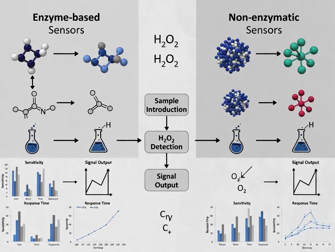
Diagram 1: H2O2 Sensor Operational Principles
The Scientist's Toolkit: Essential Research Reagents and Materials
Table 1: Core Materials for H2O2 Sensor Development
| Material Category | Specific Examples | Primary Function | Considerations |
|---|---|---|---|
| Biological Enzymes | Horseradish Peroxidase (HRP), Glucose Oxidase | Biological recognition element; provides specificity | Cold chain storage required; limited functional lifespan [19] [16] |
| Noble Metal Nanoparticles | Gold Nanoparticles (Au NPs), Platinum Nanoparticles | Catalytic nanomaterial for non-enzymatic sensing; electron transfer facilitation | Aggregation tendency requires stabilizers or supports [17] |
| Transition Metal Oxides | NiO, CeO₂, TiO₂ | Redox-active centers for H2O2 catalysis; enzyme-mimetic behavior | Ce³⁺:Ce⁴⁺ ratio critically impacts catalytic efficiency [5] [16] |
| Carbon Nanostructures | 3D Graphene Hydrogel, Carbon Nanotubes | High surface area support; enhances electron transfer kinetics | Prevents nanomaterial aggregation; maintains electrical conductivity [5] |
| Immobilization Matrices | Chitosan, Metal-Organic Frameworks (MOFs) | Enzyme/nanomaterial stabilization on electrode surface | Prevents leaching; crucial for long-term stability [20] [17] |
| Template Materials | Mesoporous Silica (SBA-15) | Controls nanomaterial morphology during synthesis | Creates defined nanostructures (e.g., NiO octahedrons) [5] |
Comparative Performance Analysis: Experimental Data
Quantitative Sensor Performance Metrics
Table 2: Performance Comparison of Representative H2O2 Sensors
| Sensor Type | Linear Range | Detection Limit | Sensitivity | Stability / Lifetime | Key Challenges |
|---|---|---|---|---|---|
| Enzymatic (HRP-based) | Varies by design | ~nM range | High (enzyme-dependent) | Days to weeks; loses >60% activity at pH extremes or >40°C [16] | Denaturation at non-physiological pH/temperature; complex immobilization [16] [17] |
| Au NPs / TiO₂ NTs [17] | Not specified | 104 nM | 519 µA/mM | >60 days | NP aggregation without proper support |
| 3DGH / NiO Octahedrons [5] | 10 µM – 33.58 mM | 5.3 µM | 117.26 µA mM⁻¹ cm⁻² | Excellent reproducibility & stability | Complex, multi-step synthesis required |
| Ceria Nanoparticles [16] | Wide range | 0.1 pM (Ultra-low) | Correlation with Ce⁴⁺ content | Functional in blood serum; broad pH/temperature tolerance | Performance depends critically on Ce³⁺:Ce⁴⁺ ratio |
Experimental Protocol: Critical Methodologies
Protocol 1: Synthesis of NiO Octahedron/3D Graphene Hydrogel Non-Enzymatic Sensor [5]
Step 1: Hard-Template Synthesis of NiO Octahedrons
- Dissolve 10 mg mesoporous silica (SBA-15) in 100 mL ethanol containing 10 mg Ni(NO₃)₂·6H₂O.
- Stir for 24 hours at room temperature for sufficient infiltration.
- Dry mixture at 80°C for 48 hours, grind powder, and repeat impregnation cycle.
- Calcinate final product at 550°C for 3 hours (2°C/min ramp rate).
- Remove silica template by washing with 2M NaOH at 60°C, followed by ethanol/water rinses.
Step 2: Self-Assembly of 3D Nanocomposite
- Disperse 48 mg graphene oxide (GO) in 32 mL DI water with 12 mg synthesized NiO octahedrons.
- Sonicate mixture (bath sonication: 2 hours; probe sonication: 1.5 hours) to achieve homogeneous distribution.
- Transfer solution to 45 mL Teflon-lined autoclave and maintain at 180°C for 12 hours for hydrothermal self-assembly.
- Wash final 3DGH/NiO25 hydrogel repeatedly with DI water and freeze-dry to preserve porous architecture.
Step 3: Electrochemical Characterization
- Perform cyclic voltammetry (CV) and chronoamperometry in 0.1 M PBS (pH 7.4) with successive H₂O₂ additions.
- Validate sensor selectivity using interfering agents (uric acid, dopamine, ascorbic acid, glucose) at physiological levels.
Protocol 2: Fabrication of Au NPs/TiO₂ Nanotubes Composite Sensor [17]
Step 1: TiO₂ Nanotubes Array Preparation
- Clean titanium foil (0.8 × 1.0 × 0.05 cm) sequentially with acetone, ethanol, and DI water via ultrasonic treatment.
- Etch foil in 18% (v/v) HCl at 85°C for 10 minutes to enhance surface uniformity.
- Anodize pre-treated Ti foil at 40 V for 8 hours in electrolyte containing DMSO + 2% HF using platinum counter electrode.
- Anneal synthesized TiO₂ nanotubes at 450°C for 1 hour in ambient atmosphere to crystallize anatase phase.
Step 2: Citrate-Reduced Au NPs Synthesis
- Add 1 mL of 1% (w/w) sodium citrate to 100 mL of 0.01% (w/w) HAuCl₄ aqueous solution under continuous stirring.
- After 1 minute, slowly introduce 1.6 mL of 0.075% (w/w) NaBH₄ (prepared in 1% sodium citrate).
- Continue stirring until solution color turns deep red, indicating Au NPs formation.
- Store synthesized Au NPs at 4°C until electrode modification.
Step 3: Composite Electrode Assembly
- Immobilize 16 µL Au NPs colloidal solution onto TiO₂ NT surface with 9 µL chitosan (2 mg/mL) as binding agent.
- Air-dry modified electrode before electrochemical measurements to ensure stable film formation.
Critical Analysis: Stability Versus Design Complexity
Diagram 2: Challenge Comparison Framework
Enzyme Stability Limitations: Beyond Operational Lifespan
The intrinsic instability of enzymes represents the most significant constraint for biosensor applications. Horseradish peroxidase experiences dramatic activity loss (>60%) when environmental pH shifts from optimal conditions or temperature exceeds 40°C, fundamentally limiting deployment in non-physiological environments [16]. This instability stems from protein denaturation and irreversible structural changes that disable catalytic function. Furthermore, enzymes like HRP can undergo H₂O₂-induced inactivation during the sensing process itself, creating an operational paradox where the target analyte progressively degrades sensor functionality [17]. These factors collectively constrain sensor lifetime to days or weeks, necessitating frequent recalibration or replacement that increases long-term operational costs despite the initial benefit of high biological specificity.
Non-Enzymatic Design Complexity: The Nanomaterial Engineering Hurdle
While non-enzymatic sensors overcome stability limitations, they introduce substantial design complexity at the nanomaterial level. Effective systems require precise control over multiple parameters: the Ce³⁺:Ce⁴⁺ ratio in ceria nanoparticles directly determines catalytic efficiency [16], while nanoscale morphology (e.g., NiO octahedrons) must be carefully engineered using hard templates like SBA-15 silica [5]. Preventing noble metal nanoparticle aggregation necessitates sophisticated supports such as TiO₂ nanotubes or 3D graphene hydrogels [5] [17]. Each additional material component introduces potential failure points and fabrication challenges. Unlike enzymes evolved for specific molecular recognition, nanomaterials often lack inherent selectivity, requiring additional design strategies to minimize interference from competing electroactive species like ascorbic acid, uric acid, or glucose in biological samples [5].
The choice between enzymatic and non-enzymatic sensing platforms involves fundamental trade-offs between biological precision and engineered robustness. Enzymatic sensors currently remain preferable for applications requiring high specificity under controlled physiological conditions, particularly where cost constraints permit regular replacement. Non-enzymatic approaches offer superior stability for long-term monitoring in challenging environments, but require significant investment in nanomaterial design and characterization. Emerging research focusing on hybrid approaches—such as integrating nanozymes with porous stabilizers or developing biomimetic materials that merge enzymatic selectivity with inorganic stability—promises to bridge this divide [20]. The optimal sensor selection ultimately depends on the specific application requirements regarding operating environment, required lifespan, accuracy tolerance, and resource constraints, with both technological pathways continuing to evolve toward addressing their inherent limitations.
In the field of electrochemical biosensing, the performance of hydrogen peroxide (H₂O₂) sensors is quantitatively assessed through three fundamental parameters: sensitivity, selectivity, and limit of detection (LOD). These metrics provide researchers with standardized criteria for evaluating and comparing sensor technologies, particularly in the ongoing research dialogue comparing enzyme-based and non-enzymatic approaches [2]. Hydrogen peroxide detection holds significant importance across biomedical, pharmaceutical, and environmental applications, as H₂O₂ serves as both a crucial biological metabolite and an industrial chemical [2] [21]. While enzymatic sensors have traditionally dominated with their exceptional biological recognition capabilities, non-enzymatic alternatives have emerged leveraging nanomaterial catalysts that offer enhanced stability and reduced cost [2] [22]. This comparison guide objectively examines both sensor paradigms through the lens of standardized performance metrics, providing experimental data and methodological details to facilitate informed technological selection for research and development applications.
Performance Metrics Comparison: Enzymatic vs. Non-Enzymatic H₂O₂ Sensors
The quantitative comparison of recent enzymatic and non-enzymatic H₂O₂ sensors reveals distinct performance advantages across different applications. The following table summarizes key performance metrics for recently developed non-enzymatic sensors:
Table 1: Performance Metrics of Recent Non-Enzymatic H₂O₂ Sensors
| Sensor Material | Sensitivity | Limit of Detection (LOD) | Linear Range | Selectivity Characteristics |
|---|---|---|---|---|
| Cu₂O@Cu₉S₅ yolk-shell nanospheres [23] | 299.7 μA mM⁻¹ cm⁻² | 28.83 nM | 0.1 μM to 3.5 mM | Minimal interference from UA, AA, DA, NaCl, glucose |
| CuO petal nanostructures [21] | 439.19 μA mM⁻¹ | 1.34 μM | 10 to 1800 μM | No interference from AA, UA, DA, glucose, acetaminophen, NaCl |
| Nanoporous gold (NPG) [24] | 159 μA mM⁻¹ cm⁻² (0.002-5 mM)64 μA mM⁻¹ cm⁻² (5-37.5 mM) | 0.3 μM | 0.002-37.5 mM | Minimal interference from AA, UA, glucose |
| Ag-doped CeO₂/Ag₂O nanocomposite [25] | 2.728 μA cm⁻² μM⁻¹ | 6.34 μM | 1×10⁻⁸ to 0.5×10⁻³ M | Minimal interference from common analytes |
Enzymatic sensors typically exhibit excellent selectivity due to the specific catalytic activity of enzymes like horseradish peroxidase, but they suffer from inherent stability limitations related to enzyme denaturation under suboptimal environmental conditions [21]. The operational lifespan of enzymatic sensors is typically limited to one to two weeks, whereas non-enzymatic sensors demonstrate significantly longer lifetime due to the absence of biological components [26]. Non-enzymatic sensors achieve their selectivity through material science approaches rather than biological recognition, utilizing specific electrocatalytic properties of nanomaterials that preferentially catalyze H₂O₂ oxidation or reduction while minimizing response to interfering species [2] [23].
Experimental Protocols for Key Sensor Technologies
Nanostructured Copper Oxide Sensor Fabrication
The synthesis of CuO petal nanostructures via a one-step hydrothermal oxidation method represents a straightforward approach to non-enzymatic sensor fabrication [21]. The methodology begins with preparation of a working solution containing 10 mL of 10 M NaOH solution, 5 mL of 1 M (NH₄)₂S₂O₈ solution, and 26 mL of H₂O. Copper wire substrates are first rinsed with water and ethanol to remove surface contaminants, then immersed in the working solution contained in a heat-resistant glass beaker with a lid. The beaker is placed in an oven preheated to 90°C for 3 hours, then allowed to cool naturally. The resulting nanostructured samples are covered with a uniform oxide layer and are repeatedly washed with distilled water to remove residual reagents, followed by drying in an oven at 90°C for 3 hours to remove moisture [21]. For electrochemical measurements, the obtained wire samples are cut into 2 cm pieces, with one end stripped to pure copper over a 5 mm length to ensure electrical contact. Characterization through field-emission scanning electron microscopy (FESEM) and X-ray diffractometry (XRD) confirms the formation of petal-like nanostructures with high surface area, which contributes significantly to the enhanced sensitivity of the sensor [21].
Nanoporous Gold (NPG) via Solid-Phase Reaction Method
The fabrication of NPG sensors employs a modified solid-phase reaction method based on a metal-induced crystallization process [24]. A triple-layer precursor structure consisting of amorphous Ge (top)/Au (middle)/amorphous Ge (bottom) is deposited onto Si(100) wafers (with 50 nm-thick amorphous SiO₂) using magnetron sputtering in a high-vacuum system. The substrates are maintained at 120°C during deposition to encourage the metal-induced crystallization process. The samples are continuously rotated at 20 rpm during sputtering to ensure uniformity. The as-sputtered samples are then immersed in hydrogen peroxide solution (30 vol%) for 5 minutes at 25°C to selectively remove Ge, followed by thorough rinsing with ultrapure water and drying under nitrogen stream [24]. This process results in NPG with a bicontinuous porous structure featuring grain and nanopore sizes of approximately 14 nm, significantly smaller than structures obtained using bilayer precursors. The electrochemical performance is evaluated using a standard three-electrode system with NPG-modified glassy carbon electrode as working electrode, saturated calomel reference electrode, and platinum wire counter electrode. Before testing, the electrode is activated through 20 cycles of cyclic voltammetry between -0.8 and 0.8 V in 0.1 M KOH solution at 50 mV s⁻¹ [24].
Yolk-Shell Nanosphere Synthesis
The preparation of Cu₂O@Cu₉S₅ yolk-shell nanospheres utilizes a facile wet chemical method based on Cu₂O nanosphere templates [23]. Cu₂O nanospheres are first synthesized through a facile reduction reaction using copper hydroxide as both copper source and morphology controlling reagent. The structural transformation from solid nanospheres to yolk-shell architectures occurs through a controlled sulfidation process, where the outer layer of Cu₂O is converted to Cu₉S₅ while maintaining the inner Cu₂O core. The key advantage of this structure is the combination of high surface area and synergistic catalytic effects between the core and shell components [23]. Materials characterization through SEM, TEM, and XRD confirms the successful formation of the yolk-shell structure with well-defined interior voids. Electrochemical testing demonstrates that this unique architecture significantly enhances electrocatalytic activity toward H₂O₂ reduction compared to pristine Cu₂O nanospheres or Cu₉S₅ hollow nanospheres [23].
Signaling Pathways and Sensor Mechanisms
The fundamental operational principles of H₂O₂ sensors differ significantly between enzymatic and non-enzymatic approaches, as illustrated in the following diagram:
Diagram 1: Signaling Pathways in H₂O₂ Detection Mechanisms
Enzyme-based detection relies on biological recognition elements such as horseradish peroxidase (HRP) that specifically catalyze the reduction of H₂O₂ while simultaneously oxidizing a substrate [21]. The electron transfer from this reaction is then measured at the electrode surface. In contrast, non-enzymatic detection utilizes nanomaterial catalysts that directly adsorb and catalyze the oxidation or reduction of H₂O₂, with electrons transferring directly between the analyte and electrode surface [2] [23]. This fundamental difference in mechanism explains the contrasting performance characteristics: enzymatic sensors achieve high selectivity through biological specificity but suffer from limited stability, while non-enzymatic sensors achieve robustness through inorganic materials but may face greater challenges with interfering species [22] [21].
The experimental workflow for developing and evaluating H₂O₂ sensors follows a systematic process from material synthesis to performance validation:
Diagram 2: Experimental Workflow for H₂O₂ Sensor Development
Essential Research Reagents and Materials
The development and fabrication of high-performance H₂O₂ sensors requires specific research reagents and functional materials. The following table details essential components and their functions in sensor construction:
Table 2: Essential Research Reagents for H₂O₂ Sensor Development
| Reagent/Material | Function/Application | Examples from Literature |
|---|---|---|
| Metal Precursors | Source for catalytic nanomaterials | Copper hydroxide (Cu(OH)₂) for CuO nanostructures [21], Cerium nitrate (Ce(NO₃)₃·6H₂O) for CeO₂ nanocomposites [25] |
| Nanostructuring Agents | Control morphology and surface area | Polyacrylic acid (PAA) for nanosphere formation [23], Polyvinylpyrrolidone (PVP) for nanocomposite synthesis [25] |
| Electrode Materials | Serve as sensing substrates | Glassy carbon electrodes [24] [25], Copper wires [21], Gold-sputtered substrates [26] |
| Buffer Solutions | Maintain optimal pH conditions | Phosphate buffer solutions (PBS, 0.1 M, pH 7.4) [24], NaOH solutions for alkaline conditions [21] |
| Interference Compounds | Selectivity testing | Ascorbic acid, uric acid, dopamine, glucose, acetaminophen [23] [21] [25] |
| Reference Electrodes | Provide stable potential reference | Saturated calomel electrode (SCE) [24], Ag/AgCl wire [21] [26] |
The selection of appropriate reagents and materials directly impacts sensor performance. For instance, the use of specific morphology-controlling agents like polyacrylic acid enables the formation of advanced nanostructures such as yolk-shell architectures that significantly enhance sensitivity [23]. Similarly, the choice of buffer system is critical, as pH dramatically influences the catalytic activity of both enzymatic and non-enzymatic sensing materials [26]. For real-sample applications, additional reagents may be required for sample pretreatment and matrix effect minimization.
The comparative analysis of enzymatic and non-enzymatic H₂O₂ sensors through the fundamental metrics of sensitivity, selectivity, and LOD reveals a clear technological landscape where each approach occupies distinct application spaces. Enzymatic sensors provide exceptional selectivity and remain valuable for clinical diagnostics where biological recognition is paramount, despite their limitations in long-term stability and environmental susceptibility [22] [21]. Non-enzymatic sensors demonstrate superior stability, wider linear ranges, and increasingly competitive sensitivity metrics, making them particularly suitable for industrial monitoring, environmental sensing, and continuous monitoring applications [2] [23] [21]. Recent advancements in nanotechnology have substantially bridged the performance gap, with novel materials such as yolk-shell nanostructures, nanoporous metals, and metal oxide nanocomposites achieving detection limits rivaling their enzymatic counterparts [23] [24] [25]. The strategic selection between these technologies should be guided by specific application requirements: enzymatic sensors for scenarios demanding exceptional specificity in controlled environments, and non-enzymatic approaches for applications requiring robustness, longevity, and cost-effectiveness. Future research directions will likely focus on further enhancing the selectivity of non-enzymatic sensors through advanced material engineering while simultaneously addressing the stability limitations of enzymatic systems through immobilization strategies and enzyme stabilization techniques.
Material Innovations and Sensing Architectures: From Laboratory to Real-World Applications
The detection and quantification of hydrogen peroxide (H₂O₂) represents a critical analytical challenge across biomedical research, clinical diagnostics, and industrial monitoring. As a significant reactive oxygen species and a common byproduct of oxidase enzymes, H₂O₂ concentration serves as a key indicator in numerous biochemical pathways and analytical assays. The sensor landscape is broadly divided between enzyme-based systems leveraging biological catalysts and non-enzymatic approaches utilizing synthetic nanomaterials. Enzyme-based sensors traditionally employ horseradish peroxidase (HRP) and catalase, prized for their high specificity and catalytic efficiency. Recently, emerging players like cholesterol oxidase (ChOx) have demonstrated unexpected utility in H₂O₂ biosensing architectures, expanding the toolkit available to researchers and developers. This guide provides a systematic comparison of these enzymatic workhorses, evaluating their performance characteristics, operational parameters, and suitability for different analytical contexts, with a specific focus on the evolving research concerning enzyme-based versus non-enzymatic H₂O₂ sensor performance.
Performance Comparison of H₂O₂ Sensing Platforms
The table below summarizes the key performance metrics of various enzymatic and non-enzymatic sensing platforms for H₂O₂ detection, as reported in recent literature.
Table 1: Performance Comparison of Enzymatic and Non-Enzymatic H₂O₂ Sensors
| Sensor Type | Sensitivity | Linear Range | Detection Limit | Reference & Year |
|---|---|---|---|---|
| HRP-based (HEPNP/rGO/Au) | Not Specified | 0.01–100 µM | 0.01 µM | [27] (2020) |
| ChOx-based (PMWCNT/ChOx) | 26.15 µA/mM | 0.4–4.0 mM | 0.43 µM | [11] (2025) |
| ChOx/nCuFe/nPt/GCE | 3960 A·M⁻¹·m⁻² | 2–50 µM | Not Specified | [28] (2023) |
| Non-enzymatic (Fe₃O₄/CNT Ink) | 1040 µA cm⁻² mM⁻¹ | Up to 2 mM | 0.5 µM | [29] (2022) |
| Non-enzymatic (Ag-CuI-exGRc) | 760 mA·M⁻¹·cm⁻² | Not Specified | 1.2 µM | [30] (2025) |
| Non-enzymatic (3DGH/NiO₂₅) | 117.26 µA mM⁻¹ cm⁻² | 10 µM–33.58 mM | 5.3 µM | [5] (2025) |
Detailed Analysis of Enzymatic Workhorses
Horseradish Peroxidase (HRP)
HRP is a classical oxidoreductase that catalyzes the reduction of H₂O₂ while oxidizing a variety of organic substrates. Its well-characterized structure and commercial availability have made it a cornerstone of enzymatic biosensing.
- Catalytic Mechanism: HRP operates via a ping-pong mechanism, where it first reacts with H₂O₂ to form an oxidized intermediate (Compound I), which is then reduced back to its native state by an electron-donating substrate [31]. This electron transfer can be monitored electrochemically.
- Recent Advancements: Research has focused on enhancing HRP's stability and electron transfer efficiency. A notable approach involves encapsulating HRP in protein nanoparticles (HEPNP). These nanoparticles create a protective three-dimensional structure that can contain a large amount of enzyme, significantly amplifying the electrochemical signal. When combined with a reduced graphene oxide (rGO)-modified gold electrode to improve electron transfer, this configuration achieved a remarkably low detection limit of 0.01 µM for H₂O₂ [27].
- Single-Entity Studies: Cutting-edge research using electrochemical collision technique has provided insights into the catalytic activity of single HRP molecules. Studies have calculated the maximum turnover number (kcat) for single HRP molecules to be on the order of ~10³ s⁻¹, with variations depending on the electrolyte environment and the presence of mediators like ABTS or K₄Fe(CN)₆ [31].
Cholesterol Oxidase (ChOx) as an Emerging Player
While traditionally used for cholesterol sensing, ChOx is gaining attention for its utility in H₂O₂ biosensing. ChOx is a FAD-containing enzyme that catalyzes the oxidation of cholesterol to cholest-4-en-3-one, simultaneously producing H₂O₂ [28] [11].
- Direct H₂O₂ Sensing: A 2025 study revealed that a multi-walled carbon nanotube paste electrode modified with ChOx (PMWCNT/ChOx) could directly enhance the sensitivity of H₂O₂ electrochemical detection by 21 times compared to an unmodified electrode [11]. This suggests ChOx itself can participate in or facilitate the electrocatalytic reduction of H₂O₂.
- Molecular Interactions: In silico studies, including molecular dynamics simulations and docking assays, have confirmed that the binding between ChOx and H₂O₂ is spontaneous. This labile interaction promotes the rapid electrochemical reduction of H₂O₂, validating the experimental findings [11].
- Traditional Use in H₂O₂ Generation: In its conventional biosensing role, ChOx is paired with a H₂O₂-detecting element, such as HRP or a nanozyme. For instance, a highly sensitive cholesterol bionanosensor was developed by immobilizing ChOx on a nano-platinized electrode decorated with bimetallic CuFe nanoparticles (a peroxidase mimetic). This setup detects the H₂O₂ generated by ChOx's oxidation of cholesterol [28].
Non-Enzymatic Platforms
Non-enzymatic sensors utilize nanomaterials with intrinsic peroxidase-like activity to mimic natural enzymes, offering an alternative with often superior stability and lower cost.
- Nanocomposite Materials: Common materials include magnetite (Fe₃O₄) loaded onto carbon nanotubes [29], silver-copper nanoparticles embedded in an oxidized carbonate green rust matrix [30], and nickel oxide (NiO) octahedrons decorated on 3D graphene hydrogel [5].
- Performance Highlights: These sensors are characterized by high sensitivity, wide linear ranges, and excellent tolerance to interfering substances. The 3DGH/NiO₂₅ nanocomposite, for example, boasts a wide linear range from 10 µM to 33.58 mM [5], while the Fe₃O₄/CNT sensor loses only 20% of its activity after three weeks [29].
Experimental Protocols & Methodologies
This protocol outlines the creation of a PMWCNT/ChOx electrode for direct H₂O₂ sensing.
- MWCNT Activation: Purify multi-walled carbon nanotubes (MWCNTs) by sonicating them sequentially in 1 M nitric acid and 1 M sulfuric acid, followed by washing with ethanol and acetone until neutral pH.
- Paste Electrode Preparation: Mix the activated MWCNTs with mineral oil in a 70/30 (w/w) ratio to form a paste (PMWCNT).
- Electrode Assembly: Pack the PMWCNT into a glassy carbon electrode sleeve with an electrical contact.
- Enzyme Immobilization: Drop-cast 10 µL of ChOx solution (20 U/mL) onto the PMWCNT surface.
- Curing: Allow the modified electrode (PMWCNT/ChOx) to dry for 10 minutes at room temperature before use.
- Electrochemical Measurement: Perform amperometric detection of H₂O₂ in 0.050 M phosphate buffer (pH 7.4) at room temperature.
This method details the synthesis of HEPNP and its integration into an rGO-modified electrode.
- HEPNP Synthesis (Ethanol Desolvation):
- Mix 100 µL of BSA (50 mg/mL) with 20 µL of HRP (25 mg/mL).
- Add 400 µL of pure ethanol at a slow flow rate (1 mL/min) under constant stirring. The solution will turn opaque.
- Add 10 µL of 4% glutaraldehyde to crosslink the proteins and incubate for 12 hours.
- Wash the formed HEPNP via centrifugation (9000 rpm, 15 min) and re-disperse in a washing buffer.
- Electrode Modification:
- Clean a gold electrode with piranha solution and rinse.
- Immobilize cysteamine as a linker onto the clean Au surface.
- Modify the electrode with rGO (100 µg/mL in NMP) and incubate at 75°C to form the rGO/Au electrode.
- Drop-cast 40 µL of the HEPNP suspension onto the rGO/Au electrode and incubate to form the final HEPNP/rGO/Au working electrode.
- H₂O₂ Detection: Use a three-electrode system (HEPNP/rGO/Au as working electrode, Pt counter electrode, Ag/AgCl reference) for cyclic voltammetry and amperometric i-t measurements to detect H₂O₂.
Comparative Signaling Pathways and Workflows
The diagrams below illustrate the core catalytic mechanisms and experimental workflows for the key sensors discussed.
Catalytic Mechanisms in H₂O₂ Sensing
Biosensor Fabrication Workflow
The Scientist's Toolkit: Essential Research Reagents
Table 2: Key Reagents and Materials for H₂O₂ Sensor Development
| Reagent/Material | Function/Application | Examples from Research |
|---|---|---|
| Carbon Nanotubes (MWCNTs) | Conductive support; enhances electron transfer and surface area. | Activated MWCNTs in paste electrodes [11]; Fe₃O₄/CNT ink [29]. |
| Graphene & Derivatives (rGO) | High-conductivity support; promotes direct electron transfer. | rGO-modified Au electrode for HRP immobilization [27]; 3D graphene hydrogel [5]. |
| Metal/Metal Oxide Nanoparticles | Peroxidase nanozymes; provide catalytic activity and signal amplification. | CuFe NPs (nanozymes) [28]; Fe₃O₄ NPs [29]; NiO octahedrons [5]; Ag & CuI NPs [30]. |
| Cross-linking Agents (Glutaraldehyde) | Immobilize enzymes on sensor surfaces; form stable covalent bonds. | Cross-linking ChOx on electrodes [28]; forming HEPNP [27]. |
| Enzymes (HRP, ChOx) | Biological recognition elements; provide high specificity and catalysis. | HRP for H₂O₂ reduction [27]; ChOx for H₂O₂ generation/detection [28] [11]. |
| Polyelectrolytes & Stabilizers | Create a biocompatible microenvironment; enhance enzyme stability. | Bovine Serum Albumin (BSA) in HEPNP [27]; compartmentalization with polyelectrolytes [32]. |
The comparative analysis presented in this guide underscores a dynamic and evolving field. HRP remains a powerful and well-understood tool, especially when engineered into nanostructured formats like HEPNP, which push the boundaries of sensitivity. Simultaneously, the emergence of ChOx in non-canonical H₂O₂ sensing roles broadens the functionality of oxidase enzymes beyond their primary substrates. However, the significant progress in non-enzymatic sensors cannot be overlooked. With their robust stability, high sensitivity, and wide linear ranges, they present a compelling alternative, particularly for applications in complex matrices where enzyme stability is a concern.
Future research will likely focus on several key areas:
- Hybrid Systems: Combining the best attributes of enzymatic specificity and non-enzymatic stability in a single sensor.
- Single-Molecule Kinetics: Utilizing techniques like electrochemical collision to deepen the fundamental understanding of catalytic mechanisms, thereby informing better sensor design [31].
- Advanced Materials: Exploring novel nanocomposites and nanostructures to further enhance electron transfer and catalytic efficiency.
- Point-of-Care Applications: Translating these advanced platforms into robust, miniaturized, and cost-effective devices for real-world clinical and environmental monitoring.
The choice between an enzymatic workhorse and a non-enzymatic alternative ultimately depends on the specific requirements of the application, weighing factors such as required sensitivity, specificity, stability, and cost.
The accurate detection of hydrogen peroxide (H₂O₂) is critically important across biomedical, environmental, and industrial fields. As a key reactive oxygen species, H₂O₂ serves as a vital biomarker in physiological processes and disease states, while also being a common industrial agent. Traditionally, enzymatic biosensors (e.g., those using horseradish peroxidase) have been favored for their high specificity and catalytic efficiency under mild conditions. However, their inherent drawbacks—including high cost, structural instability during storage, tedious immobilization procedures, and sensitivity to environmental conditions (pH and temperature)—have limited their widespread practical application [33] [5].
Non-enzymatic sensors, particularly those leveraging nanozymes (nanomaterials with enzyme-like activity), have emerged as powerful alternatives. These materials offer the advantages of broad linear detection ranges, superior stability, lower cost, and ease of manufacturing [33] [34]. This guide provides a comparative analysis of three leading classes of nanozymes—Noble Metal Hybrids, Metal Oxides, and Carbon-Based Nanocomposites—objectively evaluating their performance, mechanisms, and suitability for different H₂O₂ sensing applications.
Performance Comparison of Nanozyme Platforms
The table below summarizes the key performance metrics of recent advanced H₂O₂ sensors based on different nanozyme materials.
Table 1: Performance Comparison of Non-Enzymatic H₂O₂ Nanozyme Sensors
| Nanozyme Category | Specific Material & Structure | Sensitivity (μA mM⁻¹ cm⁻²) | Linear Range (μM or mM) | Limit of Detection (LOD) | Key Advantages |
|---|---|---|---|---|---|
| Noble Metal Hybrids | 3D Porous Au/CuO/Pt [33] | 25,836 μA/mM·cm² | Not Specified | 9.8 nM | Ultra-high sensitivity, excellent selectivity |
| Cu₁.₈Se Nanosheets [35] | Not Specified | 1.25 - 10,000 μM | 1.25 μM | Dual-mode (SERS & Electrochemical), rapid response | |
| Metal Oxides | Porous CeO₂ Hollow Microspheres (CeO₂-phm) [34] | 2,161.6 & 2,070.9 | 0.5 - 450 μM | 0.017 μM (17 nM) | Wide linear range, excellent stability & reproducibility |
| NiO Octahedrons/3D Graphene Hydrogel [5] | 117.26 | 10 μM - 33.58 mM | 5.3 μM | Wide linear range, good for real-sample (milk) analysis | |
| MOF-Based & Carbon Hybrids | Mesoporous Core-Shell Co-MOF/PBA (Electrochemical Mode) [36] | Not Specified | 1 - 2,041 nM | 0.47 nM | Ultra-low LOD, dual-mode (colorimetric & electrochemical) |
| Mesoporous Core-Shell Co-MOF/PBA (Colorimetric Mode) [36] | Not Specified | 1 - 400 μM | 0.59 μM | Visual detection, suitable for in-situ cell monitoring |
Experimental Protocols and Sensing Mechanisms
Fabrication and Testing of Noble Metal Hybrid Sensors
Protocol for 3D Porous Au/CuO/Pt Electrode [33]:
- Fabrication: The sensor is fabricated using a combined physiochemical method.
- A porous copper layer is first electrodeposited onto a substrate using dynamic hydrogen bubbling as a template.
- This layer is then thermally oxidized in air at 200°C for 2 hours to form a porous CuO framework.
- Platinum Nanoparticles (Pt NPs) are subsequently sputtered onto the CuO surface.
- Finally, Au nano- and micro-particles (NMPs) are decorated onto the structure via sputtering.
- Electrochemical Testing: Sensor performance is evaluated in a standard three-electrode electrochemical cell using a phosphate buffer saline (PBS) electrolyte. Amperometric (current-time) measurements are conducted at a fixed potential while successively adding H₂O₂ to the solution. The resulting current response is measured to determine sensitivity and linear range.
Protocol for Cu₁.₈Se Nanosheet Electrode [35]:
- Fabrication: A two-step process is employed.
- Step 1: Cu(OH)₂ nanowires are synthesized on a copper foil substrate via electrochemical oxidation in a 1 M NaOH solution at a constant current density of 4.5 mA/cm² for 20 minutes.
- Step 2: The Cu(OH)₂/Cu foil is immersed in an aqueous solution containing selenium (Se) and sodium borohydride (NaBH₄) at room temperature for 1-24 hours for a selenization process, converting the nanowires into flower-like Cu₁.₈Se nanosheets.
- Dual-Mode Testing:
- Electrochemical: The nanosheet electrode is used as a working electrode in a three-electrode system with H₂SO₄ electrolyte for amperometric H₂O₂ detection.
- SERS: The same substrate is used for Surface-Enhanced Raman Scattering by adsorbing a probe molecule (Rhodamine B) and measuring the enhanced Raman signal, demonstrating its dual-functionality.
Synthesis and Characterization of Metal Oxide and MOF-Based Sensors
Protocol for NiO Octahedrons/3D Graphene Hydrogel (3DGH) [5]:
- Synthesis of NiO Octahedrons: A hard templating method is used. Nickel nitrate hexahydrate is dissolved in ethanol with mesoporous silica (SBA-15) as a template. After stirring and drying, the powder is calcined at 550°C for 3 hours. The silica template is then removed by treatment with NaOH.
- Self-Assembly of 3DGH/NiO: Graphene oxide (GO) is dispersed in water with a specific amount of NiO octahedrons (e.g., 25% by weight). The mixture undergoes hydrothermal treatment at 180°C for 12 hours, resulting in a self-assembled 3D hydrogel composite.
- Characterization & Sensing: The material is characterized by FE-SEM, TEM, XRD, and Raman spectroscopy. The electrochemical sensing performance is evaluated via cyclic voltammetry and chronoamperometry in PBS.
Protocol for Mesoporous Core-Shell Co-MOF/PBA Probe [36]:
- Synthesis: A 3D Co-MOF precursor is first synthesized. This precursor is then dispersed in ethanol and rapidly mixed with an aqueous solution of K₃[Fe(CN)₆] under stirring. A cation-exchange reaction driven by the Kirkendall effect leads to the formation of the core-shell Co-MOF/Prussian Blue Analogue (PBA) structure.
- Dual-Mode Detection Mechanism:
- Colorimetric Mode: The probe exhibits peroxidase-like activity. In the presence of H₂O₂, a Fenton-like reaction is catalyzed, generating hydroxyl radicals (•OH) that oxidize a colorless chromogenic substrate (e.g., TMB) into a colored product.
- Electrochemical Mode: The material is drop-cast on an electrode. It catalyzes the electrochemical reduction of H₂O₂, significantly amplifying the current signal. The acceleration of Co³⁺/Co²⁺ and Fe³⁺/Co²⁺ redox cycles enhances the electron transfer efficiency.
Signaling Pathways and Catalytic Mechanisms
The following diagram illustrates the general catalytic mechanisms and electron transfer pathways employed by the different nanozyme categories for H₂O₂ detection.
Figure 1: Catalytic Mechanisms of Nanozyme Classes for H₂O₂ Sensing. Noble metal hybrids leverage synergistic effects for direct electron transfer. Metal oxides utilize reversible redox couples (e.g., Ce⁴⁺/Ce³⁺). MOF-based probes mimic natural enzymes for catalytic and electrochemical signaling.
The Scientist's Toolkit: Essential Research Reagents
This table lists key reagents and materials required for the fabrication and testing of the nanozyme sensors discussed.
Table 2: Essential Reagents and Materials for Nanozyme H₂O₂ Sensor Research
| Reagent/Material | Function/Application | Example from Protocols |
|---|---|---|
| Metal Salts | Precursors for nanozyme synthesis | Copper sulfate (CuSO₄), Cerium nitrate (Ce(NO₃)₃·6H₂O), Nickel nitrate (Ni(NO₃)₂·6H₂O) [33] [5] [34] |
| Noble Metal Targets/Salts | Catalytic nanoparticle decoration | Pt, Au, Pd sputtering targets or salts [33] [37] |
| 2D/3D Carbon Supports | High-surface-area conductive support | Graphene Oxide (GO), carboxylated multi-walled carbon nanotubes (cMWCNTs) [5] [34] |
| MOF Ligands & Precursors | Construction of metal-organic framework structures | 2-methylimidazole (2-Hmim), K₃[Fe(CN)₆] (for PBA formation) [36] |
| Structural Templates | Creating defined porosity and morphology | Silica templates (SBA-15), dynamic hydrogen bubbling [5] [33] |
| Buffer Solutions (PBS) | Electrolyte for electrochemical testing | Phosphate Buffered Saline (PBS, 0.1 M, pH 7.4) for simulating physiological conditions [33] [5] |
| Interferent Analytes | Testing sensor selectivity | Ascorbic Acid (AA), Dopamine (DA), Uric Acid (UA), Glucose, Citric Acid, Fructose, NaCl [33] [5] [34] |
| Chromogenic Substrates | Colorimetric signal generation | TMB (3,3',5,5'-Tetramethylbenzidine) for peroxidase-mimic activity detection [36] |
The detection of hydrogen peroxide (H₂O₂) is critically important across diverse fields including medical diagnostics, environmental monitoring, and food safety. H₂O₂ serves as a vital biomarker for oxidative stress and is implicated in various diseases including diabetes, cancer, and neurodegenerative disorders [38] [5]. Additionally, as a common intermediate in industrial processes and a product of oxidase enzymes, accurate monitoring of H₂O₂ concentrations is essential for both research and practical applications [2] [18]. Traditional electrochemical biosensors have predominantly relied on enzyme-based detection mechanisms, leveraging the high specificity and catalytic efficiency of biological recognition elements such as glucose oxidase, cholesterol oxidase, and acetylcholinesterase [18]. These biosensors integrate biological components with physicochemical transducers to convert biochemical reactions into measurable signals, offering remarkable specificity for their target analytes [18].
Despite their widespread use, enzyme-based sensors face significant challenges including high cost, complicated fabrication processes, sensitivity to environmental conditions (pH, temperature), and limited operational stability due to enzyme denaturation [2] [5]. These limitations have stimulated intense research into non-enzymatic alternatives, particularly those utilizing advanced heterostructure materials. Heterostructures, comprising interfaces between different semiconductor materials or between semiconductors and conductors, offer revolutionary advantages for signal transduction in electrochemical sensing [6]. By engineering p-n junctions and composite materials at the nanoscale, researchers have developed sensing platforms with enhanced charge transfer capabilities, improved catalytic activity, and superior stability compared to both conventional electrodes and enzyme-based systems [6] [2]. This review comprehensively compares the performance of emerging heterostructure-based non-enzymatic sensors against traditional enzymatic approaches, providing researchers and drug development professionals with objective experimental data to guide sensor selection and development.
Performance Comparison: Quantitative Analysis of Sensor Technologies
The advancement of non-enzymatic H₂O₂ sensors utilizing heterostructures has yielded significant improvements in key performance metrics compared to both traditional enzymatic sensors and earlier non-enzymatic approaches. The tables below summarize experimental data for various heterostructure-based sensors, highlighting their enhanced capabilities.
Table 1: Performance Comparison of Representative Heterostructure-Based Non-enzymatic H₂O₂ Sensors
| Sensor Material | Sensitivity (μA mM⁻¹ cm⁻²) | Linear Range (mM) | Detection Limit (μM) | Response Time | Stability |
|---|---|---|---|---|---|
| SnO₂@CuO/CF p-n heterojunction [6] | Not specified | 0.0015 - 8.27 | 0.29 | Not specified | Good anti-interference ability |
| 3DGH/NiO25 nanocomposite [5] | 117.26 | 0.01 - 33.58 | 5.3 | Not specified | Excellent long-term stability |
| ZIF-67/CNFs composite [39] | 323 | 0.0025 - 0.19 | 0.62 | Not specified | Satisfactory long-term stability |
| PdSe₂-MoS₂ heterostructure (IR photodetector) [40] | Not applicable | Not applicable | Not applicable | Rapid response | Enhanced stability in ambient conditions |
Table 2: Comparative Analysis of Sensor Paradigms
| Parameter | Enzyme-Based Sensors | Non-Enzymatic Heterostructure Sensors |
|---|---|---|
| Specificity | Very high (enzyme-substrate specificity) [18] | Moderate to high (depends on material selectivity) [6] [2] |
| Stability | Limited (enzyme denaturation) [2] [5] | Excellent (robust inorganic materials) [6] [5] |
| Cost | High (enzyme purification) [5] | Lower (synthetic materials) [2] |
| Fabrication Complexity | Moderate to high [18] | Varies (some are simple) [2] |
| Environmental Tolerance | Sensitive to pH, temperature [2] [18] | Generally broader operating windows [6] |
| Signal Transduction Mechanism | Enzyme-product detection (e.g., H₂O₂) [18] | Direct electron transfer, heterojunction effects [6] [38] |
The data reveal that heterostructure-based sensors achieve remarkably low detection limits, with the SnO₂@CuO/CF p-n heterojunction and ZIF-67/CNFs composite reaching sub-micromolar levels (0.29 μM and 0.62 μM respectively) [6] [39]. These values meet or exceed the sensitivity requirements for most biological applications, including tracking H₂O₂ fluctuations in cellular environments. Furthermore, the wide linear range demonstrated by the 3DGH/NiO25 nanocomposite (0.01-33.58 mM) [5] highlights the dynamic response capability of these materials across concentration ranges relevant to both physiological and industrial contexts.
Experimental Protocols: Methodologies for Sensor Fabrication and Evaluation
Fabrication of SnO₂@CuO/CF p-n Heterojunction Sensor
The SnO₂@CuO heterostructure on copper foam (CF) was synthesized through a multi-step process combining electro-oxidation and electrodeposition techniques [6]. Initially, Cu(OH)₂ nanofiber arrays were prepared on copper foam substrates via electrochemical anodization in a 2 M NaOH solution at a current density of 20 mA·cm⁻² for 600 seconds [6]. The resulting Cu(OH)₂/CF precursor was subsequently annealed in a N₂ atmosphere at 200°C for 120 minutes to convert Cu(OH)₂ to CuO nanofibers, establishing the p-type semiconductor framework [6]. The n-type SnO₂ component was then incorporated through electrochemical deposition onto the CuO/CF substrate using a solution containing 2.5 mM SnSO₄ and 50 mM H₂SO₄, with deposition performed at a constant potential of -1.1 V (vs. SCE) for 180 seconds [6]. The final SnO₂@CuO/CF heterostructure material was thoroughly rinsed with deionized water and dried at 60°C for 12 hours before characterization and electrochemical testing [6].
Preparation of 3D Graphene Hydrogel/NiO Octahedron Composite
This protocol involved the template-assisted synthesis of NiO octahedrons followed by their integration with three-dimensional graphene hydrogel (3DGH) [5]. The NiO octahedrons were first prepared using mesoporous silica (SBA-15) as a hard template, with nickel nitrate hexahydrate serving as the nickel precursor in an ethanol solution [5]. After impregnation and drying, the material was calcined at 550°C for 3 hours, followed by template removal using 2 M NaOH treatment [5]. For composite formation, 48 mg of graphene oxide (synthesized via modified Hummers' method) was dispersed in 32 mL deionized water containing 12 mg of the prepared NiO octahedrons [5]. The mixture was subjected to bath sonication for 2 hours followed by probe sonication for 1.5 hours to achieve homogeneous dispersion. The resulting suspension was transferred to a 45 mL Teflon-lined autoclave and maintained at 180°C for 12 hours to facilitate self-assembly of the 3DGH/NiO nanocomposite through hydrothermal treatment [5]. The final product was washed repeatedly with deionized water and freeze-dried to preserve the porous structure [5].
Electrochemical Testing Protocols
Standard electrochemical characterization for all sensor materials included cyclic voltammetry (CV) and chronoamperometry measurements, typically performed using a conventional three-electrode system with the modified material as working electrode, Ag/AgCl as reference electrode, and platinum wire as counter electrode [6] [5] [39]. Phosphate buffer solution (PBS, 0.1 M, pH 7.4) served as the supporting electrolyte for most measurements, with some studies utilizing NaOH solution (0.1 M) for enhanced H₂O₂ reduction signals [6] [39]. Sensitivity calculations were based on the slope of the current density versus H₂O₂ concentration plot, while detection limits were determined using the formula LOD = 3σ/S, where σ represents the standard deviation of the blank signal and S denotes the sensitivity of the calibration curve [5]. Selectivity assessments were conducted by challenging the sensors with potential interfering species including uric acid, ascorbic acid, dopamine, glucose, and various ions (Na⁺, K⁺, Cl⁻) at physiological concentrations [6] [5].
Signaling Pathways and Transduction Mechanisms
p-n Heterojunction Signaling in Metal Oxide Systems
In heterostructure sensors, the interface between p-type and n-type semiconductors creates a built-in electric field that dramatically enhances charge separation and transfer efficiency, leading to improved electrochemical responses [6]. The p-n junction formed between p-type CuO and n-type SnO₂ generates a powerful built-in electric field at their interface [6]. This field promotes the adsorption of H₂O₂ molecules and facilitates electron transfer during the electrocatalytic reduction of H₂O₂, significantly enhancing the sensor's sensitivity and lowering its detection limit [6]. The heterojunction effect not only improves charge carrier separation but also creates synergistic catalytic sites at the material interfaces, where the combined electronic properties of both semiconductors enhance the overall electrocatalytic activity beyond what either material could achieve independently [6].
Comparative Signal Transduction Pathways
The fundamental difference between enzymatic and non-enzymatic sensing approaches lies in their signal transduction mechanisms. Enzyme-based biosensors rely on the catalytic activity of biological recognition elements, where the enzyme catalyzes a specific reaction that generates or consumes electroactive species [18]. For example, glucose oxidase catalyzes the oxidation of glucose while producing H₂O₂ as a byproduct, which can then be detected electrochemically [18]. In contrast, non-enzymatic heterostructure sensors facilitate direct electron transfer between the analyte and electrode surface through the engineered material interfaces, bypassing the need for biological recognition elements [6] [38]. The heterostructure design creates optimized electronic environments that lower activation energies for H₂O₂ reduction or oxidation, resulting in enhanced electrochemical signals without enzymatic amplification [6].
The Scientist's Toolkit: Essential Research Reagents and Materials
Table 3: Key Research Reagent Solutions for Heterostructure Sensor Development
| Material/Reagent | Function in Sensor Development | Representative Examples |
|---|---|---|
| Transition Metal Oxides | Provide electrocatalytic activity, form heterojunctions | NiO, SnO₂, CuO, Co₃O₄ [6] [5] |
| Carbon Nanomaterials | Enhance conductivity, provide support structure, prevent aggregation | Carbon nanofibers (CNFs), 3D graphene hydrogel (3DGH), graphene [5] [39] |
| Metal-Organic Frameworks (MOFs) | Offer tunable porous structures, catalytic metal centers | ZIF-67 (Co-based MOF) [39] |
| Conductive Substrates | Serve as electrode material, provide 3D structure for material growth | Copper foam (CF), glassy carbon electrode (GCE) [6] [39] |
| Template Materials | Control morphology and pore structure during synthesis | Mesoporous silica (SBA-15) for NiO octahedrons [5] |
| Electrochemical Cell Components | Enable standardized testing and performance evaluation | Phosphate buffer solution (PBS), Ag/AgCl reference electrode, Pt counter electrode [6] [5] |
The toolkit for developing advanced heterostructure sensors centers on strategic combinations of metal oxides, carbon nanomaterials, and porous frameworks. Transition metal oxides like NiO, SnO₂, and CuO provide the fundamental semiconductor properties necessary for creating p-n junctions and offer inherent electrocatalytic activity toward H₂O₂ reduction [6] [5]. These are frequently combined with carbon nanomaterials including carbon nanofibers (CNFs) and 3D graphene hydrogels (3DGH) that address conductivity limitations while providing high surface area support structures [5] [39]. The integration of metal-organic frameworks (MOFs) such as ZIF-67 further enhances sensing capabilities through their exceptionally high surface areas and tunable chemical environments that facilitate analyte access and reaction [39]. For practical implementation, conductive substrates including copper foam and glassy carbon electrodes provide the foundational platform for material integration into functional devices [6] [39].
The comprehensive comparison presented in this review demonstrates that heterostructure-based non-enzymatic sensors represent a significant advancement in H₂O₂ detection technology, offering compelling advantages over traditional enzymatic approaches in terms of stability, cost-effectiveness, and simplified fabrication. While enzyme-based sensors maintain superiority in specific recognition applications, the development of sophisticated p-n junctions and composite materials has narrowed this gap substantially while providing enhanced operational robustness [6] [18]. The experimental data confirm that carefully engineered heterostructures can achieve detection limits, sensitivities, and linear ranges that meet the demanding requirements of both biological monitoring and industrial applications [6] [5] [39].
Future research directions will likely focus on further optimizing interfacial engineering in heterostructure materials to enhance charge transfer efficiency and specificity. The integration of biomimetic recognition elements with heterostructure transduction platforms may yield next-generation sensors combining the specificity of biological systems with the stability of inorganic materials. Additionally, scaling fabrication processes and improving the reproducibility of heterostructure sensors will be crucial for their translation from research laboratories to commercial applications. As these advanced materials continue to evolve, they hold exceptional promise for addressing growing demands for reliable, cost-effective sensing across biomedical, environmental, and industrial domains.
The accurate detection of hydrogen peroxide (H₂O₂) in complex biological and environmental samples is crucial for biomedical research, food safety, and clinical diagnostics. H₂O₂ plays dual roles in physiological processes, acting as a key signaling molecule at low concentrations and a harmful agent at elevated levels [21] [36]. While enzymatic biosensors have historically dominated this field, their susceptibility to denaturation under non-ideal conditions (extreme pH, temperature variations, and proteolytic digestion) has driven the development of robust non-enzymatic alternatives [17] [41]. This guide objectively compares the recent performance of non-enzymatic electrochemical sensors across three challenging media: serum, milk, and live cell cultures, providing researchers with validated experimental data and protocols for implementation.
Performance Comparison of Non-Enzymatic H₂O₂ Sensors
The tables below summarize the quantitative performance of various non-enzymatic sensors validated in complex media, highlighting their sensitivity, detection limits, and operational stability.
Table 1: Sensor Performance in Food & Environmental Samples (e.g., Milk)
| Sensor Material | Sensitivity (μA mM⁻¹ cm⁻²) | Linear Range | Limit of Detection (LOD) | Real Sample Tested | Recovery Rate | Key Advantages |
|---|---|---|---|---|---|---|
| CuO Nanostructures on Wire [21] | 439.19 μA·mM⁻¹ | 10–1800 μM | 1.34 μM | Milk | >95% | Excellent adhesion, one-step fabrication |
| Polypyrrole-CeO₂ Nanocomposite [42] | Not specified | Not specified | Not specified | Full-fat & low-fat milk | Good accuracy & precision | Enhanced ECL signal, high selectivity in complex food matrix |
| Au NPs/TiO₂ NTs [17] | 519 μA/mM | Not specified | ~104 nM | Milk | Excellent recovery | High selectivity against interferents, robust in real samples |
| 3DGH/NiO25 [5] | 117.26 μA mM⁻¹ cm⁻² | 10 μM–33.58 mM | 5.3 μM | Milk | Not specified | Wide linear range, excellent reproducibility |
Table 2: Sensor Performance in Biological Samples (e.g., Serum, Cell Cultures)
| Sensor Material | Sensitivity | Linear Range | LOD | Real Sample / Application | Response Time / Stability | Key Advantages |
|---|---|---|---|---|---|---|
| Mesoporous Core-Shell Co-MOF/PBA [36] | Colorimetric: N/AElectrochemical: N/A | Colorimetric: 1–400 μMElectrochemical: 1–2041 nM | Colorimetric: 0.59 μMElectrochemical: 0.47 nM | H₂O₂ from prostate cancer cells | Not specified | Dual-mode detection, ultra-low LOD, real-time cell secretion monitoring |
| Au NPs/TiO₂ NTs [17] | 519 μA/mM | Not specified | ~104 nM | Blood serum, Lactobacillus plantarum | Excellent stability over 60 days | Retains performance in protein-rich serum, good for bacterial H₂O₂ detection |
| Ceria Nanoparticles (CNPs) [43] | Not specified | Not specified | 0.1 pM (LOQ) | Blood serum | Functional across pH & temperature ranges | Picomolar detection, operates in harsh environments where enzymes fail |
Experimental Protocols for Key Sensor Platforms
1. Electrode Fabrication:
- Substrate Preparation: Clean a 2 mm thick copper wire sequentially with water, ethanol, and 18% HCl at 85°C for 10 minutes.
- Hydrothermal Synthesis: Immerse the cleaned wire in a solution containing 10 M NaOH and 1 M (NH₄)₂S₂O₈. Heat the mixture at 90°C for 3 hours in a sealed container.
- Post-processing: Remove the wire, wash with distilled water, and dry at 90°C for 3 hours. This yields a uniform, adherent coating of CuO "petal" nanostructures.
2. H₂O₂ Detection in Milk:
- Sample Preparation: Dilute commercial milk samples with a 0.1 M NaOH supporting electrolyte.
- Electrochemical Measurement: Use the prepared CuO wire as the working electrode in a standard three-electrode system.
- Detection Technique: Apply differential pulse voltammetry (DPV) or chronoamperometry (i-t).
- Quantification: Measure the reduction current peak, which exhibits a linear relationship with H₂O₂ concentration from 10 to 1800 μM.
1. Probe Synthesis:
- Precursor Formation: Uniformly disperse 22 mg of 3D Co-MOF precursor in 15 mL of ethanol.
- Cation Exchange: Rapidly add a transparent solution of 50 mg K₃[Fe(CN)₆] to the suspension under constant stirring. The mesoporous core-shell structure forms via the Kirkendall effect.
2. Cell Culture and H₂O₂ Detection:
- Cell Preparation: Culture prostate cancer cells (e.g., PC-3 or LNCaP) in standard media under controlled conditions (37°C, 5% CO₂).
- Stimulation: Stimulate cells to induce H₂O₂ production.
- Dual-Mode Sensing:
- Colorimetric Mode: Mix the probe with a chromogenic substrate (e.g., TMB). The catalytic generation of •OH radicals oxidizes the substrate, producing a color change measurable by absorbance.
- Electrochemical Mode: Modify an electrode with the Co-MOF/PBA probe. Monitor the electrocatalytic current associated with H₂O₂ oxidation using amperometry.
1. Nanotube Synthesis (Anodic Oxidation):
- Ti Foil Preparation: Clean titanium foil (0.8 × 1.0 × 0.05 cm) ultrasonically in acetone and ethanol, then etch in 18% HCl at 85°C.
- Anodization: Use the Ti foil as an anode in a two-electrode cell with a platinum cathode. Apply 40 V for 8 hours in an electrolyte containing dimethyl sulfoxide (DMSO) and 2% HF.
- Annealing: Anneal the formed TiO₂ nanotubes at 450°C for 1 hour to crystallize the structure.
2. Au NPs Decoration and Sensor Fabrication:
- Au NPs Synthesis: Prepare ~5 nm Au NPs by reducing a 0.01% HAuCl₄ solution with sodium citrate and NaBH₄.
- Composite Formation: Immobilize 16 µL of Au NP solution onto the TiO₂ NTs surface using 9 µL of chitosan (2 mg/mL) as a binder.
3. H₂O₂ Detection in Serum:
- Measurement: Use chronoamperometry at a fixed reducing potential.
- Validation: Test sensor accuracy in blood serum by standard addition methods, showing excellent recovery and minimal fouling.
Signaling Pathways and Experimental Workflows
The following diagrams illustrate the core catalytic mechanisms and experimental workflows for the sensor platforms discussed.
Catalytic Mechanisms of Major H₂O₂ Sensor Classes
General Workflow for H₂O₂ Sensor Development & Validation
The Scientist's Toolkit: Essential Research Reagents and Materials
Table 3: Key Reagents and Materials for H₂O₂ Sensor Development
| Item | Function/Application | Example Use Cases |
|---|---|---|
| Copper Wire (2 mm, 99.9%) | Substrate for in-situ growth of CuO nanostructures. Forms robust, direct-contact working electrode. | CuO wire sensor [21] |
| Titanium Foil | Substrate for anodic oxidation to create highly ordered TiO₂ nanotube arrays. | Au NPs/TiO₂ NTs composite [17] |
| Metal Salt Precursors | Source of catalytic metal ions (e.g., Ni, Co, Cu, Ce). | Ni(NO₃)₂•6H₂O for NiO [5], Co-MOF synthesis [36] |
| Ammonium Persulfate ((NH₄)₂S₂O₈) | Oxidizing agent in hydrothermal synthesis of metal oxide nanostructures. | CuO nanowire growth [21] |
| Chitosan | Biocompatible polymer binder for immobilizing nanomaterials on electrode surfaces. | Stabilizing Au NPs on TiO₂ NTs [17] |
| Structure-Directing Agents | Templates for creating controlled morphologies (mesoporous, yolk-shell). | SBA-15 silica for NiO octahedrons [5] |
| Chromogenic Substrates (e.g., TMB) | Produce visible color change upon oxidation by •OH radicals in colorimetric detection. | Co-MOF/PBA dual-mode probe [36] |
| Phosphate Buffered Saline (PBS) | Standard physiological pH electrolyte for electrochemical measurements and sample dilution. | Used across nearly all sensor studies [5] [17] |
Non-enzymatic sensors have demonstrated exceptional capability for detecting H₂O₂ in complex media, overcoming the environmental fragility of their enzymatic counterparts. For food safety applications (e.g., milk), CuO-based wires and Au/TiO₂ composites offer robust, sensitive, and cost-effective solutions. In the realm of biomedical research, particularly for monitoring H₂O₂ secretion from live cells, Co-MOF/PBA dual-mode probes provide unprecedented sensitivity and verification through multiple signaling pathways.
Future research will likely focus on further improving the selectivity in ultra-complex matrices like undiluted serum, the development of fully integrated wearable formats for continuous monitoring, and the creation of multi-analyte arrays that can detect H₂O₂ alongside other biomarkers. The experimental protocols and performance benchmarks outlined in this guide provide a foundation for researchers to select, optimize, and deploy these powerful tools in their own investigations.
Overcoming Practical Hurdles: Strategies for Enhancing Sensor Stability and Performance
The performance of hydrogen peroxide (H₂O₂) sensors is critically dependent on the stability and activity of their sensing elements. This guide provides a comparative analysis of enzymatic and non-enzymatic approaches, focusing on how immobilization techniques and precise environmental control dictate operational parameters in research and drug development. Enzymatic biosensors leverage biological recognition elements for superior specificity, while non-enzymatic alternatives offer enhanced stability under harsh conditions. The strategic stabilization of enzymatic activity through advanced immobilization and controlled reaction environments is a pivotal research area, directly impacting sensitivity, shelf life, and reproducibility of biosensing platforms [18]. This document objectively compares the performance of these systems based on recent experimental data, providing detailed methodologies and a toolkit for researchers.
Performance Comparison: Enzymatic vs. Non-Enzymatic H₂O₂ Sensors
The choice between enzymatic and non-enzymatic sensors involves a fundamental trade-off between biological specificity and physicochemical robustness. The following table summarizes quantitative performance data from recent studies for direct comparison.
Table 1: Performance Comparison of Enzymatic and Non-Enzymatic H₂O₂ Sensors
| Feature | Enzymatic Sensor (ChOx-Based) [11] | Enzymatic System (DAAO/CAT-Based) [44] | Non-Enzymatic Sensor (NPG-Based) [45] |
|---|---|---|---|
| Sensing Element | Cholesterol Oxidase (ChOx) | D-amino acid Oxidase (DAAO) & Catalase (CAT) | Nanoporous Gold (NPG) |
| Detection Method | Amperometric Reduction | Optical O₂ Sensing | Amperometric Reduction |
| Linear Range | 0.4 - 4.0 mM | N/A (Reaction Engineering) | 0.002 - 5 mM & 5 - 37.5 mM |
| Sensitivity | 26.15 µA/mM | ~1.55-fold rate enhancement vs. bubble aeration | 159 & 64 µA mM⁻¹ cm⁻² (two ranges) |
| Limit of Detection (LOD) | 0.43 µM | N/A | 0.3 µM |
| Key Advantage | High specificity, spontaneous H₂O₂ binding | Bubble-free O₂ supply, spatiotemporal control | High stability, wide linear range, no enzyme denaturation |
Analysis of Comparative Data
- Sensitivity and Range: The non-enzymatic NPG sensor [45] demonstrates a remarkably wide linear range and high sensitivity, making it suitable for applications requiring the detection of H₂O₂ across vastly different concentrations. In contrast, the enzymatic ChOx sensor [11] operates effectively within a more defined, physiologically relevant range.
- Functional Stability: The immobilized DAAO/CAT system [44] highlights a key benefit of enzymatic systems: the ability to be engineered for specialized environments. By co-immobilizing catalase to generate O₂ from H₂O₂ internally, the system overcomes gas-liquid transfer limitations, enhancing the reaction rate of the oxidase by 1.55-fold compared to traditional bubble aeration.
- Application Context: Non-enzymatic sensors avoid pitfalls like enzyme denaturation and complex immobilization procedures, offering superior reproducibility and longevity [45]. Enzymatic sensors, however, provide unmatched biomolecular recognition, which is crucial for detecting H₂O₂ in complex biological matrices where specificity is paramount [11] [18].
Experimental Protocols for Sensor Construction and Evaluation
To ensure reproducibility and facilitate comparative research, this section outlines detailed experimental protocols for constructing a representative enzymatic biosensor and a non-enzymatic sensor.
Protocol 1: Construction of an Enzymatic Cholesterol Oxidase (ChOx) Biosensor
This protocol is adapted from the development of a multi-walled carbon nanotube paste (PMWCNT) and ChOx platform for H₂O₂ detection [11].
- Materials: Multi-walled carbon nanotubes (MWCNTs), mineral oil, Cholesterol oxidase (ChOx) lyophilized powder, hydrogen peroxide (30% v/v), phosphate buffer (0.050 M, pH 7.4), nitric acid, sulfuric acid.
- Step 1: Activation of MWCNTs. MWCNTs are activated by sequential sonication. First, they are placed in 1 M nitric acid and sonicated for 30 minutes. After filtration, the process is repeated with 1 M sulfuric acid for another 30 minutes. This acid treatment is performed twice. Finally, the activated MWCNTs are washed extensively with ethanol and acetone until the washing residues reach a neutral pH.
- Step 2: Preparation of Carbon Nanotube Paste (PMWCNT). The activated MWCNTs are thoroughly mixed with mineral oil in a 70/30 (w/w) ratio to form a homogeneous paste.
- Step 3: Electrode Assembly. A glassy carbon electrode (GCE) surface is polished with 1 µm and 0.5 µm alumina slurry, rinsed with deionized water, sonicated for 1 minute, and dried under a nitrogen stream. The PMWCNT paste is then firmly packed onto the cleaned GCE surface.
- Step 4: Enzyme Immobilization. The enzymatic sensing platform (PMWCNT/ChOx) is prepared by drop-casting 10 µL of ChOx solution (20 U/mL in phosphate buffer) onto the PMWCNT surface. The electrode is then allowed to dry for 10 minutes at room temperature before use.
- Step 5: Electrochemical Characterization and H₂O₂ Quantification. The sensor is characterized by cyclic voltammetry and electrochemical impedance spectroscopy. For H₂O₂ quantification, amperometric measurements are performed by applying a constant potential and recording the current change upon successive additions of H₂O₂ standard solutions (0.4 to 4.0 mM) in a stirred phosphate buffer (pH 7.4).
Protocol 2: Fabrication of a Non-Enzymatic Nanoporous Gold (NPG) Sensor
This protocol details the fabrication of a highly sensitive NPG sensor using a solid-phase reaction method [45].
- Materials: Silicon wafers (with SiO₂ layer), high-purity Gold and Germanium targets for sputtering, hydrogen peroxide (30% v/v) for etching, nitrogen gas, phosphate-buffered saline (PBS, 0.1 M, pH 7.4).
- Step 1: Deposition of Triple-Layer Precursor. A triple-layer precursor film (a-Ge/Au/a-Ge) is deposited onto a substrate using magnetron sputtering in a high-vacuum system. First, a 4 nm-thick amorphous Germanium (a-Ge) sublayer is deposited. Next, a 20 nm Gold (Au) sublayer is deposited on top. Finally, a 40 nm a-Ge layer is deposited on the Au film. The substrate is maintained at 120°C during the entire process.
- Step 2: Metal-Induced Crystallization and Selective Etching. The as-sputtered sample is immersed in a 30 vol% hydrogen peroxide solution for 5 minutes at 25°C. This selectively etches the Germanium content from the precursor film, leaving behind a bicontinuous nanoporous gold (NPG) structure.
- Step 3: Sensor Assembly and Activation. The NPG-modified substrate is rinsed with ultrapure water, dried under a nitrogen stream, and used as the working electrode (NPG/GCE). The electrode is activated by performing 20 cycles of cyclic voltammetry between -0.8 and 0.8 V in a 0.1 M KOH solution at a scan rate of 50 mV s⁻¹.
- Step 4: Amperometric Detection of H₂O₂. Using a standard three-electrode system, the amperometric (I-t) response is measured at an applied potential of -0.8 V in a continuously stirred, deoxygenated PBS solution (pH 7.4). The current change is recorded as H₂O₂ is added to the solution to achieve concentrations from 0.002 mM to 37.5 mM.
The Scientist's Toolkit: Essential Research Reagent Solutions
Successful execution of the aforementioned protocols relies on key reagents and materials. The following table lists these essential items and their critical functions in sensor development.
Table 2: Key Research Reagents and Their Functions in H₂O₂ Sensor Development
| Reagent/Material | Function in Research and Development |
|---|---|
| Cholesterol Oxidase (ChOx) | Serves as the biological recognition element; catalyzes reactions that involve H₂O₂, enabling specific detection [11]. |
| Multi-Walled Carbon Nanotubes (MWCNTs) | Form the conductive backbone of the electrode paste; provide a high surface area for enzyme immobilization and enhance electron transfer [11]. |
| Nanoporous Gold (NPG) | Acts as the electrocatalytic material in non-enzymatic sensors; its high surface area and structural defects provide active sites for H₂O₂ reduction [45]. |
| ReliSorb SP400 Carrier | A polymethacrylate-based porous carrier used for enzyme co-immobilization; its anionic sulfonate surface groups enable strong binding of engineered enzymes [44]. |
| Hydrogen-Bonded Organic Framework (HOF) | Used for advanced enzyme immobilization; protects the enzyme structure, enhances stability, and maintains high catalytic activity in harsh conditions [46]. |
| Ru(II) Luminophore | An optical sensing dye co-immobilized within carriers; enables real-time, spatiotemporal monitoring of O₂ concentrations during reactions [44]. |
Visualization of Workflows and Signaling Pathways
The following diagrams, generated using Graphviz DOT language, illustrate the core working principles and experimental workflows for the two primary sensor types discussed.
Enzymatic H₂O₂ Sensor Working Principle
Non-Enzymatic H₂O₂ Sensor Fabrication
Controlled O2 Supply for Oxidase Enzymes
The performance of hydrogen peroxide (H2O2) sensors, whether enzymatic or non-enzymatic, is critically dependent on the stability and accessibility of their active sites. Nanomaterial aggregation poses a fundamental challenge, reducing effective surface area, impeding mass transport, and decreasing catalytic activity over time [5] [47]. This comparison guide examines two predominant strategies to mitigate aggregation: porous supports that provide stable, high-surface-area frameworks, and core-shell designs that create protective layers around catalytic nanomaterials. Within the broader context of sensor research, the choice between enzyme-based and non-enzymatic approaches often influences which stabilization strategy is employed. Enzyme-based sensors frequently utilize porous supports for enzyme immobilization, while non-enzymatic sensors increasingly leverage core-shell nanozymes for enhanced durability [18] [48]. This analysis objectively compares the performance, experimental data, and implementation of these two distinct approaches.
Performance Comparison: Porous Supports vs. Core-Shell Designs
The table below summarizes key performance metrics for H2O2 sensors employing porous support and core-shell design strategies, based on recent experimental studies.
Table 1: Performance Comparison of Stabilization Strategies for H2O2 Sensing
| Material Architecture | Sensitivity (μA·mM⁻¹·cm⁻²) | Linear Range (μM) | Detection Limit (μM) | Stability / Reusability | Key Advantages |
|---|---|---|---|---|---|
| 3D Porous Au/CuO/Pt Hybrid [33] | 25,836 | Not specified | 0.00053 | Excellent selectivity, good stability | Ultra-high sensitivity, abundant active sites |
| Porous Ceria Hollow Microspheres (CeO₂-phm) [49] [34] | ~2,100 | 0.5 - 450 | 0.017 | Excellent repeatability and stability | High surface area (168.6 m²/g), efficient charge transport |
| NiO Octahedron/3D Graphene Hydrogel [5] | 117.26 | 10 - 33,580 | 5.3 | Good long-term stability | Wide linear range, good reproducibility |
| Core-Shell Ag@Fe₃O₄ Nanozyme [50] | Colorimetric / Kinetic parameters measured | - | - | Enhanced stability vs. bare NPs, magnetic separation | Tunable activity via shell thickness, improved biocompatibility |
| Core-Shell SiO₂@Au@Ag Alloy NPs [51] | SERS-based detection | - | - | High stability, reusable substrate | SERS fingerprinting, high selectivity, works with TMB intermediary |
Analysis of Comparative Performance Data
The quantitative data reveals distinct performance trade-offs. The 3D Porous Au/CuO/Pt sensor exhibits exceptional sensitivity, orders of magnitude higher than other configurations, which is attributed to its synergistic hybrid framework that provides abundant active sites and enhanced electron transfer pathways [33]. In contrast, architectures like the NiO Octahedron/3D Graphene Hydrogel sacrifice some sensitivity for a remarkably wide linear range, making them suitable for applications where H2O2 concentration can vary significantly [5].
Porous supports generally provide a physical scaffold that prevents the aggregation of catalytic nanoparticles and facilitates electrolyte penetration. The high specific surface area of materials like porous ceria hollow microspheres directly contributes to increased numbers of active catalytic sites [49] [34].
Core-shell designs focus on protecting the catalytic core. The Ag@Fe₃O₄ system demonstrates how shell thickness can be systematically tuned to optimize catalytic kinetics (Km and Vmax) for different substrates, offering a unique lever for performance optimization not available in porous supports [50]. The SiO₂@Au@Ag system enables detection via Surface-Enhanced Raman Spectroscopy (SERS), which provides distinct advantages in overcoming interference issues common in electrochemical methods [51].
Experimental Protocols and Methodologies
Fabrication of Porous Support-Based Sensors
Protocol 1: Synthesis of Porous Ceria Hollow Microspheres (CeO₂-phm) for Electrochemical Sensing [49] [34]
- Objective: To create a high-surface-area porous metal oxide structure for non-enzymatic H2O2 detection.
- Materials: Cerium nitrate hexahydrate, ethylene glycol, glacial acetic acid, carboxylated multi-walled carbon nanotubes (cMWCNTs), screen-printed carbon electrodes (SPCE).
- Procedure:
- Solvothermal Synthesis: Dissolve 2.0 g of cerium nitrate hexahydrate in 80 mL of ethylene glycol. Add 4 mL each of deionized water and glacial acetic acid to form a homogeneous precursor solution.
- Reaction: Transfer the solution to a Teflon-lined autoclave and maintain at 180°C for 6 hours.
- Work-up: Upon cooling, collect the yellow precipitate by centrifugation. Wash repeatedly with deionized water and ethanol, then dry at 80°C overnight to obtain the final CeO₂-phm powder.
- Sensor Fabrication: Integrate the CeO₂-phm powder with cMWCNTs and immobilize the composite onto a SPCE to create the working electrode (CeO₂-phm/cMWCNTs/SPCE).
- Key Characterization: N₂ adsorption-desorption (BET surface area), FE-SEM/TEM (morphology), XRD (crystallinity), electrochemical tests (sensitivity, LOD).
Protocol 2: Construction of a 3D Porous Au/CuO/Pt Hybrid Framework [33]
- Objective: To fabricate an ultra-sensitive 3D porous electrode with metallic nano-/micro-particle decoration.
- Materials: Copper sulfate, sulfuric acid, precursor solutions for Pt and Au deposition.
- Procedure:
- Porous Cu Formation: Use electrochemical deposition with dynamic hydrogen bubbling to create a 3D porous copper foam template.
- Oxidation: Thermally oxidize the porous copper to form a porous CuO layer.
- Nanoparticle Decoration: Sequentially deposit Pt nanoparticles and Au nano/micro-particles (NMPs) onto the porous CuO scaffold using physical vapor deposition or similar techniques.
- Key Characterization: FE-SEM, electrochemical active surface area (ECSA) measurements, DFT simulations, amperometry for sensitivity and selectivity.
Synthesis of Core-Shell Nanozymes
Protocol 3: Synthesis of Ag@Fe₃O₄ Core-Shell Nanozymes for Colorimetric Detection [50]
- Objective: To prepare core-shell nanoparticles with peroxidase-mimic activity for H2O2 and dopamine detection.
- Materials: Silver nitrate, iron(III) nitrate nonahydrate, ethylene glycol, sodium acetate, polyvinylpyrrolidone.
- Procedure:
- Ag Core Synthesis: Reduce silver nitrate in ethylene glycol to form monodisperse silver nanoparticle cores.
- Fe₃O₄ Shell Growth: Using a solvothermal method, grow a shell of iron oxide (Fe₃O₄) on the Ag cores. The thickness of the shell is controlled by varying the concentration of iron precursor and reaction conditions.
- Magnetic Separation: Recover the resulting Ag@Fe₃O₄ nanoparticles using an external magnet, wash, and dry.
- Key Characterization: SEM/HRTEM (core-shell morphology and size), XRD (phase), kinetic assays (Km, Vmax for H2O₂ and TMB).
Protocol 4: Preparation of SiO₂@Au@Ag Core-Shell-Assembled Nanostructures for SERS Detection [51]
- Objective: To develop a stable SERS substrate for the enzyme-free detection of H2O₂.
- Materials: Aminated silica nanoparticles, gold salt solution, silver nitrate, ascorbic acid, polyvinylpyrrolidone (PVP), 3,3',5,5'-tetramethylbenzidine (TMB).
- Procedure:
- Seeding: Incubate aminated silica nanoparticles with small gold nanoparticles to create a SiO₂@Au seed.
- Shell Formation: Reduce silver ions onto the SiO₂@Au seeds in the presence of PVP and ascorbic acid to form a SiO₂@Au@Ag alloy shell.
- SERS Detection: The nanozyme catalyzes the oxidation of TMB in the presence of H₂O₂. The oxidized TMB product absorbs onto the nanoparticle surface, generating a quantifiable SERS signal.
- Key Characterization: UV-Vis, TEM, SERS mapping, SERS intensity analysis vs. H₂O₂ concentration.
Signaling Pathways and Workflow Visualizations
The following diagrams illustrate the fundamental operational principles and experimental workflows for the two sensor stabilization strategies.
Mechanism of a Core-Shell Nanozyme-Based Sensor
Diagram 1: Core-Shell Nanozyme SERS Sensing
Workflow for Porous Support-Based Sensor Fabrication
Diagram 2: Porous Sensor Fabrication
The Scientist's Toolkit: Essential Research Reagents and Materials
Table 2: Key Reagents and Materials for Sensor Development
| Item Name | Function / Application | Examples from Research |
|---|---|---|
| Cerium Nitrate Hexahydrate | Metal precursor for synthesizing ceria (CeO₂) nanozymes. | Porous ceria hollow microsphere synthesis [49] [34]. |
| Silver Nitrate (AgNO₃) | Precursor for silver nanoparticle cores or shells. | Core in Ag@Fe₃O₄ [50], shell in SiO₂@Au@Ag [51]. |
| Iron(III) Nitrate Nonahydrate | Iron precursor for magnetic Fe₃O₄ shells. | Formation of the protective shell in Ag@Fe₃O₄ nanozymes [50]. |
| 3,3',5,5'-Tetramethylbenzidine (TMB) | Chromogenic substrate for peroxidase-like nanozymes. | Colorimetric and SERS-based detection of H₂O₂ [50] [51]. |
| Ethylene Glycol | Solvent and mild reducing agent in solvothermal synthesis. | Used in the synthesis of both CeO₂-phm [49] and Ag@Fe₃O₄ [50]. |
| Aminated Silica Nanoparticles | Spherical template for core-shell nanostructures. | Core material for SiO₂@Au@Ag alloy NPs [51]. |
| Carboxylated MWCNTs | Conductive additive to enhance electron transport in composites. | Used in the CeO₂-phm/cMWCNTs/SPCE electrode [49] [34]. |
| Screen-Printed Carbon Electrodes | Disposable, miniaturized platform for flexible electrochemical sensors. | Base electrode for the porous ceria sensor [49] [34]. |
Both porous supports and core-shell designs offer effective, yet distinct, pathways to mitigate nanomaterial aggregation and enhance sensor performance. Porous supports excel in creating high-surface-area architectures that maximize active site exposure and facilitate mass transport, leading to exceptionally high sensitivities and low detection limits in electrochemical sensing [33] [49]. Conversely, core-shell designs provide superior capabilities for tuning catalytic properties and protecting the active core, enabling versatile applications in colorimetric and SERS-based detection with enhanced stability and biocompatibility [50] [51].
The choice between these strategies is fundamentally guided by the application requirements: porous supports are ideal for developing ultra-sensitive electrochemical sensors, while core-shell designs offer greater flexibility for creating robust, tunable nanozymes for optical detection platforms. Future research will likely focus on hybrid approaches that combine the topological advantages of porous structures with the protective and tunable features of core-shell designs, further advancing the performance and applicability of nanomaterial-based H2O2 sensors.
In the fields of medical diagnostics, environmental monitoring, and pharmaceutical development, electrochemical sensors for detecting hydrogen peroxide (H₂O₂) play a pivotal role. H₂O₂ is not only a key biomarker for oxidative stress but also a critical byproduct of oxidase-enzyme reactions, making its accurate detection essential for glucose monitoring and other biosensing applications [52] [38] [53]. However, two persistent challenges compromise the accuracy and longevity of these sensors: biofouling, the unwanted adhesion of proteins, cells, and bacteria to sensor surfaces, and electrochemical interference from competing compounds in complex samples.
This guide objectively compares the performance of emerging solutions designed to overcome these hurdles, framing the analysis within the broader research thesis of enzyme-based versus non-enzymatic sensor architectures. While enzymatic sensors are prized for their selectivity, their susceptibility to environmental conditions and fouling has accelerated the development of robust non-enzymatic alternatives [54] [53]. We will evaluate two key technological strategies: (1) the use of selective membranes with anti-fouling surface modifications to create a physical and chemical barrier, and (2) the application of potential modulation and advanced nanomaterials to enhance selectivity electrochemically. The following sections, supported by experimental data and protocols, provide a direct performance comparison of these solutions.
Membrane-Based Strategies for Combating Biofouling
Biofouling occurs in stages, beginning with the initial attachment of microorganisms to a surface, followed by colonization and the formation of a resilient biofilm [55] [56]. This biofilm can severely impede sensor function by creating a diffusion barrier, leading to signal drift and ultimately sensor failure. Membrane-based strategies focus on preventing this initial attachment by modifying the surface to be less susceptible to fouling.
Graphene Oxide (GO) Modification of Membrane Surfaces
Experimental Protocol: A controlled study systematically fabricated thin-film composite (TFC) membranes with GO incorporated into different layers [55]. The protocol involved:
- Substrate Preparation: Creating polysulfone (PSf) support layers via phase inversion, with and without dispersed GO nanosheets.
- Interfacial Polymerization: Forming the polyamide (PA) active layer on the substrate by reacting M-phenylenediamine (MPD) with trimesoyl chloride (TMC). For GO-modified active layers, GO was added to the MPD aqueous solution.
- Membrane Testing: Evaluating the fabricated membranes (pristine TFC, TFN-S with GO in the substrate, TFN-A with GO in the active layer, and TFN-S+A with GO in both) in a forward osmosis (FO) system. A 72-hour biofouling experiment was conducted using a synthetic feed solution containing Pseudomonas aeruginosa to assess performance.
Performance Comparison: The study provided quantitative data on the efficacy of GO modification, with results summarized in the table below.
Table 1: Performance comparison of TFC membranes with different GO modifications
| Membrane Type | Key Modification | Water Flux Improvement | Biofouling Resistance (after 72 h) | Key Mechanism |
|---|---|---|---|---|
| Pristine TFC | None (Control) | Baseline | Low | - |
| TFN-S | GO in PSf Substrate | ~27.2% higher than TFN-A [55] | Moderate | Improved substrate porosity & hydrophilicity [55] |
| TFN-A | GO in PA Active Layer | Lower than TFN-S | High | Bactericidal activity, higher hydrophilicity, lower surface roughness [55] |
| TFN-S+A | GO in Both Layers | High | Highest | Synergistic combination of both mechanisms [55] |
The data demonstrates that while incorporating GO into the substrate (TFN-S) enhances water flux, modifying the active surface layer (TFN-A) is far more effective at mitigating biofouling. The antibacterial property of GO, which disrupts bacterial cell membranes upon contact, was identified as a stronger influence on biofouling control than changes in hydrophilicity or roughness alone [55].
Enzyme-Mediated Grafting for Hydrophilic Surface Creation
Experimental Protocol: An alternative surface modification technique was employed to create a low-fouling polyethersulfone (PES) ultrafiltration (UF) membrane [56]. The methodology was as follows:
- Surface Activation: Using the enzyme laccase to catalyze the oxidation of 3-aminophenol (3-AP).
- Grafting: The resulting reactive radicals grafted onto the PES membrane surface, forming a brush-like, hydrophilic polymer layer.
- Performance Evaluation: The modified membrane's performance was compared to an unmodified membrane, with and without a conventional chlorination pre-treatment step. Tests involved filtering inoculated seawater and monitoring flux decline, bacterial cell retention on the membrane, and bacterial counts in the filtrate.
Performance Comparison: This study directly compared a novel membrane material against a conventional chemical anti-biofouling strategy.
Table 2: Comparison of anti-biofouling strategies for PES ultrafiltration membranes
| Anti-Biofouling Strategy | Initial Flux (m³·m⁻²·h⁻¹) | Bacterial Cells on Membrane (CFU m⁻²) | Mechanism of Action |
|---|---|---|---|
| Unmodified Membrane | Not Specified | ~12.07 × 10⁴ [56] | - |
| Chlorination Pre-treatment | Not Specified | Not Specified | Kills bacteria in feed water [56] |
| Modified PES Membrane (without chlorination) | 3.27 ± 0.13 [56] | ~8.9 × 10⁴ [56] | Prevents initial bacterial attachment via hydrophilic surface [56] |
The modified membrane without chlorination maintained the highest initial flux and demonstrated one and a half times higher water productivity than the unmodified membrane [56]. Critically, it achieved the highest removal of bacterial cells from the feed water. The study highlighted that chlorination, while killing bacteria, can lead to regrowth as the inactivated cells provide nutrients for surviving chlorine-resistant bacteria. In contrast, the surface modification strategy prevents the initial attachment phase of biofouling, offering a more sustainable and effective solution [56].
Non-Enzymatic Sensing and Interference Mitigation via Potential Modulation
For electrochemical sensors, particularly those operating in complex media like blood or serum, interference from electroactive species such as ascorbic acid, uric acid, and acetaminophen is a major concern. Non-enzymatic sensors, which often rely on the direct electrocatalytic oxidation or reduction of the target analyte, address this by using advanced nanomaterials and modulating the electrochemical potential.
Nanocomposite-Based Sensors for H₂O₂ Detection
Experimental Protocol: The fabrication of a highly sensitive non-enzymatic H₂O₂ sensor follows a multi-step electrode modification process [57] [58]:
- Electrode Preparation: A glassy carbon electrode (GCE) is polished to a mirror finish with alumina slurry and ultrasonically cleaned.
- Polymer Electropolymerization: A layer of polypyrrole (PPy) is directly electropolymerized onto the GCE surface from a solution containing pyrrole monomer [57]. In an alternative approach, a graphene oxide-polyaniline (GO-PANI) composite is synthesized and cast onto the GCE, followed by electrochemical reduction to rGO-PANI to enhance conductivity [58].
- Nanoparticle Decoration: Platinum nanoparticles (PtNPs) are electrodeposited onto the rGO-PANI composite [58], or a bimetallic mixture of silver and copper (Ag/Cu) nanoparticles is co-deposited onto the PPy layer [57].
- Sensor Testing: The modified electrode's performance is characterized using cyclic voltammetry (CV) and amperometry (i-t). Sensitivity, linear range, and limit of detection (LOD) are determined by measuring the current response to successive additions of H₂O₂. Selectivity is tested by adding common interferents.
Performance Comparison: The table below summarizes the performance metrics of different non-enzymatic H₂O₂ sensor designs.
Table 3: Performance of non-enzymatic nanocomposite-based H₂O₂ sensors
| Sensor Architecture | Linear Range | Detection Limit | Sensitivity | Key Advantages |
|---|---|---|---|---|
| PPy-Ag/Cu/GCE [57] | 0.1 - 1 mM & 1 - 35 mM | 0.027 μM (S/N=3) | 265.06 μA/(mM·cm²) & 445.78 μA/(mM·cm²) | Wide linear range, low-cost metals, good reproducibility [57] |
| rGO-PANI-PtNP/GCE [58] | Expanded range (specific values not in abstract) | Lower than many reported sensors | Higher than many reported sensors | Outstanding reproducibility & selectivity in real samples [58] |
| RGO/Au/Fe₃O₄/Ag [59] | 2 μM - 12 mM | 1.43 μM | Not Specified | High electrocatalytic efficiency, tested in pharmaceutical sample [59] |
These sensors operate at low applied potentials where common interferents do not undergo redox reactions, thus providing excellent selectivity. The synergistic effect between the conductive polymer and the metallic nanoparticles enhances the electrocatalytic reduction of H₂O₂, leading to high sensitivity and a low detection limit [57] [58]. The use of bimetallic nanoparticles (e.g., Ag/Cu) can further improve performance while reducing costs compared to noble metals like Pt [57].
The Mechanism of Enzyme-Free Glucose Sensing
The principles of non-enzymatic sensing extend directly to glucose detection, a massive market within medical diagnostics. Non-enzymatic glucose sensors are considered the fourth generation of this technology [53]. They operate via the direct electro-oxidation of glucose on the surface of a catalytically active electrode, bypassing the inherent instability of enzyme-based sensors.
Table 4: Common nanomaterials for non-enzymatic glucose sensors and their functions
| Material Category | Examples | Function in Glucose Sensing |
|---|---|---|
| Noble Metals & Alloys | Au, Pt, Pd, Ag, Pt-Pb [53] | Direct electrocatalysis of glucose oxidation; alloys can optimize binding energy and prevent poisoning [53] |
| Transition Metals & Oxides | Ni, Cu, Co, CuO, NiO, Co₃O₄ [53] | Oxidize glucose through redox couples (e.g., Ni²⁺/Ni³⁺, Cu⁺/Cu²⁺); high stability and low cost [53] |
| Carbon Materials | Graphene, Carbon Nanotubes (CNTs) [53] | Provide high surface area, excellent conductivity, and can be functionalized or doped to enhance activity [53] |
| Bimetallic Combinations | Cu-Ni, Ni-Co, Pd-Cu [53] | Create synergistic effects for enhanced sensitivity and selectivity, and to reduce catalyst poisoning [53] |
The following diagram illustrates the typical workflow for developing and operating a non-enzymatic sensor, from electrode fabrication to the final amperometric detection.
Non-enzymatic sensor development and operation workflow
The Scientist's Toolkit: Essential Research Reagents and Materials
This section details key materials and reagents essential for implementing the experimental protocols discussed in this guide.
Table 5: Essential materials and reagents for anti-fouling and sensor research
| Material/Reagent | Function/Application | Specific Examples from Research |
|---|---|---|
| Graphene Oxide (GO) | Nanomaterial dopant to enhance hydrophilicity, porosity, and provide bactericidal properties in composite membranes and sensors. [55] [58] | Modifying polyamide active layer of TFC membranes [55]; component in GO-PANI composite for electrodes [58] |
| Polyethersulfone (PES) | Polymer used as a base material for ultrafiltration membranes. [56] | Low-fouling UF membrane modified with 3-aminophenol [56] |
| Conductive Polymers | Electropolymerizable layers for electrode modification; provide a stable matrix with good electron transfer properties. [57] [58] | Polypyrrole (PPy) [57]; Polyaniline (PANI) [58] |
| Metal Nanoparticles | Electrocatalysts for the reduction/oxidation of H₂O₂ or glucose; significantly enhance sensitivity and lower operating potential. [57] [53] [58] | Ag, Cu nanoparticles on PPy [57]; Pt nanoparticles on rGO-PANI [58]; Au, Pt, Ni, Cu in glucose sensors [53] |
| Enzymes (for Biocatalysis) | Used for eco-friendly surface modification of membranes or as the recognition element in enzymatic biosensors. [56] | Laccase for grafting 3-AP onto PES membranes [56]; Glucose Oxidase (GOx) in enzymatic sensors [53] |
| Interdigitated Electrodes (IDEs) | Microfabricated electrode structures used for conductometric sensing and electrochemical characterization. [38] | Used in conductometric sensors to measure solution conductivity changes [38] |
The comparative data presented in this guide demonstrates that both surface-modified membranes and potential-modulating non-enzymatic sensors offer robust solutions to the critical challenges of biofouling and electrochemical interference. For long-term operational stability in complex biological environments, the integration of anti-fouling membranes as an outer protective layer can significantly extend sensor lifespan by preventing the initial attachment of foulants. For core sensing performance, non-enzymatic architectures utilizing nanocomposites of conductive polymers and catalytic nanoparticles provide superior stability, sensitivity, and selectivity by operating at optimized, low potentials.
The trend in sensor research points toward the convergence of these two strategies: developing multifunctional sensing interfaces that incorporate anti-fouling surface chemistry with advanced electrocatalytic nanomaterials. This synergistic approach, grounded in the comparative performance data outlined herein, paves the way for the next generation of reliable electrochemical sensors for healthcare, industrial, and research applications.
The accurate detection of hydrogen peroxide (H₂O₂) is critically important across biomedical research, clinical diagnostics, and industrial processes. As a key reactive oxygen species, H₂O₂ functions as a essential signaling molecule in physiological processes at controlled concentrations but induces oxidative stress and cellular damage at dysregulated levels, contributing to pathologies including Alzheimer's disease, cardiovascular conditions, and cancer [60] [2]. Conventional detection methodologies have historically relied on natural enzymes like horseradish peroxidase (HRP) for their exceptional catalytic specificity. However, these biological recognition elements suffer from fundamental limitations including poor stability under varying environmental conditions, susceptibility to denaturation, complex production processes, and high costs [60] [18] [43].
The emergence of nanotechnology has catalyzed the development of innovative alternatives, particularly nanozymes—nanomaterials engineered to mimic enzymatic activity. These inorganic counterparts offer enhanced stability, tunable catalytic properties, and simplified manufacturing [60] [43]. Yet, they frequently lack the precise molecular recognition capabilities inherent to natural enzymes. This review examines the evolving paradigm of hybrid sensing systems that strategically integrate enzymatic specificity with nanomaterial robustness, creating synergistic platforms that overcome the limitations of either component alone. We present a comprehensive comparison of performance metrics, detailed experimental protocols, and an analysis of how these hybrid designs are advancing H₂O₂ sensing capabilities for research and clinical applications.
Performance Comparison of H₂O₂ Sensing Platforms
The landscape of H₂O₂ sensing technologies encompasses enzymatic, non-enzymatic (nanozyme-based), and hybrid systems, each with distinct operational principles and performance characteristics. The table below provides a quantitative comparison of representative platforms from recent research.
Table 1: Performance Metrics of Representative H₂O₂ Sensing Platforms
| Sensor Type | Specific Material/Platform | Detection Principle | Linear Range | Detection Limit | Stability & Robustness |
|---|---|---|---|---|---|
| Enzymatic | Cholesterol Oxidase/MWCNT [11] | Amperometric (H₂O₂ reduction) | 0.4 - 4.0 mM | 0.43 µM | Stable immobilization; validated in silico. |
| Nanozyme | Ceria Nanoparticles (CNPs) [43] | Electrochemical reduction | Not Specified | 0.1 pM | Functional across broad pH/temperature ranges; works in blood serum. |
| Nanozyme | WS₂ Nanosheets/rGO [60] | Colorimetric (Peroxidase mimic) | Not Specified | 82 nM | High selectivity; validated in human urine. |
| Hybrid | Pt NPs/Fe-MOF (MIL-88B-NH₂) [61] | Colorimetric (Peroxidase mimic) | Not Specified | Not Specified | Enhanced stability and activity via synergistic catalysis. |
| Hybrid | CeO₂:Eu³⁺ Nanocrystals [62] | Luminescence quenching | Not Specified | nM range | Robust; suitable for real-time, in situ monitoring in cell cultures. |
Analysis of Comparative Data
The data reveals a clear performance gradient. Enzymatic sensors, exemplified by the Cholesterol Oxidase (ChOx) platform, leverage the inherent specificity of biological enzymes. The ChOx-based sensor demonstrates a well-defined linear range and good sensitivity, achieving a detection limit of 0.43 µM. Its design, which incorporates multi-walled carbon nanotubes (MWCNTs) for enhanced electron transfer, represents a traditional yet effective bio-electrochemical approach [11]. In contrast, nanozyme-based sensors push the boundaries of sensitivity and robustness. Ceria nanoparticles (CNPs) achieve an extraordinary detection limit of 0.1 pM, outperforming many enzymatic systems by orders of magnitude. Crucially, their enzyme-mimetic activity remains functional across a wide range of pH levels and temperatures, and in complex media like blood serum, conditions under which natural enzymes would readily denature [43]. Similarly, the WS₂/rGO nanocomposite functions as an effective peroxidase mimic for colorimetric detection with high sensitivity (82 nM) and selectivity in human urine samples, highlighting the utility of transition metal dichalcogenides in harsh environments [60].
Hybrid systems are engineered to capture the best attributes of both worlds. The Pt nanoparticle-supported metal-organic framework (MOF) exemplifies this, where the synergistic effect between the Pt and Fe atoms accelerates the redox cycling, thereby significantly boosting the peroxidase-like activity beyond what either component could achieve alone [61]. Similarly, the CeO₂:Eu³⁺ nanocrystal platform combines the enzyme-mimetic, H₂O₂-sensitive properties of ceria with the stable luminescent output of a rare-earth dopant. This design enables robust, real-time sensing in dynamic biological environments like bacterial cell cultures, a task challenging for conventional organic dyes or standalone enzymes [62].
Experimental Protocols for Key Hybrid Systems
The fabrication and characterization of hybrid sensing platforms require meticulous methodology. Below are detailed protocols for two representative systems.
Objective: To synthesize WS₂ nanosheets anchored on reduced graphene oxide (rGO) and evaluate their peroxidase-mimetic activity for the colorimetric detection of H₂O₂.
Synthesis Procedure:
- Preparation of GO: Graphene oxide (GO) is first synthesized from graphite flakes using a modified Hummers' method.
- Hydrothermal Synthesis: A homogeneous mixture of GO is combined with 0.01 mM tungsten hexachloride (WCl₆) and 0.04 mM thioacetamide (C₂H₅NS) as tungsten and sulfur sources, respectively.
- The mixture is stirred for 1 hour, after which 3 mL of hydrazine hydrate is added as a reducing agent.
- The pH of the solution is adjusted to 2 using dilute HCl.
- The final mixture is transferred to a Teflon-lined autoclave and subjected to a hydrothermal reaction at 220°C for 24 hours. During this process, GO is reduced to rGO, and WS₂ nanosheets are simultaneously formed and anchored onto the rGO matrix, creating the WS₂/rGO hybrid.
Characterization and Sensing Assay:
- Material Characterization: The synthesized nanocomposite is characterized using X-ray diffraction (XRD), field-emission scanning electron microscopy (FESEM), and Raman spectroscopy to confirm crystal structure, morphology, and successful integration.
- Colorimetric Test:
- The catalytic activity is tested using 3,3',5,5'-tetramethylbenzidine (TMB) as a chromogenic substrate.
- In a standard assay, the WS₂/rGO catalyst is added to a solution containing TMB and H₂O₂ in acetate buffer (pH 4).
- The enzymatic reaction catalyzes the oxidation of colorless TMB to a blue-colored product, which can be measured quantitatively by UV-Vis spectroscopy at 652 nm.
Objective: To fabricate a hybrid nanozyme by loading platinum nanoparticles (Pt NPs) on a Fe-based MOF (MIL-88B-NH₂) for ultrasensitive colorimetric detection of glucose via a H₂O₂ intermediary.
Synthesis Procedure:
- Synthesis of MIL-88B-NH₂: The Fe-MOF is synthesized from iron salts and the organic ligand 2-aminoterephthalic acid.
- Loading of Pt Nanoparticles: Pt nanoparticles are deposited onto the pre-formed MOF structure using a suitable reduction method, resulting in the Pt/Fe-MOF hybrid material.
Characterization and Sensing Assay:
- Characterization: The composite is characterized by techniques such as transmission electron microscopy (TEM) and X-ray photoelectron spectroscopy (XPS) to verify the successful loading of Pt NPs and to study the material's morphology and composition.
- Mechanism Investigation: Density Functional Theory (DFT) calculations are employed to validate the proposed synergistic mechanism, where electron transfer from Pt to Fe accelerates the Fe³⁺/Fe²⁺ redox cycling, enhancing peroxidase-like activity.
- Cascade Sensing Application:
- The detection of glucose is a two-step process. First, glucose oxidase (GOx) catalyzes the oxidation of glucose, producing gluconic acid and H₂O₂.
- The generated H₂O₂ then serves as the substrate for the Pt/Fe-MOF nanozyme.
- In the presence of the nanozyme and a chromogen like TMB, H₂O₂ is decomposed, oxidizing TMB and generating a colorimetric signal proportional to the original glucose concentration.
Signaling Pathways and Workflow Visualizations
The following diagrams illustrate the operational principles and experimental workflows for the hybrid systems discussed.
Diagram 1: Enzyme-MOF hybrid nanozyme mechanism for cascade sensing.
Diagram 2: Operational modes of passive and active hybrid nanostructures.
The Scientist's Toolkit: Essential Research Reagents and Materials
The development and implementation of hybrid H₂O₂ sensors rely on a specific set of functional materials and reagents. The table below details key components and their roles in these advanced sensing platforms.
Table 2: Essential Reagents and Materials for Hybrid H₂O₂ Sensor Development
| Category | Item | Primary Function in Hybrid Sensors | Example Application |
|---|---|---|---|
| Nanomaterials | Reduced Graphene Oxide (rGO) | Conductivity enhancer and support matrix; provides high surface area for enzyme/nanozyme immobilization and electron transfer. | WS₂/rGO nanocomposite [60] |
| Transition Metal Dichalcogenides (WS₂, MoS₂) | Serve as peroxidase-like nanozymes; abundant active edges and catalytic sites enable high intrinsic activity. | WS₂/rGO nanocomposite [60] | |
| Metal-Organic Frameworks (MOFs) | Porous scaffolds with tunable chemistry for hosting enzymes or nanoparticles; multi-functional sites enhance catalytic performance. | Pt NP/Fe-MOF hybrid [61] | |
| Ceria Nanoparticles (CNPs) | Multi-valent nanozymes (Ce³⁺/Ce⁴⁺) that mimic catalase/superoxide dismutase; interact with H₂O₂ for sensing and scavenging. | CNP-based sensor [43] [62] | |
| Enzymes | Cholesterol Oxidase (ChOx) | Flavoenzyme with redox-active FAD cofactor; catalyzes substrate reactions generating H₂O₂ as a detectable by-product. | ChOx/MWCNT platform [11] |
| Glucose Oxidase (GOx) | Model enzyme for biosensing; catalyzes glucose oxidation to produce H₂O₂, enabling indirect metabolite detection. | Cascade sensing systems [63] | |
| Chemicals & Substrates | TMB (3,3',5,5'-Tetramethylbenzidine) | Chromogenic substrate; oxidation by peroxidase or nanozyme in presence of H₂O₂ produces a blue color for colorimetric detection. | Standard colorimetric assay [60] [61] |
| Hydrazine Hydrate | Strong reducing agent used in synthesis to convert graphene oxide (GO) to reduced graphene oxide (rGO). | WS₂/rGO synthesis [60] | |
| Supporting Materials | Multi-Walled Carbon Nanotubes (MWCNTs) | Nanostructured electrode material; high conductivity and surface area improve sensitivity and enzyme loading. | PMWCNT/ChOx electrode [11] |
Benchmarking Sensor Performance: A Rigorous Framework for Selection and Validation
The accurate detection and quantification of hydrogen peroxide (H₂O₂) is critically important across diverse fields including medical diagnostics, environmental monitoring, food safety, and industrial processes [2] [64]. H₂O₂ plays dual roles in biological systems—functioning as a crucial signaling molecule at physiological concentrations while contributing to oxidative stress and cellular damage at elevated levels [2] [5]. The development of reliable sensing methodologies for H₂O₂ detection represents a significant focus in electroanalytical chemistry, primarily divided into enzymatic and non-enzymatic approaches.
Enzyme-based electrochemical biosensors leverage the exceptional specificity and catalytic efficiency of biological recognition elements such as glucose oxidase, cholesterol oxidase, and horseradish peroxidase [18]. These biosensors integrate biological components with physicochemical transducers to convert biochemical reactions into measurable signals, offering high sensitivity and selectivity for target analytes [18]. However, their practical application is often constrained by inherent limitations including enzyme instability under varying environmental conditions, susceptibility to inhibition by reaction products, high cost, and complex immobilization requirements [2] [58] [41].
In response to these challenges, non-enzymatic electrochemical sensors have emerged as promising alternatives, utilizing nanomaterial-based catalysts to facilitate H₂O₂ oxidation or reduction reactions directly at electrode surfaces [2] [41]. These sensors benefit from advancements in nanotechnology, exploiting the unique properties of nanomaterials including precious metals, metal oxides, carbon-based structures, and their composites [2] [5] [65]. Non-enzymatic platforms typically offer enhanced operational stability, simplified fabrication procedures, and reduced cost compared to their enzymatic counterparts [41].
This data-driven analysis provides a comprehensive comparison of analytical figures of merit for both enzymatic and non-enzymatic H₂O₂ sensors, drawing upon recent experimental studies. The performance evaluation encompasses critical parameters including sensitivity, detection limit, linear dynamic range, selectivity, and stability, providing researchers with objective criteria for sensor selection and development.
Experimental Protocols and Methodologies
Sensor Fabrication and Modification Protocols
The performance of electrochemical H₂O₂ sensors is fundamentally governed by their construction and the materials employed at the working electrode. The following protocols represent common methodologies extracted from recent studies:
Non-enzymatic Nanocomposite Electrode Fabrication: For the Ag-doped CeO₂/Ag₂O modified glassy carbon electrode (GCE), researchers employed a chemical co-precipitation method for nanocomposite synthesis [65]. The modification process involved dispersing 5 mg of the active electrocatalyst in 1 mL deionized water, followed by 2 hours of sonication. Subsequently, 10 μL of this suspension was drop-cast onto a meticulously pre-cleaned GCE surface and dried at ambient temperature [65]. Similarly, for the NiO octahedron decorated 3D graphene hydrogel (3DGH/NiO) sensor, the nanocomposite was self-assembled through a hydrothermal method at 180°C for 12 hours after thorough dispersion of graphene oxide and NiO octahedrons [5].
Enzymatic Biosensor Preparation: The cholesterol oxidase (ChOx)-based biosensor was fabricated using a multi-walled carbon nanotube paste (PMWCNT) platform [11]. The PMWCNT was prepared by mixing activated MWCNTs with mineral oil in a 70/30 w/w ratio. The enzymatic layer was formed by drop-casting 10 μL of ChOx solution (20 U/mL) onto the PMWCNT surface and allowing it to dry for 10 minutes at room temperature before use [11].
Hybrid Material Sensor Construction: The PtNP/Poly(Brilliant Green)/SPCE (PtPBG-aSPCE) sensor was prepared through a one-pot, one-step process that simultaneously combined electropolymerization of brilliant green and electrodeposition of platinum nanoparticles, effectively integrating PtNPs within the three-dimensional structure of the polymer film [66].
Electrochemical Measurement Techniques
Performance characterization of H₂O₂ sensors predominantly utilizes established electrochemical techniques:
Cyclic Voltammetry (CV): CV experiments are conducted to investigate the electrochemical behavior and redox properties of modified electrodes [58]. Typical parameters involve scanning potentials from -0.80 V to 0.20 V at scan rates of 0.10 V/s in phosphate buffer (PB) solution [11]. This technique helps confirm successful electrode modification and provides preliminary information about electrocatalytic activity toward H₂O₂.
Amperometry: Amperometric detection represents the primary method for analytical quantification of H₂O₂, offering high sensitivity and temporal resolution [66] [65]. Measurements are performed by applying a constant optimal potential while sequentially adding aliquots of H₂O₂ standard solution into a continuously stirred electrochemical cell containing supporting electrolyte [5] [65]. The resulting current response is recorded as a function of time, generating calibration data for determining sensitivity, linear range, and detection limit.
Electrochemical Impedance Spectroscopy (EIS): EIS provides characterization of electron transfer resistance and interfacial properties at modified electrode surfaces, often confirming successful immobilization of catalytic materials or enzymes [11].
Comparative Performance Analysis: Enzymatic vs. Non-Enzymatic Platforms
The quantitative comparison of analytical figures of merit reveals distinct performance characteristics and practical trade-offs between enzymatic and non-enzymatic sensing platforms.
Table 1: Comparative Analytical Figures of Merit for H₂O₂ Sensors
| Sensor Platform | Sensitivity | Linear Range | Detection Limit | Selectivity Characteristics | Stability |
|---|---|---|---|---|---|
| ChOx/PMWCNT (Enzymatic) [11] | 26.15 μA/mM | 0.4-4.0 mM | 0.43 μM | Enhanced specificity from enzyme-substrate recognition; Validated with in silico studies | Good operational stability |
| Ag-CeO₂/Ag₂O/GCE (Non-enzymatic) [65] | 2.728 μA cm⁻² μM⁻¹ | 1×10⁻⁸ - 0.5×10⁻³ M | 6.34 μM | Excellent selectivity with minimal interference from common analytes | Outstanding storage stability, reproducibility, and repeatability |
| 3DGH/NiO (Non-enzymatic) [5] | 117.26 μA mM⁻¹ cm⁻² | 10 μM - 33.58 mM | 5.3 μM | Good selectivity demonstrated in milk samples | Excellent long-term stability |
| PtPBG-aSPCE (Non-enzymatic) [66] | Not specified | 0.5-117.5 μM (H₂O₂) 1.0-112.5 μM (TBHP) | 0.15 μM (H₂O₂) 0.29 μM (TBHP) | Can discriminate between H₂O₂ and organic hydroperoxides by potential adjustment | High stability |
| rGO-PANI-PtNP/GCE (Non-enzymatic) [58] | Expanded sensitivity compared to previous sensors | Expanded linear range | Lower detection limit | Outstanding selectivity in real-sample examination | Excellent reproducibility |
Sensitivity and Detection Limits
Non-enzymatic sensors frequently demonstrate exceptional sensitivity metrics, with the Ag-CeO₂/Ag₂O/GCE platform achieving remarkable sensitivity of 2.728 μA cm⁻² μM⁻¹, significantly outperforming its undoped counterpart (0.0404 μA cm⁻² μM⁻¹) [65]. Similarly, the 3DGH/NiO nanocomposite electrode exhibited high sensitivity of 117.26 μA mM⁻¹ cm⁻², attributed to the synergistic effects between NiO octahedrons and the conductive 3D graphene hydrogel matrix [5]. Enzymatic sensors like the ChOx/PMWCNT platform offer competitive detection limits down to 0.43 μM, leveraging the catalytic efficiency of biological recognition elements [11].
Dynamic Range and Selectivity
Non-enzymatic sensors typically provide exceptionally wide linear dynamic ranges, with the Ag-CeO₂/Ag₂O/GCE operating across an impressive eight orders of magnitude (1×10⁻⁸ - 0.5×10⁻³ M) [65] and the 3DGH/NiO sensor functioning from 10 μM to 33.58 mM [5]. The PtPBG-aSPCE platform demonstrates unique capability for discriminating between H₂O₂ and organic hydroperoxides simply by adjusting the applied potential, a valuable feature for complex sample analysis [66]. Enzymatic sensors benefit from inherent biological specificity, with the ChOx/PMWCNT interface showing spontaneous binding interactions with H₂O₂ confirmed through molecular dynamics simulations [11].
Stability and Practical Application
Non-enzymatic platforms generally excel in long-term stability and reproducibility due to their inorganic composition and resistance to environmental variations [65]. The 3DGH/NiO sensor demonstrated successful application in real milk sample analysis [5], while the PtPBG-aSPCE sensor effectively quantified H₂O₂ and organic hydroperoxides in aqueous extracts from air quality monitoring filters [66]. Enzymatic sensors, while offering excellent specificity, may exhibit compromised stability in complex sample matrices due to enzyme susceptibility to denaturation and inhibition [18].
The Scientist's Toolkit: Essential Research Reagents and Materials
Successful development of advanced H₂O₂ sensors requires carefully selected materials and reagents, each serving specific functions in sensor fabrication and performance.
Table 2: Essential Research Reagents and Materials for H₂O₂ Sensor Development
| Material/Reagent | Function in Sensor Development | Example Applications |
|---|---|---|
| Glassy Carbon Electrode (GCE) | Versatile working electrode substrate with wide potential window and good conductivity | Base transducer for modified electrodes [65] [58] |
| Screen-Printed Carbon Electrodes (SPCEs) | Disposable, miniaturizable platforms for point-of-care testing | PtPBG-aSPCE sensor for hydroperoxide discrimination [66] |
| Metal/Metal Oxide Nanoparticles (Pt, Ag, NiO, CeO₂) | Provide electrocatalytic activity for H₂O₂ oxidation/reduction; Enhance electron transfer kinetics | PtNPs in rGO-PANI-PtNP/GCE [58]; NiO octahedrons in 3DGH [5]; Ag-doped CeO₂/Ag₂O [65] |
| Carbon Nanomaterials (Graphene, CNTs) | Increase surface area; Improve electrical conductivity; Provide scaffold for catalyst immobilization | 3D graphene hydrogel in 3DGH/NiO [5]; MWCNTs in enzymatic platform [11] |
| Conductive Polymers (Polyaniline, Poly(Brilliant Green)) | Enhance charge transfer; Provide matrix for nanoparticle incorporation; Improve stability | Polyaniline in rGO-PANI-PtNP [58]; Poly(Brilliant Green) in PtPBG-aSPCE [66] |
| Enzymes (ChOx, GOx, HRP) | Biological recognition elements providing high specificity through catalytic reactions | Cholesterol oxidase for H₂O₂ reduction in PMWCNT/ChOx platform [11] |
| Layered Double Hydroxides (LDHs) | Inorganic materials with layered structure for enhanced electrocatalytic performance | Zn-Al-NO₃ LDH in proline-assisted sensors [64] |
Signaling Pathways and Experimental Workflows
The operational principles of H₂O₂ sensors involve distinct signaling pathways and experimental sequences that can be visualized through standardized workflows.
H₂O₂ Sensor Signaling Pathways
Sensor Fabrication and Testing Workflow
This systematic comparison of analytical figures of merit reveals a nuanced landscape for H₂O₂ sensor selection. Non-enzymatic platforms demonstrate clear advantages in operational stability, wide dynamic range, and simplified fabrication protocols, making them particularly suitable for applications requiring robust continuous monitoring in complex matrices [66] [5] [65]. Their capacity for discriminating between different peroxide species further expands their utility in environmental and industrial monitoring scenarios [66].
Enzymatic sensors maintain their position as gold standards for applications demanding exceptional specificity and low detection limits in controlled environments [11] [18]. The integration of computational approaches for validating molecular recognition events represents a promising advancement in understanding and optimizing enzymatic sensor interfaces [11].
Future developments in H₂O₂ sensing will likely focus on hybrid approaches that combine the specificity of biological recognition elements with the stability of nanomaterial catalysts. Additionally, the integration of sensor platforms into wearable devices and continuous monitoring systems will drive requirements for miniaturization, wireless connectivity, and enhanced selectivity in complex sample matrices [18]. The ongoing innovation in advanced nanomaterials, coupled with sophisticated fabrication techniques, promises to further bridge the performance gap between enzymatic and non-enzymatic platforms while addressing their respective limitations.
The detection of hydrogen peroxide (H2O2) is critically important across biomedical, industrial, and environmental fields. In biological systems, H2O2 serves as a vital signaling molecule but also poses health risks at elevated concentrations, with links to conditions such as Alzheimer's disease, cardiovascular disease, and cancer [5]. Traditional enzyme-based electrochemical biosensors, while offering high specificity and sensitivity, face practical limitations including high cost, complicated fabrication processes, and instability under varying environmental conditions [5] [8]. These drawbacks have accelerated the development of non-enzymatic sensors that utilize advanced nanomaterials to directly catalyze H2O2 reactions, offering superior stability, lower cost, and simpler production [23] [8].
A significant challenge in both approaches, particularly for applications in complex biological fluids like blood, sweat, or serum, is achieving high selectivity against common electroactive interferents. Ascorbic acid (AA), uric acid (UA), and dopamine (DA) are three such compounds that coexist with H2O2 in physiological environments and exhibit similar electrochemical oxidation potentials, often leading to overlapping signals and inaccurate readings [67] [68]. This review objectively compares the anti-interference performance of recent non-enzymatic H2O2 sensors, providing structured experimental data and methodologies to guide researchers and drug development professionals in selecting and developing robust sensing platforms.
Performance Comparison of Non-enzymatic H2O2 Sensors
The table below summarizes the anti-interference performance and key sensing metrics of several recently developed non-enzymatic H2O2 sensors.
Table 1: Performance Comparison of Non-enzymatic H2O2 Sensors Against Common Interferents
| Sensor Material | Sensitivity (μA mM⁻¹ cm⁻²) | Linear Range | Limit of Detection | Anti-Interference Performance | Reference |
|---|---|---|---|---|---|
| 3DGH/NiO Octahedrons | 117.26 | 10 μM – 33.58 mM | 5.3 μM | Stable current response in the presence of AA, UA, DA, and glucose [5]. | [5] |
| CF/PB-FeOOH | Not Specified | 1.2 to 300 μM | 0.36 μM | Excellent selectivity for H2O2 in the presence of DA, UA, and AA [69]. | [69] |
| Cu₂O@Cu₉S₅ Yolk-Shell | 299.7 | 0.1 μM – 3.5 mM | 28.83 nM (0.02883 μM) | High selectivity against UA, AA, DA, and NaCl [23]. | [23] |
| AgNPs/rGO Nanocomposite | 49 | 5 μM – 620 μM | 3.19 μM | Capable of simultaneous detection of H2O2 and DA without interference [68]. | [68] |
Detailed Experimental Protocols for Key Sensor Platforms
3DGH/NiO Octahedrons Nanocomposite Sensor
- Synthesis of NiO Octahedrons: NiO octahedrons were prepared using a hard template method. Briefly, silica (SBA-15) was dissolved in anhydrous ethanol containing nickel nitrate hexahydrate (Ni(NO₃)₂·6H₂O) and stirred for 24 hours at room temperature. The mixed solution was dried at 80°C for 48 hours, ground into a powder, and then calcined in a muffle furnace at 550°C for 3 hours. The silica template was subsequently removed by treating the product with 2 M NaOH at 60°C [5].
- Self-Assembly of 3D Hydrogel: Graphene oxide (GO) was dispersed in deionized water along with a specific amount of the as-synthesized NiO octahedrons (e.g., 12 mg for the 3DGH/NiO25 composite). The mixture was subjected to bath sonication followed by probe sonication. The homogeneous dispersion was then transferred to a Teflon-lined autoclave and maintained at 180°C for 12 hours to form the 3D graphene hydrogel (3DGH) via a one-step hydrothermal method. The final product was washed and freeze-dried [5].
- Interference Test: The selectivity of the optimized 3DGH/NiO25 electrode was investigated using chronoamperometry. The current response to successive additions of H₂O₂ was recorded and compared to the responses elicited by the subsequent addition of equivalent concentrations of common interfering species, including ascorbic acid (AA), uric acid (UA), dopamine (DA), and glucose [5].
CF/PB-FeOOH Electrode Sensor
- Electrode Preparation: Carbon felt (CF) was cut into small ribbons with an area of 2.5 cm². Prussian blue (PB) was electrochemically synthesized and deposited directly onto the CF substrate from an acid suspension containing δ-FeOOH and potassium hexacyanoferrate (K₃[Fe(CN)₆]) using cyclic voltammetry (CV). This process resulted in a CF/PB-FeOOH hybrid electrode [69].
- Selectivity Assessment: The selectivity of the CF/PB-FeOOH electrode was evaluated in a neutral phosphate buffer solution (PBS, pH 7.4). The amperometric response to H₂O₂ was measured, and then high concentrations of potential interferents—dopamine (DA), uric acid (UA), and ascorbic acid (AA)—were introduced individually into the solution to observe any significant change in the sensor's current output [69].
Visualization of Sensor Development and Selectivity Mechanisms
The following diagram illustrates the general workflow for developing and evaluating non-enzymatic H₂O₂ sensors, highlighting key strategies for achieving selectivity.
Diagram 1: Sensor Development Workflow and Selectivity Strategies. This chart outlines the key stages in sensor development, culminating in interference testing. The mechanisms by which sensors achieve selectivity, such as charge exclusion, synergistic catalysis, and molecular sieving, are highlighted.
The Scientist's Toolkit: Essential Research Reagents
The table below lists key reagents and materials commonly used in the fabrication and evaluation of non-enzymatic H₂O₂ sensors.
Table 2: Key Reagents for Sensor Fabrication and Testing
| Reagent/Material | Function in Research | Example Application |
|---|---|---|
| Graphene Oxide (GO) | Provides a high-surface-area substrate for anchoring catalytic nanoparticles; can be reduced to form conductive 3D hydrogels or aerogels. | Base material in 3DGH/NiO composite [5] and AgNPs/rGO nanocomposite [68]. |
| Transition Metal Salts | Precursors for synthesizing metal oxide (e.g., NiO, Cu₂O) or bimetallic nanocatalysts. | Ni(NO₃)₂·6H₂O for NiO octahedrons [5]; Cu(OH)₂ for Cu₂O nanospheres [23]. |
| Nafion Solution | A cation-exchange polymer used to form a selective membrane on the electrode surface, repelling anionic interferents like AA and UA. | Used to modify electrodes for selective UA detection in neutral pH [67] and to bind materials to electrode surfaces [70] [71]. |
| Phosphate Buffer Solution (PBS) | A common electrolyte that maintains a stable pH (typically 7.4) to simulate physiological conditions during electrochemical testing. | Standard supporting electrolyte for sensor evaluation in most cited studies [5] [69] [68]. |
| Interferent Standards | Pure analytical standards of Ascorbic Acid (AA), Uric Acid (UA), and Dopamine (DA) used to quantitatively assess sensor selectivity. | Used in amperometric interference tests to validate sensor specificity [5] [69] [68]. |
The advancement of non-enzymatic H₂O₂ sensors is steadily addressing the critical challenge of interference from ascorbic acid, uric acid, and dopamine. Research strategies are increasingly focused on designing composite materials that leverage synergistic effects and physical selectivity mechanisms. As the field progresses, the integration of these sensors into wearable platforms for continuous health monitoring and their application in complex real-world samples like serum [69] represent the next frontier. Future work will likely concentrate on further improving long-term stability in biological fluids, scaling up nanomaterial synthesis, and integrating sensors with electronics for point-of-care diagnostic devices.
The detection and quantification of hydrogen peroxide (H₂O₂) are critical in pharmaceutical, clinical, environmental, and food industries due to its role as a vital biomarker and a common product of oxidase-catalyzed reactions. A persistent challenge in this field lies in choosing between traditional enzyme-based sensors, known for their high specificity under ideal conditions, and emerging non-enzymatic sensors, which promise greater stability and simpler fabrication. This guide objectively compares the performance of these two sensor classes, with a specific focus on a critical benchmark: their accuracy and reliability when analyzing complex, real-world samples. Validation through recovery rate experiments in serum, food, and environmental matrices is the ultimate test for any sensor's practical utility, moving beyond idealized laboratory conditions to assess performance in the face of potential interferents and complex sample backgrounds.
Performance Comparison: Recovery Rates in Real Samples
The most telling indicator of a sensor's practical value is its performance in real samples, measured by recovery rates. This metric evaluates the sensor's accuracy and its resistance to interference from a complex sample matrix. The following tables summarize the experimental recovery rates for various non-enzymatic and enzymatic sensors reported in recent studies.
Table 1: Recovery Rates of Non-Enzymatic H₂O₂ Sensors in Real Samples
| Sensor Type | Real Sample | Spiked Concentration | Recovery Rate | Reference |
|---|---|---|---|---|
| MoS₂@CoTiO₃ Nanocomposite (Colorimetric) | Milk | 10 µM | 99.8% | [72] |
| Tap Water | 10 µM | 101.5% | [72] | |
| FePc/GNP Self-Powered Sensor | Blood Serum | 10 µM | 98.3% | [73] |
| 100 µM | 102.1% | [73] |
Table 2: Performance Comparison of Enzymatic and Non-Enzymatic H₂O₂ Sensors
| Sensor Type | Detection Principle | Key Advantage | Key Limitation | Typical Linear Range | Reference |
|---|---|---|---|---|---|
| Enzyme-Based | Catalytic activity of enzymes like Horseradish Peroxidase (HRP) | High specificity and excellent selectivity in biological fluids. | Susceptibility to denaturation by temperature, pH; complicated immobilization; limited lifetime. | Not specified in results | [74] [75] |
| Non-Enzymatic | Direct electrocatalysis or nanozyme activity | High stability, simple fabrication, not susceptible to environmental denaturation. | Can suffer from interference from other electroactive species, though this can be mitigated. | 0.01–2.0 mM (MWCNTs/Pt) | [72] [75] [73] |
The data in Table 1 demonstrates that modern non-enzymatic sensors can achieve excellent accuracy, with recovery rates very close to the ideal 100% in complex matrices like milk, tap water, and blood serum. This indicates a strong resistance to matrix effects. Table 2 highlights the fundamental trade-off: enzymatic sensors offer superb specificity but inherent fragility, whereas non-enzymatic sensors provide robust and stable platforms, making them suitable for applications where long-term stability and cost are primary concerns.
Experimental Protocols for Key Studies
The high recovery rates reported for non-enzymatic sensors are achieved through specific and carefully optimized experimental protocols. Below are the detailed methodologies for two key sensors from the comparison.
Protocol for MoS₂@CoTiO₃ Nanocomposite Colorimetric Sensor
This protocol outlines the synthesis of the nanocomposite and its application in colorimetric detection on a filter paper platform [72].
- Synthesis of MoS₂ Nanoparticles: A hydrothermal method was employed. Briefly, 0.5 g of L-cysteine and a predetermined amount of sodium molybdate were dissolved in 30 mL of deionized water. The mixture was stirred vigorously and then transferred into a Teflon-lined stainless-steel autoclave, heated at 200°C for 24 hours. The resulting black precipitate was centrifuged, washed with water and ethanol, and dried to obtain MoS₂ nanoparticles.
- Synthesis of CoTiO₃ Nanoparticles: A sol-gel method was used. Titanium (IV) n-butoxide and cobalt acetate were used as precursors. The precursors were dissolved in ethanol with a small amount of acetic acid as a catalyst. The mixture was stirred to form a gel, which was then dried and calcined at high temperature (e.g., 600-800°C) to form the crystalline CoTiO₃ phase.
- Formation of MoS₂@CoTiO₃ Nanocomposite: The as-synthesized MoS₂ and CoTiO₃ nanoparticles were combined in a solvent and subjected to prolonged stirring or sonication to form a homogeneous nanocomposite.
- Sensor Fabrication: A gel solution was prepared by mixing sodium alginate (1.5% w/v) with the synthesized MoS₂@CoTiO₃ nanocomposite (0.5 mg/mL). This mixture was then drop-cast onto a filter paper and cross-linked with calcium chloride (2% w/v) to form a stable gel-based sensor patch.
- Colorimetric Detection: For H₂O₂ detection, the sensor patch was incubated with a solution of the chromogenic substrate 3,3',5,5'-Tetramethylbenzidine (TMB, 5 mM) and the H₂O₂-containing sample (e.g., milk or tap water) for 10 minutes. The oxidation of TMB in the presence of H₂O₂ and the peroxidase-mimic nanozyme produced a blue color, the intensity of which was quantitatively measured using a UV-Vis spectrophotometer or a smartphone-based colorimetric analyzer.
Protocol for FePc/GNP Self-Powered Sensor for Serum Analysis
This protocol describes the creation of a self-powered electrochemical sensor that does not require an external power source for H₂O₂ detection in blood serum [73].
- Electrode Modification (Cathode): A glassy carbon electrode (GCE) was polished and cleaned. Graphene nanoplatelets (GNP, 3 mg/mL) and Iron Phthalocyanine (FePc, 0.6 mg/mL) were co-dispersed in dimethylformamide (DMF). A 7 µL aliquot of this GNP-FePc mixture was drop-cast onto the GCE surface and dried at 60°C. A 7 µL Nafion solution (0.33% in DMF) was then applied as a protective coating and dried.
- Sensor Assembly (Fuel Cell): The self-powered sensor was constructed as a two-electrode fuel cell system. The GNP-FePc modified GCE served as the cathode. A nickel (Ni) wire was used as the sacrificial anode. Both electrodes were immersed in a phosphate buffer solution (pH 3.0) containing H₂O₂.
- Measurement Principle: The sensor operates based on the following spontaneous reactions:
- Anode (Oxidation): Ni → Ni²⁺ + 2e⁻
- Cathode (Reduction): H₂O₂ + 2H⁺ + 2e⁻ → 2H₂O The overall cell reaction produces a measurable electrical current without any applied external potential.
- Serum Analysis: For the determination of H₂O₂ in heat-inactivated human blood serum, the standard addition method was used. Known concentrations of H₂O₂ were spiked into the diluted serum sample, and the generated current was measured. The recovery rate was calculated by comparing the measured concentration to the spiked concentration.
Signaling Pathways and Workflows
The following diagrams illustrate the operational principles of the two primary sensor types discussed in this guide, highlighting the critical differences in their signaling mechanisms.
Non-Enzymatic Nanozyme Sensing Pathway
This diagram visualizes the peroxidase-mimic catalytic mechanism used by the MoS₂@CoTiO₃ nanocomposite for colorimetric H₂O₂ detection [72].
Self-Powered Sensor Workflow
This diagram outlines the operational workflow and electron flow path in the FePc/GNP self-powered electrochemical sensor [73].
The Scientist's Toolkit: Essential Research Reagents
The development and deployment of high-performance H₂O₂ sensors rely on a specific set of materials and reagents. The following table details key components used in the featured non-enzymatic sensors.
Table 3: Essential Reagents for Non-Enzymatic H₂O₂ Sensor Research
| Reagent/Material | Function in Research | Example Use Case |
|---|---|---|
| Dicarboxylic Acids (Succinic/Malonic Acid) | Acts as a structure-directing and capping agent to control the morphology and prevent agglomeration of metal nanostructures during synthesis. | Morphological control of silver microflowers (rose, water-lily) for enhanced electrocatalytic performance [74]. |
| Metallophthalocyanines (e.g., FePc) | Serves as a biomimetic electrocatalyst, mimicking the active site of peroxidase enzymes to catalyze the reduction of H₂O₂. | Cathode catalyst in self-powered sensors for selective H₂O₂ reduction in blood serum [73]. |
| Graphene Nanoplatelets (GNP) | Used as a high-surface-area conductive support material to disperse and stabilize catalyst nanoparticles, enhancing electron transfer and preventing aggregation. | Improving the conductivity and dispersity of FePc in the self-powered sensor cathode [73]. |
| Chitosan | A biopolymer used to create a stable hydrogel matrix for immobilizing sensing elements on the electrode surface, providing biocompatibility and preventing sintering. | Forming a stable dispersion for drop-casting silver microstructures onto electrodes [74]. |
| 3,3',5,5'-Tetramethylbenzidine (TMB) | A chromogenic substrate that undergoes a clear color change (colorless to blue) upon oxidation, enabling visual and spectrophotometric detection of H₂O₂. | The colorimetric probe in the MoS₂@CoTiO₃ nanocomposite-based sensor for milk and water testing [72]. |
| Nafion | A perfluorosulfonated ionomer used as a permselective membrane coating on electrodes. It helps to repel interfering negatively charged species (e.g., ascorbate, urate) in complex samples. | Coating the GNP-FePc cathode to improve selectivity in serum analysis [73]. |
The detection of hydrogen peroxide (H₂O₂) is critical across biomedical research, clinical diagnostics, and drug development, as it serves as a key biomarker in numerous cellular processes and disease states. Enzymatic sensors rely on biological recognition elements, such as horseradish peroxidase (HRP) or glucose oxidase (GOx), which catalyze specific reactions with H₂O₂ to produce a measurable signal [21] [18]. In contrast, non-enzymatic sensors utilize catalytic nanomaterials like metal oxides or carbon-based structures to directly oxidize or reduce H₂O₂ at the electrode surface, eliminating the biological component [76] [21] [5]. This guide provides an objective, data-driven comparison to help researchers select the optimal sensor technology for their specific application, particularly within the context of H₂O₂ detection.
Performance Comparison: Quantitative Data Analysis
Direct comparison of published performance metrics from peer-reviewed studies provides a foundation for sensor selection. The table below summarizes key electrochemical performance data for enzymatic and non-enzymatic H₂O₂ sensors.
Table 1: Performance Comparison of Enzymatic and Non-Enzymatic H₂O₂ Sensors
| Sensor Type | Sensitivity | Linear Range | Detection Limit | Stability/ Lifetime | Key Materials |
|---|---|---|---|---|---|
| Enzymatic [18] | Varies with enzyme load and transducer | Limited by enzyme kinetics and stability | ~µM range | Days to weeks; susceptible to denaturation | Horseradish Peroxidase, Glucose Oxidase |
| Non-enzymatic (CuO Wire) [21] | 439.19 µA·mM⁻¹ | 10 to 1800 µM | 1.34 µM | >95% recovery in real samples; high mechanical stability | Nanostructured Copper Oxide |
| Non-enzymatic (3DGH/NiO) [5] | 117.26 µA mM⁻¹ cm⁻² | 10 µM – 33.58 mM | 5.3 µM | Good long-term stability and reproducibility | NiO Octahedrons/3D Graphene Hydrogel |
| Non-enzymatic (3D Graphene) [77] | Wide linear range and low detection limit reported | Fast response time; high stability | Surface-bound mediation enables low LOD | Stable performance; resistant to environmental changes | Thionine-functionalized 3D Graphene |
Operational Principles and Experimental Workflows
Understanding the fundamental signaling mechanisms and standard experimental protocols is essential for designing reproducible experiments.
Signaling Pathways and Transduction Mechanisms
The core difference between the two sensor types lies in their recognition and transduction mechanisms. Enzymatic sensors rely on a bio-catalytic reaction cycle, while non-enzymatic sensors function via direct electrocatalysis.
Diagram 1: Enzymatic H₂O₂ Sensor Signaling Pathway
Diagram 2: Non-enzymatic H₂O₂ Sensor Signaling Pathway
Detailed Experimental Protocols
This protocol describes a hydrothermal method for creating a robust, wire-based non-enzymatic sensor.
- Step 1: Substrate Preparation. Cut a 2 mm thick copper wire into 2 cm lengths. Clean the wires sequentially with distilled water and ethanol in an ultrasonic bath to remove surface contaminants.
- Step 2: Hydrothermal Synthesis. Prepare a growth solution by combining 10 mL of 10 M NaOH, 5 mL of 1 M ammonium persulfate, and 26 mL of distilled water. Immerse the cleaned copper wires in the solution within a Teflon-lined autoclave.
- Step 3: Reaction and Drying. Heat the autoclave in an oven at 90°C for 3 hours. After natural cooling, remove the wires, wash them thoroughly with distilled water, and dry them in an oven at 90°C for 3 hours. This results in a wire electrode coated with a dense layer of CuO petal-like nanostructures.
- Step 4: Electrochemical Measurement. Use the modified wire as the working electrode in a standard three-electrode electrochemical cell. Employ techniques such as cyclic voltammetry (CV) and chronoamperometry (i-t) in a deaerated 0.1 M NaOH buffer solution for H₂O₂ detection.
This protocol outlines the process of creating a mediator-functionalized 3D graphene electrode.
- Step 1: 3D Graphene Preparation. Use chemical vapor deposition (CVD) to grow a monolithic, macroporous 3D graphene foam scaffold.
- Step 2: Polydopamine Coating. Immerse the 3D graphene foam in a mild basic solution (e.g., 10 mM Tris-HCl, pH 8.5) containing dopamine. The in-situ polymerization deposits a thin, hydrophilic polydopamine (pDA) layer on the graphene surface, which serves as a universal linker for further functionalization.
- Step 3: Thionine Immobilization. Immerse the pDA-coated graphene into an aqueous solution of thionine (TH). The thionine molecules covalently bind to the pDA layer, creating a surface-immobilized redox mediator.
- Step 4: Sensor Operation. The TH-3D graphene electrode is used as the working electrode. The reduction of H₂O₂ is mediated by the surface-bound thionine, which produces a measurable amperometric current.
The Scientist's Toolkit: Essential Research Reagents and Materials
Selecting the appropriate materials is critical for sensor fabrication and performance. The following table details key components used in the featured experiments.
Table 2: Key Research Reagent Solutions for H₂O₂ Sensor Development
| Item Name | Function/Description | Exemplary Use Case |
|---|---|---|
| Copper Wire (2 mm) [21] | Serves as both substrate and copper source for in-situ growth of CuO nanostructures. | Working electrode base for hydrothermal synthesis of CuO petal nanostructures. |
| Ammonium Persulfate ((NH₄)₂S₂O₈) [21] | Strong oxidizing agent used in hydrothermal synthesis to convert copper metal to copper oxide. | Key reagent in the hydrothermal growth solution for forming CuO nanostructures on wires. |
| Graphene Oxide (GO) [5] | Precursor for forming 3D hydrogel structures; provides high surface area and conductivity. | Self-assembly of 3D graphene hydrogel (3DGH) with metal oxides via hydrothermal treatment. |
| Nickel Nitrate Hexahydrate [5] | Metal precursor for synthesizing nickel oxide (NiO) nanocrystals with specific morphologies. | Synthesis of NiO octahedrons using a hard template (SBA-15 silica). |
| Thionine (TH) [77] | Redox mediator that facilitates electron transfer between H₂O₂ and the electrode surface. | Immobilized onto 3D graphene via polydopamine linker to mediate H₂O₂ reduction. |
| Polydopamine (pDA) [77] | Versatile biopolymer linker that enables strong adhesion of functional molecules to solid surfaces. | Creates a hydrophilic, functional layer on 3D graphene for subsequent mediator immobilization. |
| Phosphate Buffered Saline (PBS) [21] [5] | Standard physiological buffer solution for maintaining stable pH during electrochemical testing. | Electrolyte for electrochemical detection of H₂O₂, typically at 0.1 M concentration and pH 7.4. |
Application-Based Decision Matrix
The optimal sensor choice is dictated by the specific requirements of the research goal. The following matrix provides a structured framework for this decision.
Table 3: Sensor Selection Decision Matrix for Research Applications
| Research Goal / Priority | Recommended Sensor Type | Rationale and Supporting Evidence |
|---|---|---|
| Maximum Specificity in Simple Matrices | Enzymatic | Enzymes like HRP provide high bio-recognition specificity for H₂O₂, minimizing interference in controlled buffers [18]. |
| Long-Term Stability & Extended Use | Non-enzymatic | Nanomaterial-based electrodes (e.g., CuO, NiO/3DGH) are not susceptible to thermal or chemical denaturation, offering superior operational lifespan [21] [5]. |
| Operation in Harsh Conditions | Non-enzymatic | Metal oxides maintain catalytic activity across a wider range of temperatures and pH levels, where enzymes would rapidly denature [76] [78]. |
| Minimizing Cost & Complexity | Non-enzymatic | Hydrothermal synthesis of metal oxides (e.g., CuO wires) is a single-step, scalable process, avoiding costly enzyme purification and immobilization [21]. |
| Detection of Low Concentrations (High Sensitivity) | Both (Context-Dependent) | Both types can achieve µM-to-nM detection limits. Enzymatic sensors leverage catalytic amplification, while non-enzymatic sensors benefit from high-surface-area nanomaterials (e.g., 3D graphene) [21] [5] [77]. |
| Use in Complex Biological Samples | Non-enzymatic (with selectivity testing) | Non-enzymatic sensors based on metal oxides (e.g., CuO) have demonstrated high selectivity against common interferents like ascorbic acid, uric acid, and dopamine, and show high recovery rates in real samples like milk [21]. |
The choice between enzymatic and non-enzymatic H₂O₂ sensors is not a matter of declaring a universal winner but of aligning technology capabilities with research parameters. Enzymatic sensors remain the gold standard for applications demanding extreme specificity in controlled, mild environments. Conversely, non-enzymatic sensors are unequivocally superior for studies requiring robust, long-term monitoring in complex or harsh conditions, with emerging material designs continuously overcoming historical challenges of selectivity.
Future developments will likely focus on hybrid approaches that incorporate the stability of nanomaterials with the specificity of bio-recognition elements. Furthermore, the integration of these sensors into wearable and implantable devices for continuous health monitoring and personalized medicine represents a rapidly advancing frontier, heavily reliant on the ruggedness provided by non-enzymatic architectures [79] [8]. By applying the data and decision framework provided in this guide, researchers can make informed, strategic choices to accelerate their scientific discoveries.
Conclusion
The choice between enzymatic and non-enzymatic H₂O₂ sensors is not a matter of declaring a universal winner but of strategic selection based on application requirements. Enzymatic sensors remain the gold standard for unmatched specificity in controlled environments, while non-enzymatic sensors, powered by advanced nanomaterials, offer superior stability, tunability, and cost-effectiveness for long-term or harsh condition monitoring. The future of H₂O₂ sensing lies in the convergence of these fields—developing robust hybrid systems and leveraging smart materials that mimic enzyme-like specificity. These advancements will be pivotal in unlocking new capabilities in real-time, in vivo monitoring, point-of-care diagnostics, and high-throughput drug screening, ultimately translating laboratory research into tangible clinical and pharmaceutical breakthroughs.
References
- 1. Hydrogen peroxide [https://en.wikipedia.org/wiki/Hydrogen_peroxide]
- 2. Advances in Non-Enzymatic electrochemical materials for ... [https://www.sciencedirect.com/science/article/abs/pii/S1572665724000365]
- 3. Hydrogen Peroxide: A Potential Wound Therapeutic Target? [https://pmc.ncbi.nlm.nih.gov/articles/PMC5768111/]
- 4. Hydrogen peroxide: a signaling messenger - PubMed - NIH [https://pubmed.ncbi.nlm.nih.gov/16677071/]
- 5. Enzymeless electrochemical detection of hydrogen ... [https://www.nature.com/articles/s41598-025-10472-6]
- 6. Enzyme-free hydrogen peroxide sensing based on ... [https://www.sciencedirect.com/science/article/abs/pii/S0013468624004055]
- 7. Hydrogen Peroxide: the body's best defence system [https://www.mcgill.ca/oss/article/general-science-you-asked/hydrogen-peroxide-bodys-best-defence-system]
- 8. Recent Advancement in Non-Enzymatic Electrochemical ... [https://pmc.ncbi.nlm.nih.gov/articles/PMC12526768/]
- 9. Progress of Advanced Nanomaterials in the Non ... [https://pubmed.ncbi.nlm.nih.gov/33105571/]
- 10. A Novel Hydrogen Peroxide Sensor via the Direct ... [https://www.mdpi.com/1424-8220/3/9/350]
- 11. Experimental and In Silico Studies on the Development of ... [https://pmc.ncbi.nlm.nih.gov/articles/PMC12109943/]
- 12. Electrocatalytic investigation of H2O2 reduction and ... [https://www.sciencedirect.com/science/article/abs/pii/S0927775723020101]
- 13. A review on recent advances in hydrogen peroxide ... [https://www.sciencedirect.com/science/article/abs/pii/S1001841722001590]
- 14. Enhanced Colorimetric Immunoassay Accompanying with ... [https://www.nature.com/articles/srep03966]
- 15. Hydrogen Peroxide Fuel Cells and Self-Powered ... [https://pmc.ncbi.nlm.nih.gov/articles/PMC11852683/]
- 16. Picomolar Detection of Hydrogen Peroxide using Enzyme-free ... [https://pmc.ncbi.nlm.nih.gov/articles/PMC5431009/]
- 17. Ultrasensitive Nonenzymatic Real-Time Hydrogen ... [https://pmc.ncbi.nlm.nih.gov/articles/PMC10377226/]
- 18. Advances in Enzyme-Based Biosensors: Emerging Trends ... [https://www.mdpi.com/2673-4591/106/1/5]
- 19. Challenges and Strategies in Developing an Enzymatic ... [https://pmc.ncbi.nlm.nih.gov/articles/PMC8781831/]
- 20. Improving Biosensors: Scientists Solve Significant Enzyme ... [https://scitechdaily.com/improving-biosensors-scientists-solve-significant-enzyme-challenge-using-special-material/]
- 21. A non-enzymatic electrochemical hydrogen peroxide ... [https://pmc.ncbi.nlm.nih.gov/articles/PMC9086496/]
- 22. Recent Advances in Enzymatic and Non ... [https://pmc.ncbi.nlm.nih.gov/articles/PMC8309655/]
- 23. High catalytic performance non-enzymatic H2O2 sensor ... [https://www.sciencedirect.com/science/article/abs/pii/S0169433222003452]
- 24. Highly sensitive non-enzymatic hydrogen peroxide monitoring ... [https://pubs.rsc.org/en/content/articlehtml/2021/ra/d1ra03184h]
- 25. Sensitive and Selective Electrochemical Detection of ... [https://www.mdpi.com/2079-6374/15/9/617]
- 26. A non-enzymatic glucose sensor enabled by bioelectronic ... [https://www.nature.com/articles/s41598-019-46302-9]
- 27. Electrochemical H 2 O 2 biosensor based on horseradish ... [https://nanoconvergencejournal.springeropen.com/articles/10.1186/s40580-020-00249-0]
- 28. The Peroxidase-like Nanocomposites as Hydrogen Peroxide ... [https://pmc.ncbi.nlm.nih.gov/articles/PMC10299448/]
- 29. High-performance non-enzymatic hydrogen peroxide ... [https://www.sciencedirect.com/science/article/abs/pii/S1572665722003642]
- 30. nanoparticles-oxidized green rust nanohybrids [https://www.sciencedirect.com/science/article/abs/pii/S0026265X25031753]
- 31. Revisiting the catalytic activity of single horseradish ... [https://www.sciencedirect.com/science/article/abs/pii/S0039914024013304]
- 32. Co-immobilization and compartmentalization of cholesterol ... [https://www.sciencedirect.com/science/article/pii/S0376738822007529]
- 33. Ultra-sensitive H2O2 sensing with 3-D porous Au/CuO/Pt ... [https://www.sciencedirect.com/science/article/pii/S0925400523012273?dgcid=rss_sd_all]
- 34. A Flexible Electrochemical Sensor Based on Porous Ceria ... [https://pmc.ncbi.nlm.nih.gov/articles/PMC12563342/]
- 35. H2O2 Electrochemical Detection and SERS Substrate - PMC [https://pmc.ncbi.nlm.nih.gov/articles/PMC12250787/]
- 36. High-sensitivity dual-mode detection of H2O2 secreted by ... [https://www.sciencedirect.com/science/article/abs/pii/S0925400524015752]
- 37. A high-performance H2 gas sensor based on PtOx and ... [https://pubs.rsc.org/en/content/articlelanding/2025/nj/d4nj05357e]
- 38. Solid State Sensors for Hydrogen Peroxide Detection - PMC [https://pmc.ncbi.nlm.nih.gov/articles/PMC7823577/]
- 39. A Novel Non-Enzymatic Electrochemical Hydrogen ... [https://www.mdpi.com/1420-3049/23/10/2552]
- 40. Van der Waals heterostructures for advanced infrared ... [https://www.sciencedirect.com/science/article/abs/pii/S2588842025000136]
- 41. Progress of Advanced Nanomaterials in the Non ... [https://www.mdpi.com/2079-6374/10/11/151]
- 42. Rapid and sensitive detection of hydrogen peroxide in milk ... [https://www.sciencedirect.com/science/article/abs/pii/S0925400518309687]
- 43. Picomolar Detection of Hydrogen Peroxide using Enzyme- ... [https://www.nature.com/articles/s41598-017-01356-5]
- 44. Monitoring and control of the release of soluble O2 from H2O2 ... [https://pmc.ncbi.nlm.nih.gov/articles/PMC9545842/]
- 45. Highly sensitive non-enzymatic hydrogen peroxide ... [https://pmc.ncbi.nlm.nih.gov/articles/PMC9043567/]
- 46. Enzyme Immobilization by Hydrogen-Bonded Organic ... [https://pubmed.ncbi.nlm.nih.gov/41133921/]
- 47. Enzyme Immobilization on Nanomaterials and Their ... [https://pmc.ncbi.nlm.nih.gov/articles/PMC12430663/]
- 48. Recent trends in core/shell nanoparticles: their enzyme ... [https://link.springer.com/article/10.1007/s00604-024-06305-4]
- 49. A Flexible Electrochemical Sensor Based on Porous Ceria ... [https://www.mdpi.com/2079-6374/15/10/664]
- 50. Rational design of Ag@Fe3O4 core-shell nanozymes with ... [https://www.sciencedirect.com/science/article/abs/pii/S1386142525013678]
- 51. Nonenzymatic Hydrogen Peroxide Detection Using ... [https://www.mdpi.com/2079-4991/11/10/2748]
- 52. Enzyme-free detection of hydrogen peroxide with a hybrid ... [https://www.sciencedirect.com/science/article/abs/pii/S0003267021004906]
- 53. The Future of Enzyme-Less Nanoparticle-Based Glucose ... [https://pmc.ncbi.nlm.nih.gov/articles/PMC11571395/]
- 54. Current advancements and prospects of enzymatic ... [https://www.sciencedirect.com/science/article/abs/pii/S0141813023035778]
- 55. Comparison of performance and biofouling resistance ... [https://pmc.ncbi.nlm.nih.gov/articles/PMC9060938/]
- 56. Bacterial Anti-Attachment, Water Quality and Productivity [https://www.mdpi.com/2077-0375/10/9/227]
- 57. Hydrogen Peroxide Electrochemical Sensor Based on Ag ... [https://pmc.ncbi.nlm.nih.gov/articles/PMC10611109/]
- 58. A nanocomposite-based electrochemical sensor for non- ... [https://pmc.ncbi.nlm.nih.gov/articles/PMC5355075/]
- 59. Efficiency improvement in non-enzymatic H 2 O 2 detection ... [https://pubs.rsc.org/en/content/articlelanding/2020/nj/d0nj00526f]
- 60. Enzyme like-colorimetric sensing of H 2 O 2 based on ... [https://www.sciencedirect.com/science/article/abs/pii/S0925838821030784]
- 61. Synergistic effect enhances the peroxidase-like activity in ... [https://www.sciopen.com/article/10.1007/s12274-021-3406-z]
- 62. Luminescent CeO2:Eu3+ nanocrystals for robust in situ ... [https://pmc.ncbi.nlm.nih.gov/articles/PMC6629545/]
- 63. Synergistic convergence of materials and enzymes for ... [https://www.nature.com/articles/s43246-024-00557-6]
- 64. Novel Electrochemical Sensors Based on L-Proline Assisted LDH for... [https://pmc.ncbi.nlm.nih.gov/articles/PMC9572033/]
- 65. Sensitive and Selective Electrochemical Detection of ... [https://pmc.ncbi.nlm.nih.gov/articles/PMC12467245/]
- 66. Selective and highly sensitive measurement of H 2 O 2 and ... [https://www.sciencedirect.com/science/article/abs/pii/S0039914024014619]
- 67. Non-enzymatic electrochemical detection of uric acid with ... [https://www.sciencedirect.com/science/article/abs/pii/S157266571930284X]
- 68. Hydrogen Peroxide and Dopamine Sensors Based on ... [https://www.mdpi.com/1424-8220/24/2/355]
- 69. A Novel Non-Enzymatic Efficient H2O2 Sensor Utilizing δ- ... [https://www.mdpi.com/2311-5629/10/3/82]
- 70. Enhancing Electrochemical Non-Enzymatic Dopamine ... [https://pmc.ncbi.nlm.nih.gov/articles/PMC10820133/]
- 71. Selective non-enzymatic electrochemical detection of ... [https://pubs.rsc.org/en/content/articlehtml/2025/ra/d5ra05187h]
- 72. Sensitive and cost-effective colorimetric sensor based on ... [https://www.sciencedirect.com/science/article/abs/pii/S088915752300563X]
- 73. A Study on the Mechanism and Properties of a Self-Powered ... [https://pmc.ncbi.nlm.nih.gov/articles/PMC11202192/]
- 74. Efficient 3D-Silver Flower-like Microstructures for Non - Enzymatic ... [https://www.nature.com/articles/s41598-017-11965-9?error=cookies_not_supported&code=a0fb0f8b-640a-4bd0-8c3b-28bfd16443b1]
- 75. - Non Hydrogen Peroxide enzymatic on Multi-wall... Sensors Based [https://pmc.ncbi.nlm.nih.gov/articles/PMC5453378/]
- 76. Non-enzymatic electrochemical sensors for point-of-care ... [https://www.sciencedirect.com/science/article/pii/S0039914025003406]
- 77. Non-enzymatic detection of hydrogen peroxide using a ... [https://www.sciencedirect.com/science/article/abs/pii/S1388248112004468]
- 78. Enzymatic vs Non-enzymatic Glucose Biosensors [https://synapse.patsnap.com/article/enzymatic-vs-non-enzymatic-glucose-biosensors-pros-and-cons]
- 79. Advancements in enzyme-based wearable sensors for ... [https://www.sciencedirect.com/science/article/abs/pii/S0026265X2400362X]