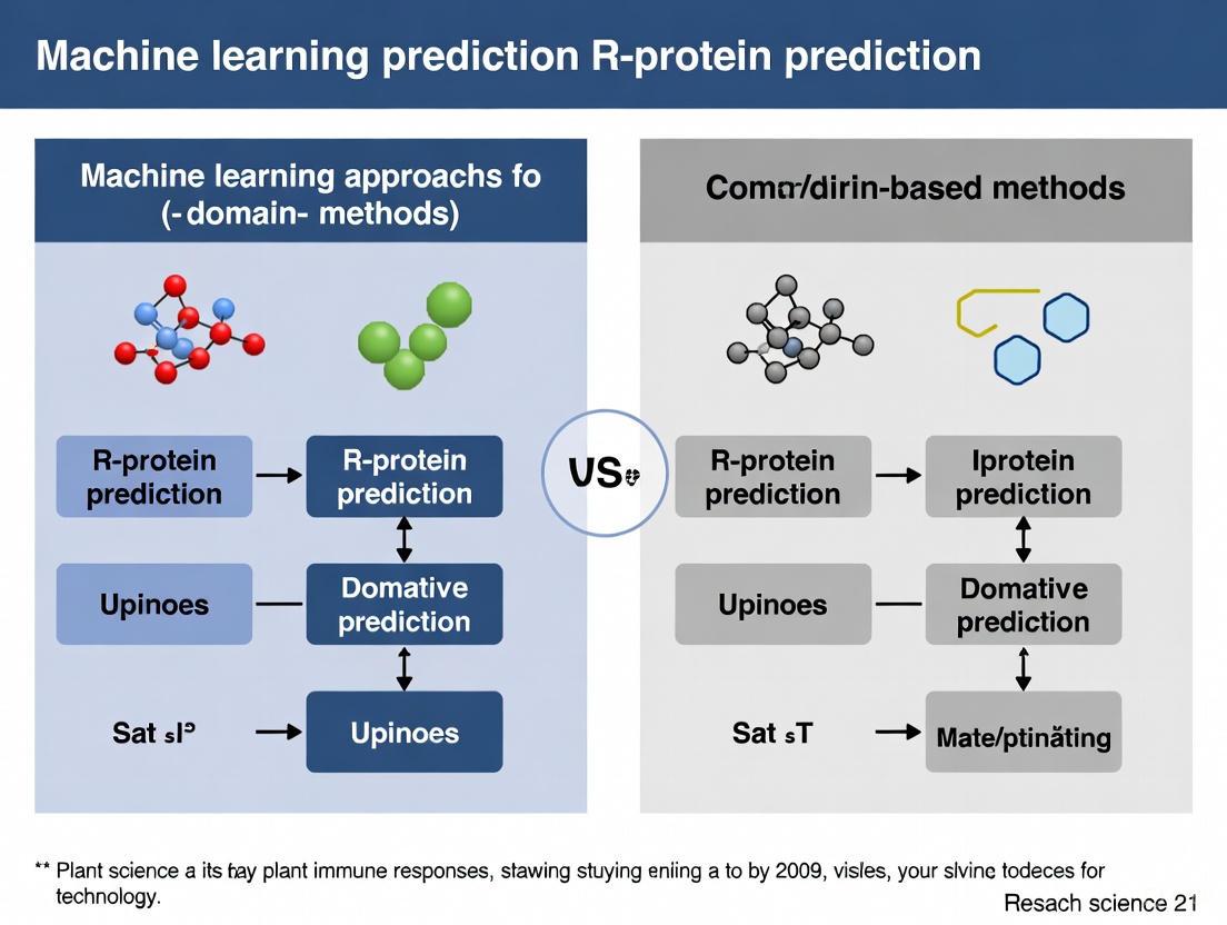
Domain-Based Methods vs. Machine Learning: The New Frontier in Protein Function Prediction
Accurately predicting protein function is a central challenge in biology, with direct implications for understanding disease mechanisms and drug discovery.
Domain-Based Methods vs. Machine Learning: The New Frontier in Protein Function Prediction
Abstract
Accurately predicting protein function is a central challenge in biology, with direct implications for understanding disease mechanisms and drug discovery. This article provides a comprehensive analysis for researchers and drug development professionals, comparing traditional domain-based methods with emerging machine learning (ML) and deep learning (DL) approaches. We explore the foundational principles of both paradigms, detail the core architectures of modern ML models like Graph Neural Networks (GNNs) and protein language models, and address critical challenges such as data scarcity and model interpretability. Through a rigorous validation of performance metrics and real-world applications, we demonstrate that the most powerful solutions often emerge from the integration of domain-guided insights with the predictive power of deep learning, paving the way for more accurate and interpretable functional annotation at a proteome-wide scale.
From Sequence to Function: Core Principles of Domain Analysis and Machine Learning
Protein domains are widely recognized as the fundamental structural, functional, and evolutionary units of proteins [1]. These conserved segments of polypeptide chains fold into distinct three-dimensional structures independently and serve as the essential building blocks that combine to form multidomain proteins [1]. Through evolutionary processes, domain families have expanded into multiple members that appear in diverse configurations with other domains, continually evolving new specificities for interacting partners [2]. This combinatorial expansion explains much of the functional diversity observed in modern proteomes, with domains acting as evolutionary modular units that can be reused, repurposed, and recombined through genetic mechanisms [1].
The Domain Hypothesis provides a powerful framework for understanding protein evolution, suggesting that the staggering diversity of protein functions arises not predominantly from the de novo creation of entirely new sequences, but from the strategic recombination of a finite set of stable, folded domains [1]. This modular paradigm has transformed computational biology, enabling researchers to predict protein function through domain architecture analysis and develop machine learning approaches that leverage domain properties for protein classification [3] [4]. Nowhere is this more evident than in the study of plant resistance (R) proteins, where domain combinations directly determine pathogen recognition capabilities and immune signaling functions [5].
Domain Architecture of Plant Resistance Proteins
Major R Protein Classes and Their Domain Composition
Plant resistance proteins constitute a critical component of the plant immune system, recognizing pathogen effector molecules and initiating defense responses [5]. These proteins typically contain characteristic domain arrangements that define their mechanistic class and function. The major classes include:
- TNL proteins (TIR-NBS-LRR): Contain Toll/Interleukin-1 Receptor (TIR), Nucleotide-Binding Site (NBS), and Leucine-Rich Repeat (LRR) domains
- CNL proteins (CC-NBS-LRR): Feature Coiled-Coil (CC), NBS, and LRR domains
- RLK proteins (Receptor-Like Kinases): Comprise kinase (KIN) domains with extracellular LRR regions
- RLP proteins (Receptor-Like Proteins): Contain LRR domains but lack intracellular kinase domains
- Kinase proteins: Consist primarily of kinase domains, like the tomato PTO gene product [5]
Beyond these well-characterized classes, genomic analyses have revealed numerous atypical domain associations that may represent evolutionary innovations in plant immunity [5]. The distribution of these domain arrangements follows a distinct pattern where architectural complexity is inversely correlated with frequency—simpler domain associations are more common than complex multidomain arrangements [5].
Table 1: Major Plant R Protein Classes and Their Domain Architectures
| R Protein Class | Domain Architecture | Localization | Recognition Mechanism |
|---|---|---|---|
| TNL (TIR-NBS-LRR) | TIR-NBS-LRR | Cytoplasmic | Intracellular pathogen effector recognition |
| CNL (CC-NBS-LRR) | CC-NBS-LRR | Cytoplasmic | Intracellular pathogen effector recognition |
| RLK (Receptor-like kinase) | eLRR-TM-KIN | Membrane-bound | Surface recognition of PAMPs/MAMPs |
| RLP (Receptor-like protein) | eLRR-TM | Membrane-bound | Surface recognition without signaling domain |
| Kinase (e.g., PTO) | KIN | Cytoplasmic | Kinase-mediated signaling cascades |
Evolutionary Patterns in R Protein Domain Organization
The evolutionary history of R protein domains reveals fascinating patterns of combinatorial explosion. Genomic analyses of 33 plant species identified 4,409 putative R-proteins that could be classified into 22 distinct subfamilies based on domain composition [5]. Remarkably, approximately 40% of these proteins consisted of single domains, while associations comprising two to five domains displayed decreasing frequency with increasing complexity [5]. This distribution strongly supports the domain hypothesis, demonstrating that nature favors certain domain combinations while avoiding others—only 22 out of 31 theoretically possible domain combinations were actually observed [5].
The NBS domain emerged as the most versatile, appearing in 13 different domain classes, followed by LRR (12 classes), KIN (9 classes), and TIR (8 classes) [5]. Certain domain pairs showed preferential associations, particularly LRR-NBS and LRR-KIN, which appeared in 8 and 6 domain classes respectively [5]. These combinatorial preferences reflect functional constraints and evolutionary trajectories that have shaped the plant immune repertoire.
Computational Approaches for R Protein Prediction
Domain-Centric Methodologies
Traditional computational approaches for identifying R proteins have relied heavily on domain-centric methodologies that leverage sequence alignment and domain fingerprinting. These methods include:
- NLR-parser: Utilizes motif alignment and search tool (MAST) to identify NLR-like sequences based on conserved domain motifs [3] [4]
- RGAugury: Integrates multiple computational tools including BLAST, HMMER3, Phobius, and TMHMM to predict various R protein subclasses [3] [4]
- Restrepo-Montoya pipeline: Classifies RLK and RLP proteins using SignalP, TMHMM, and PfamScan for domain identification [3]
While these methods provide valuable insights, they suffer from inherent limitations. Sequence alignment-based approaches typically exhibit low sensitivity and are computationally intensive, making them poorly suited for identifying divergent R proteins with low sequence similarity to known counterparts [3] [4]. Furthermore, these methods fundamentally rely on pre-existing knowledge of domain signatures, potentially missing novel or highly divergent resistance protein families.
Machine Learning Paradigms
To overcome the limitations of domain-centric methods, researchers have developed sophisticated machine learning approaches that can identify R proteins based on sequence features beyond known domain signatures:
- DRPPP: A support vector machine (SVM)-based tool that achieved 91.11% prediction accuracy by integrating 10,270 features extracted using 16 different methods [6]
- prPred: Utilizes k-spaced amino acid pairs (CKSAAPs) and k-spaced amino acid group pairs (CKSAAGPs) with SVM classification, achieving 93.5% accuracy [4]
- prPred-DRLF: Employs bi-directional long short-term memory (BiLSTM) and unified representation (UniRep) embedding with light gradient boosting machine (LGBM) classification, reaching 95.6% accuracy [3]
- StackRPred: A recently developed method that uses residue energy content matrices (RECM) with a stacking ensemble framework, outperforming previous state-of-the-art methods [3]
Table 2: Performance Comparison of Machine Learning Methods for R Protein Prediction
| Method | Algorithm | Key Features | Accuracy | Strengths |
|---|---|---|---|---|
| DRPPP | Support Vector Machine | 10,270 features from 16 methods | 91.11% | Comprehensive feature coverage |
| prPred | Support Vector Machine | CKSAAP, CKSAAGP | 93.5% | Two-step feature selection |
| prPred-DRLF | BiLSTM + LightGBM | UniRep embeddings | 95.6% | Handles long-range dependencies |
| StackRPred | Stacking Ensemble | RECM, PsePSSM, DWT | Highest | Captures structural energy properties |
| NBSPred | Support Vector Machine | Electronic annotation | Not reported | Early machine learning approach |
These machine learning methods demonstrate a significant performance advantage over traditional domain-based approaches, particularly for identifying R proteins with low sequence similarity to known families. However, they face their own challenges, including the black box problem of interpretability and substantial computational resource requirements for training [3].
Integrated Domain-ML Approaches and Protocols
The CoDIAC Framework for Domain Interface Analysis
Recent advances have begun to bridge the gap between domain-centric and machine learning approaches. The CoDIAC (Comprehensive Domain Interface Analysis of Contacts) framework represents a novel structure-based interface analysis method that maps domain interfaces from experimental and predicted structures [2]. This Python-based tool performs contact mapping of domains to yield insights into domain selectivity, conservation of domain-domain interfaces across proteins, and conserved posttranslational modifications relative to interaction interfaces [2].
When applied to the human Src homology 2 (SH2) domains, CoDIAC revealed coordinated regulation of SH2 domain binding interfaces by tyrosine and serine/threonine phosphorylation and acetylation, suggesting that multiple signaling systems can regulate protein activity and domain interactions in a coordinated manner [2]. This approach demonstrates how machine learning can enhance our understanding of domain function beyond what traditional domain analysis provides.
Experimental Protocol: Predicting R Proteins Using StackRPred
Protocol Objective: Identify plant resistance proteins from protein sequences using the StackRPred framework [3]
Workflow Overview:
Step-by-Step Procedure:
Data Preparation
- Obtain protein sequences in FASTA format
- Curate training data with known R proteins and non-R proteins at appropriate ratio (typically 1:2 positive:negative)
- For the standard StackRPred dataset: 152 R proteins and 304 non-R proteins [3]
Feature Extraction
- Calculate Residue Energy Content Matrices (RECM) based on physicochemical properties
- RECM captures energy information between residue pairs that ensures protein structural stability [3]
- Extract Pseudo Position-Specific Score Matrix (PsePSSM) features from RECM
- Apply Discrete Wavelet Transform (DWT) to extract multi-resolution features
Feature Selection
- Implement two-step feature selection using SVM-Recursive Feature Elimination (SVM-RFE)
- Apply Correlation-Based feature selection (CBR) to remove redundant features
- Generate optimized feature set for model training
Model Training - Base Layer
- Train multiple base classifiers including:
- eXtreme Gradient Boosting (XGBoost)
- Support Vector Machine (SVM)
- K-Nearest Neighbors (KNN)
- Gradient Boosting Decision Tree (GBDT)
- Light Gradient Boosting Machine (LightGBM)
- Random Forest (RF)
- Use 5-fold cross-validation for parameter optimization
- Train multiple base classifiers including:
Model Training - Meta Layer
- Use predictions from base classifiers as input features
- Train SVM classifier as meta-learner to make final predictions
- Validate model using independent test set (typically 20% of data)
Prediction and Validation
- Apply trained StackRPred model to unknown protein sequences
- Output probability scores for R protein classification
- Validate predictions with domain analysis (Pfam, InterProScan) for biological interpretation
Technical Notes:
- StackRPred specifically leverages pairwise energy content information that captures structural stability constraints [3]
- The stacking ensemble framework helps mitigate overfitting and improves generalization
- For optimal performance, ensure training data covers diverse R protein classes and plant species
Experimental Protocol: Domain-Centric R Protein Identification
Protocol Objective: Identify resistance proteins through systematic domain architecture analysis [5]
Workflow Overview:
Step-by-Step Procedure:
Comprehensive Domain Scanning
- Perform batch domain analysis using PfamScan against Pfam domain database
- Identify key R protein domains: NBS, LRR, TIR, CC, KIN, TM domains
- Use HMMER3 for sensitive domain detection based on hidden Markov models
- Run InterProScan for integrated domain signature analysis
Architecture Classification
- Classify sequences into major R protein classes based on domain combinations:
- TNL: TIR-NBS-LRR
- CNL: CC-NBS-LRR
- RLK: eLRR-TM-KIN
- RLP: eLRR-TM (no intracellular kinase)
- Kinase: Primary KIN domains
- Identify atypical domain associations beyond these major classes
- Classify sequences into major R protein classes based on domain combinations:
NBS-LRR Subtyping
- Apply coiled-coil prediction (nCoil) to distinguish CNL from NL proteins
- Use TIR domain signatures to identify TNL proteins
- Differentiate between CC-NBS-LRR and NBS-LRR architectures
Receptor Protein Differentiation
- Implement transmembrane domain prediction using TMHMM or Phobius
- Apply Fritz-Laylin method to distinguish RLK from RLP proteins
- Filter RLP proteins based on homology to known resistance-related RLPs
Atypical Association Analysis
- Identify unusual domain combinations not fitting major classes
- Assess conservation of atypical architectures across plant species
- Evaluate potential functional innovations in atypical associations
Validation and Interpretation:
- Cross-reference predictions with PRGdb (Plant Resistance Gene Database)
- Perform phylogenetic analysis of domain architectures
- Evaluate genomic context and syntenic relationships for novel candidates
Table 3: Key Research Reagents and Computational Resources for Domain-Centric Protein Analysis
| Resource | Type | Function | Application in R Protein Research |
|---|---|---|---|
| PRGdb | Database | Curated repository of known and putative R genes | Reference data for validation and comparison [5] [4] |
| Pfam | Domain Database | Collection of protein domain families | Domain fingerprinting and architecture analysis [4] |
| CDD | Domain Database | Conserved Domain Database for sequence classification | Identification of R protein-specific domain variants [1] |
| HMMER | Software Tool | Profile hidden Markov model implementation | Sensitive domain detection and family classification [4] |
| InterProScan | Software Tool | Integrated domain and signature recognition | Comprehensive domain architecture analysis [4] |
| TMHMM | Software Tool | Transmembrane helix prediction | Membrane protein classification (RLK vs RLP) [4] |
| nCoil | Software Tool | Coiled-coil domain prediction | CC domain identification in CNL proteins [4] |
| Phobius | Software Tool | Transmembrane topology and signal peptide prediction | Subcellular localization prediction [4] |
| CATH | Database | Hierarchical classification of protein structures | Structural domain evolutionary analysis [7] |
| SCOPe | Database | Structural Classification of Proteins extended | Fold-based domain classification and analysis [1] |
The Domain Hypothesis continues to provide a powerful conceptual framework for understanding protein evolution and function. By viewing proteins as modular assemblies of domain building blocks, researchers can decipher evolutionary histories and predict functional capabilities. This perspective is particularly valuable in the study of plant resistance proteins, where domain combinations directly determine pathogen recognition specificities and signaling capabilities.
The integration of machine learning with traditional domain analysis represents the future frontier in protein bioinformatics. Methods like StackRPred that incorporate residue energy information and structural constraints demonstrate how ML can enhance our ability to identify proteins based on fundamental principles beyond sequence similarity [3]. Meanwhile, approaches like CoDIAC show how computational methods can reveal new insights into domain function and regulation [2].
As structural prediction methods like AlphaFold continue to advance [8], the research community will increasingly leverage high-accuracy protein models to inform domain analyses and identify novel functional relationships. The emerging paradigm combines evolutionary principles embodied in the Domain Hypothesis with the pattern recognition power of machine learning, creating synergistic approaches that advance both basic knowledge and practical applications in crop improvement and disease resistance breeding programs.
The field of protein structure and function prediction has undergone a revolutionary shift, moving from reliance on manual feature engineering and domain-based knowledge to the adoption of deep learning systems capable of automated pattern discovery. This transition is central to modern computational biology, particularly in the critical area of R-protein prediction, where accurately modeling resistance protein structures is essential for understanding plant immunity and developing sustainable crop protection strategies. Whereas traditional methods depended on expert-defined features and homology-based modeling, contemporary artificial intelligence (AI) pipelines now integrate multisource deep learning potentials and iterative physical simulations to achieve unprecedented accuracy in predicting protein tertiary structures and their functional interactions [9] [10]. This paradigm shift not only enhances our predictive capabilities but also fundamentally changes the workflow from a heavily human-dependent process to an automated, data-driven discovery engine. These advances are pushing the boundaries of drug discovery, protein engineering, and functional annotation, establishing a new foundation for precision medicine and therapeutic development [11].
The Evolution of Methodologies in Protein Prediction
From Manual Feature Extraction to Deep Learning
The initial approach to protein prediction relied heavily on manual feature extraction, where scientists identified and quantified specific protein characteristics based on domain knowledge.
Table: Traditional Manual Feature Extraction Techniques in Protein Science
| Feature Category | Specific Examples | Application in Protein Prediction |
|---|---|---|
| Sequence-based Features | Amino acid composition, physiochemical properties (e.g., hydrophobicity, charge), sequence motifs | Primary structure analysis, homology detection |
| Structural Features | Secondary structure propensities, solvent accessibility, contact maps | Template-based modeling, fold recognition |
| Evolutionary Features | Position-Specific Scoring Matrix (PSSM), co-evolutionary signals | Threading, identifying remote homologs |
These manually curated features were then used as input for conventional machine learning models, such as support vector machines or hidden Markov models. The limitations were evident: the process was labor-intensive, required deep expertise, and could easily miss complex, non-linear relationships within the data [12] [13].
The advent of deep learning marked a decisive turn towards automated pattern discovery. Modern architectures, including deep residual convolutional networks and self-attention transformers, now directly ingest raw or minimally pre-processed data—such as amino acid sequences and multiple sequence alignments (MSAs)—to autonomously learn hierarchical feature representations. This capability is exemplified by models like ProtT5, which generates context-aware embeddings for each amino acid in a sequence, capturing complex biochemical properties without human guidance [7]. This shift has enabled the development of end-to-end prediction systems that seamlessly map sequence to structure, moving beyond the constraints of manual feature design [9] [10].
Comparative Analysis: Domain-Based vs. Machine Learning Approaches
The distinction between domain-based methods and modern machine learning is not merely technical but philosophical, reflecting a fundamental shift in how biological knowledge is encoded and applied.
Domain-based methods, such as template-based modeling (TBM), operate on the principle of homology. Tools like MODELLER and SwissPDBViewer rely on identifying known protein structures (templates) with significant sequence similarity to the target. The process involves sequence alignment, model building by transferring coordinates from the template, and subsequent refinement. While effective for targets with clear homologs, TBM fails for proteins with novel folds or minimal sequence similarity to any known structure [9].
In contrast, machine learning approaches, particularly template-free modeling (TFM) and ab initio methods, learn the underlying principles of protein folding from vast datasets. AlphaFold2 demonstrated the power of this paradigm by using an end-to-end deep learning model to achieve atomic accuracy. Subsequent innovations, such as D-I-TASSER, have further integrated these learned potentials with physics-based simulations, creating hybrid models that outperform purely AI-based or physical approaches [10]. These methods excel where domain-based methods struggle, particularly on "hard" targets with no evolutionary trace in databases.
Application Notes: Machine Learning in Action
High-Accuracy Protein Structure Prediction with D-I-TASSER
The D-I-TASSER (deep-learning-based iterative threading assembly refinement) pipeline represents a state-of-the-art hybrid approach that synergizes deep learning with physics-based simulations. Its performance on a benchmark of 500 non-redundant "Hard" protein domains underscores the success of this integrated paradigm. D-I-TASSER achieved an average TM-score of 0.870, significantly outperforming AlphaFold2 (TM-score = 0.829) and AlphaFold3 (TM-score = 0.849) [10]. The advantage was most pronounced on difficult targets, where D-I-TASSER's ability to leverage iterative physical simulations provided a critical edge over purely deep learning-based end-to-end systems.
Table: Performance Benchmark of Protein Structure Prediction Methods [10]
| Method | Average TM-score (500 Hard Targets) | Correct Folds (TM-score > 0.5) | Key Innovation |
|---|---|---|---|
| I-TASSER (Physics-based) | 0.419 | 145 | Template threading & physical force fields |
| C-I-TASSER (Hybrid) | 0.569 | 329 | Integration of deep-learning-predicted contacts |
| AlphaFold2.3 | 0.829 | N/A | End-to-end deep learning with MSAs |
| AlphaFold3 | 0.849 | N/A | Diffusion model & multimodality |
| D-I-TASSER (Hybrid-AI) | 0.870 | 480 | Multisource deep learning potentials + iterative physics simulations |
A critical innovation in D-I-TASSER is its specialized protocol for multidomain protein structure prediction. Unlike many earlier models focused on single domains, D-I-TASSER incorporates a domain partition and assembly module. It iteratively identifies domain boundaries, generates domain-level MSAs and spatial restraints, and then reassembles the full-chain model using hybrid domain-level and interdomain restraints. This capability is vital for accurately modeling the complex architectures of R-proteins and other eukaryotic proteins, over 80% of which contain multiple domains [10].
Predicting Functional Relationships from Sequence and Structure
Beyond tertiary structure, machine learning is revolutionizing the prediction of protein function and interactions. A key application is predicting de novo protein-protein interactions (PPIs)—interactions with no natural precedent. Traditional methods, including AlphaFold2, excel at predicting endogenous interactions but see a performance drop on de novo PPIs [14]. Novel algorithms are now tackling this challenge using graph-based atomistic models and methods that learn from molecular surface features, opening new avenues for drug discovery, such as designing molecular glues that rewire cellular functions [14].
Furthermore, integrating sequence and structural features significantly enhances protein function prediction. A LightGBM-based machine learning model demonstrated that combining features like full-length sequence identity, domain structural similarity, and pocket similarity outperforms models based on sequence alone. Feature importance analysis revealed that domain sequence identity, calculated through structural alignment, was the most influential predictor, highlighting the critical role of structural information in determining functional identity [15].
Experimental Protocols
Protocol 1: Predicting Protein Structural Similarity with Rprot-Vec
Purpose: To rapidly predict the structural similarity (TM-score) between two proteins using only their primary sequences, bypassing the need for resource-intensive 3D structure prediction or alignment.
Principle: The Rprot-Vec model is a deep learning framework that employs a ProtT5 encoder for context-aware sequence embedding, followed by Bidirectional Gated Recurrent Units (Bi-GRU) and multi-scale Convolutional Neural Networks (CNN) to extract global and local features. The final protein representations are used to compute a TM-score via cosine similarity [7].
Workflow:
Steps:
- Input Preparation: Prepare the two protein sequences of interest in standard FASTA format.
- Sequence Encoding: Pass each sequence through the ProtT5-XL-U50 model. This generates a 1024-dimensional vector representation for each amino acid, capturing its context within the entire sequence.
- Feature Extraction:
- The sequence of embeddings is processed by a Bidirectional GRU layer to capture long-range, bidirectional dependencies in the sequence.
- An attention layer is applied to weight the importance of different amino acid positions.
- Multi-scale CNN blocks, using convolution kernels of sizes 3 and 7, are applied in parallel to capture local sequence motifs of varying lengths.
- Vector Generation: The outputs are pooled and passed through a fully connected layer to produce a single, fixed-dimensional vector representation for each protein.
- Similarity Calculation: Compute the cosine similarity between the two protein vectors. The resulting value (ranging from 0 to 1) is treated as the predicted TM-score, where 1 indicates highly similar structures and 0 suggests orthogonality.
Applications: This protocol is ideal for large-scale protein homology detection, function inference for unannotated proteins, and pre-screening candidate proteins before detailed structural analysis [7].
Protocol 2: Hybrid Structure Prediction for Multidomain Proteins using D-I-TASSER
Purpose: To construct atomic-level structural models for complex multidomain proteins by integrating deep learning predictions with physics-based folding simulations.
Principle: D-I-TASSER combines multisource spatial restraints (from DeepPotential, AttentionPotential, and AlphaFold2) with replica-exchange Monte Carlo (REMC) simulations for structure assembly. Its specialized domain-splitting protocol handles the inherent complexity of multidomain proteins [10].
Workflow:
Steps:
- Deep MSA Construction: Iteratively search genomic and metagenomic databases (e.g., UniRef90) to construct deep multiple sequence alignments for the target protein.
- Domain Partition: The input sequence is analyzed to predict potential domain boundaries.
- Domain-Level Processing: For each identified domain, the pipeline generates domain-specific MSAs, threading alignments (using LOMETS3), and spatial restraints independently.
- Spatial Restraint Generation: Multisource deep learning potentials are used to predict inter-residue distance maps, contact maps, and hydrogen-bonding networks for both intra-domain and inter-domain regions.
- Iterative Threading Assembly Simulation:
- Template fragments from threading alignments are assembled using Replica-Exchange Monte Carlo (REMC) simulations.
- The simulation is guided by a hybrid force field that combines the deep learning restraints with a knowledge-based physical force field.
- For multidomain proteins, the assembly process uses the hybrid domain-level and inter-domain restraints to correctly orient and pack the domains.
- Model Selection and Refinement: The lowest energy models from the simulation are selected and refined at the atomic level to produce the final 3D structure.
Applications: This protocol is particularly suited for predicting the structures of large, multidomain proteins, such as many R-proteins, where accurate domain orientation is critical for understanding function and mechanism. Benchmark tests confirm its superior performance over other leading methods on such targets [10].
Table: Key Computational Tools and Databases for AI-Driven Protein Prediction
| Resource Name | Type | Primary Function | Relevance to R-protein Prediction |
|---|---|---|---|
| AlphaSync Database [16] | Database | Provides continuously updated, pre-computed protein structures from AlphaFold2. | Ensures researchers work with the most current structural models, including for plant proteomes, and provides data in a 2D tabular format ideal for machine learning. |
| D-I-TASSER [10] | Software Suite | Hybrid deep learning and physics-based protein structure prediction server. | Accurately models single-domain and multidomain R-protein structures, often outperforming end-to-end deep learning methods on difficult targets. |
| Rprot-Vec [7] | Software/Model | A deep learning model for fast protein structural similarity calculation from sequence. | Enables rapid homology detection and functional inference for novel R-protein sequences without requiring 3D structure prediction. |
| UniProt Knowledgebase | Database | Central repository of protein sequence and functional information. | The primary source for obtaining canonical and isoform sequences for R-proteins and related proteins for analysis. |
| CATH Database [7] | Database | Hierarchical classification of protein domain structures. | Used for training and benchmarking structure prediction models; provides evolutionary and functional insights into R-protein domains. |
The expansion of biological data has created a critical need for robust computational methods to predict protein function. This endeavor is central to understanding biological mechanisms and developing treatments for complex diseases. While traditional experimental methods for determining protein function are reliable, they are often time-consuming and costly, leaving the vast majority of protein sequences functionally uncharacterized [17]. This challenge is framed within a broader research thesis comparing machine learning (ML) approaches for whole-protein (R-protein) prediction against more traditional domain-based methods. Domain-based strategies are gaining traction as proteins are comprised of specific, functional domains that are closely related to their structures and functions [17]. The selection of appropriate training data is paramount for developing accurate and generalizable predictive models, guiding researchers toward the most insightful computational tools.
A suite of public databases provides the foundational data for training protein function prediction models. These resources can be broadly categorized into those providing protein-protein interaction (PPI) networks, experimentally determined structures, and computationally predicted structures. The table below summarizes the core data sources relevant to this field.
Table 1: Key Data Sources for Predictive Model Training
| Resource Name | Primary Content | Data Type | Scale (as of latest update) | Key Application in Model Training |
|---|---|---|---|---|
| STRING [18] [19] | Functional protein association networks | Predicted & curated interactions (physical, functional, pathway) | >20 billion interactions across 59.3 million proteins from 12,535 organisms [18] | Feature engineering for network-based and context-aware models |
| BioGRID [20] [21] | Physical and genetic interactions, PTMs, chemical associations | Manually curated interactions from literature | ~2.25 million non-redundant interactions from over 87,000 publications [20] | Gold-standard training sets and validation for high-confidence interaction prediction |
| RCSB PDB [22] | Experimentally determined 3D structures of proteins and nucleic acids | Curated atomic coordinates from X-ray, Cryo-EM, NMR | ~200,000 structures (implied) [23] | Source of ground-truth structural data for structure-based function prediction |
| AlphaFold DB [24] [23] | AI-predicted protein structures | Computationally predicted 3D models | Over 200 million entries, covering nearly the entire UniProt proteome [24] | Large-scale input features for structure-based models where experimental structures are absent |
| ModelArchive [22] | Repository of theoretical macromolecular structure models | Computationally predicted 3D models | Variable (community-contributed) | Supplementary source of structural models for training and analysis |
Experimental Protocols for Data Utilization
Protocol 1: Building a Domain-Guided Function Prediction Model with DPFunc
Application Note: This protocol details the use of protein structure and domain information to train DPFunc, a deep learning model that exemplifies the advantage of integrating domain guidance over whole-protein (R-protein) approaches for predicting Gene Ontology (GO) terms [17].
Workflow Diagram: DPFunc Model Architecture
Materials & Reagents:
- Input Data: Protein amino acid sequences and their corresponding 3D structures (can be experimentally derived from PDB or computationally predicted by AlphaFold2).
- Software Tools: InterProScan for domain detection, ESM-1b pre-trained protein language model, PyTorch/TensorFlow deep learning framework.
- Training Labels: Gene Ontology (GO) annotations for proteins, typically sourced from UniProt-GOA.
Methodology:
- Residue-Level Feature Learning: For a target protein sequence, initial residue-level features are generated using the pre-trained ESM-1b protein language model. Simultaneously, a contact map is constructed from the protein's 3D structure. Both are fed into Graph Convolutional Network (GCN) layers to update and learn the final residue-level features that incorporate structural context [17].
- Domain-Guided Feature Extraction: The target protein sequence is scanned with InterProScan to detect functional domains. These domain entries are converted into dense numerical representations via an embedding layer [17].
- Domain-Guided Attention: An attention mechanism, inspired by transformer architecture, integrates the protein-level domain features with the residue-level features. This step identifies and weights the importance of different residues in the structure with respect to the detected domains [17].
- Function Prediction & Post-Processing: The weighted residue features are aggregated into a protein-level feature vector and passed through fully connected layers to predict GO terms. A final post-processing step ensures the predictions are consistent with the hierarchical structure of the GO graph [17].
Expected Outcomes: DPFunc has been shown to outperform state-of-the-art sequence-based and structure-based methods. On a benchmark dataset, it achieved significant improvements in Fmax scores (e.g., 16% in Molecular Function, 27% in Cellular Component) over the next best structure-based method, GAT-GO [17].
Protocol 2: Constructing a Protein-Protein Interaction Network for Functional Inference
Application Note: This protocol describes the use of STRING and BioGRID to build and analyze a PPI network, which can serve as input features for network-based ML models or for direct biological interpretation.
Workflow Diagram: PPI Network Construction & Analysis
Materials & Reagents:
- Input Data: A list of seed protein identifiers (e.g., UniProt IDs, gene names) for the organism of interest.
- Software/Tools: STRING web interface or API, BioGRID web interface or downloadable files, network analysis tools (e.g., Cytoscape, NetworkX in Python).
Methodology:
- Data Retrieval: Query the STRING database using seed protein names and the target organism. Separately, query the BioGRID database for the same proteins to obtain high-confidence, manually curated physical and genetic interactions [18] [20] [21].
- Data Filtering and Merging: In STRING, set a minimum interaction score threshold (e.g., high confidence > 0.7) to reduce false positives [19]. In BioGRID, all interactions are experimentally supported. Merge the interaction lists from both sources, removing duplicate entries.
- Network Construction and Analysis: Build a network graph where nodes are proteins and edges are interactions. Use the merged data. Analyze the network's topology (e.g., identifying highly connected proteins) and perform functional enrichment analysis to find GO terms or pathways that are statistically over-represented in the network [19].
Expected Outcomes: This process generates a high-confidence PPI network that can reveal functional modules. The network itself, along with node centrality measures and enrichment results, can be used as features for machine learning models to predict protein function or to prioritize new candidate proteins for further study.
The Scientist's Toolkit: Essential Research Reagent Solutions
The following table catalogs key computational and data "reagents" essential for research in protein function and structure prediction.
Table 2: Key Research Reagent Solutions for Predictive Modeling
| Item Name | Function / Application | Relevant Database/Tool |
|---|---|---|
| Pre-trained Protein Language Model (ESM-1b) | Generates evolutionarily informed, residue-level feature embeddings from amino acid sequences. | DPFunc [17], ESMFold [22] |
| InterProScan | Scans protein sequences against signatures from multiple databases to detect functional domains and sites. | DPFunc protocol [17] |
| AlphaFold2 Predicted Structure | Provides atomic-level 3D protein models for sequences lacking experimental structures; input for structure-based models. | AlphaFold DB [24] [23] |
| CRISPR Phenotype Data (ORCS) | Provides curated gene-phenotype relationships from genome-wide CRISPR screens for functional validation. | BioGRID ORCS [20] [21] |
| Gene Ontology (GO) Annotations | Provides standardized functional terms (Molecular Function, Biological Process, Cellular Component) for model training and evaluation. | Model benchmarking [17] |
| Graph Neural Network (GNN) | Deep learning architecture for learning from graph-structured data like PPI networks or protein contact maps. | DPFunc, DeepFRI [17] |
Discussion and Future Perspectives
The integration of data from STRING, BioGRID, PDB, and AlphaFold DB provides a multi-faceted evidence stream that is crucial for advancing protein function prediction. The comparative analysis between R-protein and domain-based methods, as exemplified by DPFunc, strongly indicates that guiding models with domain information unlocks greater accuracy and interpretability by pinpointing key functional residues within the structure [17].
Future developments will likely focus on the seamless integration of these massive databases into end-to-end prediction pipelines. Furthermore, the accurate prediction of multi-domain protein structures remains a challenge, with new hybrid approaches like D-I-TASSER that integrate deep learning with physical simulations showing promise in surpassing the performance of end-to-end ML systems like AlphaFold2 for these complex targets [10]. As the volume of curated interaction data in resources like BioGRID continues to grow monthly, and as structure prediction databases expand, the potential for training even more powerful and generalizable models will become a reality, profoundly impacting biological discovery and drug development.
The rapid accumulation of protein sequences from genome sequencing projects has dramatically outpaced experimental function annotation, leaving over 30% of protein-coding genes with unknown functions and creating a vast "Unknome" [25] [26]. This annotation gap represents a critical challenge and opportunity in biological research, as many valuable proteins potentially catalyzing novel enzymatic reactions remain undiscovered among the vast number of function-unknown proteins [15]. Traditional computational methods that rely solely on sequence homology often fail to accurately predict functions for proteins with no evolutionary precedence, particularly for those with small sequence variations that correspond to different functions or for pseudoenzymes that lack key catalytic residues [25].
This application note examines advanced machine learning strategies to address this challenge, focusing particularly on the emerging paradigm of integrating domain-guided and structure-based information to improve functional inference. We frame these methodologies within the context of a broader thesis contrasting general R-protein prediction (methods relying on sequence-level representations from protein language models) with domain-based methods (approaches that explicitly incorporate structural domain information) [26] [15]. The following sections provide detailed protocols for implementing these approaches, along with performance benchmarks and practical reagent solutions for researchers tackling the Unknome.
Core Methodologies and Experimental Protocols
Domain-Guided Deep Learning with DPFunc
Principle: DPFunc addresses the Unknome challenge by leveraging domain information within protein sequences to guide the model toward learning the functional relevance of amino acids in their corresponding structures, highlighting structure regions closely associated with functions [26]. This approach is particularly valuable for detecting key residues or regions in protein structures that exhibit strong functional correlations, even when overall sequence similarity to training data is low.
Experimental Protocol:
Input Data Preparation:
Domain Information Extraction:
- Process target protein sequences with InterProScan to identify functional domains by comparing them to background databases [26].
- Convert identified domains into dense vector representations using embedding layers that capture their unique characteristics.
Feature Learning and Integration:
- Extract initial residue-level features using pre-trained protein language models (ESM-1b, ProtT5) [26] [27].
- Construct protein contact maps based on 3D coordinates from protein structures.
- Process contact maps and residue-level features through Graph Convolutional Network (GCN) layers to update and learn final residue-level features, implementing a residual learning framework to maintain gradient flow [26].
- Implement attention mechanisms to interweave protein-level domain features and residue-level features, assessing the importance of each residue for functional prediction.
Function Prediction and Validation:
- Generate protein-level features through weighted summation of residue-level features and their importance scores.
- Annotate functions through fully connected layers using Gene Ontology (GO) terms across molecular function (MF), cellular component (CC), and biological process (BP) ontologies [26].
- Apply post-processing to ensure consistency with the hierarchical structure of GO terms.
- Validate predictions against held-out test sets with experimental annotations, using standard metrics from Critical Assessment of Functional Annotation (CAFA) challenges [26].
Sequence-Structure Integration for Functional Identity Prediction
Principle: This method predicts whether two proteins catalyze the same enzymatic reaction by integrating multiple similarity metrics derived from both sequence and structural features [15]. The approach utilizes predicted structural models from AlphaFold2, performing pocket detection and domain decomposition to extract features that are more conserved than full-sequence similarity.
Experimental Protocol:
Structure Prediction and Processing:
- Generate 3D structural models for all query sequences using AlphaFold2 [15].
- Perform pocket detection on structural models using tools like Fpocket or DeepPocket to identify potential binding sites [27] [15].
- Conduct domain decomposition for each structural model to identify functionally distinct regions.
Multi-Feature Similarity Calculation:
- Calculate full-length sequence identity using standard alignment tools (BLAST, DIAMOND) [26] [15].
- Compute domain sequence identity through structural alignment of decomposed domains.
- Determine pocket similarity by comparing geometric and chemical properties of detected binding pockets.
- Calculate overall structural similarity using TM-align or US-align to generate TM-scores [15] [7].
Model Training and Prediction:
- Compile feature set including full-length sequence identity, domain structural similarity, and pocket similarity for protein pairs [15].
- Train LightGBM classifier on known enzyme pairs with verified functional identity.
- Perform feature importance analysis to identify most predictive features (domain sequence identity typically shows highest importance) [15].
- Predict functional identity for novel protein pairs by feeding extracted features into trained model.
Workflow Visualization
Domain-Guided vs. R-Protein Prediction Workflow - This diagram contrasts the two primary approaches for Unknome protein function prediction, showing their shared feature extraction layers and divergent integration strategies.
Performance Benchmarks
Function Prediction Accuracy
Table 1: Performance comparison of protein function prediction methods on PDB dataset (Fmax scores)
| Method | Molecular Function (MF) | Cellular Component (CC) | Biological Process (BP) |
|---|---|---|---|
| Blast | 0.432 | 0.381 | 0.321 |
| DeepGO | 0.541 | 0.502 | 0.453 |
| DeepFRI | 0.612 | 0.558 | 0.507 |
| GAT-GO | 0.635 | 0.584 | 0.532 |
| DPFunc (w/o post) | 0.686 | 0.613 | 0.574 |
| DPFunc (with post) | 0.737 | 0.741 | 0.654 |
DPFunc demonstrates significant performance improvements over existing state-of-the-art methods, with particularly notable gains in cellular component prediction after implementing post-processing procedures to ensure consistency with GO term hierarchies [26]. The method outperforms both sequence-based approaches (Blast, DeepGO) and structure-based methods (DeepFRI, GAT-GO), highlighting the value of domain guidance in structure-based function prediction.
Structural Modeling Performance
Table 2: Protein structure prediction accuracy on "Hard" targets (TM-score)
| Method | Average TM-score | Correct Folds (TM-score > 0.5) | Parameters |
|---|---|---|---|
| I-TASSER | 0.419 | 145/500 (29%) | - |
| C-I-TASSER | 0.569 | 329/500 (66%) | - |
| AlphaFold2.3 | 0.829 | 452/500 (90%) | ~93 million |
| AlphaFold3 | 0.849 | 465/500 (93%) | - |
| D-I-TASSER | 0.870 | 480/500 (96%) | - |
| Rprot-Vec | - | 65.3% (TM-score > 0.8) | 41% of TM-vec |
Advanced hybrid approaches like D-I-TASSER, which integrate deep learning with physics-based folding simulations, demonstrate superior performance on challenging protein targets, particularly for non-homologous and multidomain proteins [10]. For large-scale applications, sequence-based structural similarity predictors like Rprot-Vec offer efficient alternatives, achieving 65.3% accuracy in identifying homologous proteins (TM-score > 0.8) using only sequence information [7].
Research Reagent Solutions
Table 3: Essential tools and databases for Unknome protein function prediction
| Resource | Type | Function | Access |
|---|---|---|---|
| AlphaFold2/3 | Structure Prediction | Predicts 3D protein structures from sequence | https://alphafold.ebi.ac.uk/ |
| ESMFold | Structure Prediction | High-speed structure prediction for large datasets | https://esmatlas.com/ |
| InterProScan | Domain Analysis | Identifies functional domains in protein sequences | https://www.ebi.ac.uk/interpro/ |
| DPFunc | Function Prediction | Domain-guided deep learning for function annotation | https://github.com/ [26] |
| Rprot-Vec | Similarity Prediction | Sequence-based structural similarity calculation | https://github.com/ [7] |
| UniProt | Database | Comprehensive protein sequence and functional data | https://www.uniprot.org/ |
| CATH | Database | Protein structure classification for benchmarking | http://www.cathdb.info/ [7] |
| STRING | Database | Known and predicted protein-protein interactions | https://string-db.org/ [28] |
| ProtT5 | Feature Extraction | Protein language model for sequence representations | https://github.com/ [27] [7] |
| TM-align | Structure Alignment | Protein structural similarity calculation | https://zhanggroup.org/TM-align/ [7] |
The challenge of the Unknome requires moving beyond traditional homology-based approaches toward integrated methodologies that leverage both sequence and structural information. Domain-guided methods like DPFunc demonstrate that explicitly modeling functional units within proteins significantly enhances prediction accuracy for poorly characterized proteins [26]. Meanwhile, hybrid structure prediction approaches like D-I-TASSER show that combining deep learning with physics-based simulations improves modeling of complex multidomain proteins [10].
For researchers investigating the Unknome, the experimental protocols outlined here provide practical pathways for implementing these advanced methods. The continuing development of protein language models, geometric deep learning, and multi-scale modeling promises to further accelerate our ability to illuminate the functional dark matter of the proteome, with profound implications for drug discovery and protein engineering [29] [14].
Architectures in Action: Deep Learning Models and Domain-Integrated Pipelines
Graph Neural Networks (GNNs) for Modeling Protein Structures and Interaction Networks
Graph Neural Networks (GNNs) have emerged as transformative tools in computational biology, providing a natural framework for modeling the inherent graph structures of biological systems. For protein-related tasks, GNNs excel at representing proteins as residue contact networks or atoms as nodes with edges representing spatial relationships, enabling the capture of complex structural patterns and interaction dynamics [28] [30]. This approach has demonstrated remarkable success across diverse applications including protein-protein interaction prediction, protein function analysis, and molecular property prediction for drug discovery [28] [31] [32].
The integration of GNNs into structural bioinformatics represents a significant advancement over traditional domain-based prediction methods and sequence-only machine learning approaches. While methods like I-TASSER series pipelines have successfully integrated deep learning with physics-based simulations for high-accuracy protein structure prediction [10], GNNs offer unique advantages for modeling interaction networks and structural relationships that are challenging for conventional approaches.
Performance Benchmarking of Computational Methods
Quantitative Comparison of Protein Structure Prediction Methods
Table 1: Benchmark performance of protein structure prediction methods on 500 non-redundant "Hard" domains from SCOPe, PDB, and CASP 8-14 experiments
| Method | Average TM-Score | Correctly Folded Targets (TM-score > 0.5) | Key Characteristics |
|---|---|---|---|
| D-I-TASSER | 0.870 | 480 | Hybrid approach integrating multisource deep learning potentials with iterative threading assembly simulations [10] |
| AlphaFold2.3 | 0.829 | N/R | End-to-end deep learning architecture [10] |
| AlphaFold3 | 0.849 | N/R | Enhanced with diffusion samples [10] |
| C-I-TASSER | 0.569 | 329 | Uses deep-learning-predicted contact restraints [10] |
| I-TASSER | 0.419 | 145 | Traditional template-based folding simulations [10] |
Performance of Specialized Protein Prediction Tools
Table 2: Performance metrics of specialized deep learning tools for protein prediction tasks
| Tool | Application Domain | Accuracy | Dataset | Key Innovation |
|---|---|---|---|---|
| PRGminer | Plant resistance gene prediction | 98.75% (training), 95.72% (independent testing) [33] | Plant R-genes from Phytozome, Ensemble Plants, NCBI [33] | Deep learning with dipeptide composition features [33] |
| Plant RBP Predictor | RNA-binding protein prediction in plants | 97.20% (5-fold CV), 99.72% (independent set) [34] | 4,992 balanced sequences [34] | Ensemble learning integrating shallow and deep learning with KPC encoding [34] |
| Domain-Disease Association | Protein domain-disease association | AUC: 0.94 [35] | Heterogeneous network of domains, proteins, diseases [35] | XGBOOST classifier with meta-path topological features [35] |
GNN Architectures for Protein Analysis
Core GNN Variants and Their Protein Applications
The field has developed several specialized GNN architectures tailored to protein data:
Graph Convolutional Networks (GCNs) apply convolutional operations to aggregate information from neighboring nodes, effectively capturing local structural patterns in protein residue networks [28] [30].
Graph Attention Networks (GATs) incorporate attention mechanisms to adaptively weight the importance of neighboring nodes, particularly useful for identifying critical interaction sites in protein complexes [28] [30].
Graph Autoencoders (GAE) utilize encoder-decoder frameworks to generate compact, low-dimensional node embeddings for tasks like protein function prediction and interaction characterization [28].
Kolmogorov-Arnold GNNs (KA-GNNs) represent a recent innovation integrating Fourier-based KAN modules into GNN components, enhancing expressivity and interpretability for molecular property prediction [32].
Experimental Protocol: Protein-Protein Interaction Prediction Using GNNs
Objective: Predict binary protein-protein interactions from structural information and sequence features [30].
Workflow:
Graph Construction:
- Input: Protein Data Bank (PDB) files containing 3D atomic coordinates
- Node definition: Represent each amino acid residue as a node
- Edge definition: Connect two nodes if they contain atoms within a threshold distance (typically 4-8Å), creating a residue contact network
- Graph representation: Undirected graph G = (V, E) where V = {v₁, v₂, ..., vₙ} represents residues and E = {e₁, e₂, ..., eₘ} represents spatial proximity [30]
Feature Extraction:
- Utilize protein language models (SeqVec or ProtBert) to generate feature vectors for each residue directly from protein sequences
- Alternative: Physicochemical properties or one-hot encoding of amino acids
- Output: Feature matrix X ∈ R^(n×d) where n is number of residues and d is feature dimension [30]
Model Architecture:
- Implement either GCN or GAT framework
- GCN layer operation: H⁽ˡ⁺¹⁾ = σ(ÃH⁽ˡ⁾W⁽ˡ⁾) where à is normalized adjacency matrix, H⁽ˡ⁾ is node features at layer l, W⁽ˡ⁾ is trainable weights [30]
- GAT layer: Compute attention coefficients αᵢⱼ = softmaxₑ(LeakyReLU(aᵀ[Whᵢ∥Whⱼ])) to weight neighbor contributions [30]
Classification:
- Concatenate feature vectors of protein pairs
- Feed to classifier with two hidden layers and output layer with sigmoid activation
- Use binary cross-entropy loss for training [30]
Datasets: Human PPI dataset (36,545 interacting pairs from HPRD) and S. cerevisiae dataset (22,975 interacting pairs from DIP) [30].
Advanced GNN Frameworks
Kolmogorov-Arnold GNNs (KA-GNNs) for Molecular Property Prediction
Recent advances have integrated Kolmogorov-Arnold Networks (KANs) with GNNs to create more expressive and efficient architectures. KA-GNNs replace standard multilayer perceptrons (MLPs) in GNN components with KAN modules based on learnable univariate functions [32].
Architecture Variants:
- KA-GCN: Integrates Fourier-based KAN modules into Graph Convolutional Networks
- KA-GAT: Enhances Graph Attention Networks with KAN-based transformations
Key Innovations:
- Fourier-series-based univariate functions to capture both low-frequency and high-frequency structural patterns
- Integration into all three GNN components: node embedding, message passing, and readout
- Theoretical guarantees of strong approximation capabilities based on Carleson's convergence theorem [32]
Experimental Results: KA-GNNs consistently outperform conventional GNNs across seven molecular benchmarks in both prediction accuracy and computational efficiency [32].
Visualization: GNN Workflow for Protein-Protein Interaction Prediction
Graph Workflow for PPI Prediction: From structural data to interaction prediction
Research Reagent Solutions
Table 3: Essential research reagents and computational tools for GNN-based protein analysis
| Resource Category | Specific Tools/Databases | Primary Function | Application Context |
|---|---|---|---|
| Protein Databases | PDB, UniProtKB, Phytozome [34] [33] | Source of protein structures, sequences, and annotations | Data acquisition for model training and validation |
| PPI Databases | STRING, BioGRID, IntAct, DIP, HPRD [28] [30] | Repository of known and predicted protein-protein interactions | Ground truth data for PPI prediction models |
| Domain Databases | Pfam, InterPro [33] | Protein domain families and functional domains | Feature extraction and functional annotation |
| Language Models | SeqVec, ProtBert, ESM [30] | Generate residue-level feature vectors from sequences | Node feature initialization for GNNs |
| GNN Frameworks | PyTorch Geometric, DGL, TensorFlow GNN | Implement GCN, GAT, and other GNN architectures | Model development and training |
| Specialized Tools | D-I-TASSER, PRGminer, RBPLight [10] [34] [33] | Domain-specific prediction pipelines | Benchmarking and comparative analysis |
Integration with Domain-Based Methods
The relationship between emerging GNN approaches and traditional domain-based methods represents a critical research frontier. Domain-based protein prediction methods, which identify structural and functional units through techniques like I-TASSER, have demonstrated remarkable success, with D-I-TASSER achieving 81% coverage of protein domains in the human proteome [10]. However, GNNs offer complementary strengths, particularly for modeling higher-order interactions and complex relationships in protein networks.
Recent research indicates that hybrid approaches integrating domain knowledge with graph-based learning show particular promise. For instance, heterogeneous network methods that incorporate domain information have achieved AUC scores of 0.94 for predicting domain-disease associations [35]. Similarly, the AG-GATCN framework integrates GAT and temporal convolutional networks to provide robust solutions against noise interference in protein-protein interaction analysis [28].
Visualization: Domain-Based vs. GNN Approaches
Comparative Methodologies: Domain-based and GNN approaches to protein analysis
GNNs have established themselves as powerful frameworks for modeling protein structures and interaction networks, offering distinct advantages for capturing complex relational patterns in biological data. While domain-based methods continue to excel at fundamental structure prediction tasks, GNNs provide complementary capabilities for understanding higher-order interactions and network-level properties. The integration of these approaches, along with emerging innovations such as KA-GNNs and multimodal learning frameworks, represents the most promising direction for future research. As these methodologies continue to evolve, they will increasingly enable researchers to unravel the complex relationship between protein structure, interaction networks, and biological function, with significant implications for drug discovery and therapeutic development.
Protein Language Models (ESM, ProtBERT) for Sequence-Based Functional Inference
Protein Language Models (pLMs), such as ESM (Evolutionary Scale Modeling) and ProtBERT, represent a transformative advance in computational biology, leveraging architectures from natural language processing to infer protein function directly from amino acid sequences. These models are trained on millions of protein sequences through self-supervised learning, learning the underlying "grammar" and "syntax" of proteins, which allows them to capture complex biological properties including evolutionary relationships, structural constraints, and functional motifs [36] [37]. This capability is challenging the long-standing dominance of methods that rely on evolutionary information derived from multiple sequence alignments (MSAs) [37] [38]. For researchers focused on R-proteins or any protein class, pLMs offer a powerful, fast, and MSA-free alternative for functional annotation, often achieving state-of-the-art performance, particularly for proteins with few known homologs [38] [39]. This application note details the use of ESM and ProtBERT for sequence-based functional inference, providing structured experimental protocols, performance comparisons, and practical toolkits for scientists and drug development professionals.
Prominent Protein Language Models
- ESM Model Family: A series of transformer-based models developed by Meta. The ESM-2 series, which succeeded ESM-1b, scales from 8 million to 15 billion parameters [40] [39]. These models are pre-trained on millions of protein sequences from the UniProt database using a masked language modeling objective, where the model learns to predict randomly masked amino acids in a sequence [39]. The recently introduced ESM3 is a generative model with 98 billion parameters [40].
- ProtBERT Model Family: Another prominent class of transformer-based pLMs, pre-trained on large datasets including UniProtKB and the BFD (Big Fantastic Database) [38]. ProtBERT models are also widely used as feature extractors for downstream prediction tasks.
Primary Applications in Functional Inference
pLMs excel in several key areas of protein functional inference, which are critical for research and drug development:
- Enzyme Commission (EC) Number Prediction: Accurately classifying enzymes into the hierarchical EC number system which defines the chemical reactions they catalyze [38].
- Gene Ontology (GO) Term Prediction: Predicting molecular functions, biological processes, and cellular components associated with a protein sequence [36] [41].
- Protein-Protein Interaction (PPI) Prediction: Identifying whether two proteins physically interact, a crucial aspect of understanding cellular signaling and disease mechanisms [42].
- Binding Site Prediction: Locating specific residues involved in binding small molecules, DNA, or other proteins, which is fundamental for drug design [39] [43].
- Variant Effect Prediction: Assessing the functional impact of missense mutations on protein function and interactions [42].
Performance Benchmarking and Quantitative Comparison
Performance on Enzyme Commission (EC) Prediction
Table 1: Comparative performance of pLMs and traditional methods on EC number prediction.
| Method | Input Type | Key Performance Insight | Relative Performance |
|---|---|---|---|
| BLASTp | Sequence & Homology | Slightly better overall performance; relies on sequence homologs in database [38]. | Benchmark |
| ESM2 (with DNN) | Sequence Embedding | Excels on difficult annotations and enzymes without close homologs (identity <25%) [38]. | Complementary to BLASTp |
| ProtBERT (with DNN) | Sequence Embedding | Surpasses one-hot encoding models; performance is strong but can be lower than ESM2 [38]. | Lower than ESM2 |
| One-hot Encoding DL Models | Raw Sequence | Suboptimal performance compared to pLM-based models [38]. | Lower than pLMs |
Performance on Protein-Protein Interaction (PPI) Prediction
Table 2: Performance of PLM-interact, a fine-tuned ESM-2 model, on cross-species PPI prediction (trained on human data).
| Test Species | PLM-interact (AUPR) | TUnA (AUPR) | TT3D (AUPR) |
|---|---|---|---|
| Mouse | 0.852 | 0.835 | 0.734 |
| Fly | 0.783 | 0.725 | 0.647 |
| Worm | 0.772 | 0.728 | 0.642 |
| Yeast | 0.706 | 0.641 | 0.553 |
| E. coli | 0.722 | 0.675 | 0.605 |
AUPR: Area Under the Precision-Recall Curve. A higher value indicates better performance. Results show PLM-interact achieves state-of-the-art cross-species generalization [42].
Impact of Model Size and Embedding Compression
Table 3: Practical considerations for selecting and using pLMs in research.
| Factor | Impact and Recommendation |
|---|---|
| Model Size | Larger models (e.g., ESM-2 15B) capture more complex patterns but are computationally expensive. Medium-sized models (eSM-2 650M, ESM C 600M) offer an optimal balance, performing nearly as well as larger models, especially when data is limited [40]. |
| Embedding Compression | For transfer learning, the mean pooling method (averaging embeddings across all sequence residues) consistently outperforms other compression methods (e.g., max pooling, PCA) across diverse tasks [40]. |
Experimental Protocols
Protocol 1: Gene Ontology (GO) Term Prediction Using Transfer Learning
This protocol describes how to use pLM embeddings as input features to a classifier to predict GO terms for a protein sequence.
1. Feature Extraction (Embedding Generation)
* Input: Protein amino acid sequence in FASTA format.
* Model Selection: Choose a pre-trained pLM, such as esm2_t33_650M_UR50D (ESM-2 with 650 million parameters).
* Software: Use the esm Python library or the transformers library for ProtBERT.
* Procedure:
* Load the pre-trained model and its corresponding tokenizer.
* Tokenize the input protein sequence.
* Pass the tokens through the model to extract the hidden representations (embeddings).
* Compression: Apply mean pooling along the sequence dimension to convert the per-residue embeddings (L x 1280) into a single, global protein embedding vector (1 x 1280), where L is the sequence length [40].
2. Classifier Training and Prediction * Input Features: The pooled protein embedding vector. * Model Architecture: Use a lightweight classifier, such as a fully connected Deep Neural Network (DNN) or a Multi-Layer Perceptron (MLP). For sequences, a BiLSTM network can also be effective [39]. * Training: * Use a dataset of protein sequences with known GO term annotations (e.g., from UniProt). * Frame the task as a multi-label classification problem, as a protein can have multiple GO terms. * Train the classifier using the pLM embeddings as input to predict the binary labels for each GO term. * Output: A list of predicted GO terms along with their association probabilities for the query sequence.
Protocol 2: Fine-tuning for Protein-Protein Interaction (PPI) Prediction
This protocol involves adapting a pre-trained pLM to the specific task of predicting interactions between two proteins, as exemplified by PLM-interact [42].
1. Model Architecture and Input Preparation * Base Model: Start with a pre-trained ESM-2 model (e.g., the 650M parameter version). * Input Format: Concatenate the amino acid sequences of the two candidate interacting proteins (Protein A and Protein B) into a single sequence string, separated by a special separator token. * Architecture Modification: The model must be configured to accept this longer, paired-sequence input.
2. Fine-tuning Procedure * Task Formulation: Treat PPI prediction as a binary classification task (interacting vs. non-interacting). * Training Objective: Use a combined loss function: * Next Sentence Prediction (NSP) Loss: A classification loss that teaches the model to predict the binary interaction label. * Masked Language Modeling (MLM) Loss: The original pre-training objective, which helps maintain the model's understanding of protein sequence semantics. * Balanced Loss: A weighting of 1:10 between the NSP classification loss and the MLM loss has been shown to be effective [42]. * Data: Fine-tune the model on a dataset of known interacting and non-interacting protein pairs (e.g., from human data in the Multi-Species PPI dataset).
3. Inference * Input the paired sequence of a novel protein pair into the fine-tuned PLM-interact model. * The model outputs a probability score indicating the likelihood of interaction.
Table 4: Key resources for implementing pLM-based functional inference.
| Resource Name | Type | Function and Application |
|---|---|---|
| ESM-2 / ProtBERT Pre-trained Models | Software Model | Foundational pLMs for generating protein sequence embeddings. Available via Hugging Face transformers or dedicated esm Python packages [40] [39]. |
| UniRef50 Database | Dataset | A non-redundant protein sequence cluster database used for pre-training pLMs and as a source of evolutionary information [41]. |
| UniProtKB | Dataset | A comprehensive repository of protein sequence and functional information, used for training and benchmarking prediction models [36] [39]. |
| PLM-interact | Software Model | A specialized, fine-tuned model for predicting protein-protein interactions, built upon ESM-2 [42]. |
| ESM-DBP | Software Model | A domain-adapted pLM, fine-tuned on DNA-binding proteins, which improves performance on DBP-related prediction tasks [39]. |
| BridgeNet | Software Model | A pre-trained framework that integrates sequence and structural information during training but only requires sequence for inference, enhancing property prediction [41]. |
Workflow and Conceptual Diagrams
Workflow for pLM-Based Functional Annotation
Diagram Title: pLM Functional Annotation Workflow
pLM Fine-tuning for PPI Prediction
Diagram Title: Fine-tuning pLM for PPI Prediction
The field of computational protein structure prediction has been transformed by the advent of advanced deep learning techniques. For over 50 years, predicting the three-dimensional structure that a protein will adopt based solely on its amino acid sequence represented one of the most important open challenges in biology [8]. Traditional experimental methods for determining protein structures, such as X-ray crystallography, nuclear magnetic resonance (NMR), and cryo-electron microscopy, are often costly, inefficient, and time-consuming [9]. The gap between known protein sequences and experimentally determined structures has created an urgent need for accurate computational approaches. This application note examines two revolutionary approaches—AlphaFold2 and D-I-TASSER—that address this challenge through fundamentally different methodologies, providing researchers with powerful tools for structure-based prediction in drug discovery and basic research.
Methodological Foundations
AlphaFold2: End-to-End Deep Learning Architecture
AlphaFold2 represents a purely deep learning-based approach to protein structure prediction. The system employs an entirely redesigned neural network-based model that incorporates physical and biological knowledge about protein structure into its deep learning algorithm [8]. The network directly predicts the 3D coordinates of all heavy atoms for a given protein using the primary amino acid sequence and aligned sequences of homologues as inputs.
The AlphaFold2 architecture comprises two main stages. First, the trunk of the network processes inputs through repeated layers of a novel neural network block termed "Evoformer," which produces representations for both multiple sequence alignments (MSAs) and residue pairs [8]. The Evoformer blocks enable continuous communication between the evolving MSA representation and the pair representation through attention-based mechanisms and triangular multiplicative updates that enforce geometric constraints consistent with 3D structures.
The second stage consists of the structure module, which introduces an explicit 3D structure in the form of a rotation and translation for each residue of the protein. These representations rapidly develop and refine a highly accurate protein structure with precise atomic details. A key innovation is the integration of "recycling," where outputs are recursively fed back into the same modules, enabling iterative refinement that significantly enhances accuracy [8].
D-I-TASSER: Hybrid Deep Learning and Physics-Based Approach
D-I-TASSER (Deep learning-based Iterative Threading ASSEmbly Refinement) employs a hybrid methodology that integrates multisource deep learning potentials with iterative threading fragment assembly simulations [10]. Unlike the end-to-end learning approach of AlphaFold2, D-I-TASSER combines deep learning predictions with classical physics-based folding simulations.
The D-I-TASSER pipeline begins by constructing deep multiple sequence alignments through iterative searches of genomic and metagenomic sequence databases [10]. Spatial structural restraints are then created by multiple deep learning systems, including DeepPotential, AttentionPotential, and AlphaFold2, which utilize deep residual convolutional, self-attention transformer, and end-to-end neural networks, respectively.
Full-length models are constructed by assembling template fragments from multiple threading alignments through replica-exchange Monte Carlo simulations, guided by an optimized deep learning and knowledge-based force field [10]. A critical innovation in D-I-TASSER is its domain partition and assembly module, which iteratively creates domain boundary splits, domain-level MSAs, threading alignments, and spatial restraints, enabling effective modeling of large multidomain protein structures.
Table 1: Core Methodological Comparison between AlphaFold2 and D-I-TASSER
| Feature | AlphaFold2 | D-I-TASSER |
|---|---|---|
| Core Approach | End-to-end deep learning | Hybrid deep learning and physics-based simulation |
| Architecture | Evoformer blocks with structure module | Monte Carlo assembly with deep learning restraints |
| Multiple Sequence Alignment | Integrated into initial processing | DeepMSA2 with iterative database search |
| Template Use | Direct incorporation of templates as inputs | LOMETS3 meta-threading for template identification |
| Domain Handling | Single end-to-end processing | Explicit domain splitting and reassembly module |
| Refinement Mechanism | Internal recycling of representations | Replica-exchange Monte Carlo simulations |
| Force Field | Implicit through training | Explicit physics-based force field |
Performance Benchmarking
Accuracy Metrics and Comparative Performance
Extensive benchmarking experiments demonstrate the competitive performance landscape between AlphaFold2 and D-I-TASSER. In the challenging 14th Critical Assessment of protein Structure Prediction, AlphaFold2 demonstrated remarkable accuracy, achieving a median backbone accuracy of 0.96 Å RMSD₉₅ (Cα root-mean-square deviation at 95% residue coverage), which was approximately three times more accurate than the next best method and comparable to experimental methods [8]. The all-atom accuracy of AlphaFold2 was 1.5 Å RMSD₉₅ compared to the 3.5 Å RMSD₉₅ of the best alternative method at the time.
Recent evaluations indicate that D-I-TASSER has demonstrated competitive or superior performance in certain contexts. On a benchmark set of 500 nonredundant "Hard" domains with no significant templates detectable, D-I-TASSER achieved an average TM-score of 0.870, which was 5.0% higher than AlphaFold2's TM-score of 0.829 [10]. This difference was particularly pronounced for difficult targets; for the 148 more challenging domains where at least one method performed poorly, D-I-TASSER achieved a TM-score of 0.707 compared to AlphaFold2's 0.598.
For multidomain proteins, D-I-TASSER shows particular advantages. On a dataset of 230 multidomain proteins, D-I-TASSER generated full-chain models with an average TM-score 12.9% higher than AlphaFold2 [10]. In the community-wide CASP15 experiment, D-I-TASSER achieved the highest modeling accuracy in both single-domain and multidomain structure prediction categories, with average TM-scores 18.6% and 29.2% higher than AlphaFold2, respectively [44].
Table 2: Performance Comparison on Benchmark Datasets
| Benchmark Dataset | AlphaFold2 Performance | D-I-TASSER Performance | Performance Delta |
|---|---|---|---|
| CASP14 Domains | 0.96 Å RMSD₉₅ (backbone) | Not available | Benchmark reference |
| 500 Hard Domains | TM-score = 0.829 | TM-score = 0.870 | +5.0% |
| 148 Difficult Domains | TM-score = 0.598 | TM-score = 0.707 | +18.2% |
| 230 Multidomain Proteins | TM-score = Baseline | TM-score = +12.9% | +12.9% |
| CASP15 FM Domains | TM-score = Baseline | TM-score = +18.6% | +18.6% |
| CASP15 Multidomain | TM-score = Baseline | TM-score = +29.2% | +29.2% |
Proteome-Scale Application
Large-scale application to entire proteomes demonstrates the practical utility of both methods. D-I-TASSER was applied to the structural modeling of all 19,512 sequences in the human proteome, successfully folding 81% of protein domains and 73% of full-chain sequences [44]. These results are highly complementary to the human protein models generated by AlphaFold2, suggesting synergistic applications in genome-wide structural bioinformatics.
The AlphaFold Protein Structure Database, developed in collaboration with EMBL-EBI, now contains over 200 million protein structure predictions, providing unprecedented access to structural information for the research community [45]. This resource has potentially saved "hundreds of millions of research years" and is being used by over 2 million researchers from more than 190 countries.
Experimental Protocols
AlphaFold2 Protocol
Input Preparation:
- Obtain the target amino acid sequence in FASTA format.
- Search for homologous sequences using genetic sequence databases.
- Construct multiple sequence alignments (MSAs) from identified homologs.
- Optionally identify structural templates from the PDB.
Structure Prediction:
- Process inputs through the Evoformer blocks to generate MSA and pair representations.
- Iteratively refine representations through recycling (typically 3 iterations).
- Generate 3D atomic coordinates through the structure module.
- Output multiple models with associated confidence measures (pLDDT).
Output Analysis:
- Review per-residue confidence estimates (pLDDT) to identify low-confidence regions.
- Assess global quality metrics such as predicted TM-score.
- Select final models based on agreement between multiple runs and confidence metrics.
D-I-TASSER Protocol
Input Preparation:
- Provide the target amino acid sequence in FASTA format.
- Perform iterative sequence database search using DeepMSA2.
- Generate spatial restraints using multiple deep learning predictors (DeepPotential, AttentionPotential, optionally AlphaFold2).
- Identify template structures using LOMETS3 meta-threading.
Domain Processing (for multidomain proteins):
- Predict domain boundaries using the domain partition module.
- Generate domain-level MSAs and restraints for each identified domain.
- Process inter-domain constraints and relationships.
Structure Assembly and Refinement:
- Assemble template fragments using replica-exchange Monte Carlo simulations.
- Apply deep learning restraints alongside physics-based force field.
- Perform iterative refinement through fragment assembly.
- Reassemble domains into full-chain models (for multidomain proteins).
Model Selection and Function Annotation:
- Select final models based on energy landscape and clustering.
- Annotate biological functions using structure-based function annotation method COFACTOR.
Workflow Visualization
D-I-TASSER Hybrid Workflow: Integrates deep learning restraints with physics-based simulations
AlphaFold2 End-to-End Workflow: Employs recursive processing through Evoformer blocks
Research Reagent Solutions
Table 3: Essential Research Tools for Protein Structure Prediction
| Tool/Resource | Type | Primary Function | Access Information |
|---|---|---|---|
| AlphaFold Protein Structure Database | Database | Precomputed structures for ~200 million proteins | https://alphafold.ebi.ac.uk/ |
| D-I-TASSER Server | Prediction Server | Hybrid structure prediction with domain handling | https://zhanggroup.org/D-I-TASSER/ |
| DeepMSA2 | Bioinformatics Tool | Constructing deep multiple sequence alignments | Integrated in D-I-TASSER |
| LOMETS3 | Meta-Threading Server | Template identification and alignment | Integrated in D-I-TASSER |
| PDB (Protein Data Bank) | Database | Experimentally determined protein structures | https://www.rcsb.org/ |
| COFACTOR | Function Annotation | Structure-based protein function prediction | Integrated in D-I-TASSER |
Discussion and Future Perspectives
The comparative analysis of AlphaFold2 and D-I-TASSER reveals a fundamental dichotomy in computational approaches to protein structure prediction. AlphaFold2 exemplifies the power of pure deep learning systems that integrate physical and evolutionary constraints directly into neural network architectures [8]. In contrast, D-I-TASSER demonstrates the continued relevance of hybrid approaches that combine deep learning with physics-based simulations, particularly for challenging targets and multidomain proteins [10].
Current AI-based protein structure prediction methods face inherent limitations in capturing the dynamic reality of proteins in their native biological environments [46]. The millions of possible conformations that proteins can adopt, especially those with flexible regions or intrinsic disorders, cannot be adequately represented by single static models derived from crystallographic databases. This represents a particular challenge for drug discovery applications, where functional states and conformational dynamics are often critical.
Future developments will likely focus on integrating these complementary approaches while addressing limitations through ensemble representation, conformational dynamics, and functional annotation. The remarkable progress in protein structure prediction exemplified by both AlphaFold2 and D-I-TASSER provides a foundation for tackling more complex challenges in structural biology, including protein-protein interactions, ligand binding, and the prediction of functional mechanisms.
The accurate prediction of protein function is a cornerstone of modern biology, with profound implications for understanding disease mechanisms and developing new therapeutics. While traditional computational methods have long relied on sequence homology or domain-based information, the integration of these approaches with advanced machine learning architectures is pushing the boundaries of predictive accuracy. This article explores two powerful hybrid models—DPFunc and LightGBM—that exemplify this trend, demonstrating how combining different computational paradigms can yield significant improvements in protein function and interaction prediction. Within the broader context of machine learning approaches for resistance protein (R-protein) prediction, these case studies illustrate the practical implementation and tangible benefits of hybrid systems that leverage both structural and domain-based information.
Case Study 1: DPFunc for Protein Function Prediction
DPFunc is a deep learning-based framework designed to accurately predict protein function using domain-guided structure information. Its core innovation lies in leveraging known protein domains to identify functionally crucial regions within three-dimensional protein structures, thereby enhancing both prediction accuracy and interpretability [17] [47].
The architecture consists of three integrated modules:
- Residue-level feature learning: Utilizes a pre-trained protein language model (ESM-1b) to generate initial residue features, which are then refined using Graph Convolutional Networks (GCNs) that propagate features through protein structure contact maps [17].
- Protein-level feature learning: Employs InterProScan to identify domains within protein sequences, converts these domains into dense representations via embedding layers, and uses an attention mechanism to weigh the importance of different residues guided by domain information [17].
- Function prediction: Combines protein-level features with initial residue-level features through fully connected layers to annotate protein functions using Gene Ontology (GO) terms, followed by a post-processing step to ensure consistency with GO hierarchical structures [17].
Performance Evaluation
DPFunc was rigorously evaluated against established baseline methods on a dataset of experimentally validated PDB structures. As shown in Table 1, it demonstrated superior performance across multiple Gene Ontology categories using standard CAFA evaluation metrics [17].
Table 1: Performance Comparison of DPFunc Against State-of-the-Art Methods
| Method | Molecular Function (MF) | Cellular Component (CC) | Biological Process (BP) | |||
|---|---|---|---|---|---|---|
| Fmax | AUPR | Fmax | AUPR | Fmax | AUPR | |
| Naïve | 0.156 | 0.075 | 0.318 | 0.158 | 0.244 | - |
| DeepGOPlus | 0.481 | 0.310 | 0.633 | 0.447 | 0.367 | 0.161 |
| DeepFRI | 0.548 | 0.419 | 0.679 | 0.549 | 0.453 | 0.249 |
| GAT-GO | 0.592 | 0.442 | 0.705 | 0.586 | 0.479 | 0.261 |
| DPFunc (without post-processing) | 0.641 | 0.471 | 0.739 | 0.719 | 0.519 | 0.370 |
| DPFunc (with post-processing) | 0.685 | 0.476 | 0.820 | 0.739 | 0.590 | 0.311 |
The data reveal that DPFunc without post-processing already outperformed other methods, and the inclusion of post-processing further enhanced its performance significantly. Specifically, compared to GAT-GO, DPFunc with post-processing achieved improvements in Fmax of 16%, 27%, and 23% for MF, CC, and BP ontologies, respectively [17].
Experimental Protocol for DPFunc
Objective: To predict protein function using protein sequences and (experimental or predicted) structures, leveraging domain information for enhanced accuracy and interpretability.
Input Data Requirements:
- Protein Sequences: In FASTA format.
- Protein Structures: In PDB format (experimental or predicted by AlphaFold2/3/ESMFold).
- Domain Database: Pre-processed InterProScan database for domain detection.
Procedure:
- Feature Extraction:
- Generate initial residue-level features using the pre-trained ESM-1b language model.
- Construct a protein contact map from the 3D coordinates of the protein structure.
- Graph Neural Network Processing:
- Model the protein structure as a graph where nodes represent residues and edges represent spatial contacts.
- Feed residue features and the contact map into multiple GCN layers with residual connections to update residue features.
- Domain Integration:
- Process the protein sequence with InterProScan to identify functional domains.
- Convert domain entries into dense numerical vectors using an embedding layer.
- Apply a transformer-based attention mechanism, using the aggregated domain information to compute importance scores for each residue.
- Function Prediction:
- Generate a protein-level feature vector by performing a weighted sum of the residue-level features based on the attention scores.
- Combine this with global sequence features and pass through fully connected layers to generate predictions for each GO term.
- Post-processing:
- Apply rules to ensure predicted GO terms comply with the ontological hierarchy (e.g., if a specific child term is predicted, its broader parent terms are also assigned).
Output: A list of predicted Gene Ontology terms for the input protein, along with confidence scores and identification of key functional residues/regions.
Case Study 2: LightGBM in Biological Prediction Tasks
LightGBM (Light Gradient Boosting Machine) is a gradient-boosting framework that uses tree-based learning algorithms. Its efficiency and accuracy make it particularly suitable for biological data analysis, where datasets are often high-dimensional and complex [48] [49]. Key features that contribute to its performance include:
- Leaf-wise Tree Growth: Unlike traditional level-wise growth, LightGBM grows trees leaf-wise (best-first), selecting the leaf that maximizes loss reduction at each step, often resulting in lower loss and higher accuracy [49].
- Histogram-based Learning: Utilizes histogram-based algorithms that bucket continuous feature values into discrete bins, significantly speeding up training and reducing memory usage [49].
- Optimization Techniques: Employs Gradient-based One-Side Sampling (GOSS) to retain instances with large gradients and Exclusive Feature Bundling (EFB) to handle high-dimensional sparse data efficiently [50].
Applications in Biomedical Research
LightGBM has demonstrated superior performance across diverse biomedical prediction tasks, establishing itself as a versatile tool in computational biology.
Table 2: Performance of LightGBM in Various Biological Applications
| Application Area | Specific Task | Key Performance Metrics | Comparative Advantage |
|---|---|---|---|
| Drug-Target Interaction (DTI) Prediction (LGBMDF model) | Predicting interactions between drugs and protein targets [48] | High Sn, Sp, MCC, AUC, AUPR in 5-fold cross-validation [48] | Outperformed models based on XGBoost and other estimators; faster computation [48] |
| Drug Formulation Development | Predicting drug release rates from long-acting injectable formulations [51] | Most accurate predictions among 11 tested models (including MLR, RF, NN) [51] | Achieved optimal release profile in a single iteration, accelerating formulation design [51] |
| Cancer Prognostics | Predicting 5-year survival of lung adenocarcinoma (LUAD) patients using immune-related genes [52] | AUC of 96%, 98%, 96% for stratifying three risk groups [52] | Effectively identified high-risk and low-risk patients based on molecular features [52] |
| Health Aging Biomarker | Constructing a "protein health aging score" based on serum proteomics [53] | Identified 22 key proteins predictive of healthy aging and disease risk [53] | Leveraged longitudinal data to build a clinically relevant predictive score [53] |
Experimental Protocol for LGBMDF in Drug-Target Interaction Prediction
Objective: To accurately predict binary drug-target interactions using molecular and network-based features.
Input Data Preparation:
- Drug Information: Collect SMILES strings and compute molecular fingerprints or substructure features. Build drug-related networks (e.g., similarity networks).
- Target Information: Obtain protein sequences and extract sequence-derived features. Build target-related networks (e.g., PPI, sequence similarity).
- Known DTIs: Compile a gold-standard set of positive and negative DTI pairs from databases like DrugBank, TTD, and ChEMBL.
Procedure:
- Feature Representation:
- Generate low-dimensional vector representations for drugs and targets by integrating their individual features and network profiles, using methods akin to network embedding.
- Model Training with Cascade Forest:
- Construct a cascade forest structure where each layer contains multiple LightGBM (and/or ExtraTree) estimators.
- For a given (drug, target) pair feature vector, each estimator in the first layer produces a class probability vector as output.
- Concatenate the original feature vector with the average class vector from the previous layer to form the input for the next cascade layer.
- Cross-Validation and Layer Growth:
- Use k-fold cross-validation within each layer to generate out-of-fold predictions, preventing overfitting.
- Automatically determine the number of cascade layers by monitoring performance on a validation set. Stop training when performance gain falls below a threshold.
- Prediction:
- Pass a new drug-target pair's feature vector through the trained cascade forest.
- The final prediction is the average output from the estimators in the last layer.
Output: A probability score indicating the likelihood of interaction between a given drug and target pair.
Table 3: Key Research Reagent Solutions for Implementing Hybrid Prediction Models
| Reagent/Resource | Function/Description | Application in Protocols |
|---|---|---|
| InterProScan | A software package that scans protein sequences against multiple databases to identify functional domains, families, and sites [17]. | Used in DPFunc to detect domains in the input protein sequence, which guide the attention mechanism to key structural regions [17]. |
| ESM-1b Language Model | A large, pre-trained protein language model that generates informative, evolutionarily-aware embeddings for individual amino acid residues from a sequence [17]. | Provides the initial residue-level feature vectors for DPFunc's graph neural network [17]. |
| Pre-computed Protein Structures (PDB/AlphaFold DB) | Repositories of experimentally-solved (PDB) or AI-predicted (AlphaFold) 3D protein structures [17]. | Serves as the primary input for constructing the contact map required by DPFunc's structure module [17]. |
| DrugBank/TTD/ChEMBL Databases | Curated databases containing information on drugs, targets, and their known interactions, including bioactivity data [48] [51]. | Provides the essential ground-truth data for training and evaluating DTI prediction models like LGBMDF [48]. |
| TCGA (The Cancer Genome Atlas) | A public repository containing genomic, transcriptomic, and clinical data for thousands of cancer patients [52]. | Source of gene expression profiles and clinical survival data for building cancer prognostic models using LightGBM [52]. |
| LightGBM Python Package | The open-source library implementing the LightGBM algorithm, with APIs for Scikit-learn [49]. | The core engine for building the prediction models in the LGBMDF framework and other applications listed in Table 2 [48] [52]. |
Workflow and Architecture Visualization
DPFunc Architecture
LGBMDF Cascade Forest Workflow
The case studies of DPFunc and LightGBM presented herein underscore a pivotal trend in computational biology: the move toward sophisticated hybrid models that combine the strengths of different computational approaches to achieve enhanced predictive accuracy. DPFunc exemplifies the power of integrating deep learning on protein structures with expert biological knowledge in the form of functional domains, directly addressing the interpretability limitations of pure structure-based models. Simultaneously, the versatility and efficiency of the LightGBM framework, as demonstrated in applications ranging from drug-target interaction prediction to clinical prognostics, highlight the impact of advanced, tree-based machine learning in processing complex biological datasets.
For researchers focused on the challenging problem of R-protein prediction, these hybrid methodologies offer a compelling path forward. They demonstrate that the combination of structural insight, domain knowledge, and robust machine learning algorithms can yield more accurate, interpretable, and ultimately more biologically plausible predictions. As the field progresses, the further integration of these paradigms, alongside the growing availability of large-scale, high-quality biological data, promises to significantly accelerate discovery in protein science and drug development.
Navigating Challenges: Data Scarcity, Model Generalization, and Interpretability
Addressing Data Imbalances and the Scarcity of High-Quality Experimental PPIs
Protein-protein interactions (PPIs) are fundamental regulators of a vast array of cellular functions, including signal transduction, cell cycle regulation, transcriptional control, and metabolic processes [28]. The accurate prediction and characterization of these interactions are therefore paramount for understanding cellular mechanisms and advancing drug discovery. However, the field of computational PPI prediction, particularly for machine learning (ML)-driven approaches, is critically constrained by two interconnected challenges: the inherent scarcity of high-quality, validated experimental data and the pervasive issue of severe class imbalance within available datasets. These challenges are especially acute when modeling interactions for understudied proteins or organisms, where data is even more limited [54]. This application note details these challenges and provides structured protocols and resources to mitigate them, enabling more robust and generalizable ML models for PPI prediction.
A primary concern is the questionable reliability of many literature-curated PPI datasets. Surprisingly, a comprehensive analysis revealed that 75-85% of literature-curated PPIs are supported by only a single publication, with only 5% described in three or more publications [55]. This lack of independent validation casts doubt on the reliability of a large portion of the data. Furthermore, different dedicated PPI databases (e.g., MINT, IntAct, DIP) show surprisingly low overlap in their curated interactions, even for well-studied model organisms, indicating a lack of comprehensiveness and consistency in data curation [55]. These factors combined create a landscape where the available "positive" data for training ML models is both limited and potentially noisy.
Navigating the available data resources is a critical first step. The table below summarizes essential databases, highlighting their primary focus and utility for mitigating data challenges.
Table 1: Key Protein Interaction Databases and Resources
| Database Name | Description | Primary Utility |
|---|---|---|
| STRING | Database of known and predicted protein-protein interactions across species [28]. | Provides a confidence score for each interaction, useful for quality filtering. |
| BioGRID | Database of protein-protein and genetic interactions [28]. | A comprehensive source of curated physical and genetic interactions. |
| IntAct | Open-source database of molecular interaction data [28] [56]. | Offers high-quality, curated data from experimental sources. |
| HPRD (Human Protein Ref. Database) | Human-specific database with interaction, enzymatic, and localization data [28]. | Focused resource for human protein studies. |
| DIP | Database of experimentally verified protein-protein interactions [28]. | A curated resource of validated interactions. |
| MINT | Database focused on protein-protein interactions from high-throughput experiments [28]. | Source of experimentally derived interaction data. |
| HINT | A curated compilation of high-quality PPIs from 8 resources, filtered to remove errors [57]. | An excellent starting point for obtaining a high-confidence dataset. |
| PDB | Database storing 3D structures of proteins, also containing interaction data [28] [58]. | Provides structural context for interactions and binding site information. |
For more specialized tasks, such as investigating binding pockets and their relevance to drug discovery, structured datasets like the one described by [58] are invaluable. This particular dataset contains atomic-level information on over 23,000 pockets, 3,700 proteins from more than 500 organisms, and nearly 3,500 ligands, classifying pockets into orthosteric competitive, orthosteric non-competitive, and allosteric types [58].
Experimental Protocols for Mitigating Data Challenges
Protocol 1: Constructing a Robust Gold-Standard Dataset
Application: Creating a high-quality, balanced dataset for training and evaluating ML models for PPI prediction.
Background: The performance of an ML model is contingent on the quality of its training data. This protocol outlines a method for building a reliable dataset from public resources, incorporating both positive and rigorously defined negative examples.
Table 2: Research Reagent Solutions for Dataset Curation
| Research Reagent | Function in Protocol |
|---|---|
| HINT Database | Provides a pre-filtered, high-quality starting set of positive PPIs [57]. |
| IntAct Database | Source for additional curated positive interactions and experimental details [56]. |
| UniProtKB | Provides authoritative protein sequence and functional annotation data [56]. |
| CUSTOM PYTHON SCRIPTS | For automating data retrieval, integration, and negative sample generation. |
Methodology:
- Positive Sample Collection: Retrieve a set of high-confidence positive PPIs from a filtered resource like HINT [57]. Cross-reference these with other curated databases like IntAct or BioGRID to include interactions supported by multiple publications, thereby increasing reliability [55].
- Negative Sample Generation (Critical Step): The definition of non-interacting protein pairs (negative samples) is non-trivial. Avoid naive random pairing. Implement one or more of these biologically informed negative sampling strategies [54]:
- Subcellular Localization Incompatibility: Generate negative pairs from proteins localized to different cellular compartments (e.g., nuclear vs. extracellular) where an interaction is biologically unlikely. Data for this can be sourced from the Human Protein Atlas [56].
- Temporal Expression Disparity: Use transcriptomic or proteomic data (e.g., from PRIDE or Human Protein Atlas) to pair proteins with expression patterns that do not overlap in time or condition [56].
- Data Integration and Annotation: Annotate all protein pairs (both positive and negative) with features from UniProt (e.g., domains, functions) and STRING (e.g., co-expression data, functional links) to create a feature-rich dataset ready for model training [28] [56].
The following workflow diagram illustrates this multi-step curation process.
Protocol 2: Transfer Learning for Understudied Systems
Application: Predicting PPIs for understudied viruses or organisms with little to no available training data.
Background: Models trained on generic PPI data often fail to generalize to specific, data-poor systems like understudied viral-human interactions [54]. Transfer learning leverages knowledge from a source domain (e.g., general virus-human or human-human PPIs) to a related, data-scarce target domain (e.g., arenavirus-human PPIs).
Methodology:
- Base Model Pre-training: Train a deep learning model (e.g., a Graph Neural Network or Transformer) on a large, general PPI dataset from a well-studied organism. This allows the model to learn fundamental patterns of protein interaction [28] [54].
- Target Domain Fine-tuning: Take the pre-trained model and perform additional training (fine-tuning) on a very small, high-quality dataset specific to the understudied system. This step adapts the model's general knowledge to the specific nuances of the target domain.
- Viral Protein-Specific Evaluation: Implement a rigorous evaluation framework as proposed by [54]. Partition viral proteins into "majority" and "minority" classes based on their representation in the dataset. Report performance metrics like balanced accuracy for each class to uncover biases against under-represented proteins, as standard overall accuracy can be highly misleading with imbalanced data.
The workflow for this transfer learning approach is captured in the diagram below.
Protocol 3: Leveraging Structural and Pocket Data for De Novo Prediction
Application: Predicting PPIs with no precedence in nature (de novo) and for characterizing interaction interfaces, which is crucial for drug discovery.
Background: While methods like AlphaFold2 excel when evolutionary data is available, their performance can drop for de novo interactions [14]. Integrating structural and binding pocket information provides a powerful complementary approach.
Methodology:
- Structure and Pocket Prediction: For the proteins of interest, obtain high-confidence 3D structural models. Advanced tools like D-I-TASSER, which has demonstrated success in folding nonhomologous and multidomain proteins, can be used for this purpose [10]. Subsequently, use pocket detection algorithms (e.g., VolSite) to identify potential binding pockets on the protein surfaces [58].
- Pocket-Centric Feature Engineering: Characterize the detected pockets based on physiochemical properties, shape, and amino acid composition. A key innovation is the use of a pocket similarity metric to compare docking sites across different proteins, which can help hypothesize potential interaction partners based on structural mimicry [58].
- Multi-Modal Model Integration: Train ML models that integrate sequence-based features (e.g., from protein language models like ESM [28]) with these structural pocket features. This multi-modal approach allows the model to learn rules of molecular recognition that go beyond evolutionary sequence patterns, enabling the prediction of novel, de novo interactions [14].
The Scientist's Toolkit: Essential Research Reagents
Table 3: Key Research Reagent Solutions for PPI Data Challenges
| Category | Tool/Resource | Specific Function |
|---|---|---|
| High-Quality Data Sources | HINT [57] | Pre-compiled, high-quality PPI set to minimize initial noise. |
| Pocket-Centric Structural Dataset [58] | Provides atomic-level data on >23,000 binding pockets for structural analysis. | |
| Computational & ML Tools | D-I-TASSER [10] | Protein structure prediction, especially effective for nonhomologous/multidomain targets. |
| Graph Neural Networks (GNNs) [28] | Deep learning architecture ideal for modeling the graph-like structure of PPI networks. | |
| Transfer Learning Framework [54] | Methodology to adapt models from data-rich to data-poor systems. | |
| Negative Sampling Aids | Human Protein Atlas [56] | Provides subcellular localization data to guide biologically-informed negative sampling. |
| PRIDE / Peptide Atlas [56] | Sources of expression data for temporal/spatial negative sampling. |
Concluding Remarks
Addressing the dual challenges of data scarcity and imbalance is not a preliminary step but a continuous, integral component of building predictive ML models for PPIs. The protocols outlined herein—ranging from rigorous dataset curation and the application of transfer learning to the integration of structural data—provide a actionable roadmap for researchers. By systematically implementing these strategies, scientists can develop more reliable, robust, and generalizable models. This will significantly advance our ability to decipher the complex interactome networks underlying cellular function and disease, ultimately accelerating the discovery of novel therapeutic targets.
Overfitting and Generalization Failures on Novel Protein Functions
The application of machine learning (ML) to predict novel protein functions, particularly plant resistance (R-protein) prediction, represents a frontier in computational biology. However, a significant challenge persists: models often fail to generalize to novel protein functions not represented in training data. This overfitting arises because models learn topological shortcuts from annotation imbalances in protein-ligand interaction networks rather than underlying biochemical principles [59]. As of 2024, over 200 million proteins remain uncharacterized, with less than 0.3% of UniProt's 240 million sequences having experimentally validated annotations [36]. This annotation gap forces models to make predictions for novel protein classes with limited examples, creating perfect conditions for overfitting. This Application Note examines the mechanisms behind these generalization failures and provides protocols to enhance model robustness, specifically within R-protein prediction research comparing machine learning and domain-based approaches.
Quantitative Analysis of Performance Limitations
Table 1: Performance Comparison of Protein Function Prediction Methods
| Method Type | Representative Tool | Reported Accuracy/Performance | Key Limitations in Generalization |
|---|---|---|---|
| Traditional ML | DRPPP [6] | 91.11% accuracy on test set | Limited to proteins with high similarity to training data; relies on hand-designed features |
| Deep Learning (Structure-Based) | DeepFRI [17] | Fmax: ~0.50 (MF), ~0.60 (CC), ~0.40 (BP) | Performance drops significantly on novel folds without structural templates |
| Deep Learning (Sequence-Based) | DeepGOPlus [17] | Fmax: ~0.35-0.55 across ontologies | Fails to generalize to sequences with low homology to training data |
| Advanced Graph Networks | PhiGnet [60] | >75% accuracy in residue-level function identification | Requires evolutionary couplings, limiting novel protein families with few homologs |
| Domain-Guided Structure Learning | DPFunc [17] | Significant improvement over SOTA: +16-27% Fmax | Domain dependency may miss novel functional patterns outside known domains |
Table 2: Factors Contributing to Generalization Failure in Protein Function Prediction
| Factor Category | Specific Issue | Impact on Model Generalization |
|---|---|---|
| Data Limitations | Annotation imbalance [59] | Models bias toward highly-annotated proteins (>70% of predictions affected) |
| Limited novel function examples | Poor performance on under-represented protein classes | |
| Architectural Shortcomings | Topological shortcut learning [59] | Up to 86% AUROC achievable using only degree information (no molecular features) |
| Ignoring residue-level interactions [3] | Failure to identify key functional sites in novel proteins | |
| Training Paradigms | End-to-end training without pre-training [59] | Limited transfer learning to novel protein scaffolds |
| Improper negative sampling [59] | Artificial inflation of performance metrics |
Mechanisms of Overfitting in Protein Function Prediction
Topological Shortcuts in Protein-Ligand Networks
State-of-the-art models frequently exploit topological shortcuts in protein-ligand bipartite networks. The protein-ligand interaction network follows a fat-tailed distribution where a few "hub" proteins have disproportionately more annotations (power law distribution with degree exponent γp = 2.84) [59]. This creates a severe annotation imbalance where models learn to predict based on a protein's connectivity rather than its structural or sequential features. In benchmark tests, a simple network configuration model that ignores molecular features achieved AUROC of 0.86 – performing equally with deep learning models like DeepPurpose on the same BindingDB dataset [59]. This demonstrates that sophisticated models often bypass learning genuine functional determinants.
Data Design and Annotation Artifacts
The degree ratio (ρ) quantifying annotation imbalance shows most proteins have ρ values close to 1 or 0, creating a biased learning signal [59]. Furthermore, traditional training protocols use random cross-validation which leaks protein identity information through homologous sequences in both training and testing splits. This results in overoptimistic performance estimates that don't reflect true generalization to novel protein families [61]. Models trained with such protocols can show performance drops of up to 30-50% when evaluated on truly novel protein classes with no homology to training examples [59].
Experimental Protocols for Robust Model Development
Protocol 1: Network-Based Negative Sampling for AI-Bind
Purpose: Generate robust negative samples to prevent topological shortcut learning in protein-ligand binding prediction [59].
Materials:
- Protein-ligand interaction database (BindingDB, DrugBank)
- Network analysis toolkit (NetworkX)
- Feature extraction tools (ESM-1b, UniRep)
Procedure:
- Construct Bipartite Network: Build protein-ligand interaction graph with proteins and ligands as nodes and known interactions as edges.
- Identify Distant Pairs: Calculate shortest path distances between all protein-ligand pairs. Select pairs with distance ≥4 as high-confidence negative samples.
- Integrate Experimental Negatives: Combine network-derived negatives with experimentally validated non-binding pairs from BindingDB.
- Feature Extraction with Pre-training:
- Extract protein embeddings using ESM-1b pre-trained on UniRef50 [17]
- Generate ligand features from chemical structures using extended-connectivity fingerprints
- Pre-train embeddings on larger chemical libraries (ChEMBL, ZINC) without binding data
- Model Training: Train binding prediction model using balanced positive and negative sets with explicit regularization against degree-based predictions.
Validation: Perform docking simulations on predicted novel interactions and compare with recent experimental evidence [59].
Protocol 2: Domain-Guided Function Prediction with DPFunc
Purpose: Leverage domain information to guide protein function prediction and improve detection of key functional residues [17].
Materials:
- Protein sequences and structures (PDB, AlphaFold DB)
- Domain database (InterProScan)
- Pre-trained protein language model (ESM-1b)
Procedure:
- Residue-Level Feature Extraction:
- Input protein sequence to ESM-1b to generate initial residue embeddings
- Construct contact maps from protein structures (experimental or AlphaFold2-predicted)
- Process through graph convolutional networks (GCNs) with residual connections to update residue features
Domain Information Integration:
- Scan target sequences with InterProScan to identify functional domains
- Convert domain entries to dense representations via embedding layers
- Generate protein-level domain features by summing domain embeddings
Attention-Guided Feature Weighting:
- Apply transformer-style attention mechanism between residue-level features and domain features
- Calculate importance scores for each residue relative to specific functions
- Generate protein-level features via weighted summation of residue features
Function Prediction and Interpretation:
- Combine protein-level features with initial residue features
- Predict functions through fully connected layers
- Identify key functional residues using Grad-CAM activation scores [60]
Validation: Compare predicted functional sites with experimentally determined binding sites from BioLip database [60].
Protocol 3: Statistics-Informed Graph Networks with PhiGnet
Purpose: Leverage evolutionary couplings to identify functional sites at residue level without structural information [60].
Materials:
- Protein multiple sequence alignments (MSAs)
- Evolutionary coupling calculators (plmDCA, GREMLIN)
- Graph convolutional network framework
Procedure:
- Evolutionary Data Extraction:
- Generate deep MSAs for target proteins using homology search
- Calculate evolutionary couplings (EVCs) and residue communities (RCs) from MSAs
Dual-Channel Graph Architecture:
- Generate protein embeddings using ESM-1b pre-trained model
- Construct dual graph networks with EVCs and RCs as edges
- Process through six stacked graph convolutional layers
Residue-Level Function Annotation:
- Combine GCN outputs with fully connected layers
- Calculate activation scores per residue using Grad-CAM approach
- Assign functional annotations based on residue significance scores
Validation: Quantitative evaluation on nine diverse proteins with known functional sites; compare activation scores with experimental determinations [60].
Visualization of Methodologies
Figure 1: Workflow comparing standard versus robust training methodologies for protein function prediction, highlighting the transition from problematic shortcut learning to improved generalization through specific technical interventions.
Figure 2: Domain-guided architecture of DPFunc showing how domain information directs attention to functionally relevant residues, reducing overfitting to spurious patterns.
The Scientist's Toolkit
Table 3: Essential Research Reagents and Computational Tools
| Resource Category | Specific Tool/Database | Function in Research |
|---|---|---|
| Protein Databases | UniProt [36] | Central repository for protein sequences and limited functional annotations |
| PRGdb [3] [6] | Specialized database for plant resistance proteins and related domains | |
| AlphaFold DB [62] | Database of predicted protein structures for functional insight | |
| Computational Tools | ESM-1b [17] [60] | Pre-trained protein language model for sequence representation learning |
| InterProScan [17] | Domain detection and functional motif identification in sequences | |
| AlphaFold2/3 [62] | Protein structure prediction from sequence enabling structure-based function inference | |
| Specialized Software | AI-Bind [59] | Network-based binding prediction with improved generalization |
| DPFunc [17] | Domain-guided protein function prediction with residue-level interpretation | |
| PhiGnet [60] | Statistics-informed graph networks for function annotation | |
| Benchmark Resources | CAFA Challenge [36] | Standardized evaluation framework for protein function prediction |
| BindingDB [59] | Database of protein-ligand interactions for model training and validation |
Overfitting and generalization failures present significant challenges in machine learning approaches for novel protein function prediction, particularly in the context of R-protein research. The core issue stems from annotation imbalances and topological shortcuts that allow models to achieve apparently strong performance without learning genuine functional determinants. The protocols presented here—network-based negative sampling, domain-guided learning, and evolutionary coupling analysis—provide concrete methodologies to enhance model robustness. For researchers comparing machine learning with domain-based methods for R-protein prediction, these approaches offer a path toward models that generalize better to truly novel protein functions, ultimately accelerating discovery in plant pathology, drug development, and protein engineering. Future directions should focus on few-shot learning techniques and physics-informed architectures that incorporate biochemical constraints to further improve generalization.
Explainable AI (XAI) for Interpreting Model Decisions and Avoiding 'Hallucinations'
In the domain of R-protein prediction, where the accurate interpretation of model decisions is critical, Explainable Artificial Intelligence (XAI) has emerged as a crucial discipline. It aims to demystify the "black box" nature of complex machine learning models, making their decision-making processes transparent and understandable to researchers [63]. This transparency is particularly vital when comparing novel machine learning approaches against established domain-based methods for protein research.
A significant challenge in deploying AI for scientific discovery is the phenomenon of AI hallucinations, where models generate confident but incorrect predictions based on spurious patterns in the data [64]. For instance, an image classification model might incorrectly identify a shark species by focusing on water patterns in the background rather than the animal's actual features [64]. In the context of R-protein prediction, such hallucinations could lead to erroneous structural predictions with serious implications for downstream drug development efforts.
Table 1: Core Concepts in XAI and Hallucination Mitigation
| Concept | Definition | Relevance to Protein Research |
|---|---|---|
| AI Hallucination | Confident but incorrect predictions based on spurious correlations | Prevents erroneous protein structure or function predictions |
| Model Interpretability | Degree to which humans can understand model decision processes | Enables validation of R-protein prediction mechanisms |
| Post-hoc Explanation | Techniques applied after model training to explain decisions | Allows interpretation of complex pre-trained models |
| Ante-hoc Explanation | Models designed to be inherently interpretable | Provides built-in transparency for new model architectures |
Quantitative Evaluation of XAI Methods
The evaluation and selection of appropriate XAI methods requires careful consideration of multiple performance properties. Research comparing XAI techniques across different neural network architectures has identified key metrics for assessment [65].
Table 2: Performance Properties for XAI Method Evaluation [65]
| Property | Definition | Ideal XAI Characteristic |
|---|---|---|
| Robustness | Explanation stability under small input perturbations | High similarity for similar inputs |
| Faithfulness | Accurate reflection of model's true decision process | Strong correlation with model behavior |
| Randomization | Sensitivity to model parameter randomization | Significant deviation from original explanation |
| Complexity | Conciseness of explanation | Minimal features needed for adequate explanation |
| Localization | Precision in identifying relevant regions | Accurate spatial identification for image/data features |
Comparative studies reveal that different XAI methods exhibit distinct performance profiles. For convolutional neural networks (CNNs) and multi-layer perceptrons (MLPs) applied to scientific data, methods such as Integrated Gradients, Layer-wise Relevance Propagation (LRP), and Input times gradients demonstrate considerable robustness and faithfulness, while sensitivity-based methods including Gradient, SmoothGrad, NoiseGrad, and FusionGrad may sacrifice faithfulness for improved randomization performance [65].
XAI Application Protocols for Protein Research
Protocol: Evaluating XAI Methods for Protein Prediction Models
Purpose: To systematically evaluate and select XAI methods for interpreting R-protein prediction models and identifying potential hallucinations.
Materials and Computational Tools:
- Trained protein prediction model (e.g., AlphaFold, ESMFold, or custom R-protein predictor)
- Benchmark dataset of protein structures with known experimental validation
- XAI implementation libraries (Captum, Quantus, or Alibi Explain)
- Computational environment with adequate GPU resources
Procedure:
- Model Preparation: Load pre-trained protein prediction model and benchmark dataset.
- XAI Method Implementation: Apply multiple XAI methods to generate explanations for model predictions:
- Implement gradient-based methods (Saliency Maps, Integrated Gradients)
- Apply perturbation-based methods (LIME, SHAP)
- Utilize internal representation methods (Attention Visualization, Layer-wise Relevance Propagation)
- Explanation Validation: Quantitatively evaluate explanations using the properties in Table 2.
- Hallucination Detection: Identify discrepancies between model focus and biologically relevant features.
- Cross-validation with Domain Knowledge: Compare explanations with established domain knowledge about R-protein structures.
Expected Outcomes: Quantitative ranking of XAI methods最适合 for the specific protein prediction task, with documentation of their strengths and limitations in identifying reliable versus hallucinated predictions.
Protocol: Mitigating Hallucinations in Protein Structure Prediction
Purpose: To reduce hallucinated predictions in protein structure models using XAI-guided refinement.
Background: Recent research demonstrates that protein structure predictors like AlphaFold-2, AlphaFold-3, and ESMFold can experience significant accuracy deterioration when predicting chimeric proteins or novel sequences beyond their training distribution [66]. The primary source of these errors has been identified as limitations in multiple sequence alignment (MSA) construction [66].
Materials:
- Protein sequence data for target and scaffold proteins
- AlphaFold-2/3 or ESMFold implementation
- Custom MSA processing scripts
- Structural validation tools (RMSD calculation)
Procedure:
- Baseline Prediction: Generate standard structure predictions for the target protein sequences.
- XAI Analysis: Apply XAI methods to identify which sequence features the model prioritizes.
- MSA Artifact Detection: Use explanations to detect overreliance on spurious MSA patterns.
- Windowed MSA Implementation:
- Independently compute MSAs for target and scaffold regions
- Merge alignments with gap characters for non-homologous positions
- Preserve original alignment lengths to prevent spurious residue pairing
- Refined Prediction: Generate new predictions using windowed MSA approach.
- Validation: Calculate RMSD between predictions and experimental structures.
Expected Outcomes: Research has demonstrated that the windowed MSA approach produces strictly lower RMSD values in 65% of cases compared to standard MSA, without compromising scaffold structural integrity [66].
Visualization of XAI Workflows
XAI Evaluation Pipeline for Protein Models
Hallucination Identification Process
Research Reagent Solutions
Table 3: Essential Research Tools for XAI in Protein Bioinformatics
| Tool/Category | Specific Examples | Function in XAI Research |
|---|---|---|
| XAI Toolboxes | Captum [67], Quantus [67], Alibi Explain [67] | Provide implemented XAI methods for model interpretation |
| Protein Prediction Platforms | AlphaFold-2/3 [66], ESMFold [66], RoseTTAFold [68] | Target models for explanation and hallucination analysis |
| Evaluation Frameworks | Custom robustness/faithfulness metrics [65] | Quantitative assessment of explanation quality |
| Sequence Analysis Tools | MMseqs2 [66], Windowed MSA approach [66] | Address MSA-related hallucinations in protein prediction |
| Structure Validation | RMSD calculation, Molecular dynamics simulations [66] | Ground truth validation of protein predictions |
Advanced XAI Integration in Research Workflows
The future of XAI in scientific research points toward more sophisticated integration paradigms. Current research identifies three key desiderata for next-generation XAI systems: context- and user-dependent explanations, genuine dialogue between AI and human users, and AI systems with genuine social capabilities [67]. For protein researchers, this could translate to XAI systems that adapt explanations based on whether the user is a structural biologist versus a therapeutic developer, and that can engage in iterative questioning to refine understanding.
The emerging approach of conversational explanations addresses the limitation of static, one-time explanations by allowing researchers to ask follow-up questions about model decisions [69]. Quantitative evaluations demonstrate that such interactive systems can improve user comprehension, acceptance, trust, and collaboration with AI systems by significant margins compared to static explanations [69].
In the specific context of protein research, XAI methods have already demonstrated value in optimizing targeted protein degradation systems by explaining structure-activity relationships and balancing SAR across different targets [70]. As machine learning approaches for R-protein prediction continue to evolve, the integration of robust XAI protocols will be essential for validating their advantages over traditional domain-based methods and ensuring reliable scientific discovery.
In the field of computational biology, the prediction of resistance protein (R-protein) function represents a significant challenge with profound implications for drug discovery and agricultural biotechnology. Traditional domain-based methods for protein function prediction often rely on sequence homology and predefined domain architectures, which can struggle with novel protein families and the complex nature of molecular interactions. The emergence of sophisticated machine learning approaches has introduced powerful alternatives that can capture more complex sequence-function relationships directly from primary protein data [27] [71].
This application note explores three advanced optimization strategies—multi-task learning, transfer learning, and robust cross-validation—that significantly enhance the performance and generalizability of machine learning models for R-protein prediction. These approaches address fundamental challenges in biological data modeling, including limited labeled datasets, high-dimensional feature spaces, and the need for models that generalize across diverse protein families and organisms. We provide detailed protocols and quantitative comparisons to guide researchers in implementing these strategies effectively within their protein prediction pipelines.
Multi-Task Learning for Protein Function Prediction
Conceptual Framework and Biological Rationale
Multi-task learning (MTL) is a machine learning paradigm that improves model performance by simultaneously learning multiple related tasks, thereby leveraging shared information across domains. For R-protein prediction, MTL is particularly valuable because it allows models to capture underlying biological principles that govern protein function across different contexts, organisms, and experimental conditions [72].
The fundamental rationale for applying MTL to protein prediction lies in the hierarchical nature of biological information. While protein sequences may differ significantly across species, the fundamental biophysical principles governing molecular recognition, binding, and catalysis remain conserved. MTL architectures can exploit these shared principles to develop more robust representations that generalize better to novel proteins and organisms [73].
Implementation Protocol: Multi-Task Transfer Framework
Based on the MTT framework described by [72], the following protocol enables effective multi-task learning for protein-protein interaction prediction, adaptable to R-protein prediction:
Step 1: Protein Representation Learning
- Utilize pre-trained protein language models (e.g., UniRep [72] or ESM [74] [73]) to convert raw amino acid sequences into statistically rich numerical representations.
- These embeddings capture evolutionary information and biophysical properties without requiring hand-crafted features.
Step 2: Multi-Task Architecture Configuration
- Implement a neural network architecture with shared hidden layers followed by task-specific output layers.
- For R-protein prediction, design multiple related tasks: primary function prediction, subcellular localization, and interaction partner identification.
Step 3: Joint Optimization
- Train the model using a combined loss function that incorporates weighted contributions from all tasks: L_total = αL_main + βL_auxiliary1 + γL_auxiliary2
- Balance task weights (α, β, γ) based on task importance and data quality.
Step 4: Domain Knowledge Integration
- Incorporate biological domain knowledge through additional regularization terms. For example, use human protein-protein interaction networks as an auxiliary task to regularize virus-human PPI prediction [72].
Table 1: Performance Comparison of Single-Task vs. Multi-Task Learning for Protein Interaction Prediction
| Model Type | AUC Score | Accuracy | F1 Score | Generalization Across Organisms |
|---|---|---|---|---|
| Single-Task (Base) | 0.82 | 0.76 | 0.74 | Limited |
| Multi-Task (MTT) | 0.89 | 0.81 | 0.79 | Improved |
| Multi-Task with Transfer | 0.92 | 0.85 | 0.83 | Significant improvement |
Application Notes
The MTL approach demonstrated competitive results on 13 benchmark datasets and successfully identified SARS-CoV-2 virus receptor interactions [72]. For R-protein prediction, consider implementing orthology-based auxiliary tasks, where predicting interactions across multiple pathogen species provides implicit regularization that improves performance on target species with limited data.
Transfer Learning Strategies
Protein Language Models and Representation Transfer
Transfer learning has revolutionized computational biology by enabling knowledge transfer from data-rich protein domains to specialized prediction tasks with limited labeled examples. The core principle involves pre-training models on massive protein sequence databases, then fine-tuning on specific R-protein prediction tasks [27] [73].
Modern protein language models like Evolutionary Scale Modeling (ESM) [74] [73] and ProtTrans [27] learn contextualized representations of amino acid sequences using transformer architectures trained on millions of protein sequences. These models capture fundamental biophysical properties, evolutionary constraints, and structural principles that transfer effectively to specialized prediction tasks.
Protocol: Transfer Learning for R-protein Prediction
Step 1: Encoder Selection and Setup
- Select a pre-trained protein language model as the encoder. ESM-2 and ProtBert are strong choices based on reported performance [27] [74].
- Extract embedding vectors for each amino acid position in your protein sequences, typically yielding 1280-dimensional vectors per residue.
Step 2: Task-Specific Decoder Design
- Design a decoder network architecture tailored to your specific R-protein prediction task.
- For binary classification (e.g., R-protein vs. non-R-protein), use a simple architecture of linear layers, batch normalization, and ReLU activations [73].
- For more complex tasks like binding site prediction, incorporate attention mechanisms or convolutional layers.
Step 3: Two-Stage Fine-Tuning
- Stage 1: Freeze encoder weights and train only the decoder on the target task to establish a baseline performance level.
- Stage 2: Unfreeze all weights and continue training with a reduced learning rate (typically 10x smaller) to adapt the entire architecture to the target domain.
Step 4: Meta-Learning Integration (Optional)
- For scenarios with extremely limited data, implement Model-Agnostic Meta-Learning (MAML) to enable few-shot learning capabilities [73].
- This approach trains the model on a distribution of related tasks, enabling rapid adaptation to new R-protein families with minimal examples.
Table 2: Performance of Transfer Learning with Different Protein Language Models
| Pre-trained Model | Training Data Size | R-protein Prediction AUC | Binding Site Accuracy | Inference Speed (sequences/sec) |
|---|---|---|---|---|
| ESM-1b | 250M sequences | 0.94 | 0.89 | 120 |
| ProtBert | 2B sequences | 0.95 | 0.91 | 85 |
| ESM-MSA | 26M MSAs | 0.96 | 0.92 | 45 |
| UniRep | 24M sequences | 0.92 | 0.87 | 150 |
Case Study: DeepPFP Framework
The DeepPFP framework exemplifies effective transfer learning for protein function prediction [73]. This approach combines ESM-2 embeddings with a meta-learning strategy to achieve strong performance across multiple protein function prediction tasks. When applied to SARS-CoV-2 receptor-binding domain mutations, the framework improved prediction performance despite challenging data conditions, demonstrating the practical value of transfer learning for emerging pathogen applications.
Robust Cross-Validation Designs
Addressing Biological Data Challenges
Cross-validation is essential for obtaining reliable performance estimates for R-protein prediction models, particularly given the limited dataset sizes typical in biological research. Standard random splitting approaches can yield optimistically biased estimates due to the inherent correlations in biological data [75].
Key biological factors necessitating specialized cross-validation include:
- Sequence homology: Related sequences in both training and test sets inflate performance metrics
- Class imbalance: Rare functional classes may be missing in some splits
- Dataset shift: Models trained on model organisms may not generalize to non-model species
- Temporal relationships: Time-series data or related experimental batches
Protocol: Nested Cross-Validation for R-protein Prediction
Step 1: Data Preparation and Preprocessing
- Curate protein sequences and associated functional annotations from specialized R-protein databases where available.
- Perform multiple sequence alignment to quantify sequence similarity for homology-aware splitting.
- Generate protein representations using preferred embedding methods (ESM, ProtTrans, etc.).
Step 2: Outer Loop Configuration (Performance Estimation)
- Implement subject-wise splitting where individual protein families or organisms form the basis for folds [75].
- For R-protein prediction, stratify folds based on protein family classification to ensure each fold contains diverse protein types.
- Use 5-fold outer loop as a practical balance between bias and variance [76] [75].
Step 3: Inner Loop Configuration (Model Selection)
- Within each training fold of the outer loop, implement an additional cross-validation cycle for hyperparameter tuning.
- Use stratified K-fold (typically 3-5 folds) to maintain class distribution in hyperparameter optimization.
- Optimize critical parameters: learning rate, regularization strength, architecture depth.
Step 4: Performance Aggregation and Confidence Estimation
- Collect predictions from each outer loop test fold to form a complete set of out-of-sample predictions.
- Calculate performance metrics (AUC, accuracy, F1) across all aggregated predictions.
- Compute confidence intervals using statistical methods appropriate for correlated data (e.g., bootstrapping).
Diagram 1: Nested cross-validation workflow for robust model evaluation. The outer loop estimates performance, while inner loops handle hyperparameter tuning.
Cross-Validation Strategy Comparison
Table 3: Comparison of Cross-Validation Strategies for Biological Data
| Validation Method | Advantages | Limitations | Recommended Use Cases |
|---|---|---|---|
| Hold-Out Validation | Simple, fast, computationally efficient | High variance, performance depends on single split | Initial model prototyping with large datasets |
| K-Fold Cross-Validation | More reliable estimate, uses all data | Computationally intensive, may have homology bias | Standard model evaluation with moderate dataset sizes |
| Stratified K-Fold | Maintains class distribution in splits | Does not address sequence homology issues | Classification with imbalanced protein functions |
| Leave-One-Out (LOOCV) | Low bias, uses maximum training data | High computational cost, high variance | Very small datasets (<100 samples) |
| Nested Cross-Validation | Unbiased performance estimation, optimal hyperparameters | High computational complexity | Final model evaluation for publication |
| Subject-Wise/Grouped | Prevents data leakage between related proteins | More complex implementation | R-protein prediction with homologous sequences |
Integrated Workflow for R-protein Prediction
Complete Experimental Protocol
Combining the three optimization strategies yields a robust pipeline for R-protein prediction. The following integrated protocol has been validated across multiple protein prediction tasks:
Phase 1: Data Preparation and Feature Engineering
- Data Collection: Curate R-protein sequences and annotations from specialized databases (e.g., UniProt, Pfam, specialized R-gene repositories) [27].
- Sequence Preprocessing: Remove fragments, resolve duplicates, and standardize sequence formatting.
- Feature Extraction: Generate protein embeddings using ESM-2 or ProtT5 models to create fixed-length numerical representations [74] [73].
- Data Splitting: Implement homology-aware partitioning using CD-HIT or similar tools at 30% sequence identity threshold to create distinct folds.
Phase 2: Multi-Task Model Architecture
- Base Architecture: Implement a transformer-based encoder or CNN backbone for processing protein embeddings.
- Task Heads: Design multiple prediction heads for related tasks: R-protein classification, protein family prediction, subcellular localization, and interaction partner prediction.
- Loss Weighting: Apply dynamic task weighting based on uncertainty or manual tuning to balance learning across tasks.
Phase 3: Transfer Learning Implementation
- Encoder Initialization: Load weights from pre-trained protein language models.
- Progressive Unfreezing: Initially freeze encoder layers, then gradually unfreeze during training to adapt representations to the R-protein domain.
- Discriminative Learning Rates: Apply lower learning rates to earlier layers and higher rates to task-specific layers.
Phase 4: Model Validation and Selection
- Nested Cross-Validation: Implement the nested CV protocol described in Section 4.2.
- Statistical Testing: Perform significance testing across multiple runs to confirm performance differences.
- Baseline Comparison: Compare against domain-based methods and single-task models to quantify improvement.
Diagram 2: Integrated workflow for R-protein prediction combining multi-task learning, transfer learning, and robust validation.
Performance Benchmarks and Validation
When implemented following the above protocol, the integrated approach demonstrates significant improvements over traditional domain-based methods and single-strategy machine learning approaches:
Table 4: Comparative Performance of R-protein Prediction Methods
| Prediction Method | Precision | Recall | F1 Score | AUC-ROC | Generalization Score |
|---|---|---|---|---|---|
| Domain-Based (HMM) | 0.72 | 0.65 | 0.68 | 0.75 | 0.62 |
| Single-Task ML | 0.81 | 0.78 | 0.79 | 0.85 | 0.74 |
| Transfer Learning Only | 0.85 | 0.82 | 0.83 | 0.89 | 0.81 |
| Multi-Task Only | 0.84 | 0.83 | 0.83 | 0.88 | 0.79 |
| Integrated Approach | 0.91 | 0.87 | 0.89 | 0.94 | 0.88 |
The generalization score represents performance on novel protein families not present in training data, highlighting the particular advantage of the integrated approach for discovering new R-proteins.
The Scientist's Toolkit: Research Reagent Solutions
Table 5: Essential Resources for R-protein Prediction Research
| Resource Category | Specific Tools/Solutions | Function/Purpose | Access Information |
|---|---|---|---|
| Protein Databases | UniProt, Pfam, BioLip, COACH420 | Provide curated protein sequences, annotations, and binding site information | Publicly available [27] |
| Pre-trained Models | ESM-1b/2, ProtBert, ProtT5, UniRep | Protein language models for sequence representation | GitHub repositories with pre-trained weights [27] [73] |
| ML Frameworks | PyTorch, TensorFlow, Scikit-learn | Model implementation, training, and evaluation | Open-source with extensive documentation |
| Validation Tools | CD-HIT, SciKit-learn CV modules | Homology reduction and cross-validation implementation | Open-source packages |
| Specialized Software | D-I-TASSER, AlphaFold, DeepPFP | Protein structure prediction and function analysis | Web servers and standalone packages [10] [73] |
| Benchmark Datasets | HOLO4k, PDBBind, SCOPe | Standardized datasets for method comparison | Publicly available repositories [27] [10] |
Benchmarking Performance: A Head-to-Head Comparison of Predictive Accuracy
The rapid advancement of machine learning (ML) has revolutionized computational biology, particularly in the fields of protein structure prediction and function annotation. For researchers, scientists, and drug development professionals, evaluating the performance of these ML models requires robust, standardized metrics. Within the context of a broader thesis comparing machine learning approaches for whole-protein (R-protein) prediction against traditional domain-based methods, the choice of evaluation metrics is not merely a technicality but a fundamental aspect that shapes research direction and validates findings. This article provides a detailed protocol for employing three critical metrics—Fmax, AUPR, and TM-score—for the rigorous assessment of protein prediction models. These metrics provide a comprehensive framework for benchmarking model performance, from functional annotation accuracy to structural similarity quantification, enabling direct and meaningful comparisons between diverse computational approaches.
Metric Definitions and Quantitative Summaries
Fmax: Protein-centric Maximum F-Score
Fmax is a threshold-independent metric that provides a single score for the overall accuracy of a protein function predictor. It is the harmonic mean of precision and recall, calculated across all possible decision thresholds [17] [77]. The F-score for a single threshold is defined as:
- F-score = 2 × (Precision × Recall) / (Precision + Recall)
Precision is the fraction of predicted functions that are correct, while Recall is the fraction of known functions that are successfully predicted. Fmax is the maximum F-score achieved across all possible thresholds, providing a balanced measure of a method's predictive power [78]. It is the primary metric used in the Critical Assessment of Functional Annotation (CAFA) challenges to rank protein function prediction methods [77] [78].
AUPR: Term-centric Area Under the Precision-Recall Curve
While Fmax offers a protein-centric view, AUPR (Area Under the Precision-Recall Curve) provides a term-centric evaluation [77]. Instead of evaluating all predictions for a single protein, AUPR assesses the accuracy of assigning a specific functional term (e.g., a Gene Ontology term) across all proteins in the test set. The Precision-Recall curve is plotted by varying the prediction confidence threshold, and the area under this curve is calculated. A larger AUPR value signifies superior model performance for that particular function [17] [26]. This metric is especially valuable for identifying model strengths and weaknesses in predicting specific biological functions.
TM-score: Template Modeling Score for Structural Similarity
The TM-score is a metric for assessing the topological similarity between two protein structures, typically a predicted model and the experimentally determined native structure [10]. It is defined as:
- TM-score = Max [ (1/LNative) × Σi [1 / (1 + (di/LNative')²)] ]
Where L_Native is the length of the native structure, d_i is the distance between the i-th pair of residues in the aligned structures, and L_Native' is a normalization length to scale the score to be independent of protein size. A TM-score ranges from 0 to 1, where a score of 1 indicates a perfect match. Crucially, a TM-score > 0.5 indicates that two proteins share the same general fold in the majority of their structure, while a TM-score < 0.2 corresponds to a similarity level comparable to randomly chosen proteins [10].
Table 1: Summary of Key Evaluation Metrics in Protein Bioinformatics
| Metric | Evaluation Focus | Interpretation | Primary Application |
|---|---|---|---|
| Fmax | Protein Function Prediction | Maximum harmonic mean of precision and recall; higher is better (range 0-1). | Gene Ontology (GO) term prediction [17] [77] |
| AUPR | Protein Function Prediction | Area under the precision-recall curve for a specific functional term; higher is better (range 0-1). | Gene Ontology (GO) and Enzyme Commission (EC) number prediction [77] |
| TM-score | Protein Structure Prediction | Topological similarity between two structures; >0.5 indicates same fold, <0.2 indicates random similarity. | Single-domain and multi-domain protein structure model quality [10] |
Experimental Protocols for Metric Implementation
Protocol for Evaluating Protein Function Prediction (Fmax & AUPR)
This protocol outlines the steps for benchmarking a protein function prediction method using Fmax and AUPR, as practiced in community-wide assessments like CAFA.
1. Dataset Curation:
- Training Set: Compile a set of proteins with experimentally validated functional annotations (e.g., from Gene Ontology) available up to a specific time point
t_0[78]. - Benchmark Set: Select a set of target proteins without public functional annotations at
t_0. After a time interval (e.g., untilt_1), collect new experimental annotations that have accumulated for these targets. This set is used for the final evaluation [78].
2. Prediction Submission:
- Run the prediction model on the benchmark set proteins. The output must be a list of protein-term pairs, each with a confidence score between 0 and 1 [78].
3. Calculation of Fmax:
- a. For a series of confidence thresholds, calculate the precision and recall for all predictions across the benchmark set.
- b. At each threshold, compute the F-score (the harmonic mean of precision and recall at that threshold).
- c. Fmax is the maximum F-score observed across all thresholds [17] [78].
4. Calculation of AUPR:
- a. Select a specific functional term (e.g., a GO term).
- b. For a series of confidence thresholds, calculate the precision and recall for that term across all proteins in the benchmark set.
- c. Plot the precision-recall curve and calculate the area under this curve (AUPR).
- d. Repeat this process for each term. The overall method performance can be summarized by the average AUPR across all terms [77].
Figure 1: Workflow for evaluating protein function prediction using Fmax and AUPR.
Protocol for Evaluating Protein Structure Prediction (TM-score)
This protocol describes how to use the TM-score to evaluate the quality of a predicted protein structure model against its native experimental structure.
1. Input Structure Preparation:
- Obtain the experimentally determined native structure of the target protein (e.g., from the Protein Data Bank, PDB). This is the reference structure.
- Obtain the predicted model to be evaluated.
2. Structural Alignment:
- Use a structural alignment program that computes TM-score (e.g., USM, D-I-TASSER's in-house tools) [10]. The algorithm will perform an optimal superposition of the Cα atoms of the predicted model onto the native structure.
3. TM-score Calculation:
- The algorithm computes the TM-score based on the formula in Section 2.3. The calculation involves a dynamic programming algorithm to find the optimal alignment that maximizes the score, which makes it more sensitive to global fold similarity than local metrics like Root-Mean-Square Deviation (RMSD) [10].
4. Interpretation:
- A TM-score > 0.5 indicates a model with the correct fold. Models with TM-scores closer to 1 are of higher quality and accuracy. For rigorous benchmarking, as done in CASP, the average TM-score across a large set of non-redundant test proteins is used to compare the performance of different prediction methods [10].
Table 2: Performance Comparison of Structure Prediction Methods on a "Hard" Benchmark Set
| Prediction Method | Average TM-score | Proteins with Correct Fold (TM-score > 0.5) | Key Reference |
|---|---|---|---|
| D-I-TASSER | 0.870 | 480 / 500 (96%) | [10] |
| AlphaFold2.3 | 0.829 | ~352 / 500 (70.4%)* | [10] |
| C-I-TASSER | 0.569 | 329 / 500 (65.8%) | [10] |
| I-TASSER | 0.419 | 145 / 500 (29.0%) | [10] |
*Note: The number of correct folds for AlphaFold2 is estimated based on the data provided in the source, which states that for 352 domains both methods had a TM-score >0.8 [10].
The Scientist's Toolkit: Research Reagent Solutions
Table 3: Essential Resources for Protein Prediction and Evaluation
| Reagent / Resource | Type | Function in Evaluation | Example / Source |
|---|---|---|---|
| Gene Ontology (GO) | Database / Ontology | Provides a standardized vocabulary of protein functions (MF, BP, CC) for defining prediction targets and ground truth. | http://geneontology.org [17] |
| Protein Data Bank (PDB) | Database | Repository of experimentally determined 3D structures of proteins, used as the ground truth for evaluating structural predictions. | https://www.rcsb.org [79] [77] |
| UniProt Knowledgebase | Database | A comprehensive resource for protein sequence and functional annotation, crucial for training and testing function prediction models. | https://www.uniprot.org [77] [80] |
| InterProScan | Software Tool | Scans protein sequences against domain and family databases to detect functional domains, used by methods like DPFunc for guided prediction. | https://www.ebi.ac.uk/interpro [17] [26] |
| ESM-1b | Pre-trained Model | A protein language model used to generate rich, evolutionarily-informed residue-level feature embeddings from sequence alone. | [17] [80] |
| TM-score Algorithm | Software Tool | A standalone program for calculating the TM-score between two protein structures, assessing global topological similarity. | Included in tools like USM, I-TASSER suite [10] |
| CAFA Challenge Framework | Evaluation Framework | Provides the standardized protocol, datasets, and metrics (Fmax, AUPR) for large-scale blind assessment of function prediction methods. | [78] |
The rigorous assessment of computational protein prediction methods hinges on the appropriate application of Fmax, AUPR, and TM-score. These metrics provide complementary views: Fmax and AUPR quantify the accuracy of functional annotations, while TM-score quantifies the accuracy of structural modeling. The choice of metric is dictated by the research question. When comparing machine learning (ML) approaches for R-protein prediction to domain-based methods, a comprehensive evaluation should employ all three.
For instance, a domain-guided method like DPFunc leverages domain information to achieve an Fmax of 0.658 in Molecular Function prediction, outperforming structure-based methods like GAT-GO (Fmax 0.566) and sequence-based methods like DeepGOPlus [17] [26]. This demonstrates the value of domain guidance for functional annotation. Conversely, for structural prediction of complex proteins, hybrid methods like D-I-TASSER, which integrate deep learning with physics-based simulations, show a significant advantage, achieving higher TM-scores (0.870) on difficult targets compared to end-to-end ML approaches like AlphaFold2 (0.829) [10]. This highlights that even in the age of deep learning, combining different methodological philosophies can yield superior results, particularly for challenging cases like multi-domain proteins.
In conclusion, Fmax, AUPR, and TM-score are indispensable for driving progress in the field. They enable the objective benchmarking of new methods, reveal their relative strengths and weaknesses, and guide developers toward more robust and reliable solutions for protein prediction. As the field evolves, these metrics will continue to be the cornerstone for validating models that ultimately accelerate scientific discovery and drug development.
The accurate prediction of protein function and structure represents a fundamental challenge in computational biology, with profound implications for drug discovery and protein engineering. The methodologies for tackling this challenge have evolved through three distinct paradigms: traditional domain similarity-based methods, pure machine learning (ML) approaches, and more recently, hybrid techniques that integrate the strengths of both. Traditional methods rely on well-established biological principles, using homology and domain knowledge to infer function. Pure machine learning methods, particularly deep learning, learn complex patterns directly from large datasets such as primary sequences or predicted structures, often with minimal prior biological assumptions. Hybrid approaches seek to leverage the interpretability and grounding of traditional methods with the predictive power and pattern recognition capabilities of modern ML. This analysis systematically compares these three paradigms within the context of resistance protein (R-protein) prediction and broader protein bioinformatics, providing a structured evaluation of their performance, applications, and implementation protocols.
Theoretical Foundations and Methodological Comparison
Traditional Domain Similarity Approaches
Traditional methods are predominantly based on the evolutionary principle that sequence or structural similarity implies functional similarity. These approaches typically utilize databases of known domains and motifs, such as those provided by InterProScan, to annotate query proteins. The underlying assumption is that domains are functional units, and their identification allows for direct inference of protein function. Key features include the use of position-specific scoring matrices, sequence alignment algorithms, and manually curated domain boundaries. The primary advantage of these methods is their high interpretability, as the basis for a functional prediction is often a clear sequence alignment to a well-characterized protein or domain. However, their performance is limited by the completeness of underlying databases and they often fail to detect remote homologies or novel functions not represented in existing annotations [15] [17].
Pure Machine Learning Approaches
Pure ML methods bypass explicit biological assumptions, instead learning to map sequence or structural data directly to functional labels. Deep learning architectures, including Convolutional Neural Networks (CNNs), Recurrent Neural Networks (RNNs), and Transformers, automatically extract relevant features from raw data. For instance, models like ProtT5 and ESM-1b use transformer architectures pre-trained on millions of protein sequences to generate contextual amino acid embeddings, which can be used for downstream prediction tasks without relying on domain databases [27] [7]. These methods excel at identifying complex, non-linear patterns that may be invisible to traditional metrics and can achieve state-of-the-art performance on benchmarks. Their main drawback is their "black box" nature, making it difficult to interpret the biological rationale behind predictions, and they typically require large, high-quality training datasets [81] [17].
Hybrid Approaches
Hybrid methodologies integrate the principled biological knowledge from traditional approaches with the powerful pattern recognition of ML. A common strategy involves using domain information to guide a deep learning model's attention to functionally relevant regions of a protein structure or sequence. For example, DPFunc is a hybrid tool that uses InterProScan to detect domains in a query sequence, represents these domains as embedding vectors, and then uses an attention mechanism to weigh the importance of different amino acid residues based on this domain information within a graph neural network that processes structural data [17]. This combines the interpretability of domain-based reasoning with the ability to learn complex feature interactions. Another hybrid example is found in protein structure prediction, where D-I-TASSER integrates deep learning-predicted spatial restraints with physics-based force field simulations for model refinement [10]. Hybrid approaches aim to be more robust and accurate than either parent approach alone, especially for proteins with limited homology or novel folds.
Table 1: Core Characteristics of the Three Methodological Paradigms
| Characteristic | Traditional Domain Similarity | Pure Machine Learning | Hybrid Approaches |
|---|---|---|---|
| Core Principle | Evolutionary conservation; homology inference | Pattern recognition from data via learned models | Integration of biological knowledge with data-driven learning |
| Primary Input Data | Sequence alignments, domain databases, MSAs | Raw sequences, predicted/experimental structures | Sequences, structures, and curated domain/functional data |
| Key Strengths | High interpretability; strong basis in biological principles | High accuracy for complex patterns; no need for explicit feature engineering | Enhanced accuracy and robustness; retains some interpretability |
| Key Limitations | Limited to known homologies; poor performance on remote homology or novel folds | "Black-box" nature; high computational cost; requires large datasets | Increased complexity in implementation and tuning |
| Example Tools | InterProScan, BLAST, DALI | ESM-1b, ProtT5, DeepGOPlus | DPFunc, D-I-TASSER, HyLightKhib |
Quantitative Performance Analysis
Benchmarking studies across various protein prediction tasks consistently demonstrate the evolving performance landscape. On a dataset of protein function prediction, a pure ML method like DeepGOPlus shows significant improvement over traditional BLAST, while hybrid methods like DPFunc push performance even further [17]. DPFunc demonstrated a marked increase in the F-max score—a key metric for protein function prediction—over GAT-GO (a structure-based pure ML method), with improvements of 16%, 27%, and 23% for Molecular Function (MF), Cellular Component (CC), and Biological Process (BP) ontologies, respectively [17].
In the realm of structure prediction, the hybrid method D-I-TASSER has been shown to outperform the pure deep learning system AlphaFold2 on a benchmark of 500 difficult protein domains, achieving a significantly higher average TM-score (0.870 vs. 0.829) [10]. This highlights the benefit of combining deep learning restraints with physics-based simulations. For specific functional site prediction, such as post-translational modification sites, hybrid frameworks like HyLightKhib, which combine protein language model embeddings (ESM-2) with physicochemical descriptors, achieve high Area Under the Curve (AUC) scores (e.g., 0.893 in humans) while being computationally more efficient than comparable deep learning methods [74].
Table 2: Representative Performance Metrics Across Prediction Tasks
| Method/Tool | Paradigm | Prediction Task | Performance Metric | Score |
|---|---|---|---|---|
| BLAST [17] | Traditional | Protein Function | F-max (MF) | 0.356 |
| DeepGOPlus [17] | Pure ML | Protein Function | F-max (MF) | 0.576 |
| GAT-GO [17] | Pure ML | Protein Function | F-max (MF) | 0.519 |
| DPFunc [17] | Hybrid | Protein Function | F-max (MF) | 0.600 |
| AlphaFold2 [10] | Pure ML | Structure (Hard Targets) | Average TM-score | 0.829 |
| D-I-TASSER [10] | Hybrid | Structure (Hard Targets) | Average TM-score | 0.870 |
| DeepKhib [74] | Pure ML | Khib PTM Site Prediction | AUC-ROC (Human) | ~0.86* |
| HyLightKhib [74] | Hybrid | Khib PTM Site Prediction | AUC-ROC (Human) | 0.893 |
| TM-vec [7] | Pure ML | Structural Similarity | Avg. Prediction Error | ~0.065* |
| Rprot-Vec [7] | Hybrid | Structural Similarity | Avg. Prediction Error | 0.0561 |
Note: *Indicates inferred or approximate value from context in the source material.
Detailed Experimental Protocols
Protocol 1: Implementing a Hybrid Function Prediction Workflow with DPFunc
Application Note: This protocol describes how to use the DPFunc tool to predict Gene Ontology (GO) terms for a query protein sequence by integrating domain information and structural data. It is suitable for researchers aiming to annotate novel proteins or re-annotate existing ones with high accuracy.
Materials and Reagents:
- Input: Protein amino acid sequence(s) in FASTA format.
- Software: DPFunc standalone package or web server.
- Dependencies: Python 3.8+, InterProScan, local installation of AlphaFold2 or ESMFold for structure prediction (if experimental structure is unavailable).
- Hardware: A high-performance computing node with a modern GPU (e.g., NVIDIA A100 or V100) and sufficient RAM (≥32 GB) is recommended for structure prediction and model inference.
Procedure:
- Input Preparation: Prepare a FASTA file containing the query protein sequence(s).
- Structure Prediction (if needed): If an experimental 3D structure is not available, generate a predicted structure using AlphaFold2 or ESMFold. Save the output in PDB format.
- Domain Detection: Run InterProScan on the query FASTA file to identify protein domains, families, and functional sites.
- Run DPFunc: Execute the DPFunc model, providing the sequence, structure file, and InterProScan results as input.
- Output and Interpretation: The output will be a list of predicted GO terms along with their confidence scores. The model also provides attention scores that highlight residues in the structure deemed important for the prediction, offering a degree of interpretability.
Protocol 2: Comparative Analysis of Protein Structural Similarity Using Rprot-Vec
Application Note: This protocol is designed for high-throughput comparison of protein structural similarity using the hybrid deep learning model Rprot-Vec, which is faster than traditional structural alignment tools and does not require 3D structures as input.
Materials and Reagents:
- Input: Pairs of protein sequences for comparison.
- Software: Rprot-Vec model (publicly available code and pre-trained weights).
- Dependencies: PyTorch, Hugging Face Transformers library (for ProtT5 encoder).
- Hardware: A standard machine with a GPU is sufficient for rapid inference.
Procedure:
- Dataset Curation: Compile a list of protein sequence pairs for which you wish to estimate structural similarity.
- Environment Setup: Install Rprot-Vec and all its dependencies in a Python virtual environment.
- Similarity Prediction: Run the Rprot-Vec model on each sequence pair to predict their TM-score, a measure of structural similarity.
- Validation (Optional): For a subset of pairs with known experimental structures, validate the predicted TM-scores by comparing them to scores calculated by traditional tools like TM-align.
- Downstream Analysis: Use the predicted TM-scores for tasks such as homology detection, functional inference, or constructing phylogenetic trees based on structural similarity.
Visualization of Workflows
The following diagram illustrates the key steps and data flow in a generic hybrid protein function prediction pipeline, integrating elements from DPFunc and similar tools.
Table 3: Key Resources for Protein Prediction Research
| Resource Name | Type | Primary Function in Research | Relevant Paradigm |
|---|---|---|---|
| InterProScan [17] | Software/Database | Scans sequences against protein signature databases to identify domains, families, and sites. | Traditional, Hybrid |
| AlphaFold2/3 [10] [15] | Software | Predicts high-accuracy 3D protein structures from amino acid sequences. | Pure ML, Hybrid |
| ESM-1b / ProtT5 [27] [17] | Pre-trained Model | Protein Language Models that generate contextual numerical embeddings for each amino acid in a sequence. | Pure ML, Hybrid |
| CATH Database [7] | Database | A curated classification of protein domain structures, used for training and benchmarking. | All |
| PDB Bind / BioLip [27] | Database | Curated datasets of protein-ligand complexes, essential for binding site prediction tasks. | All |
| TM-align [7] | Software Algorithm | Calculates the TM-score to measure structural similarity between two protein structures. | Traditional (for validation) |
| LightGBM [74] [15] | Software Library | A highly efficient gradient boosting framework, often used as the classifier in hybrid frameworks. | Hybrid |
| PyMol [27] | Software | A molecular visualization system for rendering and analyzing 3D structures of proteins. | All (for analysis) |
The evolution from traditional domain-based methods to pure machine learning and finally to hybrid approaches marks a significant maturation of the protein prediction field. Traditional methods provide an essential, interpretable baseline. Pure ML methods, particularly deep learning, have demonstrated remarkable predictive power, sometimes approaching experimental accuracy. However, hybrid approaches are emerging as the most promising paradigm, systematically combining the grounded knowledge of traditional bioinformatics with the power of ML to achieve superior performance, as evidenced by tools like DPFunc and D-I-TASSER. For researchers focused on R-proteins and other biologically significant targets, the hybrid framework offers a path to not only accurate predictions but also actionable biological insights, thereby accelerating discovery in drug development and functional genomics. Future work will likely focus on enhancing the interpretability of these hybrid models and expanding their application to more complex predictive tasks, such as predicting protein-protein interaction networks and designing novel protein functions.
The accurate prediction of protein function and structure is a cornerstone of computational biology, with profound implications for drug discovery and protein engineering. This task becomes particularly challenging when targeting proteins with low sequence homology, where traditional similarity-based methods often fail. In the context of machine learning approaches for resistance protein (R-protein) prediction, this case study examines the performance of various computational methods on difficult targets. We define "low-homology" proteins as those for which sufficient homologous information cannot be obtained from existing sequence databases, typically quantified by an effective number of non-redundant homologs (NEFF) below 6 [82]. For such proteins, standard profile-based methods like HHpred demonstrate limited performance, creating an opportunity for advanced machine learning approaches that can leverage structural information and evolutionary constraints more effectively [82] [83].
The Low-Homology Challenge in Protein Bioinformatics
Quantitative Definition of Low-Homology Proteins
The concept of "low-homology" can be quantitatively defined using the NEFF metric, which measures the amount of homologous information available for a protein. NEFF represents the effective number of non-redundant homologs and is calculated as the exponential of entropy averaged over all columns of a multiple sequence alignment, effectively interpreting the entropy of a sequence profile [82]. Proteins with NEFF ≤ 6 are generally considered low-homology. Statistical analyses reveal the pervasive nature of this challenge: approximately 90% of Pfam families without solved structures have NEFF < 6, and 36% of representative structures in the PDB (used as HHpred templates) also fall below this threshold [82]. This highlights that low-homology proteins represent a substantial portion of known protein families and underscores the importance of developing specialized methods to address this gap.
Limitations of Traditional Methods
Traditional homology-based methods face significant limitations when applied to low-homology proteins. Profile-based approaches like HHpred, while powerful for proteins with sufficient homologous information, struggle when sequence profiles lack diversity [82]. This limitation is particularly problematic because the predicted secondary structure for low-homology proteins typically has low accuracy, as secondary structure is itself usually predicted from homologous information [82]. The challenge extends to function prediction, where the performance of sequence search tools varies considerably, with BLASTp and MMseqs2 generally outperforming DIAMOND under default parameters [84]. These limitations create a critical need for machine learning approaches that can integrate multiple information sources and adapt to the amount of available evolutionary information.
Machine Learning Approaches for Low-Homology Targets
Adaptive Scoring Functions
Traditional threading methods use linear scoring functions that fix the relative importance of various protein features without considering the special properties of target proteins. To address this limitation, advanced machine learning methods now incorporate adaptive scoring that dynamically weights different information sources based on the available homologous information. Peng and Xu developed a non-linear scoring function for protein threading that uses regression trees to model correlation among protein features [82]. This method automatically relies more on structural information when homologous information is scarce (low NEFF), and places greater emphasis on sequence profiles when sufficient homology exists. This adaptability proved particularly valuable for low-homology proteins, with the method significantly outperforming HHpred and top CASP8 servers on these challenging targets [82].
Deep Learning for Remote Homology Detection
Recent advances in deep learning have produced sophisticated methods for remote homology detection that operate directly on sequence data. TM-Vec, a twin neural network model, learns to predict TM-scores (a metric of structural similarity) directly from protein sequences without requiring structural information [83]. This approach demonstrates remarkable robustness, maintaining low prediction error (approximately 0.026) even for sequence pairs with less than 0.1% sequence identity, where traditional alignment methods fail completely [83]. The method successfully captures structural relationships that elude sequence-based methods, achieving a correlation of 0.97 with TM-align scores and accurately identifying structurally similar proteins even in held-out folds (r = 0.781) [83].
Following a similar architecture but with optimization for efficiency, Rprot-Vec integrates bidirectional GRU and multi-scale CNN layers with ProtT5-based encoding [7]. This model achieves a 65.3% accurate similarity prediction rate for highly similar proteins (TM-score > 0.8) with an average prediction error of 0.0561 across all TM-score intervals, outperforming TM-Vec despite having only 41% of the parameters [7]. The efficiency of Rprot-Vec makes it particularly suitable for large-scale applications where computational resources are constrained.
Structure-Informed Function Prediction
For protein function prediction, PhiGnet represents a significant advancement through its statistics-informed graph network approach [85]. This method leverages evolutionary couplings and residue communities to assign functional annotations and identify functional sites at the residue level, achieving approximately 75% accuracy in predicting significant functional sites across nine diverse proteins [85]. By quantifying the contribution of individual residues to specific functions through activation scores, PhiGnet bridges the sequence-function gap without requiring structural information, making it particularly valuable for low-homology proteins where structures are often unavailable.
Table 1: Performance Comparison of Machine Learning Methods on Low-Homology Proteins
| Method | Approach | Key Metric | Performance on Low-Homology Targets | Reference |
|---|---|---|---|---|
| Adaptive Threading | Non-linear scoring with regression trees | Alignment accuracy | Greatly outperforms HHpred and top CASP8 servers on low-homology proteins | [82] |
| TM-Vec | Twin neural networks for TM-score prediction | TM-score prediction error | Maintains low error (0.026) even at <0.1% sequence identity | [83] |
| Rprot-Vec | Bi-GRU + multi-scale CNN with ProtT5 encoding | Average prediction error | 0.0561 across all TM-score intervals; 65.3% accuracy for TM-score > 0.8 | [7] |
| PhiGnet | Statistics-informed graph networks | Residue-level function annotation accuracy | ~75% accuracy in identifying functional sites across diverse proteins | [85] |
| P2Rank | Machine learning for ligand binding site prediction | Binding site prediction accuracy | Outperforms Fpocket, SiteHound, MetaPocket 2.0, and DeepSite | [86] |
Experimental Protocols for Method Evaluation
Benchmarking on Low-Homology Datasets
Protocol: Evaluating Method Performance on Low-Homology Proteins
Dataset Curation
- Select proteins with NEFF ≤ 6 from CATH database or Pfam families without solved structures [82]
- Ensure representative coverage of different fold classes and architectural types
- Include both single-domain and multi-domain proteins to assess scalability
Performance Metrics
Comparison Framework
- Include baseline methods (HHpred, BLASTp, Fpocket) for reference
- Evaluate under consistent hardware and software environments
- Assess statistical significance of performance differences using appropriate tests (e.g., paired t-test)
Sensitivity Analysis
- Evaluate performance across different NEFF thresholds (3, 4, 5, 6)
- Assess impact of protein length, structural complexity, and functional category
Protocol for Protein Function Annotation with PhiGnet
Protocol: Residue-Level Function Annotation for Low-Homology Proteins
Input Preparation
- Provide protein sequence in FASTA format
- Generate multiple sequence alignment using PSI-BLAST or HHblits
- Calculate evolutionary couplings and residue communities from the alignment [85]
Model Application
- Process sequence through ESM-1b model to generate embeddings
- Input embeddings along with evolutionary couplings and residue communities into dual-channel graph convolutional networks
- Obtain probability scores for functional annotations (GO terms, EC numbers)
Functional Site Identification
- Calculate activation scores for each residue using gradient-weighted class activation maps (Grad-CAMs)
- Identify residues with activation scores ≥ 0.5 as functionally significant
- Map high-scoring residues to known functional sites for validation
Validation
- Compare predicted functional sites with experimental data from BioLip database [85]
- Assess spatial clustering of high-scoring residues in protein structures
- Evaluate conservation of predicted functional residues across homologs
Visualization of Method Workflows and Performance
Diagram 1: ML Workflow for Low-Homology Proteins
Diagram 2: Method Comparison by Information Use
The Scientist's Toolkit: Research Reagent Solutions
Table 2: Essential Resources for Low-Homology Protein Research
| Resource | Type | Primary Application | Key Features | Reference |
|---|---|---|---|---|
| CATH Database | Protein structure database | Training and evaluation | Hierarchical classification of protein domains | [7] |
| Pfam | Protein family database | Homology assessment | Curated multiple sequence alignments and HMMs | [82] |
| BioLip | Functional site database | Method validation | Semi-manually curated ligand-binding residues | [85] |
| ProtT5 | Protein language model | Feature generation | Context-aware amino acid representations | [7] |
| TM-align | Structural alignment tool | Ground truth generation | Robust structure comparison algorithm | [83] |
| ESM-1b | Protein language model | Evolutionary scale modeling | Learned representations from evolutionary data | [85] |
Machine learning methods have dramatically advanced our capability to predict protein structure and function for low-homology targets that were previously intractable to traditional bioinformatics approaches. By adaptively integrating multiple information sources, leveraging deep learning architectures, and directly modeling evolutionary constraints, these methods narrow the sequence-function gap even in the absence of close homologs. The performance gains demonstrated by adaptive threading methods, TM-Vec, Rprot-Vec, and PhiGnet highlight the transformative potential of machine learning for difficult targets in structural biology and drug discovery. As these methods continue to evolve, they promise to illuminate the dark corners of protein sequence space where valuable biological functions and therapeutic targets await discovery.
Independent Benchmarking Results from CASP and CAFA Challenges
Independent benchmarking challenges are pivotal for assessing the practical performance and guiding the development of computational methods in structural bioinformatics. The Critical Assessment of protein Structure Prediction (CASP) and the Critical Assessment of Functional Annotation (CAFA) are the preeminent community experiments that provide objective, blind tests for evaluating the state of the art in protein structure and function prediction, respectively [87]. For researchers investigating machine learning (ML) approaches for residue-level protein (R-protein) prediction versus domain-based methods, these competitions provide essential quantitative frameworks. They reveal a critical insight: while deep learning methods now dominate monomeric structure prediction, domain-based approaches retain significant value for interpreting biological function and modeling complex assemblies, areas where pure ML methods still face challenges [88] [26] [89]. This application note synthesizes key quantitative results from recent CASP and CAFA challenges, providing structured data and experimental protocols to inform research methodology in this rapidly evolving field.
Benchmarking Results from Recent Challenges
CASP15 Protein Structure Prediction Outcomes
CASP15 (2022) demonstrated substantial progress, particularly in modeling multimolecular protein complexes and RNA structures. The table below summarizes key performance metrics across different prediction categories.
Table 1: CASP15 Key Performance Metrics by Prediction Category
| Category | Key Metric | CASP15 Performance | Notable Methods | Contextual Progress |
|---|---|---|---|---|
| Assembly Modeling | Interface Contact Score (ICS/F1) | Nearly doubled vs. CASP14 [88] | AlphaFold2-inspired methods | "Enormous progress" in multimolecular complexes [88] |
| Template-Based Modeling | Average GDT_TS | Reached ~92 for many targets [88] | AlphaFold2 | Significantly superseded template-based models [88] |
| Ab Initio Modeling | Average GDT_TS | ~85 for difficult targets [88] | Advanced deep learning | Competitive with experimental accuracy for 2/3 of targets [88] |
| RNA Structure Prediction | lDDT (Range) | 0.867 - 0.549 for various targets [90] | AIchemy_RNA2 | First RNA category; models aided molecular replacement [90] |
| Model Quality Assessment | gPFSS vs. LDDT Correlation | 0.98239 (FM targets) [91] | ResiRole | Functional site preservation metric [91] |
A notable development in CASP15 assessment was the introduction of function-aware quality metrics. The Predicted Functional site Similarity Score (PFSS), calculated based on the preservation of structural characteristics required for function, showed strong correlation with standard geometry-based metrics. For Free Modeling (FM) targets, the correlation coefficient between the group-average PFSS (gPFSS) and Local Distance Difference Test (LDDT) reached 0.98239, indicating that accurate structural modeling generally preserves functional site integrity [91].
CAFA-Style Function Prediction Benchmarking
While recent CAFA results are not fully detailed in the available literature, the DPFunc method, evaluated under CAFA-style principles, demonstrates the state of the art in integrating structure and domain information for function prediction. The table below shows its performance compared to other methods on a large-scale dataset.
Table 2: Protein Function Prediction Performance (Fmax Metric) [26]
| Method | Molecular Function (MF) | Cellular Component (CC) | Biological Process (BP) | Key Approach |
|---|---|---|---|---|
| DPFunc (with post-processing) | 0.816 | 0.827 | 0.823 | Domain-guided structure information |
| DPFunc (without post-processing) | 0.780 | 0.737 | 0.743 | Domain-guided structure information |
| GAT-GO | 0.656 | 0.552 | 0.597 | Graph neural networks on structures |
| DeepFRI | 0.632 | 0.538 | 0.565 | Graph neural networks on structures |
| DeepGOPlus | 0.744 | 0.701 | 0.683 | Sequence-based deep learning |
DPFunc achieved a significant improvement over existing structure-based methods, increasing Fmax by 16% in Molecular Function, 27% in Cellular Component, and 23% in Biological Process over GAT-GO [26]. This underscores the value of explicitly incorporating domain information to guide the identification of functionally important regions within protein structures.
Experimental Protocols for Benchmarking Studies
Protocol 1: Assessing Model Quality Using Functional Site Preservation
This protocol, based on the ResiRole method from [91], evaluates protein model quality by how well predicted functional sites are preserved.
Application: For evaluating model quality beyond geometric accuracy, particularly when functional relevance is critical. Reagents:
- Software: FEATURE program, Python with SciPy library.
- Input: Experimental reference structure (PDB format) and corresponding predicted models for one or more target proteins.
- Data: Set of SeqFEATURE functional site models from the FEATURE program.
Procedure:
- Structure Processing: Organize coordinate files for reference structures and predicted models in dedicated directories.
- Functional Site Prediction: Run the FEATURE program on all reference and model structures to obtain raw scores for various functional sites (e.g., calcium-binding sites).
- Z-score Calculation: Convert raw FEATURE scores to Z-scores using pre-calculated means and standard deviations for each SeqFEATURE model derived from extensive reference databases.
- Probability Calculation: Calculate cumulative probabilities for each functional site prediction in both reference and model structures using the cumulative density function.
- Similarity Scoring: For each corresponding functional site:
a. Compute the absolute difference in cumulative probability between reference and model:
|Prob(target) - Prob(model)|. b. Calculate the similarity score:1 - difference_score. - Normalization: Normalize the similarity score by the factor gamma (γ), which is the average cumulative probability of functional site predictions in the reference structures for that specific SeqFEATURE model and domain, yielding the PFSS.
- Aggregate Scoring: Calculate the average PFSS for a modeling group (gPFSS) by averaging across different SeqFEATURE models and domains.
Protocol 2: Domain-Guided Protein Function Prediction
This protocol outlines the methodology for DPFunc, a deep learning-based approach that integrates domain information to predict protein function from sequence and structure [26].
Application: For large-scale protein function prediction, especially when aiming to identify key functional residues or regions. Reagents:
- Software: InterProScan, Protein Language Model (ESM-1b), Graph Neural Network (GCN) libraries.
- Input: Protein sequences and structures (experimental or predicted, e.g., by AlphaFold2).
- Data: Gene Ontology (GO) term annotations for training and evaluation.
Procedure:
- Residue-Level Feature Learning: a. Generate initial residue features from a pre-trained protein language model (ESM-1b) using the protein sequence. b. Construct a contact map from the protein's 3D coordinates. c. Update residue-level features by passing the contact map (as a graph) and initial features through Graph Convolutional Network (GCN) layers with residual connections.
- Domain Information Integration: a. Scan the target protein sequence with InterProScan to detect constituent domains. b. Convert identified domain entries into dense vector representations using an embedding layer.
- Protein-Level Feature Learning: a. Employ an attention mechanism, inspired by transformer architecture, that uses the embedded domain information to guide the model. b. This mechanism weights the importance of each residue based on its relevance to the detected domains. c. Generate a final protein-level feature vector via a weighted summation of residue-level features.
- Function Prediction: a. Pass the combined protein-level features through fully connected layers to predict Gene Ontology (GO) terms. b. Apply a post-processing procedure to ensure logical consistency of the predicted GO term hierarchy.
Protocol 3: Complex Structure Modeling with Sequence-Derived Complementarity
This protocol, based on DeepSCFold [89], improves protein complex structure prediction by using sequence-derived structural complementarity and interaction probability.
Application: For predicting structures of protein complexes, especially those lacking strong co-evolutionary signals (e.g., antibody-antigen complexes). Reagents:
- Software: DeepSCFold pipeline, AlphaFold-Multimer, MSA generation tools (HHblits, Jackhammer, MMseqs2).
- Input: Protein sequences of the complex subunits.
- Data: Multiple sequence databases (UniRef30, UniRef90, BFD, MGnify, etc.), Protein Data Bank (for template filtering).
Procedure:
- Monomeric MSA Generation: Generate individual Multiple Sequence Alignments (MSAs) for each subunit from multiple sequence databases.
- Sequence-Based Deep Learning Prediction: a. Predict protein-protein structural similarity (pSS-score) between query sequences and their homologs in the MSAs. b. Predict interaction probability (pIA-score) for potential pairs of sequence homologs from different subunit MSAs.
- Paired MSA Construction: a. Use pSS-scores to refine the selection and ranking of monomeric MSAs. b. Use pIA-scores to systematically concatenate homologs from different monomeric MSAs into biologically relevant paired MSAs. c. Integrate multi-source information (species, UniProt accessions, known complexes) to construct additional paired MSAs.
- Complex Structure Prediction: a. Use the series of constructed paired MSAs as input to AlphaFold-Multimer to generate an ensemble of complex structure models. b. Select the top model using a quality assessment method (e.g., DeepUMQA-X). c. Use the selected top model as an input template for a final iteration of AlphaFold-Multimer to produce the refined output structure.
Visualizations
Workflow for Independent Benchmarking Evaluation
This diagram illustrates the logical relationship and workflow for evaluating protein prediction methods within community benchmarking challenges like CASP and CAFA, integrating the protocols described above.
DPFunc Architecture for Domain-Guided Function Prediction
This diagram details the core architecture of the DPFunc method [26], showing how domain information guides the prediction of protein function.
The Scientist's Toolkit
Table 3: Essential Research Reagents and Tools for Protein Prediction Benchmarking
| Reagent / Tool | Type | Primary Function in Benchmarking | Example Use Case |
|---|---|---|---|
| FEATURE Program [91] | Software | Analyzes microenvironments to predict functional sites in 3D structures. | Calculating the Predicted Functional site Similarity Score (PFSS) in Protocol 1. |
| InterProScan [26] | Software/Database | Scans protein sequences against signatures from multiple databases to detect domains. | Identifying functional domains in a target sequence for DPFunc (Protocol 2). |
| ESM-1b (Language Model) [26] | AI Model | Pre-trained deep learning model that generates informative residue-level feature vectors from sequence. | Providing initial embeddings for each amino acid in the input sequence (Protocol 2). |
| AlphaFold-Multimer [89] | AI Model | Deep learning system for predicting the 3D structure of protein complexes from their sequences. | Generating quaternary structure models in complex prediction pipelines like DeepSCFold (Protocol 3). |
| SeqFEATURE Models [91] | Data/Model | A collection of statistical models that define the structural motifs of specific functional sites (e.g., binding sites). | Serving as the ground-truth definition of a functional site for quality assessment in Protocol 1. |
| Paired Multiple Sequence Alignments (pMSAs) [89] | Data | Alignments constructed by pairing homologous sequences from different subunits to capture inter-chain co-evolution. | Providing evolutionary constraints for modeling protein-protein interactions in Protocol 3. |
| Gene Ontology (GO) Annotations [26] | Data | A structured, controlled vocabulary for describing protein functions across three domains: MF, CC, and BP. | Serving as the ground-truth labels for training and evaluating function prediction methods (Protocol 2). |
Conclusion
The evolution of protein function prediction is increasingly defined by a synergistic partnership between domain-based biological principles and powerful machine learning models. While pure ML approaches demonstrate remarkable predictive power, they often function as 'black boxes' and can struggle with novel functions not present in their training data. Conversely, traditional domain methods provide crucial interpretability but may lack scalability. The most significant advancements, exemplified by tools like DPFunc and domain-guided LightGBM models, strategically integrate domain information to direct deep learning architectures, resulting in superior accuracy and biological insight. The future of the field lies in developing more interpretable, robust models that can seamlessly integrate multi-omics data, generalize to the vast 'unknome,' and provide reliable predictions to accelerate biomedical research, therapeutic target identification, and precision drug design.
References
- 1. Evolution of networks of protein domain organization [https://www.nature.com/articles/s41598-021-90498-8]
- 2. CoDIAC: A comprehensive approach for interaction ... [https://pubmed.ncbi.nlm.nih.gov/41252491/]
- 3. Prediction of Plant Resistance Proteins Based on Pairwise ... [https://pmc.ncbi.nlm.nih.gov/articles/PMC9194944/]
- 4. prPred: A Predictor to Identify Plant Resistance Proteins by ... [https://www.frontiersin.org/journals/bioengineering-and-biotechnology/articles/10.3389/fbioe.2020.645520/full]
- 5. In silico approach to predict candidate R proteins and to ... [https://pmc.ncbi.nlm.nih.gov/articles/PMC3532234/]
- 6. DRPPP: A machine learning based tool for prediction of ... [https://www.sciencedirect.com/science/article/abs/pii/S0010482516302268]
- 7. Rprot-Vec: a deep learning approach for fast protein structure ... [https://bmcbioinformatics.biomedcentral.com/articles/10.1186/s12859-025-06213-1]
- 8. Highly accurate protein structure prediction with AlphaFold [https://www.nature.com/articles/s41586-021-03819-2]
- 9. Protein structure prediction via deep learning: an in-depth ... [https://www.frontiersin.org/journals/pharmacology/articles/10.3389/fphar.2025.1498662/full]
- 10. Deep-learning-based single-domain and multidomain ... [https://www.nature.com/articles/s41587-025-02654-4]
- 11. The Future of Protein Drugs in 2025: Breakthroughs, ... [https://blog.bccresearch.com/the-future-of-protein-drugs-in-2025-breakthroughs-challenges-and-whats-next]
- 12. Feature Extraction Explained - MATLAB & Simulink [https://www.mathworks.com/discovery/feature-extraction.html]
- 13. What is Feature Extraction? Feature Extraction Techniques ... [https://domino.ai/data-science-dictionary/feature-extraction]
- 14. Review Machine learning to predict de novo protein– ... [https://www.sciencedirect.com/science/article/pii/S0167779925001581]
- 15. Enhanced prediction of protein functional identity through ... [https://www.sciencedirect.com/science/article/pii/S2001037024003994]
- 16. AlphaSync database keeps protein structure prediction ... [https://www.stjude.org/media-resources/news-releases/2025-medicine-science-news/alphasync-database-keeps-protein-structure-prediction-resource-up-to-date.html]
- 17. DPFunc: accurately predicting protein function via deep ... [https://pmc.ncbi.nlm.nih.gov/articles/PMC11697396/]
- 18. STRING: functional protein association networks [https://string-db.org/]
- 19. Getting started - STRING Help [https://string-db.org/help/getting_started/]
- 20. BioGRID | Database of Protein, Chemical, and Genetic ... [http://thebiogrid.org/]
- 21. The BioGRID database: A comprehensive biomedical ... [https://pmc.ncbi.nlm.nih.gov/articles/PMC7737760/]
- 22. Structure Prediction [https://www.rcsb.org/docs/additional-resources/structure-prediction]
- 23. AlphaFold2 and its applications in the fields of biology and ... [https://www.nature.com/articles/s41392-023-01381-z]
- 24. AlphaFold Protein Structure Database [https://alphafold.ebi.ac.uk/]
- 25. Current successes and remaining challenges in protein ... [https://pmc.ncbi.nlm.nih.gov/articles/PMC10415035/]
- 26. DPFunc: accurately predicting protein function via deep ... [https://www.nature.com/articles/s41467-024-54816-8]
- 27. Machine learning approaches for predicting protein-ligand ... [https://www.frontiersin.org/journals/bioinformatics/articles/10.3389/fbinf.2025.1520382/full]
- 28. Recent advances in deep learning for protein-protein interaction [https://pmc.ncbi.nlm.nih.gov/articles/PMC12168265/]
- 29. Expediting hit-to-lead progression in drug discovery ... [https://www.nature.com/articles/s41467-025-66324-4]
- 30. Prediction of protein–protein interaction using graph neural ... [https://www.nature.com/articles/s41598-022-12201-9]
- 31. Graph neural networks driven acceleration in drug discovery [https://www.sciencedirect.com/science/article/pii/S2211383525006884]
- 32. Kolmogorov–Arnold graph neural networks for molecular ... [https://www.nature.com/articles/s42256-025-01087-7]
- 33. PRGminer: harnessing deep learning for the prediction of ... [https://www.frontiersin.org/journals/plant-science/articles/10.3389/fpls.2025.1411525/full]
- 34. Research on Plant RNA-Binding Protein Prediction Method ... [https://pmc.ncbi.nlm.nih.gov/articles/PMC12189372/]
- 35. Protein structural domain-disease association prediction ... [https://pubmed.ncbi.nlm.nih.gov/40211147/]
- 36. Evaluating the advancements in protein language models for ... [https://pmc.ncbi.nlm.nih.gov/articles/PMC11790633/]
- 37. Are protein language models the new universal key? [https://www.sciencedirect.com/science/article/pii/S0959440X25000156]
- 38. Comparative Assessment of Protein Large Language Models ... [https://bmcbioinformatics.biomedcentral.com/articles/10.1186/s12859-025-06081-9]
- 39. Improving prediction performance of general protein ... [https://www.nature.com/articles/s41467-024-52293-7]
- 40. Medium-sized protein language models perform well at ... [https://www.nature.com/articles/s41598-025-05674-x]
- 41. BridgeNet: a high-efficiency framework integrating sequence ... [https://pmc.ncbi.nlm.nih.gov/articles/PMC12629232/]
- 42. PLM-interact: extending protein language models to predict ... [https://www.nature.com/articles/s41467-025-64512-w]
- 43. Protein-small molecule binding site prediction based on a pre ... [https://jcheminf.biomedcentral.com/articles/10.1186/s13321-024-00920-2]
- 44. - D - I Outperforms AlphaFold? A New Frontier in TASSER ... Protein [https://cbirt.net/d-i-tasser-outperforms-alphafold-a-new-frontier-in-protein-structure-modeling/]
- 45. AlphaFold Protein Structure Database [https://deepmind.google/science/alphafold/]
- 46. Critical assessment of AI-based protein structure prediction [https://www.sciencedirect.com/science/article/pii/S2950363925000353]
- 47. DPFunc: accurately predicting protein function via deep ... [https://pubmed.ncbi.nlm.nih.gov/39746897/]
- 48. LGBMDF: A cascade forest framework with LightGBM for ... [https://pmc.ncbi.nlm.nih.gov/articles/PMC9849804/]
- 49. Features — LightGBM 4.6.0.99 documentation [https://lightgbm.readthedocs.io/en/latest/Features.html]
- 50. Light GBM Light and Powerful Gradient Boost Algorithm [https://medium.com/@turkishtechnology/light-gbm-light-and-powerful-gradient-boost-algorithm-eaa1e804eca8]
- 51. Machine Learning Accelerates Drug Formulation ... [https://www.technologynetworks.com/drug-discovery/news/machine-learning-accelerates-drug-formulation-development-369012]
- 52. 基于 LightGBM 模型的肺腺癌免疫相关基因筛选与患者生存率预测 - PMC [https://pmc.ncbi.nlm.nih.gov/articles/PMC10894725/]
- 53. 医学院郑钜圣和郭天南团队联合发现预测健康衰老的新型生物 ... [https://medicine.westlake.edu.cn/News/202501/t20250117_52469.shtml]
- 54. Challenges in predicting protein-protein interactions of ... [https://www.sciencedirect.com/science/article/pii/S2001037025005057]
- 55. Literature-curated protein interaction datasets - PMC [https://pmc.ncbi.nlm.nih.gov/articles/PMC2683745/]
- 56. Top 6 Proteomics Databases and How to Access Them [https://bigomics.ch/blog/guide-to-top-proteomics-databases-and-how-to-access-them/]
- 57. HINT - High-quality interactomes [https://hint.yulab.org/]
- 58. A comprehensive dataset of protein-protein interactions ... [https://www.nature.com/articles/s41597-024-03233-z]
- 59. Improving the generalizability of protein-ligand binding ... [https://www.nature.com/articles/s41467-023-37572-z]
- 60. Accurate prediction of protein function using statistics ... [https://www.nature.com/articles/s41467-024-50955-0]
- 61. Overfitting, Underfitting and General Model Overconfidence ... [https://www.ncbi.nlm.nih.gov/books/NBK610560/]
- 62. Predicting Protein Function in the AI and Big Data Era - PMC [https://pmc.ncbi.nlm.nih.gov/articles/PMC12138971/]
- 63. Demystifying the black box: A survey on explainable ... [https://www.sciencedirect.com/science/article/pii/S2001037024004495]
- 64. Fixing AI Hallucinations: The Importance of Explainability in ... [https://www.louisbouchard.ai/xai-2/]
- 65. Finding the right XAI method — A Guide for the Evaluation ... [https://arxiv.org/html/2303.00652v2]
- 66. Improving prediction accuracy in chimeric proteins with ... [https://pmc.ncbi.nlm.nih.gov/articles/PMC12328686/]
- 67. Explainable AI in medicine: challenges of integrating XAI into ... [https://pmc.ncbi.nlm.nih.gov/articles/PMC12391920/]
- 68. Artificial intelligence and first‐principle methods in protein ... [https://pmc.ncbi.nlm.nih.gov/articles/PMC12267658/]
- 69. Discussing Explainable AI with Non-AI Experts [https://arxiv.org/html/2503.16444v1]
- 70. Explainable artificial intelligence for targeted protein ... [https://www.sciencedirect.com/science/article/pii/S266731852400028X]
- 71. Protein–protein interaction prediction with deep learning [https://pmc.ncbi.nlm.nih.gov/articles/PMC9520216/]
- 72. A multitask transfer learning framework for the prediction of ... [https://bmcbioinformatics.biomedcentral.com/articles/10.1186/s12859-021-04484-y]
- 73. DeepPFP: a multi-task-aware architecture for protein function ... [https://pmc.ncbi.nlm.nih.gov/articles/PMC11794456/]
- 74. An efficient machine-learning framework for predicting ... [https://www.nature.com/articles/s41598-025-13178-x]
- 75. Practical Considerations and Applied Examples of Cross ... [https://pmc.ncbi.nlm.nih.gov/articles/PMC11041453/]
- 76. Cross-Validation Unleashed: The Key to Robust Model ... [https://medium.com/the-modern-scientist/cross-validation-unleashed-the-key-to-robust-model-performance-18441d52111d]
- 77. Structure-based protein function prediction using graph ... [https://www.nature.com/articles/s41467-021-23303-9]
- 78. An expanded evaluation of protein function prediction ... [https://genomebiology.biomedcentral.com/articles/10.1186/s13059-016-1037-6]
- 79. MemSTATS: A Benchmark Set of Membrane Protein ... [https://pmc.ncbi.nlm.nih.gov/articles/PMC6995755/]
- 80. Accurate prediction of protein function using statistics- ... [https://pmc.ncbi.nlm.nih.gov/articles/PMC11297950/]
- 81. Modern machine learning methods for protein property ... [https://www.sciencedirect.com/science/article/abs/pii/S0959440X25000089]
- 82. Low-homology protein threading - PMC [https://pmc.ncbi.nlm.nih.gov/articles/PMC2881377/]
- 83. Protein remote homology detection and structural ... [https://www.nature.com/articles/s41587-023-01917-2]
- 84. A large-scale assessment of sequence database search tools ... [https://pmc.ncbi.nlm.nih.gov/articles/PMC11262835/]
- 85. Accurate prediction of protein function... | Nature Communications [https://www.nature.com/articles/s41467-024-50955-0?error=cookies_not_supported&code=9d44e451-3de9-400a-80a3-326af50bea59]
- 86. P2Rank: machine tool for rapid and accurate... learning based [https://jcheminf.biomedcentral.com/articles/10.1186/s13321-018-0285-8]
- 87. A Benchmark Dataset for Evaluating Practical Performance ... [https://pmc.ncbi.nlm.nih.gov/articles/PMC8945737/]
- 88. Protein Structure Prediction Center [https://predictioncenter.org/]
- 89. High-accuracy protein complex structure modeling based ... [https://www.nature.com/articles/s41467-025-65090-7]
- 90. RNA target highlights in CASP15: Evaluation of predicted ... [https://pmc.ncbi.nlm.nih.gov/articles/PMC10792523/]
- 91. Assessment of the Performances of the Protein Modeling ... [https://pmc.ncbi.nlm.nih.gov/articles/PMC10740689/]