Decoding Plant Stress: Real-Time Detection of Hydrogen Peroxide and Salicylic Acid Signaling for Precision Agriculture
This article explores the validation of real-time plant stress detection by monitoring the dynamic signaling waves of hydrogen peroxide (H₂O₂) and salicylic acid (SA).
Decoding Plant Stress: Real-Time Detection of Hydrogen Peroxide and Salicylic Acid Signaling for Precision Agriculture
Abstract
This article explores the validation of real-time plant stress detection by monitoring the dynamic signaling waves of hydrogen peroxide (H₂O₂) and salicylic acid (SA). We cover the foundational science of these key signaling molecules in plant stress responses, detail the latest methodological breakthroughs in nanosensor technology—including sensor multiplexing for simultaneous, in-planta monitoring—and address troubleshooting and optimization for practical application. A comparative analysis validates this approach against conventional detection methods, highlighting its superior speed, specificity, and potential for pre-symptomatic stress diagnosis. This resource is tailored for researchers and scientists developing next-generation diagnostic tools for resilient crop systems.
The Language of Plant Stress: Foundational Roles of H₂O₂ and Salicylic Acid in Signaling Pathways
Hydrogen peroxide (H₂O₂) has emerged as a central redox signaling molecule in the initial perception of abiotic and biotic stress in plants. Unlike other reactive oxygen species (ROS), its relative stability and capacity for targeted protein oxidation allow it to function as a specific secondary messenger, orchestrating downstream defense and acclimation responses. This review objectively compares H₂O₂'s role to other stress signaling molecules and biomarkers, evaluating its detection methodologies, temporal signaling dynamics, and interplay with hormonal pathways such as salicylic acid (SA). Supported by experimental data, we posit that H₂O₂ is a universal early alarm, with its production kinetics and waves encoding stress-specific information, thereby offering immense potential for pre-symptomatic stress diagnosis in agriculture.
The concept of plant stress perception has evolved significantly, moving from a view of oxidative damage to an appreciation of sophisticated redox signaling networks. Among various ROS, hydrogen peroxide (H₂O₂) is uniquely positioned as a key initial stress perceiver and signal transducer [1] [2]. Its chemical properties—including relative stability (lifetime >1 ms) and the ability to diffuse across membranes via aquaporins—make it an ideal candidate for a rapid, systemic alarm signal [1] [2]. This review examines the evidence validating H₂O₂ as a universal early alarm, comparing its signaling efficacy to other biomarkers and highlighting its integral role in the emerging field of real-time plant stress diagnostics, often in conjunction with the hormone salicylic acid.
H₂O₂ Generation, Sensing, and Metabolism: The Foundation of Its Signaling Role
Compartmentalized Production and Stress-Specific Generation
H₂O₂ is not merely a byproduct of metabolism but is actively produced in specific cellular compartments in response to stress, which contributes to the specificity of its signaling function. The major subcellular sources include:
- Chloroplasts & Peroxisomes: Major production sites in photosynthetic tissues, particularly under high light stress and photorespiration [1] [2].
- Apoplast: NADPH oxidases (RBOHs) and cell wall peroxidases dismutate superoxide to H₂O₂, a key step in signaling cascades triggered by biotic and abiotic stresses [1].
- Mitochondria: Generated during respiratory electron transport, especially under stress conditions that perturb mitochondrial function [2].
The activation of these distinct sources creates a compartmentalized H₂O₂ signature that helps tailor the plant's response to the specific stress encountered.
Molecular Sensing and Signal Transduction
H₂O₂ signals are perceived and transduced via specific molecular mechanisms, which elevate it from a general oxidant to a precise messenger:
- Oxidative Post-Translational Modifications (OPTMs): H₂O₂ oxidizes specific cysteine and methionine residues in target proteins, altering their activity, stability, and interaction partners [1] [2]. This is a fundamental mechanism for propagating the H₂O₂ signal.
- Specific Receptors: The plasma membrane-localized leucine-rich-repeat receptor kinase HPCA1 has been identified as a hydrogen peroxide sensor. It is activated by the oxidation of extracellular cysteine residues and mediates cytosolic Ca²⁺ influx, a critical secondary messenger [1].
Antioxidant Metabolism and Signal Control
The spatiotemporal dynamics of the H₂O₂ signal are tightly controlled by the plant's antioxidant machinery. Enzymes like catalases (CATs) and ascorbate peroxidases (APXs) are crucial for metabolizing H₂O₂ to water and oxygen, preventing toxic accumulation and shaping the amplitude and duration of the signal [2] [3]. The balance between H₂O₂ production and scavenging determines whether it functions as a benign signal or a damaging agent, a concept crucial to its use as a stress biomarker.
Table 1: Key Enzymes in H₂O₂ Metabolism and Their Roles
| Enzyme | Primary Location | Function in H₂O₂ Metabolism | Role in Signaling |
|---|---|---|---|
| Catalase (CAT) | Peroxisomes | High-capacity conversion of H₂O₂ to H₂O and O₂ | Prevents H₂O₂ leakage from peroxisomes; controls baseline levels [2]. |
| Ascorbate Peroxidase (APX) | Chloroplasts, Cytosol, Peroxisomes | Uses ascorbate to reduce H₂O₂ to H₂O | Fine-tunes H₂O₂ levels in sensitive compartments; crucial for signal modulation [2]. |
| Peroxiredoxin (PRX) | Various compartments | Reduces H₂O₂ and organic hydroperoxides | Involved in H₂O₂ sensing and signal relay, often linked to thioredoxin [2]. |
| Glutathione Peroxidase (GPX) | Cytosol, Mitochondria | Uses glutathione to reduce H₂O₂ and lipid hydroperoxides | Protects against lipid peroxidation; intersects with redox state signaling [2]. |
H₂O₂ as an Early Alarm: Comparative Signaling Dynamics and Stress Encoding
Temporal Dynamics and Stress-Specific Signatures
Recent advances in nanosensor technology have enabled the real-time monitoring of H₂O₂ in living plants, revealing that its production kinetics serve as a fingerprint for different stress types.
Table 2: Stress-Specific H₂O₂ and SA Signaling Signatures in Pak Choi (Brassica rapa) [4] [5]
| Stress Type | H₂O₂ Dynamics | Salicylic Acid (SA) Dynamics | Distinct Signature |
|---|---|---|---|
| Heat Stress | Rapid increase, peaking within an hour. | Production follows H₂O₂, with a distinct timepoint. | Coupled H₂O₂-SA wave with specific lag time. |
| High Light Stress | Rapid increase, peaking within an hour. | Production follows H₂O₂, with a distinct timepoint. | Coupled H₂O₂-SA wave with a different lag profile than heat stress. |
| Pathogen Attack | Rapid increase, peaking within an hour. | Production follows H₂O₂, with a distinct timepoint. | Coupled H₂O₂-SA wave; mimics abiotic stress H₂O₂ kinetics. |
| Mechanical Wounding | Rapid increase, peaking within an hour. | No significant SA production. | H₂O₂ wave without concomitant SA production. |
This data demonstrates that the H₂O₂ waveform itself, including its interaction with other signals like SA, encodes information about the nature of the stress, allowing the plant to mount a customized response [5].
H₂O₂ in Stress Priming and Acclimation
Exogenous application of low concentrations of H₂O₂ can prime plants for enhanced tolerance to subsequent stress, a process known as acclimation or hardening. This priming effect is observed across a wide range of stresses, including salt, drought, heat, and chilling [1]. The mechanisms underlying H₂O₂-induced priming involve:
- Activation of Antioxidant Systems: Pre-treatment with H₂O₂ often leads to increased activities of antioxidant enzymes like superoxide dismutase, catalase, and ascorbate peroxidase, preparing the plant for more efficient ROS management during future stress [1].
- Modulation of Epigenetic Landscapes and Gene Expression: H₂O₂ can influence chromatin remodeling and lead to transcriptional reprogramming, establishing a "stress memory" that facilitates a faster and stronger response upon subsequent stress exposure [1].
Comparative Analysis: H₂O₂ vs. Other Stress Biomarkers
While other molecules serve as stress biomarkers, H₂O₂ offers unique advantages as an early alarm signal.
Table 3: H₂O₂ Compared to Other Plant Stress Biomarkers
| Biomarker | Role in Stress Response | Advantages | Limitations as an Early Alarm |
|---|---|---|---|
| Hydrogen Peroxide (H₂O₂) | Key early signaling molecule; directly involved in stress perception and transduction. | Rapid generation (minutes); relatively stable; cross-membrane mobility; stress-specific kinetics [1] [2] [5]. | Can transition from signal to damage agent at high concentrations; requires precise measurement. |
| Salicylic Acid (SA) | Phytohormone central to biotic stress defense and some abiotic stress responses. | Well-established role in systemic acquired resistance; strong synergy with H₂O₂ [5] [6]. | Slower production (hours) compared to H₂O₂; not produced in some stresses (e.g., wounding) [5]. |
| Abscisic Acid (ABA) | Key phytohormone in abiotic stress response (drought, salinity). | Strong, well-characterized link to stomatal closure and water conservation [7]. | Generally acts later in the stress signaling cascade; changes in concentration may not be as rapid as H₂O₂. |
| Heat Shock Proteins (HSPs) | Molecular chaperones that stabilize proteins under proteotoxic stress. | Excellent indicators of protein-folding stress (e.g., heat). | Their induction requires gene expression and protein synthesis, making them a slower response [7]. |
| Osmoregulants (e.g., Proline) | Compatible solutes that maintain cellular turgor and protect macromolecules. | Effective for monitoring osmotic stress. | Accumulation is a slower, physiological adaptation rather than a rapid signaling event. |
Experimental Protocols for H₂O₂ Detection and Validation
Protocol: Real-Time H₂O₂ and SA Monitoring using Nanosensors
This cutting-edge protocol allows for non-destructive, simultaneous monitoring of H₂O₂ and SA in living plants [4] [5].
- Nanosensor Synthesis:
- H₂O₂ Sensor: Single-walled carbon nanotubes (SWNTs) wrapped with (GT)₁₅ DNA oligomers via the corona phase molecular recognition (CoPhMoRe) technique.
- SA Sensor: SWNTs wrapped with a cationic fluorene-based copolymer (S3), identified via a CoPhMoRe screen for selective SA response.
- Plant Infiltration: The nanosensors are dissolved in an aqueous solution. The solution is applied to the abaxial side (underside) of a leaf, allowing the sensors to enter the mesophyll tissue through the stomata.
- Stress Application: The treated plant is exposed to controlled stressors (e.g., high light, heat, pathogenic bacteria, mechanical wounding).
- Signal Detection and Imaging: The near-infrared (nIR) fluorescence of the nanosensors is monitored in real-time using an infrared camera. The H₂O₂ sensor fluoresces upon binding H₂O₂, while the SA sensor's fluorescence is quenched upon binding SA, allowing simultaneous and distinct quantification.
- Data Analysis: The fluorescence dynamics are analyzed to extract the temporal waveforms of H₂O₂ and SA, which are then correlated to the specific stress applied.
Protocol: Assessing H₂O₂ in Abiotic Stress Response (e.g., Light and Iron Stress)
This laboratory-based protocol quantifies H₂O₂ and related physiological parameters under controlled abiotic stress [3].
- Plant Material and Growth: Healthy cuttings of a model submerged macrophyte (e.g., Egeria densa) are cultured under stable laboratory conditions (e.g., 25°C, 12/12h photoperiod).
- Stress Treatment:
- Factor 1 - Light: Plants are exposed to different Photosynthetically Active Radiation (PAR) intensities (e.g., 30, 100, 200 μmol m⁻² s⁻¹).
- Factor 2 - Iron: Plants are grown in media with a range of FeCl₃ concentrations (e.g., 0, 0.5, 3, 5, 7, 10 mg L⁻¹).
- Parameter Measurement:
- H₂O₂ Concentration: Quantified chemically from plant tissue extracts.
- Photosynthetic Pigments: Chlorophyll a, b, and carotenoid concentrations are measured.
- Antioxidant Enzyme Activity: Catalase (CAT), ascorbate peroxidase (APX), and peroxidase (POD) activities are assayed.
- Photosynthetic Efficiency: The maximal quantum yield of PSII (Fv/Fm) is measured.
- Growth Rate: The shoot growth rate (SGR) is monitored.
- Correlation Analysis: H₂O₂ accumulation is correlated with the other measured parameters to establish its value as an indicator of stress intensity and physiological impact.
Visualization of Signaling Pathways and Experimental Workflows
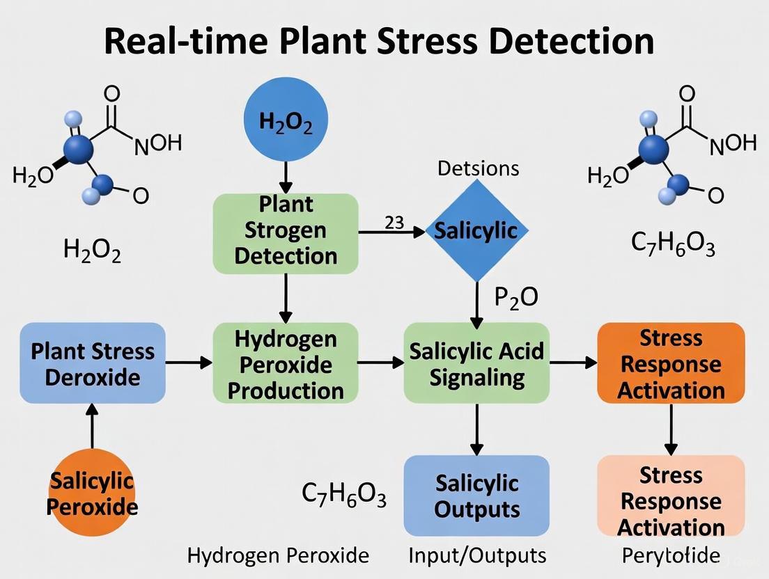
Diagram 1: H₂O₂ and SA in Early Plant Stress Signaling. This diagram illustrates the cascade from initial stress perception to a coordinated defense response, highlighting the central role of H₂O₂ and its interplay with SA and calcium signaling.
Diagram 2: Experimental Workflows for Validating H₂O₂ as a Stress Marker. This flowchart compares two primary methodological approaches for investigating H₂O₂ signaling: real-time nanosensor monitoring and traditional laboratory physiology and biochemistry.
The Scientist's Toolkit: Key Research Reagent Solutions
Table 4: Essential Reagents and Tools for H₂O₂ and Stress Signaling Research
| Research Tool / Reagent | Function / Application | Key Utility |
|---|---|---|
| Carbon Nanotube (SWNT) Nanosensors | Non-destructive, real-time monitoring of H₂O₂ and SA in living plants. | Enables decoding of stress-specific signaling kinetics and multiplexing of different signals [4] [5]. |
| Chemical H₂O₂ Donors (e.g., H₂O₂ solution) | Used for exogenous application to study priming effects and specific H₂O₂-triggered pathways. | Allows investigation of H₂O₂ as a priming agent and its direct impact on gene expression and acclimation [1]. |
| Antioxidant Enzyme Inhibitors & Scavengers | Inhibitors (e.g., IMD for NADPH oxidase) or scavengers (e.g., DMTU for H₂O₂) to perturb endogenous levels. | Essential for establishing causality and proving the requirement of H₂O₂ in a given stress response [6]. |
| Antibodies & ELISA Kits | For traditional quantification of stress hormones (SA, ABA) and oxidative damage markers (e.g., lipid peroxidation). | Provides standardized, accessible methods for validating and complementing sensor-based data [7]. |
| Redox-Sensitive Dyes (e.g., DAB, H2DCF-DA) | Histochemical and fluorometric detection of H₂O₂ and general ROS in plant tissues. | Allows spatial localization of ROS accumulation, though with potential limitations in specificity and real-time application [8]. |
The body of evidence consolidates hydrogen peroxide's status as a universal early alarm in plant stress perception. Its rapid, stress-specific production kinetics, capacity for targeted signal transduction via oxidative post-translational modifications and specific sensors, and synergistic relationship with hormones like salicylic acid make it a superior initial redox marker. The development of advanced tools, particularly multiplexed nanosensors, is transforming our ability to decipher the "H₂O₂ language" of plants in real-time. Future research should focus on mapping complete H₂O₂ signaling networks, expanding nanosensor technology to field applications for precision agriculture, and engineering crops with optimized H₂O₂ signaling capacities for enhanced climate resilience. Validating H₂O₂ as a core component of a real-time diagnostic system represents a paradigm shift from reactive to proactive crop management.
Salicylic acid (SA) is a pivotal phytohormone that orchestrates plant immune responses and systemic acquired resistance (SAR) against pathogens. Beyond its established role in biotic stress, recent research underscores its significance in mediating abiotic stress adaptation, positioning SA as a master regulator of plant defense and resilience. This review objectively compares SA's functional performance against other defense mechanisms and presents supporting experimental data, framed within the context of validating real-time plant stress detection. The emergence of sophisticated biosensors is revolutionizing our understanding of SA spatiotemporal dynamics, enabling unprecedented live visualization of SA fluxes during stress events and offering new avenues for crop improvement strategies.
SA Biosynthesis Pathways and Molecular Signaling
Biosynthetic Routes
Plants primarily synthesize SA through two distinct pathways with differential contributions across species:
The Isochorismate Synthase (ICS) Pathway: This serves as the dominant route for defense-related SA biosynthesis in Arabidopsis, tomato, and tobacco. The process initiates with chorismate in plastids, where ICS1 converts it to isochorismate [9]. The EDS5 transporter then exports isochorismate to the cytosol, where the acyl acid amido synthetase PBS3 and ENHANCED PSEUDOMONAS SUSCEPTIBILITY 1 facilitate its conversion to SA [9]. In Arabidopsis, approximately 90% of immune-elicited SA is produced via this pathway [9].
The Phenylalanine Ammonia-Lyase (PAL) Pathway: Once considered incomplete in plants, this pathway has been fully elucidated [9]. It begins with phenylalanine conversion to trans-cinnamic acid by PAL, followed by transformation to trans-cinnamoyl-CoA by cinnamoyl-CoA ligase. Subsequent β-oxidation in peroxisomes produces benzoyl-CoA, which is sequentially processed by OSD2 (producing benzyl benzoate), OSD3 (hydroxylating to benzyl salicylate), and OSD4 (releasing SA) [9]. Stable isotope labeling confirms phenylalanine as a precursor, overturning previous assumptions about benzoic acid conversion [9].
The relative importance of these pathways varies by species; for instance, the ICS pathway is not dominant in rice, where osics mutants maintain relatively high basal SA levels [9].
SA Perception and Signaling Core
The NPR proteins (NPR1, NPR3, and NPR4) serve as SA receptors in Arabidopsis [9]. NPR1 functions as the master transcriptional coactivator during SAR, interacting with TGA transcription factors to upregulate defense genes like PATHOGENESIS-RELATED 1 (PR1) [9]. In contrast, NPR3 and NPR4 operate redundantly as transcriptional corepressors whose activities are suppressed by SA binding [9]. These paralogs also exhibit E3 ubiquitin ligase activity, targeting both NPR1 and EDS1 for degradation by the 26S proteasome pathway [9]. EDS1 serves as a core signaling hub downstream of both nucleotide-binding leucine-rich repeat (NLR) receptors and pattern recognition receptors (PRRs), integrating upstream immune perception with downstream transcriptional reprogramming [9].
Table 1: Core Components of SA Biosynthesis and Signaling
| Component | Function | Localization |
|---|---|---|
| ICS1 | Key enzyme in SA biosynthesis via isochorismate conversion | Plastids |
| EDS5 | Transporter exporting isochorismate from plastids | Plastid membrane |
| PBS3 | Acyl acid amido synthetase facilitating SA production | Cytosol |
| PAL | Converts phenylalanine to trans-cinnamic acid | Cytosol |
| NPR1 | Master transcriptional coactivator, SA receptor | Nucleus |
| NPR3/NPR4 | Transcriptional corepressors, E3 ubiquitin ligases | Nucleus |
| EDS1 | Signaling hub integrating immune perception | Cytosol/Nucleus |
Figure 1: SA Signaling Pathway Integration. The diagram illustrates how biotic and abiotic stresses activate SA biosynthesis through ICS and PAL pathways, leading to diverse physiological responses via NPR protein-mediated signaling.
SA in Systemic Acquired Resistance
Establishing Long-Distance Immunity
Systemic acquired resistance represents a pathogen-induced whole-plant immunity state against secondary infections. SA serves as the central hormonal regulator coordinating SAR through complex spatial and temporal dynamics. Recent research has identified hydrogen peroxide as a SAR-inducing signal with dose-dependent effects on SA biosynthesis in systemic tissues following pathogen attack [10]. This redox-SA relationship forms a crucial signaling axis that amplifies defense responses throughout the plant.
The SALICYLIC ACID SENSOR1 (SalicS1), a genetically encoded FRET biosensor, has enabled real-time, reversible monitoring of SA levels in vivo with cellular precision [11] [12]. This technology reveals the propagation of an SA surge spreading from bacterial infection sites with spatiotemporal fidelity, demonstrating how local infections establish systemic immunity [11]. The improved nuclear-localized version (nlsSalicS1) enables stable SA detection across various organs, including roots, cotyledons, and mature leaves [12].
Comparative Signaling Efficacy
SA-mediated SAR demonstrates distinctive effectiveness compared to other defense hormones:
- Spatial Range: SA signals travel systemically from infection sites to distal tissues, establishing whole-plant resistance, whereas jasmonic acid (JA) responses are often more localized.
- Duration: SAR provides lasting protection (days to weeks), outperforming the transient protection offered by reactive oxygen species (ROS) bursts.
- Pathogen Spectrum: SA is particularly effective against biotrophic and hemibiotrophic pathogens, while JA is more effective against necrotrophs.
- Signal Fidelity: SA surges propagate with remarkable spatiotemporal precision, as visualized by SalicS1, unlike the more diffuse calcium waves associated with some defense responses.
SA-Mediated Abiotic Stress Adaptation
Thermotolerance Mechanisms
Temperature fluctuations profoundly influence SA pathways, with high temperatures suppressing SA biosynthesis and signaling while low temperatures enhance them [9]. This thermosensitivity directly impacts plant resilience under climate change scenarios. Moderately elevated temperatures (28°C) inhibit expression of key SA biosynthesis regulators, including SARD1 and CBP60g, which normally activate ICS1, EDS5, PBS3, EDS1, and PAD4 expression [9]. The heat sensitivity of SA-mediated immunity is evidenced by experiments where ics1, eds1, or pad4 mutants lose thermosensitive disease resistance, displaying similar susceptibility at both optimal and elevated temperatures [9].
Table 2: Temperature Modulation of SA Pathways and Immunity
| Temperature Condition | Effect on SA Biosynthesis | Impact on Disease Resistance | Molecular Mechanisms |
|---|---|---|---|
| Low Temperature | Enhanced SA biosynthesis | Improved resistance against biotrophic pathogens | Upregulation of ICS1, EDS5, PBS3, EDS1, PAD4 |
| Moderate Temperature (22-23°C) | Optimal SA pathway function | Balanced growth and defense | Normal expression of SARD1 and CBP60g regulators |
| High Temperature (28-30°C) | Suppressed SA biosynthesis | Compromised immunity, increased susceptibility | Inhibition of SARD1, CBP60g, and downstream biosynthetic genes |
Oxidative Stress Mitigation via Metal Chelation
SA application mitigates plant oxidative stress through metal chelation mechanisms, forming complexes with essential metals that enhance antioxidant activity [13]. In vitro experiments examining SA interactions with metal ions (Mg, Ca, Mn, Fe, Co, Ni, Cu, Zn, Mo) revealed that SA and its metal complexes exhibit higher antioxidant activity than ascorbic acid, with radical scavenging activity of 28.22% and electrode potential of -0.74 V [13]. Among these complexes, the Mn(II)-SA complex demonstrated superior antioxidant activity with 64.52% radical scavenging capacity and -0.9 V electrode potential [13].
In vivo studies on pesticide-stressed wheat plants confirmed that the Mn(II)-SA complex enhances antioxidant enzyme activities, specifically superoxide dismutase (SOD) and peroxidase (POD) [13]. Inductively Coupled Plasma Optical Emission Spectroscopy analysis confirmed that SA-treated plants had higher metal content, supporting that SA enhances metal uptake through chelation, thereby mitigating oxidative stress [13]. This chelation-based strategy proves more effective and less pH-sensitive than reduction-based nutrient uptake mechanisms [13].
Drought Stress Alleviation
Research on Scrophularia striata demonstrates that SA application enhances drought stress tolerance by modulating biochemical and molecular responses [14]. Under drought conditions (50% field capacity), SA treatment at 100 mg L⁻¹ increased expression of terpenoid pathway genes, including isopentenyl diphosphate isomerase (IPPI), and enhanced accumulation of protective metabolites like β-carotene, α-tocopherol, and beta-amyrin [14]. Combined application of SA and silicon (Si) proved particularly effective, elevating β-amyrin content by 264.4% compared to controls under drought stress [14].
SA-mediated drought protection involves transcriptional reprogramming of stress-responsive genes and enhanced antioxidant capacity, reducing oxidative damage while maintaining metabolic functionality. The synergistic effect of SA with silicon underscores the potential of combination treatments for enhancing crop resilience in water-limited environments.
Advanced Methodologies for SA Research
Real-Time SA Monitoring with Genetically Encoded Biosensors
The SalicS1 FRET biosensor represents a breakthrough technology for visualizing SA dynamics in living plant tissues [11] [12]. This sensor employs a truncated Arabidopsis NPR1 protein linked to NIMIN1, which interact in vivo. SA binding to NPR1 disrupts this interaction, decreasing the FRET emission ratio and enabling quantitative SA measurement [12]. Sensor validation through genetic manipulation confirms its specificity: plants carrying NahG (SA degradation) or mutations in EDS5 and PBS3 (blocking SA synthesis) display significantly lower FRET signals [12].
Experimental Protocol for SalicS1 Implementation:
- Plant Material: Generate stable Arabidopsis lines expressing nlsSalicS1
- Pathogen Inoculation: Apply Pseudomonas syringae at 10⁸ CFU/mL
- Imaging Setup: Use confocal microscopy with FRET capabilities
- Excitation/Emission: CFP excitation at 458nm, detect emission at 475-500nm (CFP) and 520-550nm (FRET)
- Image Analysis: Calculate FRET/CFP ratio changes over time
- Controls: Include NahG and eds5/pbs3 mutants for baseline correction
This methodology enables unprecedented spatial and temporal resolution of SA fluxes, revealing propagation patterns previously undetectable with destructive sampling methods.
Machine Learning-Assisted Stress Phenotyping
Advanced machine learning approaches complement SA research by enabling non-destructive stress detection through visual symptom analysis. Del Cioppo et al. developed a decision tree-based model that achieves 91% mean precision in stress detection using image-derived color features like "Chroma Difference" and "Chroma Ratio" [15]. These indices capture subtle color patterns invisible to the naked eye, serving as reliable digital equivalents of biochemical stress signals [15].
The model demonstrates exceptional true positive rates (0.967) for stress detection, correctly identifying nearly 97% of stressed plants, with only 3% going undetected [15]. This approach successfully classified stress intensity with 84% precision, performing best for high-stress (0.929 precision) and no-stress conditions (0.885 precision) compared to moderate stress (0.694 precision) [15]. Remarkably, image-only models achieved 88% mean precision for stress detection without biochemical data, demonstrating that plants reveal internal stress states through algorithm-detectable visual cues [15].
Figure 2: Integrated Experimental Workflow for SA Research. The diagram outlines complementary methodologies for investigating SA-mediated stress responses, combining machine learning, biosensor technology, and biochemical approaches.
Metal Chelation and Antioxidant Assessment
Experimental Protocol for SA-Metal Complex Analysis:
- Complex Synthesis: React 10mL of 0.01M aqueous metal salt solutions with 20mL ethanolic SA (0.01M)
- pH Adjustment: Maintain pH 6-7 with continuous stirring for 2 hours
- Characterization: Employ spectrophotometry and cyclic voltammetry
- Plant Treatment: Apply SA and metal complexes to wheat plants under pesticide stress
- Enzyme Assays: Measure SOD, POD, and CAT activities spectrophotometrically
- Elemental Analysis: Quantify metal content in treated shoots using ICP-OES
- Computational Validation: Perform molecular docking with AutoDock4 and AutoGrid4 software
This comprehensive approach verifies both the antioxidant potential of SA-metal complexes and their physiological efficacy in stress mitigation [13].
The Scientist's Toolkit: Essential Research Reagents
Table 3: Key Research Reagents for SA Signaling and Stress Studies
| Reagent/Resource | Function/Application | Experimental Context |
|---|---|---|
| SalicS1/nlsSalicS1 | Genetically encoded FRET biosensor for real-time SA monitoring | Live imaging of SA dynamics in Arabidopsis with cellular precision [11] [12] |
| NahG Arabidopsis | Transgenic line expressing bacterial salicylate hydroxylase for SA degradation | SA-deficient controls for biosensor validation and pathway analysis [12] |
| ics1/eds5/pbs3 mutants | SA biosynthesis-deficient mutants | Establishing SA-dependent vs independent mechanisms [9] [12] |
| npr1/npr3/npr4 mutants | SA receptor mutants | Dissecting canonical NPR-dependent signaling pathways [9] [16] |
| SA-Metal Complexes | Pre-formed complexes like Mn(II)-SA | Investigating antioxidant enhancement through chelation [13] |
| Decision Tree ML Models | Image-based stress classification using chroma indices | Non-destructive stress phenotyping without biochemical assays [15] |
Salicylic acid emerges as a master regulator integrating plant responses to both biotic and abiotic challenges, coordinating systemic acquired resistance while enhancing tolerance to temperature extremes, oxidative stress, and drought. The development of advanced tools like SalicS1 biosensors and machine learning phenotyping platforms has revolutionized our capacity to monitor SA dynamics with spatiotemporal precision, validating its central role in stress signaling networks. Future research leveraging these technologies will further elucidate SA's multifaceted functions, accelerating the development of climate-resilient crops through targeted manipulation of SA-mediated resilience pathways.
This guide compares experimental approaches for investigating the synergistic relationship between hydrogen peroxide (H₂O₂) and salicylic acid (SA) in plant stress signaling. We objectively evaluate traditional biochemical methods against emerging nanobionic sensor technology, providing supporting data from key studies. The analysis focuses on methodological capabilities, temporal resolution, and applications for validating real-time plant stress detection, addressing the critical need for advanced phenotyping tools in plant science research and agricultural innovation.
The cross-talk between hydrogen peroxide (H₂O₂) and salicylic acid (SA) represents a fundamental signaling module in plant stress physiology. Both molecules function as key signaling hubs in plant immune responses and adaptation to environmental challenges [17] [18]. H₂O₂, a reactive oxygen species (ROS), acts as a versatile signaling molecule with dual roles—at high concentrations it induces oxidative damage, while at controlled levels it functions as a secondary messenger in stress signal transduction pathways [17]. Similarly, SA is a pivotal phytohormone regulating plant defense responses to both biotic and abiotic stresses [19].
Recent research has revealed extensive synergistic interactions between H₂O₂ and SA signaling pathways, forming amplifying feedback loops that enhance plant stress resilience [19]. Understanding these complex interactions requires sophisticated methodological approaches capable of capturing the dynamic, real-time exchange of signals between these pathways. This guide compares established and emerging technologies for decoding this sophisticated chemical language, with implications for developing climate-resilient crops and precision agriculture systems.
Experimental Approaches: Methodological Comparison
Researchers employ diverse methodologies to investigate H₂O₂-SA cross-talk, each with distinct advantages and limitations. The table below summarizes the core experimental approaches used in this field.
Table 1: Comparison of Experimental Approaches for Studying H₂O₂-SA Signaling
| Method Type | Key Features | Temporal Resolution | Spatial Resolution | Primary Applications |
|---|---|---|---|---|
| Chemical Priming & Biochemical Analysis [20] [21] | Exogenous application of H₂O₂/SA followed by molecular analysis | Hours to days | Tissue/organ level | Studying synergistic effects on stress tolerance, gene expression, and metabolic changes |
| Genetic Manipulation [19] | CRISPR/Cas9 knockout, overexpression studies | Days to weeks | Cellular/organism level | Establishing causal relationships in signaling pathways and identifying key regulatory nodes |
| Nanobionic Sensors [22] [23] [5] | Real-time monitoring with carbon nanotube-based sensors | Seconds to minutes | Cellular/subcellular level | Decoding early signaling dynamics and stress-specific signatures |
Key Signaling Pathways and Experimental Findings
The Synergistic Priming Effect
Studies on maize seed germination under chilling stress (13°C) demonstrate that combined SA+H₂O₂ priming synergistically enhances chilling tolerance more effectively than individual treatments [20] [21]. This synergistic effect manifests through multiple physiological and molecular changes:
- Enhanced antioxidant defense: SA+H₂O₂ priming significantly increased activities of superoxide dismutase (SOD), catalase (CAT), ascorbate peroxidase (APX), and glutathione reductase (GR), along with upregulation of their corresponding genes (ZmSOD4, ZmCAT2, ZmAPX2, ZmGR) [20].
- Hormonal reprogramming: The treatment upregulated gibberellic acid (GA) biosynthesis genes (ZmGA20ox1, ZmGA3ox2), downregulated GA catabolism gene (ZmGA2ox1), and promoted ABA catabolism through upregulation of ZmCYP707A2 [20].
- Energy mobilization: Enhanced α-amylase activity increased soluble sugar content, providing essential energy and metabolites for germination under stress conditions [20].
Table 2: Quantitative Effects of SA+H₂O₂ Priming on Maize Seed Germination Under Chilling Stress (13°C) [20]
| Parameter | Hydropriming + Chilling Stress | SA+H₂O₂ Priming + Chilling Stress | Improvement |
|---|---|---|---|
| Germination Percentage (Day 7) | 68.5% | 89.2% | +30.2% |
| Germination Index | 28.7 | 45.3 | +57.8% |
| Mean Germination Time (days) | 4.8 | 3.5 | -27.1% |
| Vigor Index | 3.9 | 7.8 | +100% |
| SOD Activity (U/g FW) | 42.5 | 68.3 | +60.7% |
| CAT Activity (μmol H₂O₂/min/g FW) | 35.2 | 58.6 | +66.5% |
| H₂O₂ Content (nmol/g FW) | 126.8 | 215.4 | +69.9% |
| Endogenous SA Content (μg/g FW) | 0.85 | 1.42 | +67.1% |
The miR398-SlCSD1 Regulatory Module
Research in tomato (Solanum lycopersicum) has identified a key regulatory mechanism in the SA-H₂O₂ feedback loop. The miR398-SlCSD1 module participates in an amplifying cycle where [19]:
- Low SA concentrations stimulate H₂O₂ accumulation and suppress sly-miR398 expression, effects absent in SA-deficient NahG plants
- TGACG-sequence-specific binding protein 2 (TGA2) mediates SA-induced regulation of the miR398-SlCSD1 module
- Fluctuations in miR398 levels induce SA synthesis via both phenylalanine ammonia-lyase (PAL) and isochorismate synthase (ICS) pathways
- CRISPR/Cas9 knockout of SlCSD1 partially inhibits SA-induced H₂O₂ accumulation, confirming its role in SA-dependent H₂O₂ signaling
Real-Time Decoding of Stress Signatures
Groundbreaking research using nanobionic sensors has enabled real-time monitoring of H₂O₂ and SA dynamics in living plants (Brassica rapa subsp. Chinensis), revealing stress-specific signaling patterns [22] [23] [5]:
- Pathogen infection: Triggers sequential H₂O₂ production (within minutes) followed by SA accumulation (within 2 hours)
- Heat stress: Induces both H₂O₂ and SA production within 2 hours of stress application
- Light stress: Generates distinct H₂O₂ and SA waveforms different from other stressors
- Mechanical wounding: Stimulates H₂O₂ production but does not induce SA accumulation within 4 hours
These stress-specific signatures enable early diagnosis before visible symptoms appear, providing a window for intervention [22].
Diagram 1: H₂O₂-SA signaling pathway and feedback loops
Experimental Protocols: Core Methodologies
Materials: Maize seeds, salicylic acid (0.5 mM), hydrogen peroxide (50 mM), sterilization solution (0.5% NaClO), distilled water, germination paper, growth chambers.
Procedure:
- Surface-sterilize seeds with 0.5% NaClO solution for 15 minutes
- Rinse thoroughly with sterilized distilled water (3 times)
- Immerse seeds in priming solutions (1:5, w/v) at 20°C in darkness for 24 hours
- Air-dry primed seeds at 25°C for 48 hours to original moisture content
- Germinate seeds under controlled stress conditions (13°C for chilling stress)
- Assess germination parameters daily for 7 days
- Collect tissue samples at 0, 6, 24, 48, and 72 hours for molecular analysis
Key Measurements:
- Germination percentage, germination index, mean germination time
- H₂O₂ content (Doulis et al. method)
- Antioxidant enzyme activities (SOD, CAT, APX, GR)
- Gene expression analysis (qRT-PCR for stress-responsive genes)
Materials: Single-walled carbon nanotubes (SWNTs), (GT)₁₅ DNA oligomer (for H₂O₂ sensor), cationic fluorene-based copolymer S3 (for SA sensor), pak choi plants, near-infrared spectrometer, sensor injection system.
Procedure:
- Sensor Preparation:
- Suspend SWNTs with specific wrappings ((GT)₁₅ DNA for H₂O₂; S3 polymer for SA)
- Characterize sensor selectivity via photoluminescence excitation (PLE) spectroscopy
- Validate sensor response to 100 μM plant hormone analytes
Plant Preparation:
- Grow pak choi plants under controlled conditions
- Divide into experimental groups for different stress treatments
Sensor Introduction:
- Infiltrate nanosensors into plant leaf mesophyll using needle-free syringe
- Allow sensors to distribute through apoplastic space (30-60 minutes)
Stress Application & Monitoring:
- Apply specific stresses (pathogen, heat, light, mechanical wounding)
- Monitor sensor fluorescence simultaneously using multiplexed detection system
- Record H₂O₂ and SA dynamics in real-time (seconds to hours resolution)
Data Analysis:
- Convert fluorescence signals to analyte concentrations using calibration curves
- Analyze temporal patterns and waveform characteristics
- Apply biochemical kinetic modeling for stress identification
Diagram 2: Experimental workflow for H₂O₂-SA research
The Scientist's Toolkit: Essential Research Reagents
Table 3: Key Research Reagent Solutions for H₂O₂-SA Signaling Studies
| Reagent/Category | Specific Examples | Function/Application | Experimental Notes |
|---|---|---|---|
| Chemical Priming Agents | Salicylic acid (0.5 mM), Hydrogen peroxide (50 mM) | Induce cross-talk responses, study synergistic effects | Combined application shows enhanced efficacy; concentration-dependent effects observed [20] |
| Molecular Biology Tools | CRISPR/Cas9 constructs (SlCSD1 knockout), qPCR primers (ZmPAL, ZmSOD4, ZmCAT2) | Genetic manipulation, gene expression analysis | miR398-SlCSD1 module identification crucial for feedback loop understanding [19] |
| Nanobionic Sensors | (GT)₁₅ DNA-SWNT (H₂O₂ sensor), S3 polymer-SWNT (SA sensor) | Real-time monitoring of signaling molecules | Enables stress-specific signature identification; species-agnostic [22] [5] |
| Antioxidant Assay Kits | SOD activity assay, CAT activity assay, APX activity assay | Quantify antioxidant response in priming experiments | Essential for measuring redox homeostasis in H₂O₂-SA cross-talk [20] |
| Stress Application Systems | Controlled growth chambers, Pathogen inoculation tools, Mechanical wounding devices | Apply standardized stress treatments | Critical for comparing signaling responses across different stress types [22] |
Comparative Analysis: Methodological Insights
Temporal Resolution Capabilities
The choice of experimental approach dramatically affects the temporal insights achievable in H₂O₂-SA signaling research:
- Nanobionic sensors provide superior temporal resolution, capturing H₂O₂ waves within minutes of stress application and SA responses within 1-2 hours [22] [5]. This enables identification of stress-specific signatures based on waveform characteristics.
- Chemical priming studies operate on a longer timescale, measuring changes over hours to days, focusing on cumulative physiological effects rather than initial signaling events [20].
- Genetic approaches require the longest timeframe but provide causal evidence for regulatory relationships within the signaling network [19].
Applications in Stress Phenotyping
Each method offers distinct advantages for specific applications in plant stress research:
- Chemical priming is optimal for developing practical agricultural treatments to enhance crop stress tolerance [20] [21].
- Nanobionic sensors enable pre-symptomatic stress diagnosis and decoding of early signaling mechanisms [22] [23].
- Genetic manipulation identifies key regulatory nodes for developing improved crop varieties through molecular breeding [19].
The cross-talk between H₂O₂ and SA represents a sophisticated signaling network with amplifying feedback loops that enhance plant stress resilience. Methodological advancements, particularly the development of multiplexed nanobionic sensors, have revolutionized our ability to decode these complex interactions in real-time. The integration of approaches—from chemical priming and genetic manipulation to real-time sensing—provides complementary insights into these dynamic signaling processes.
For researchers validating real-time plant stress detection, nanobionic sensor technology offers unprecedented capabilities for early stress diagnosis and identification of stress-specific signatures. However, traditional methods remain invaluable for understanding downstream physiological responses and developing practical applications. The continuing refinement of these technologies promises to accelerate the development of climate-resilient crops and precision agriculture systems capable of responding to environmental challenges with unprecedented temporal and spatial resolution.
In the face of climate change and its associated biotic and abiotic stressors, understanding plant immune signaling has never been more critical. This guide compares traditional and emerging methodologies for tracing the complete signaling cascade from initial stress perception to systemic defense activation, with a specific focus on validating real-time detection using hydrogen peroxide (H₂O₂) and salicylic acid (SA) research. For researchers and scientists, selecting the appropriate tools to capture the spatiotemporal dynamics of these key signaling molecules determines the depth and applicability of findings in both basic and applied plant science.
The plant immune system functions through an intricate signaling network that transforms localized stress perception into whole-plant defense readiness. The cascade begins within seconds to minutes after stress perception, with rapid biochemical changes including calcium ion (Ca²⁺) influx and reactive oxygen species (ROS) burst, particularly H₂O₂ [24] [25]. These early signals activate downstream phytohormone pathways, with salicylic acid (SA) playing a predominant role in defense against pathogens and various abiotic stresses [26].
SA biosynthesis occurs primarily via the isochorismate pathway in chloroplasts, with ISOCHORISMATE SYNTHASE 1 (ICS1) as the critical enzyme [27]. Calcium-dependent protein kinases (CDPKs) and calmodulin-binding transcription factors decode initial Ca²⁺ signals to regulate ICS1 expression [24] [27]. The produced SA is then exported from chloroplasts via the MATE transporter EDS5 [27].
SA perception occurs through multiple mechanisms, with NPR1 (Nonexpressor of Pathogenesis-Related Genes 1) serving as a central co-activator of TGA transcription factors [27] [26]. Recent models propose both NPR1 itself and its homologs NPR3 and NPR4 as SA receptors [27]. This signaling nexus activates extensive transcriptional reprogramming, leading to the expression of Pathogenesis-Related (PR) genes and the establishment of both local resistance and Systemic Acquired Resistance (SAR) [27] [26].
The visualization below maps this complex journey from initial stress encounter to systemic immunity.
Comparative Analysis of Research Methodologies
Traditional Molecular Biology Approaches
Established methods for studying plant stress signaling rely on discrete, often destructive sampling followed by laboratory analysis.
- Molecular Bioassays: Enzyme-Linked Immunosorbent Assay (ELISA) is widely used to quantify stress-related hormones like SA and pathogen concentrations [28] [29]. Chlorophyll fluorescence imaging (Fv/Fm ratio) non-destructively assesses photosynthetic efficiency as an indicator of physiological stress [28] [29].
- Mass Spectrometry-Based Omics: Gas Chromatography-Mass Spectrometry (GC-MS) and other MS techniques enable comprehensive profiling of the ionome, metabolome (including SA and its derivatives), and proteome to characterize biochemical stress responses [29].
- Genetic and Transcriptomic Analysis: Quantitative PCR and RNA sequencing quantify expression of key signaling genes (e.g., ICS1, NPR1, PR genes) in mutant or transgenic plants to establish genetic functions within the pathway [27] [29].
Emerging Real-Time Sensing Technologies
Cutting-edge plant nanobionic sensors represent a paradigm shift, enabling non-destructive, real-time monitoring of signaling dynamics in living plants.
- Carbon Nanotube-Based Optical Sensors: These sensors use the Corona Phase Molecular Recognition (CoPhMoRe) technique, where single-walled carbon nanotubes (SWNTs) are wrapped with specific polymers or DNA oligomers to create a corona phase selective for target molecules like H₂O₂ or SA [4] [30] [5]. Upon binding, the nanosensors emit a fluorescent signal in the near-infrared (nIR) range, detectable with an infrared camera [4] [5].
- Sensor Multiplexing: This innovative approach allows simultaneous monitoring of multiple signals—such as H₂O₂ and SA—within the same plant, revealing stress-specific temporal wave patterns that function as a chemical "language" for stress communication [30] [5].
The table below provides a direct, data-driven comparison of these methodological approaches.
Table 1: Performance Comparison of Plant Stress Signaling Detection Methodologies
| Methodology | Target Analytes | Temporal Resolution | Spatial Resolution | Key Performance Metrics | Plant Disruption |
|---|---|---|---|---|---|
| ELISA | SA, ABA, Pathogens [28] [29] | Hours to days | Tissue-level | Sensitivity in ng/g FW [28] | Destructive (tissue harvest) |
| Chlorophyll Fluorescence | PSII Efficiency (Fv/Fm) [28] [29] | Minutes | Leaf-level | Fv/Fm ratio (theoretical max: 0.83) [28] | Non-destructive |
| Mass Spectrometry | Metabolites, Ions, Proteins [29] | Days | Tissue-level | Detection limits in pmol to nmol range [29] | Destructive (tissue harvest) |
| Nanosensor Multiplexing | H₂O₂, SA in real-time [4] [5] | Seconds to minutes | Cellular-mesophyll level [4] | ~35% fluorescence quenching for SA [5] | Minimally invasive |
Experimental Protocols for Key Assays
Protocol 1: Real-Time Decoding of Stress Signals using Nanosensor Multiplexing
This protocol, adapted from Saju et al. (2024) and MIT news reports, details the simultaneous detection of H₂O₂ and SA in living plants [4] [30] [5].
- Nanosensor Preparation:
- Plant Infiltration:
- Dissolve each nanosensor in an aqueous solution.
- Apply the solution to the abaxial side (underside) of a plant leaf (e.g., Pak choi), allowing infiltration into the mesophyll layer through the stomata [4].
- Stress Application:
- Expose plants to controlled stress treatments: heat (e.g., 37°C), intense light, mechanical wounding (to mimic insect attack), or bacterial pathogen infection (e.g., Pseudomonas syringae) [5].
- Signal Acquisition & Analysis:
- Use an infrared camera to monitor fluorescence signals from both nanosensors simultaneously in real-time.
- Analyze temporal dynamics: H₂O₂ typically peaks within 1 hour post-stress, while SA follows distinct, stress-specific patterns (e.g., rising within 2 hours for heat, light, and pathogen stress) [4] [5].
Protocol 2: Validating SA-Mediated Freezing Tolerance via Pharmacological Inhibition
This protocol, based on Guo et al. (2018), investigates the crosstalk between H₂O₂ and ABA in SA-induced stress tolerance [28].
- Plant Material & Pretreatment:
- Use wheat plants at the four-leaf stage.
- Foliar spray with 100 μM SA solution three times at 12-hour intervals [28].
- Inhibitor Application:
- To test H₂O₂ involvement, pretreat with 2 mM Dimethylthiourea (DMTU, a H₂O₂ scavenger) 8 hours before SA application.
- To test ABA involvement, pretreat with 1 μM Fluridone (Flu, an ABA biosynthesis inhibitor) 8 hours before SA application [28].
- Stress Treatment & Sampling:
- Apply freezing stress (-2°C for 24 h) 12 hours after the final SA treatment.
- Harvest the last fully expanded leaves for analysis [28].
- Downstream Analysis:
- Electrolyte Leakage: Measure to assess cell membrane damage.
- Hormone Quantification: Use ELISA to measure endogenous ABA levels.
- Gene Expression: Analyze transcript levels of NADPH oxidase genes (sources of H₂O₂) via qRT-PCR [28].
The workflow for the nanosensor multiplexing protocol is illustrated below.
The Scientist's Toolkit: Essential Research Reagents and Materials
Selecting appropriate reagents is fundamental for successful experimental outcomes in plant stress signaling research.
Table 2: Key Research Reagent Solutions for H₂O₂ and SA Signaling Studies
| Reagent / Material | Function / Application | Specific Example / Role in Pathway |
|---|---|---|
| Polymer-wrapped SWNTs | Core sensing element for nanobionic sensors [5] | S3 polymer wrapper confers selectivity for SA; (GT)₁₅ DNA wrapper for H₂O₂ [5]. |
| Pharmacological Inhibitors | Chemically dissect signaling pathways [28] | DMTU (H₂O₂ scavenger); Fluridone (ABA biosynthesis inhibitor); SHAM/DPI (apoplastic H₂O₂ source inhibitors) [28]. |
| ELISA Kits | Quantify hormone levels and pathogen load [28] [29] | Commercial kits for SA, ABA, and pathogen-specific antibodies. |
| Transgenic Plant Lines | Establish gene function via loss/gain-of-function studies [27] | ics1, npr1, eds5 mutants; NPR1-overexpressors; biosensor-equipped plants [27]. |
| Fluorescent Probes & Dyes | Histochemical localization of signaling molecules [28] [29] | CeCl₃ for subcellular H₂O₂ detection; DCFH₂-DA for general ROS staining. |
The journey from cellular alarm to systemic defense encapsulates one of the most sophisticated communication systems in plant biology. Traditional molecular methods provide detailed snapshots of individual components within this pathway, while emerging nanosensor technologies offer a transformative real-time, multiplexed view of the entire signaling cascade. The experimental data and comparative analysis presented herein demonstrate that the choice of methodology directly shapes our understanding of the complex interplay between H₂O₂ and SA. For researchers aiming to validate real-time plant stress detection, nanosensor multiplexing emerges as a powerful tool, capable of decoding the unique chemical "fingerprints" of different stresses. This capability is paramount for developing climate-resilient crops and implementing pre-symptomatic diagnostic strategies to safeguard global food security.
From Lab to Leaf: Methodological Advances in Real-Time H₂O₂ and SA Biosensing
Corona Phase Molecular Recognition (CoPhMoRe) is a groundbreaking synthetic method for creating molecular recognition elements by templating heteropolymers onto nanoparticle surfaces [31]. This approach mimics the selective binding capabilities of biological systems, such as antibodies, but using synthetic, non-biological materials. The fundamental principle involves adsorbing a heteropolymer onto a nanoparticle, where it adopts a specific constrained configuration or "corona" that can selectively recognize target analytes based on molecular structure [32] [31]. When the constrained polymer corona binds its target molecule, it modulates the optical properties of the underlying nanoparticle, which serves as the signal transducer.
For single-walled carbon nanotubes (SWCNTs), which fluoresce in the near-infrared (NIR) region, binding events cause measurable changes in fluorescence intensity or spectral shifts [32]. This combination creates a powerful sensing platform that has been successfully demonstrated for various targets, including small molecules, neurotransmitters, and recently, proteins and plant hormones [32] [5] [33]. The CoPhMoRe approach represents a significant advancement in nanosensor engineering, offering a generic strategy for creating selective synthetic recognition elements without requiring biological systems for production.
Comparative Analysis of Optical Biosensing Technologies
The field of optical biosensing encompasses multiple technological platforms, each with distinct mechanisms and performance characteristics. The table below provides a systematic comparison of CoPhMoRe-based sensors with other established optical biosensing technologies:
Table 1: Performance comparison of major optical biosensing technologies
| Technology Platform | Mechanism | Target Analytes | Sensitivity | Selectivity Source | Key Advantages |
|---|---|---|---|---|---|
| CoPhMoRe-SWCNT | Polymer corona-induced fluorescence modulation | Small molecules, proteins, plant hormones | 11 nM (dopamine) [33] | Polymer conformation on nanoparticle surface | No biological components required; real-time monitoring in living plants [5] |
| Plasmonic Sensors | Refractive index change affecting surface plasmon resonance | Chemical and biological molecules | Ultra-high sensitivity [34] | Functionalization layers | Compact footprint; fast reaction [34] |
| Photonic Crystal Sensors | Structural color changes from periodicity alterations | Various biomolecules | High sensitivity | Crystal functionalization | Label-free detection; high quality factor |
| Optical Fiber Sensors | Evanescent wave interactions with analytes | Physical parameters, chemicals | Medium to high | Coating materials | Electromagnetic immunity; remote sensing |
| Graphene Sensors | Charge transfer affecting optical properties | Gases, biomolecules | High | Surface functionalization | High surface-to-volume ratio; tunable properties |
| Nanozyme Colorimetric | Catalytic activity mimicking enzymes | H₂O₂, glucose, ions | Varies by application | Catalyst composition | Visual detection; simple instrumentation [35] |
CoPhMoRe technology distinguishes itself through its unique mechanism that creates synthetic recognition sites without biological components, enabling applications where traditional biological elements like antibodies may degrade or prove impractical [32]. The approach has demonstrated exceptional versatility across target classes, from small molecules like riboflavin, L-thyroxine, and estradiol to proteins like fibrinogen and plant hormones such as salicylic acid [32] [5].
Experimental Validation: CoPhMoRe for Plant Stress Detection
Sensor Design and Implementation
Recent pioneering work has demonstrated the application of CoPhMoRe-based nanosensors for real-time decoding of plant stress signaling [4] [30] [5]. Researchers developed a multiplexed sensing platform incorporating two distinct SWCNT-based sensors: one for hydrogen peroxide (H₂O₂) and another for salicylic acid (SA). The H₂O₂ sensor utilized SWCNTs wrapped with single-stranded (GT)₁₅ DNA oligomers, while the SA sensor employed SWCNTs wrapped with specifically designed cationic fluorene-based co-polymers (designated S3) containing pyrazine monomers for additional hydrogen bonding interactions with SA [5].
The experimental workflow involved introducing these nanosensors into living plants (Brassica rapa subsp. Chinensis, or pak choi) through infiltration into the leaf mesophyll layer via stomatal pores [5]. The sensors distributed within the plant tissue and provided real-time, non-destructive monitoring of stress signaling molecules through changes in their NIR fluorescence, which can be detected using an infrared camera [4].
Table 2: Key research reagents for CoPhMoRe plant stress sensing
| Research Reagent | Function/Application | Key Characteristics |
|---|---|---|
| HiPCO SWCNTs | Fluorescent transducer element | Near-infrared fluorescence; high photostability [5] |
| (GT)₁₅ DNA oligomer | Corona phase for H₂O₂ recognition | Forms selective binding pocket for H₂O₂ on SWCNT surface [5] |
| S3 copolymer | Corona phase for SA recognition | Cationic fluorene-based polymer with pyrazine co-monomers [5] |
| Pak choi plants | Model plant system | Leafy green vegetable; suitable for sensor infiltration [5] |
| Pseudomonas syringae | Pathogen stress inducer | Bacterial pathogen used to elicit immune response [5] |
Signaling Pathways and Stress Response Dynamics
The multiplexed sensor platform revealed distinctive temporal dynamics in H₂O₂ and SA signaling waves across different stress types. The following diagram illustrates the experimental workflow and the characteristic signaling patterns observed for each stress type:
The experimental results demonstrated that each stress type generated a unique temporal pattern of H₂O₂ and SA production, serving as a distinctive fingerprint for stress identification [4] [5]. Specifically, H₂O₂ production occurred within minutes of stress exposure, reaching maximum levels within approximately one hour before returning to baseline [5]. The SA response patterns, however, varied significantly by stress type: heat, light, and bacterial infection all stimulated SA production within two hours of stress but at distinct time points, while mechanical wounding did not trigger significant SA production within the observed timeframe [5].
Quantitative Sensor Performance Metrics
The CoPhMoRe-based nanosensors demonstrated exceptional performance in detecting plant stress signaling molecules. The following table summarizes the key quantitative performance metrics established through experimental validation:
Table 3: Performance characteristics of CoPhMoRe plant stress nanosensors
| Sensor Type | Target Molecule | Response Mechanism | Response Magnitude | Selectivity | Temporal Resolution |
|---|---|---|---|---|---|
| SA Sensor | Salicylic acid | Fluorescence quenching | 35% quenching at 100 μM SA [5] | High (minimal response to other plant hormones) [5] | Real-time (minutes) |
| H₂O₂ Sensor | Hydrogen peroxide | Fluorescence modulation | 58-80% enhancement at 100 μM dopamine [33] | Selective for H₂O₂ over other ROS [5] | Real-time (minutes) |
| Multiplexed Platform | Combined SA & H₂O₂ | Distinct temporal patterns | Unique wave characteristics for each stress [5] | Stress-type identification via signature patterns [5] | Continuous monitoring |
The high selectivity of the SA sensor is particularly noteworthy, as it showed minimal response to other plant hormones including jasmonic acid, gibberellic acid, abscisic acid, and various auxins and cytokinins [5]. This specificity enables precise monitoring of SA signaling dynamics without interference from related signaling pathways.
Implementation Protocols for CoPhMoRe Sensor Development
Corona Phase Screening and Optimization
The development of CoPhMoRe-based sensors follows a systematic protocol beginning with the creation of a diverse polymer library. Researchers typically assemble a collection of heteropolymers with varying hydrophobic-hydrophilic balances and functional groups, such as the phospholipid-PEG polymers, single-stranded DNA/RNA oligomers, and fluorene-based copolymers demonstrated in previous studies [32] [5] [33].
The screening process involves suspending SWCNTs with each polymer candidate and exposing them to target analytes while monitoring SWCNT fluorescence changes. For example, in developing the SA sensor, researchers screened four cationic fluorene-based co-polymers (S1-S4) against 12 plant hormones and signaling molecules, identifying S3 as the optimal corona phase for SA recognition based on its specific 35% fluorescence quenching response to SA [5]. This empirical screening approach is crucial for identifying corona phases with the requisite selectivity for target analytes.
Sensor Integration and Deployment
For plant applications, the optimized nanosensors are introduced into plant tissue through several methods. The most common approach involves dissolving the sensor complexes in an aqueous solution and applying it to the underside of leaves, allowing entry through stomatal pores and distribution into the mesophyll layer where most photosynthesis occurs [4] [5]. The following diagram illustrates the sensor integration and signal detection process:
Once integrated, the sensors provide continuous monitoring capability, with fluorescence detection typically performed using NIR imaging systems equipped with appropriate excitation sources and filters to detect the SWCNT emission while minimizing background autofluorescence from plant tissues [5].
Comparative Advantages and Technical Considerations
CoPhMoRe-based nanosensors offer several distinct advantages over alternative sensing technologies for plant science applications. Unlike genetically encoded biosensors that require plant transformation—a process that can be challenging or impossible for many crop species—CoPhMoRe sensors can be applied to virtually any plant species without genetic modification [5]. This universality significantly expands the range of addressable species and applications.
The near-infrared fluorescence emission of SWCNTs provides another key advantage, as NIR light experiences reduced scattering and absorption in plant tissues compared to visible light, enabling better signal detection from within leaves [5]. Additionally, SWCNT fluorescence is exceptionally photostable, unlike molecular fluorophores that photobleach, allowing long-term monitoring over days or weeks [5].
From a practical implementation perspective, future development should address several technical considerations. Sensor longevity and stability within plant tissues requires further investigation for long-term monitoring applications. The potential for sensor diffusion from application sites may necessitate stabilization strategies or repeated applications for extended studies. For field deployment, developing portable readout systems and automated sensor application methods will be essential for scaling this technology beyond research settings into agricultural practice [22].
CoPhMoRe represents a powerful and versatile platform for engineering nanosensors with tailored molecular recognition capabilities. The successful demonstration of multiplexed stress signaling detection in plants highlights the transformative potential of this technology for fundamental plant science and agricultural applications. By enabling real-time, non-destructive monitoring of plant physiological status, these sensors provide an unprecedented window into plant stress responses, potentially revolutionizing crop management practices and breeding programs.
Future research directions will likely focus on expanding the sensor repertoire to encompass a broader range of plant signaling molecules, developing more sophisticated multiplexing capabilities for comprehensive pathway monitoring, and creating field-deployable systems for agricultural use. The integration of CoPhMoRe sensors with other emerging technologies, such as portable imaging systems and data analytics platforms, will further enhance their utility for both basic research and practical applications in precision agriculture. As these developments progress, CoPhMoRe-based nanosensors are poised to make significant contributions to addressing challenges in food security and sustainable agriculture in the face of climate change.
The escalating impact of climate change on global agriculture has intensified the search for technologies that can decode early plant stress responses before visible damage occurs. Traditional plant stress detection methods, which often rely on visible symptom observation or single-analyte measurements, provide limited insight into the complex signaling networks that plants activate under stress. Among the earliest signaling events in plant stress response are the rapid generation of reactive oxygen species (ROS), particularly hydrogen peroxide (H₂O₂), and subsequent hormonal changes, including salicylic acid (SA) production. These signaling molecules form a complex interplay that varies depending on stress type, creating unique temporal patterns that can serve as diagnostic "fingerprints" [5].
Sensor multiplexing represents a technological paradigm shift that enables researchers to capture these dynamic interactions in real-time. By simultaneously monitoring multiple signaling molecules in living plants, this approach provides unprecedented insight into stress response mechanisms and offers the potential for pre-symptomatic stress diagnosis. This comparison guide examines the performance characteristics of emerging multiplexed sensing platforms, their experimental implementation, and their transformative potential for plant stress research and precision agriculture [5] [36].
Comparative Performance Analysis of Sensing Platforms
The development of sensing platforms for H₂O₂ and SA has progressed through multiple technological generations, from destructive single-measurement approaches to non-destructive multiplexed nanosensors. The table below compares the key performance metrics of current sensing methodologies.
Table 1: Performance comparison of plant stress sensing platforms
| Technology Platform | Detection Method | H₂O₂ LOD | SA LOD | Temporal Resolution | Spatial Resolution | Multiplexing Capability |
|---|---|---|---|---|---|---|
| Electrochemical Sensors [37] | MWCNT-Ti₃C₂Tₓ-Pd modified electrode | 3.83 μM | N/A | Minutes | Single point measurement | Limited |
| Optical Nanosensors [5] | SWNT with (GT)₁₅ DNA (H₂O₂) and cationic polymer (SA) | ~1 μM | ~100 nM | Seconds to minutes | Mesophyll tissue level | Excellent (H₂O₂ + SA) |
| Thermal-Nanosensor Hybrid [38] | Nanosensors converting H₂O₂ to thermal signals | Sub-μM | N/A | Minutes | Leaf surface mapping | Limited |
| Hyperspectral Imaging [39] | MLVI and H_VSI indices via UAV/satellite | Indirect | Indirect | Hours to days | Canopy level | Moderate (multiple stress types) |
| Conventional Methods [37] [7] | DAB staining, HPLC, ELISA | ~10 μM | ~1 μM | Hours to days | Destructive tissue sampling | Poor |
Abbreviations: LOD - Limit of Detection; MWCNT - Multi-Walled Carbon Nanotube; SWNT - Single-Walled Carbon Nanotube; MLVI - Machine Learning-Based Vegetation Index; H_VSI - Hyperspectral Vegetation Stress Index
The performance data reveals distinct advantages and limitations for each platform. Electrochemical sensors offer excellent sensitivity for H₂O₂ detection with a linear range of 0.05-18 mM, making them suitable for direct quantitative measurements but limited in multiplexing capability [37]. In contrast, optical nanosensor platforms provide superior temporal resolution and the unique ability to simultaneously monitor both H₂O₂ and SA dynamics in real-time, enabling the capture of stress-specific waveform patterns that are lost with other methods [5]. The emerging thermal-nanosensor hybrid approach demonstrates how nanotechnology can be integrated with imaging systems to create machine-learnable stress signatures [38].
Experimental Protocols for Multiplexed Stress Sensing
Nanosensor Preparation and Application
The implementation of multiplexed nanosensing for H₂O₂ and SA requires precise sensor fabrication and application protocols. For the SWNT-based optical sensors, the procedure involves:
Sensor Synthesis: H₂O₂ sensors are prepared by suspending SWNTs with single-stranded (GT)₁₅ DNA oligomers via π-stacking interactions, forming a corona phase that confers selective binding to H₂O₂. SA sensors are created using SWNTs wrapped with cationic fluorene-based co-polymers (specifically S3 polymer), which showed 35% fluorescence quenching response to 100 μM SA with minimal cross-reactivity to other plant hormones [5].
Sensor Validation: Selectivity screening against 12 key plant hormones and signaling molecules confirmed minimal interference. The sensors were validated in transgenic Arabidopsis thaliana plants before application in non-model species [5].
Plant Infiltration: Sensors in liquid solution are applied to the abaxial surface (underside) of leaves, allowing entry through stomata without mechanical damage. The sensors lodge in the mesophyll layer where most photosynthesis occurs [5] [36].
Signal Detection: Near-infrared fluorescence is monitored using customized microscopes equipped with InGaAs cameras or portable infrared imagers, capturing signals in the 900-1600 nm range to avoid chlorophyll autofluorescence interference [5].
Stress Induction and Signal Recording
Experimental protocols for stress application must be carefully controlled to generate reproducible temporal fingerprints:
Stress Treatments: Plants are subjected to controlled stress conditions including:
- Light stress: High-intensity light (1000-1500 μmol m⁻² s⁻¹)
- Heat stress: Elevated temperatures (35-42°C)
- Pathogen stress: Bacterial inoculation (e.g., Pseudomonas syringae)
- Mechanical wounding: Leaf puncture or crushing [5]
Multiplexed Imaging: Simultaneous monitoring of H₂O₂ and SA dynamics is performed using ratiometric measurements to account for sensor concentration variations. The distinct fluorescence signatures of both sensors (H₂O₂ and SA) enable simultaneous tracking without spectral overlap [5].
Data Processing: Fluorescence signals are converted to analyte concentrations using pre-established calibration curves. Temporal wave characteristics including amplitude, full-width at half-maximum, and propagation speed are quantified for each stress type [5].
Temporal Fingerprints of Stress-Specific Responses
Multiplexed sensing reveals that different stress types generate distinct temporal patterns in H₂O₂ and SA signaling. The quantitative characteristics of these stress fingerprints are summarized in the table below.
Table 2: Temporal characteristics of H₂O₂ and SA waves under different stress conditions
| Stress Type | H₂O₂ Wave Onset | SA Wave Onset | H₂O₂ Peak Amplitude | SA Peak Amplitude | Key Pattern Characteristics |
|---|---|---|---|---|---|
| Heat Stress [5] | 5-15 minutes | 60-90 minutes | Moderate | Low to moderate | Rapid H₂O₂ spike with delayed, sustained SA increase |
| Light Stress [5] | 10-20 minutes | 45-75 minutes | High | Moderate | Synchronized oscillations in both signals |
| Pathogen Attack [5] | 30-60 minutes | 90-120 minutes | Low to moderate | High | Biphasic SA response with prolonged duration |
| Mechanical Wounding [5] | 2-5 minutes | 30-60 minutes | Very high | Low | Immediate, localized H₂O₂ burst with minimal SA involvement |
The data demonstrates that each stress type generates a unique biochemical signature encoded in the timing, amplitude, and waveform of H₂O₂ and SA production. Heat stress triggers an early H₂O₂ response followed by moderate SA accumulation, while pathogen attack shows the opposite pattern with SA as the dominant signal. Mechanical wounding produces the most rapid and intense H₂O₂ burst but minimal SA involvement. These distinct temporal fingerprints enable not just stress detection but precise stress identification before visible symptoms appear [5].
The Researcher's Toolkit: Essential Reagents and Materials
Successful implementation of multiplexed stress sensing requires specific research reagents and materials. The following table details the essential components and their functions.
Table 3: Research reagent solutions for multiplexed stress sensing
| Reagent/Material | Specifications | Function | Application Notes |
|---|---|---|---|
| SWNT Raw Materials [5] | HiPco or CoMoCAT SWNTs, 0.8-1.2 nm diameter | Sensor scaffold providing nIR fluorescence | Purification required to remove catalyst residues |
| DNA Wrapping [5] | (GT)₁₅ single-stranded DNA oligomers | Creates H₂O₂-selective corona phase | HPLC purification recommended for reproducibility |
| Polymer Wrapping [5] | Cationic fluorene-based co-polymers (S3 variant) | Creates SA-selective corona phase | Specific diazine co-monomer critical for SA selectivity |
| Reference Nanosensors [5] | SWNTs with non-responsive wrappings | Internal control for ratiometric measurements | (AT)₁₅ DNA often used as reference |
| Infiltration Solution [5] | 0.1-0.5% v/v surfactant in buffer | Enables sensor entry through stomata | Low surfactant concentration prevents membrane damage |
| Calibration Standards [37] [5] | H₂O₂ (0.05-20 mM), SA (1-500 μM) | Quantitative signal conversion | Must be prepared fresh to prevent degradation |
Signaling Pathways and Experimental Workflows
The complex interplay between H₂O₂ and SA in plant stress responses can be visualized through their signaling pathways and experimental implementation. The following diagram illustrates the conceptual framework and workflow for multiplexed stress sensing.
Plant Stress Signaling and Multiplexed Sensing Workflow
The diagram illustrates both the biological signaling pathways and experimental workflow for multiplexed stress decoding. The left side shows the sequential activation from stress perception through early H₂O₂ signaling to later SA-mediated responses, culminating in various physiological outcomes. The right side depicts the experimental implementation from sensor application through stress induction, multiplexed imaging, and final fingerprint analysis. The dashed lines indicate how the experimental measurements directly capture the dynamic signaling events occurring in the biological pathway.
Multiplexed sensing of H₂O₂ and SA represents a transformative approach for decoding plant stress responses, moving beyond single-analyte detection to capture the complex temporal relationships that define stress-specific fingerprints. The comparative analysis presented here demonstrates that optical nanosensor platforms currently offer the most comprehensive solution for simultaneous monitoring of these key signaling molecules, enabling researchers to capture the dynamic interplay that characterizes different stress types.
The future of this technology lies in expanding the multiplexing capability to include additional signaling components such as jasmonic acid, abscisic acid, and calcium ions, creating increasingly detailed stress fingerprints. Integration with machine learning approaches for automated pattern recognition [38] [39] and development of field-deployable imaging systems will further enhance the practical applications in precision agriculture and climate-resilient crop development. As these technologies mature, sensor multiplexing promises to unlock new understanding of plant communication and stress adaptation mechanisms, ultimately contributing to enhanced food security in the face of climate change.
The ability to detect plant stress in real-time is paramount for advancing fundamental plant science and improving agricultural resilience. Central to this endeavor is the precise monitoring of key signaling molecules, notably hydrogen peroxide (H₂O₂) and salicylic acid (SA), which serve as crucial biomarkers in a plant's acute stress response system [40] [41]. The fidelity of this monitoring is fundamentally dependent on the methods used to integrate sensing elements into live plant tissues and to validate their function across different species. This guide provides a comparative analysis of the leading techniques for in-planta sensor integration and validation, offering a detailed examination of their protocols, performance metrics, and suitability for various research applications. The objective is to furnish researchers and product developers with the experimental data necessary to select and optimize the most appropriate technology for their specific needs in the context of plant stress signaling.
Comparative Analysis of Sensor Integration Techniques
Different sensor integration strategies offer distinct advantages and limitations concerning invasiveness, scalability, and applicability across plant species. The table below summarizes the core characteristics of three primary techniques.
Table 1: Comparison of Primary Sensor Integration Techniques
| Technique | Core Principle | Key Advantages | Key Limitations | Best-Suited Applications |
|---|---|---|---|---|
| Solution-Based Infiltration [4] | Sensors dissolved in a solution are applied to the leaf underside for uptake via stomata. | - Minimally invasive- Universally applicable to most plant species- Simple protocol requiring no genetic modification | - Limited to leaf mesophyll- Potential for variable uptake efficiency | High-throughput screening in both model (e.g., Arabidopsis) and crop (e.g., Pak Choi) species [4]. |
| Microneedle (MN) Patch [42] [41] | A hydrogel-based microneedle array penetrates the leaf surface to extract sap for analysis. | - Rapid analysis (<1 minute)- In-field applicability- Reusable (up to 10x) | - Micro-damage to epidermal cells- Measures sap from a single time point (snapshot) | Portable, on-site stress monitoring in field conditions for crops like tobacco and soybean [41]. |
| Genetic Encoding [43] | Sensor components are transiently or permanently expressed in the plant via genetic engineering. | - Continuous, whole-plant monitoring- Cell-type-specific expression possible | - Requires species-specific transformation protocol- Confined to genetically tractable species | Fundamental research in established model plants like Arabidopsis thaliana and Nicotiana benthamiana [43]. |
Detailed Experimental Protocols
Protocol 1: Solution-Based Infiltration of Nanosensors
This protocol, adapted from MIT research, details the procedure for embedding carbon nanotube-based sensors into plant leaves for real-time stress biomarker detection [4].
- Sensor Preparation: Prepare the nanosensors by suspending carbon nanotubes tailored for H₂O₂ or SA detection in a sterile aqueous solution. The polymers coating the nanotubes are engineered for specificity to each target molecule [4].
- Plant Preparation: Select healthy, mature plants. For consistency, use the second extended leaf from 1-month-old plants grown under controlled conditions to minimize biological variability [4] [43].
- Infiltration Procedure: Using a needleless syringe, gently apply the sensor solution to the abaxial (underside) of the leaf, exploiting the natural stomatal openings. Apply gentle pressure to infiltrate the solution into the mesophyll layer. A successful infiltration is indicated by a water-soaked appearance of the leaf area.
- Acclimation: Allow the treated plants to acclimatize for several hours (e.g., 4-6 hours) before applying stressors or initiating imaging to ensure proper sensor integration and stabilization.
Protocol 2: Agrobacterium-Mediated Transient Assay for Validation
This robust method uses Agrobacterium tumefaciens to transiently deliver sensor or reporter constructs into plant cells for rapid validation [43].
- Vector Construction: Clone the candidate sensor or genetic element (e.g., an enhancer or promoter) into a suitable binary vector upstream of a reporter gene, such as firefly luciferase (LUC) or β-glucuronidase (GUS).
- Agrobacterium Preparation: Transform the constructed vector into an appropriate Agrobacterium strain (e.g., GV3101). Grow a culture to mid-log phase and then resuspend the cells in an induction medium (e.g., with acetosyringone) to activate virulence genes.
- Infiltration: Using a syringe, infiltrate the bacterial suspension into the leaves of Nicotiana benthamiana plants. This method delivers the genetic material into the plant cells.
- Reporter Assay: After an incubation period (typically 48-72 hours), quantify the reporter signal. For LUC, measure bioluminescence with a low-light imaging system. For GUS, perform a colorimetric assay.
Quantitative Performance Data
The efficacy of sensor technologies is quantified through key performance indicators such as sensitivity, response time, and cost. The following table compares these metrics for H₂O₂ and SA detection methods.
Table 2: Quantitative Performance Comparison of Sensor Technologies
| Sensor Target & Technology | Reported Sensitivity | Response Time | Approx. Cost per Test | Key Experimental Evidence |
|---|---|---|---|---|
| H₂O₂ (Microneedle Patch) [42] [41] | High sensitivity (electrical current directly related to H₂O₂ concentration) | < 1 minute | < $1.00 | Accurate identification of H₂O₂ in tobacco and soybean plants infected with Pseudomonas syringae [41]. |
| H₂O₂ (Carbon Nanotube Sensor) [4] [44] | ~8 nm/ppm (for SWNT-based sensors) | Within minutes of stress onset | Information Missing | Distinctive H₂O₂ wave patterns detected in Pak Choi under heat, light, insect, and bacterial stress [4]. |
| Salicylic Acid (Carbon Nanotube Sensor) [4] | Information Missing | Max. levels within 2 hours of heat, light, or bacterial stress | Information Missing | Production of SA was triggered by heat, intense light, and bacterial infection, but not by insect bites, creating a unique stress fingerprint [4]. |
| Phytohormones (LC-MS/MS) [45] | Varies by analyte; requires rigorous validation (sensitivity, specificity, repeatability) | Hours (incl. extraction) | High (hundreds to thousands of dollars) | Considered the gold-standard validation method; provides highly accurate and specific quantification for JA, ABA, SA, IAA [45]. |
Signaling Pathways and Experimental Workflows
Understanding the plant's stress response network is crucial for interpreting sensor data. The diagram below illustrates the relationship between stressors, key signaling molecules, and the subsequent plant defense mechanisms, highlighting the points where sensors intercept these signals.
The experimental journey from sensor integration to data interpretation involves a structured workflow. The following diagram outlines the key steps for implementing and validating solution-based nanosensors, a common and versatile technique.
The Scientist's Toolkit: Essential Research Reagents
Successful implementation of the described protocols relies on a suite of specific reagents and materials. This table details the essential components for sensor integration and validation experiments.
Table 3: Essential Reagents and Materials for Sensor Integration & Validation
| Item Name | Function/Description | Example Application/Note |
|---|---|---|
| Carbon Nanotube (CNT) Sensors | The core sensing element; polymer-wrapped CNTs fluoresce upon binding target molecules (H₂O₂, SA) [4]. | Can be tailored for different targets by modifying the polymer coating [4]. |
| pCAMBIA-CRE-LUC Vector | A binary vector containing a luciferase (LUC) reporter gene downstream of a minimal promoter, used for validating genetic elements [43]. | The candidate enhancer/sensor is cloned upstream of the minimal 35S promoter. |
| Agrobacterium tumefaciens GV3101 | A disarmed strain used to deliver genetic material (e.g., sensor/reporter constructs) into plant cells via transient transformation [43]. | Standard workhorse for agroinfiltration of N. benthamiana. |
| Chitosan-based Hydrogel | A biocompatible polymer matrix used in microneedle patches to absorb and interact with leaf sap analytes like H₂O₂ [41]. | Often mixed with enzymes like horseradish peroxidase to enhance detection [41]. |
| LC-MS/MS System | Liquid Chromatography with Tandem Mass Spectrometry; the gold-standard method for precise and accurate quantification of phytohormones like SA, JA, and ABA [45]. | Used for absolute validation of sensor accuracy; requires rigorous method validation [45]. |
The escalating challenges of climate change and global food security have intensified the need for technologies that can detect plant stress before the onset of visible symptoms [5]. Traditional methods, which often rely on visual examination or destructive laboratory tests, are inherently limited by their tardiness and inability to capture the dynamic, early signaling events within plants [46]. The emerging field of plant nanobionics has introduced a transformative approach: the use of near-infrared (nIR) fluorescent nanosensors for the standoff detection of specific signaling molecules in living plants [5] [47]. This guide provides a comparative analysis of this technology, framing it within the critical context of validating real-time detection of hydrogen peroxide (H₂O₂) and salicylic acid (SA) waves, which are pivotal early indicators of plant stress [5] [22]. We objectively compare the performance of nIR and shortwave infrared (SWIR) imaging systems, detail experimental protocols, and catalog essential research tools to equip scientists with the knowledge to implement these advanced biosensing methodologies.
Technology Comparison: nIR versus SWIR Fluorescence Imaging
Selecting the appropriate imaging system is fundamental for successful standoff detection. The table below summarizes a quantitative performance comparison between Near-Infrared (NIR) and Shortwave Infrared (SWIR) imaging systems, based on calibrated working standards.
Table 1: Quantitative Performance Comparison of NIR vs. SWIR Imaging Systems for Fluorescence Detection [48]
| Performance Metric | NIR Imaging (Si-based camera) | SWIR Imaging (InGaAs-based camera) |
|---|---|---|
| Spectral Range | ~700 - 1000 nm | 1000 - 2000 nm |
| Tissue Scattering | Higher | Reduced |
| Autofluorescence | Present from biological tissues | Negligible |
| Signal-to-Noise Ratio (SNR) | Lower in comparative studies | Higher in comparative studies |
| Contrast | Lower | Superior |
| Sensitivity | Quantifiably lower measurement sensitivity | Higher measurement sensitivity |
| Resolution in Small Animals | Good | Superior |
| Deep-Tissue Penetration in Large Animals | Limited by sensitivity and SNR | Limited by water absorption |
| Key Advantage | Wider availability, lower cost | Reduced scattering and autofluorescence lead to superior resolution and contrast in pre-clinical imaging |
The data indicates that while SWIR imaging offers superior performance in terms of resolution, contrast, and signal-to-noise ratio due to reduced tissue scattering and autofluorescence, its application for deep penetration in larger subjects can be hampered by water absorption [48]. For plant sensing, where penetration depth is less of an issue than in mammalian tissue, the superior SNR and contrast of SWIR systems are highly advantageous. However, the widespread use of nIR sensors in plants is justified by the availability of robust nanosensor designs like single-walled carbon nanotubes (SWCNTs) that fluoresce brightly in the nIR region, away from plant chlorophyll autofluorescence [5] [47].
Experimental Protocols for H₂O₂ and SA Nanosensor Imaging
The following section details the methodology for using nanosensors to decode early stress signaling in living plants, with a focus on the multiplexed detection of H₂O₂ and SA.
Nanosensor Synthesis and Functionalization
- H₂O₂ Nanosensor: Single-walled carbon nanotubes (SWCNTs) are non-covalently functionalized with a corona phase of single-stranded (GT)₁₅ DNA oligomers. This specific corona phase, formed through the Corona Phase Molecular Recognition (CoPhMoRe) technique, confers selective binding to H₂O₂ [5] [22].
- SA Nanosensor: SWCNTs are wrapped with a cationic fluorene-based co-polymer (designated S3 in the source material). This polymer coating is engineered to create a corona phase that selectively binds to salicylic acid, resulting in a ~35% quenching of the SWCNT's nIR fluorescence upon SA binding [5]. A selectivity screen against other plant hormones (e.g., jasmonic acid, abscisic acid, auxins) is crucial to validate specificity.
Plant Infiltration and Sensor Deployment
A solution of the nanosensors is prepared and applied to the underside of a plant leaf (e.g., Brassica rapa subsp. Chinensis, or pak choi). The sensors are able to enter the leaf interior through the stomata and incorporate into the mesophyll layer—the primary site for photosynthesis and early stress signaling [4] [22]. A reference sensor (e.g., a nanosensor with no response to the target analytes) can be co-infiltrated to account for potential non-specific background effects [5].
Real-Time Standoff Imaging and Data Acquisition
- Setup: An nIR or SWIR camera is positioned at a standoff distance from the plant specimen. For nIR detection, an excitation laser source is used to illuminate the leaf area containing the sensors [5] [47].
- Imaging: The fluorescence emission from the nanosensors is recorded in real-time over the course of the experiment.
- Stress Application: After establishing a baseline, distinct stresses—such as high-intensity light, heat, bacterial infection, or mechanical wounding—are applied to the plant [5] [4].
- Data Processing: The fluorescence intensity of the H₂O₂ and SA sensors is tracked over time. For the H₂O₂ sensor, an increase in signal indicates H₂O₂ production. For the SA sensor, a decrease in fluorescence (quenching) indicates SA binding and production [5]. Data is normalized to the reference sensor to account for drift.
Key Findings from Multiplexed Sensing
Multiplexed experiments reveal distinct temporal waves of H₂O₂ and SA for different stress types, creating a unique "fingerprint" for each [5] [22]:
- H₂O₂: Rapidly generated within minutes of all four stress types, peaking within an hour and then returning to baseline.
- SA: Production is stimulated within two hours of heat, light, and bacterial infection, but at distinct time points for each. Mechanical wounding did not induce SA production within the observed timeframe [4] [22].
These temporal patterns provide a more robust diagnostic tool than detecting a single molecule alone and have been used to formulate biochemical kinetic models of plant stress signaling [5].
Signaling Pathways and Experimental Workflow
The following diagrams map the logical flow of the plant stress signaling process and the experimental workflow for its detection.
Plant Stress Signaling Pathway
Experimental Workflow for Stress Detection
The Scientist's Toolkit: Essential Research Reagents and Materials
Successful implementation of this technology requires a specific set of reagents and instruments. The table below catalogs the key solutions for researchers.
Table 2: Essential Research Reagents and Materials for Plant Nanosensor Experiments
| Item | Function / Description | Example / Specification |
|---|---|---|
| Single-Walled Carbon Nanotubes (SWCNTs) | The fluorescent nanoparticle scaffold for sensors. | HiPco or CoMoCAT SWCNTs [5]. |
| DNA Wrapper (for H₂O₂ sensor) | Confers selectivity to H₂O₂. | Single-stranded (GT)₁₅ DNA oligomer [5]. |
| Polymer Wrapper (for SA sensor) | Confers selectivity to Salicylic Acid. | Cationic fluorene-based co-polymer (S3) [5]. |
| Near-Infrared (nIR) Camera | Detects fluorescence from SWCNTs in the nIR range. | Silicon-based CCD camera; alternatives include InGaAs for SWIR [5] [48]. |
| Reference Nanosensor | Controls for non-specific effects. | e.g., (GT)₁₀-DNA-SWCNTs, which are insensitive to H₂O₂ and SA [5]. |
| Portable Hyperspectral Imager | An alternative for external stress symptom detection. | Captures spectral reflectance to monitor crop physiology and stress indicators [49] [46]. |
The multiplexed, real-time detection of H₂O₂ and salicylic acid in living plants using nIR fluorescent nanosensors represents a significant leap forward in our ability to decipher the complex language of plant stress. This technology moves beyond destructive, single-time-point measurements to provide a dynamic window into early signaling events. As demonstrated, the distinct temporal waves of these molecules serve as precise fingerprints for different stressors, enabling early diagnosis long before visual symptoms manifest. While challenges in large-scale deployment remain, the continued development of portable imaging systems and robust nanosensors paves the way for transformative applications in precision agriculture, high-throughput phenotyping, and the development of climate-resilient crops. For the research community, this toolkit offers a validated pathway to probe the intricate interplay of plant signaling pathways in real-time.
Optimizing Detection Systems: Troubleshooting Sensor Performance and Scalability
A comparison of biosensor performance and engineering strategies
The validation of real-time plant stress detection systems using hydrogen peroxide (H₂O₂) and salicylic acid (SA) as key biomarkers hinges on overcoming two primary challenges: ensuring the sensor responds only to the target molecule (selectivity) and maintaining its performance over time (stability). The table below compares the documented performance of several biosensor platforms, highlighting the trade-offs between these critical parameters.
| Sensor Platform | Target Analyte | Key Performance Metrics | Selectivity Features | Stability & Long-Term Performance |
|---|---|---|---|---|
| Reprogrammable PYR1-based Biosensors [50] | Synthetic cannabinoids, organophosphates | Sensitivity: nanomolar to micromolar; Output: luminescence, transcriptional circuits | Structure-guided mutagenesis of ligand-binding pocket; 10-fold higher on-target vs. off-target sensitivity demonstrated [50]. | Portable to various ligand-responsive outputs; performance maintained in in vitro and in vivo systems [50]. |
| Carbon Nanotube-based Nanosensors [4] | H₂O₂ and Salicylic Acid | Real-time, in vivo monitoring; sensitivity not quantified in results. | Distinctive temporal response patterns (H₂O₂ peaks in minutes, SA in hours) serve as a "fingerprint" for different stresses [4]. | Universal application to nearly any plant without genetic engineering; suitable for sentinel plant development [4]. |
| FRET-Based Biosensors (e.g., ABAleon) [51] | Abscisic Acid (ABA) | Dynamic range: -8.98% to -10.09%; K_D: ~79-600 nM [51]. | Specificity engineered via protein-ligand binding; detailed characterization of cross-reactivity often required. | Genetically encoded; allows for continuous, non-invasive monitoring in living plant materials [51]. |
Experimental Protocols for Assessing Sensor Performance
Rigorous experimental validation is essential to confirm that biosensors for H₂O₂ and SA are fit for purpose in real-world research applications.
Protocol for Validating Selectivity
A standard method to rule out cross-reactivity involves exposing the sensor to a panel of structurally similar molecules and potential interferents.
- Sensor Preparation: For carbon nanotube-based sensors, a solution containing the nanosensors is applied to the underside of a plant leaf, allowing them to enter through the stomata and reside in the mesophyll layer [4].
- Challenge Exposure: The sensor-treated plants or isolated sensors are exposed to a range of potential interferents. These should include:
- Other Plant Hormones: Abscisic acid (ABA), auxins, jasmonic acid, ethylene.
- Structurally Similar Molecules: Other phenolic compounds similar to SA.
- Reactive Oxygen Species: Such as superoxide, which is closely related to H₂O₂.
- Response Measurement: The sensor's signal is monitored in real-time using an appropriate method, such as an infrared camera for carbon nanotube sensors [4]. A selective sensor will show a significantly stronger response to the target molecule (H₂O₂ or SA) compared to any interferent.
Protocol for Testing Stability
Long-term performance is assessed through operational stability tests that simulate the sensor's working environment.
- In-Vivo Storage Stability: For sensors designed to function within living plants, a key test is to store the sensor-embedded plants under normal growth conditions and periodically measure the sensor's baseline signal and response to a known concentration of the target molecule over several days or weeks [4].
- In-Vitro Operational Stability: For bench-top sensors, a standard approach involves storing the sensors in a buffer solution (like phosphate-buffered saline) at a relevant physiological temperature and glucose concentration. The sensor's sensitivity, linearity, and detection limits are then assessed daily over a period of at least one week to monitor for performance degradation [52].
Signaling Pathways and Experimental Workflow
Understanding the underlying biological pathways and having a clear experimental roadmap are fundamental to developing effective sensors.
Plant Stress Signaling Pathway
The following diagram illustrates the interconnected roles of H₂O₂ and Salicylic Acid in a plant's response to different stressors, forming the biological basis for their use as biomarkers.
Biosensor Validation Workflow
This flowchart outlines the key steps for systematically developing and validating the selectivity and stability of a new biosensor.
The Scientist's Toolkit: Key Research Reagents & Materials
This table lists essential materials and reagents used in the development and deployment of H₂O₂ and SA plant stress sensors, as featured in the cited research.
| Item Name | Function / Description | Application Context |
|---|---|---|
| Carbon Nanotube (CNT) Sensors | The core sensing element; polymer-wrapped nanotubes that fluoresce upon binding target molecules (H₂O₂ or SA) [4]. | Serves as a universal, non-genetically encoded sensor for real-time, in vivo monitoring in nearly any plant species. |
| Abscisic Acid (ABA) | A key plant stress hormone; used as a primary negative control to test for cross-reactivity in H₂O₂ and SA sensors. | Essential for selectivity screening protocols to ensure sensors do not falsely respond to this common interferent. |
| PYR1 Plant Hormone Receptor | A malleable, naturally occurring plant receptor (from Arabidopsis) that can be engineered to bind new, user-defined ligands [50]. | Acts as a reprogrammable scaffold for developing highly selective biosensors for a wide range of small molecules. |
| Salicylic Acid (SA) Metal Complexes | Complexes of SA with metals like Mn(II); shown to have enhanced antioxidant activity and can be more readily taken up by plants [13]. | Used in positive control treatments to induce a known, robust SA-mediated stress response in plant validation studies. |
Publish Comparison Guides
The validation of real-time plant stress detection through hydrogen peroxide (H₂O₂) and salicylic acid (SA) signaling represents a frontier in precision agriculture. For researchers and scientists, the transition of these sensing technologies from controlled environments to durable, reliable field applications is a significant challenge. This guide objectively compares current sensor modalities, detailing experimental protocols and providing a critical analysis of strategies to enhance sensor durability and functional lifespan. By synthesizing recent advancements, we aim to provide a framework for developing robust agricultural diagnostic tools that can withstand the rigors of real-world deployment.
Plant health monitoring is evolving into a data-driven, intelligent field, where wearable and implantable sensing devices stand out for their non-invasive nature and ability to provide continuous, real-time monitoring [53]. The recent development of nanosensors that decode early stress signaling waves in plants by detecting H₂O₂ and SA has created new opportunities for pre-symptomatic stress diagnosis [5]. However, the harsh agricultural environment—with its fluctuating temperatures, humidity, UV exposure, and physical abrasion—poses substantial challenges to the structural integrity and functional stability of these sophisticated tools. For these technologies to achieve widespread adoption, strategies for maximizing their reusability and field longevity must be systematically developed and validated.
Comparative Analysis of Sensor Technologies and Their Durability Characteristics
The following table summarizes key sensor technologies for plant stress detection, with a focus on attributes affecting their durability and field readiness.
Table 1: Comparison of Plant Stress Sensor Technologies and Durability Considerations
| Sensor Technology | Target Analytes | Key Material/Formulation | Durability Advantages | Longevity Limitations |
|---|---|---|---|---|
| Carbon Nanotube (CNT) Nanosensors [4] [22] [5] | H₂O₂, Salicylic Acid | Single-walled CNTs wrapped with specific polymers (e.g., (GT)₁₅ DNA for H₂O₂, cationic fluorene-based copolymers for SA) | High photostability; Fluorescence in nIR region avoids chlorophyll auto-fluorescence [5] | Manual application limits scalability; Long-term stability and diffusion within plant tissues not fully evaluated [22] |
| Wearable Physical Sensors [53] | Strain, Temperature, Humidity | Flexible polymeric substrates, conductive composites | Non-invasive; High integration; Can be designed for high sensitivity [53] | Exposure to environmental elements (rain, wind, UV); Potential for physical damage or delamination |
| Electrophysiological Sensors [53] | Action Potentials, Variation Potentials | Electrode arrays, conductive hydrogels | Provides direct measurement of plant electrical signals [53] | Stable electrode-plant interface difficult to maintain long-term; Signal drift over time |
Experimental Protocols for Validating Sensor Performance and Longevity
To ensure the reliability of sensor data, rigorous experimental validation is required. Below are detailed protocols for key activities in sensor development and testing.
This protocol is adapted from the methodology used to validate H₂O₂ and SA nanosensors in Pak choi plants [4] [5].
- Sensor Solution Preparation: Dissolve the nanosensors (e.g., polymer-wrapped SWNTs) in a specialized solution to create a stable suspension [54] [4].
- Plant Infiltration: Carefully apply the solution to the underside of the plant leaf, allowing it to penetrate through the stomata and into the mesophyll layer using a needleless syringe for infiltration [54] [4].
- Stress Application:
- Heat Stress: Expose plants to elevated temperatures (e.g., >35°C) [5].
- Light Stress: Apply intense light exceeding the plant's saturation point [22] [5].
- Pathogen Stress: Infect leaves with a bacterial solution (e.g., Pseudomonas syringae) [4] [5].
- Mechanical Wounding: Use a sterile tool to create uniform cuts, simulating insect bites [4] [5].
- Signal Detection: Use a near-infrared (nIR) camera or photoluminescence excitation (PLE) spectrometer to detect the fluorescence signal of the sensors within the plant tissue over a time course [54] [4] [5].
Protocol: Accelerated Aging for Field Longevity Assessment
This protocol provides a framework for proactively evaluating sensor durability.
- Environmental Chamber Cycling: Subject sensor-treated plants or sensor components to repeated cycles of temperature (e.g., 10°C to 45°C) and humidity (30% to 90% RH) in a controlled environmental chamber.
- Mechanical Stability Testing: For wearable sensors, simulate wind and plant growth-induced strain through controlled vibration or repeated flexing of the sensor substrate [53].
- Optical Performance Monitoring: Periodically measure the fluorescence intensity and response kinetics of nanosensors against calibrated analyte concentrations after exposure to stress cycles to quantify signal degradation.
- Data Analysis: Model the decay in sensor performance (e.g., signal-to-noise ratio, response time) over the accelerated aging period to predict functional lifespan under field conditions.
Strategic Pathways to Enhanced Durability and Lifespan
Material and Design Innovations
Improvements at the material level are fundamental to extending sensor operational life.
- Polymer Wrapping Optimization: The corona phase molecular recognition (CoPhMoRe) strategy uses specific polymers to wrap CNTs and confer selectivity [22] [5]. Research into more robust, UV-resistant, and enzymatically stable polymer wrappings could significantly reduce sensor degradation in the apoplast.
- Encapsulation Strategies: Developing thin, analyte-permeable barrier coatings could shield sensitive sensor components from the plant's internal immune responses and external environmental fluctuations without impeding analyte diffusion.
- Flexible Substrates for Wearables: For non-invasive wearable sensors, using advanced elastomers and stretchable conductors can enhance resistance to mechanical fatigue caused by natural plant movement and growth [53].
Deployment and Systems-Level Engineering
Beyond the sensor itself, system architecture can mitigate longevity challenges.
- Sentinel Plant Concept: Instead of instrumenting entire fields, a strategic approach involves deploying a few "sentinel plants" with integrated sensors. This drastically reduces the scale and cost of sensor deployment and allows for more intensive maintenance and monitoring of a smaller number of units [22].
- Automated Readout and Injection: To address the scalability limitation of manual sensor application, research is underway to develop portable systems for automatic sensor injection and imaging in plants [22]. This automation is crucial for large-scale, long-term agricultural monitoring.
- Multiplexing for Diagnostic Robustness: Relying on a single analyte can be unreliable. Multiplexing sensors for H₂O₂, SA, and other stress signaling molecules creates a redundant, fault-tolerant diagnostic system. The distinct temporal "fingerprint" of multiple signals provides a more resilient stress identification method, even if one sensor channel degrades [4] [22].
Visualization of Signaling Pathways and Sensor Deployment
Plant Stress Signaling and Sensor Detection Workflow
This diagram illustrates the logical sequence from stress imposition to sensor-based detection and data output, highlighting the critical role of H₂O₂ and SA.
Strategic Deployment for Longevity
This chart outlines the system-level strategy for creating a durable and scalable monitoring system.
The Scientist's Toolkit: Key Research Reagent Solutions
Table 2: Essential Materials for Plant Stress Sensor Research
| Research Reagent / Material | Function in Experimentation |
|---|---|
| Single-Walled Carbon Nanotubes (SWNTs) [5] | The core transducer element; provides near-infrared fluorescence for optical detection. |
| CoPhMoRe Polymers (e.g., (GT)₁₅ DNA, Cationic Fluorene-based Copolymers) [22] [5] | Form a corona phase around SWNTs, imparting high selectivity for target analytes like H₂O₂ and SA. |
| Near-Infrared (nIR) Spectrometer/Camera [54] [5] | Detects the fluorescence signal from SWNT-based sensors, avoiding interference from plant chlorophyll autofluorescence. |
| Stress Application Tools (Pathogen Cultures, Light Sources, Thermal Chambers) [4] [5] | Used to apply controlled, reproducible biotic and abiotic stresses to plants for sensor validation. |
| Polymeric Encapsulation Materials [53] | Used to develop protective coatings for sensors, enhancing their stability against environmental and biological degradation. |
The journey to maximizing the reusability and field longevity of agricultural sensors is interdisciplinary, demanding collaboration between material science, chemical engineering, plant biology, and agricultural engineering. While significant progress has been made with the development of highly sensitive nanosensors for key stress signaling molecules like hydrogen peroxide and salicylic acid, the path forward requires a dedicated focus on engineering for durability. By adopting strategies such as material optimization, sensor multiplexing, and the deployment of automated sentinel plant systems, researchers can transform these sophisticated laboratory tools into resilient, field-ready diagnostics that will form the backbone of a sustainable and data-driven agricultural future.
The validation of real-time plant stress detection using hydrogen peroxide (H₂O₂) and salicylic acid (SA) as key biomarkers represents a significant frontier in agricultural science. These signaling molecules form the core of plants' early defense mechanisms; H₂O₂ acts as a key redox molecule in early stress signaling due to its stability within cells [5], while SA is a multifaceted hormone crucial for mediating responses to both biotic and abiotic stresses [5]. However, a significant translational gap exists between measuring these signals in individual laboratory plants and implementing monitoring across entire agricultural fields. Current quantitative laboratory methods, while highly accurate, are inherently disruptive and impractical for large-scale monitoring [55]. Conversely, qualitative remote sensing techniques that cover large areas lack the specificity for early, pre-visual stress detection [55]. This guide objectively compares emerging technologies designed to bridge this critical gap, enabling scalable, precise stress monitoring from single plants to entire crops.
Technology Performance Comparison
The following table summarizes the core performance characteristics of the primary technologies available for scaling plant stress detection.
Table 1: Performance Comparison of Scaling Technologies for Plant Stress Detection
| Technology | Key Measured Parameters | Spatial Scalability | Temporal Resolution | Key Performance Metrics |
|---|---|---|---|---|
| Wearable Plant Patches [56] | Hydrogen peroxide (H₂O₂) | Single plant level | Real-time (measurements in ~1 minute) | Direct measurement; <$1 per test; Reusable up to 9 times; Accuracy confirmed by conventional lab analyses |
| Nanosensor Multiplexing [30] [5] | H₂O₂ and Salicylic Acid (SA) simultaneously | Single plant level | Real-time | First highly selective SA nanosensor validated in living plants; Distinct stress-specific temporal waveforms for H₂O₂ and SA |
| Sentinel Plant Networks [30] | Multiple plant hormone profiles and chemical signals | From single plants to entire batches | Real-time | Not yet fully quantified; Proposed future application of multiplexed nanosensors |
| Satellite Monitoring (Sentinel-1/2) [57] [58] | Radar backscatter, optical data, phenological stages | Regional/field level (20m scale) | Days (revisit time) | R² > 0.43 for phenology prediction; MAE of 6 days for phenological stage estimation |
| Machine Learning Fusion Models [57] [59] | Fused satellite & climate data for yield estimation | Field level | Pre-harvest estimation | R² of 0.58-0.70 for sugarcane yield estimation |
Detailed Experimental Protocols
Protocol for Wearable Plant Patch Application and Measurement
The wearable patch developed by researchers provides a methodology for direct, real-time H₂O₂ sensing [56].
- Patch Fabrication: Create an array of microscopic plastic needles on a flexible base. Coat this patterned surface with a chitosan-based hydrogel mixture containing an enzyme that reacts with H₂O₂ to produce electrons and reduced graphene oxide to conduct those electrons through the sensor [56].
- Plant Application: Attach the patch directly to the underside of live plant leaves where it can interface with the plant's biochemistry [56].
- Measurement Procedure: Upon plant stress occurrence, the patch converts small changes in H₂O₂ concentration into measurable differences in electrical current. The sensor produces more electrical current on stressed leaves than on healthy ones, with current levels directly related to the amount of H₂O₂ present [56].
- Validation: Accuracy of H₂O₂ measurement should be confirmed through conventional lab analyses to validate sensor performance [56].
Protocol for Nanosensor Multiplexing in Living Plants
This protocol enables simultaneous monitoring of H₂O₂ and SA dynamics, revealing stress-specific signaling patterns [5].
- Nanosensor Preparation: For SA detection, utilize single-walled carbon nanotubes (SWNTs) wrapped with cationic fluorene-based co-polymers (specifically S3 polymer), which show selective fluorescence quenching response upon SA binding. For H₂O₂ detection, use SWNTs wrapped with single-stranded (GT)15 DNA oligomer [5].
- Plant Integration: Introduce both CoPhMoRe nanosensors into living plants (e.g., pak choi), creating "nanobionic" plants with sensing capabilities. The sensors fluoresce in the near-infrared region away from chlorophyll auto-fluorescence [5].
- Stress Application: Expose instrumented plants to distinct stress treatments including light stress, heat stress, pathogen stress (e.g., Pseudomonas syringae), and mechanical wounding [5].
- Data Acquisition & Analysis: Monitor sensor fluorescence in real-time to track temporal dynamics of both H₂O₂ and SA. Analyze the distinct wave characteristics and temporal patterns for each stress type. Formulate biochemical kinetic models based on the early H₂O₂ waveforms which encode information specific to each stress type [5].
Technology Workflows and Signaling Pathways
Stress Signaling Pathway and Detection
Table 2: Plant Stress Biomarkers and Their Detection Significance
| Biomarker | Role in Stress Response | Detection Significance |
|---|---|---|
| Hydrogen Peroxide (H₂O₂) | Key early redox signaling molecule; triggers production of ROS in neighboring cells initiating systemic signaling wave [5] | Early indicator of multiple stress types; waveform characteristics encode stress-specific information [5] |
| Salicylic Acid (SA) | Multifaceted plant hormone mediating growth, development, and response to pathogens and abiotic stress; establishes systemic acquired resistance (SAR) [5] | Provides distinction between stress types; interplay with H₂O₂ reveals customized stress response pathways |
Scaling Workflow from Single Plants to Entire Crops
The Scientist's Toolkit: Research Reagent Solutions
Table 3: Essential Research Reagents and Materials for Plant Stress Detection
| Research Tool | Function/Application | Key Characteristics |
|---|---|---|
| H₂O₂ Wearable Patch [56] | Direct detection of hydrogen peroxide stress signals in live plants | Micro-needle array with chitosan-based hydrogel; <$1 per test; reusable 9x; provides measurements in under 1 minute |
| SA Nanosensor (S3 Polymer-wrapped SWNT) [5] | Selective detection of salicylic acid in living plants | Cationic fluorene-based co-polymer with pyrazine; shows 35% fluorescence quenching upon SA binding; works in near-infrared range |
| H₂O₂ Nanosensor ((GT)15 DNA-wrapped SWNT) [5] | Real-time monitoring of endogenous H₂O₂ in plants | Single-stranded DNA oligomer corona phase; specific binding to H₂O₂; fluorescence in near-infrared away from chlorophyll auto-fluorescence |
| Plant PhysioSpace Computational Tool [60] | Quantitative analysis of stress responses from transcriptomics data | Robust against platform bias and noise; capable of inter-species or cross-platform similarity measurement; translates stress responses between species |
| Sentinel-1 Satellite Data [57] [58] | Crop monitoring using radar backscatter independent of cloud cover | C-band radar; sensitive to moisture content and structural changes; 2-day revisit time at >45° N/S; VV and VH polarization data |
| Random Forest Algorithm [59] | Machine learning for yield estimation and phenology prediction | Handles high-dimensional data and nonlinear relationships; evaluates variable importance; mitigates multicollinearity and overfitting |
The scaling from single-plant measurements to crop-wide monitoring requires strategic technology selection based on specific research or agricultural needs. For fundamental research into early stress signaling mechanisms, nanosensor multiplexing provides unparalleled insights into the temporal dynamics of H₂O₂ and SA waves [5]. For applied agricultural monitoring, wearable plant patches offer a practical, cost-effective solution for direct H₂O₂ monitoring [56]. Sentinel plant networks represent the most promising framework for bridging these scales, where individual plants equipped with advanced sensors can provide real-time data on environmental variables, pathogens, and stresses for entire batches of crops [30]. This integrated approach, combining fundamental biomarker research with scalable monitoring technologies, holds significant potential for enhancing crop resilience and enabling pre-symptomatic stress diagnosis in the face of climate change.
The accurate real-time detection of plant stress is a cornerstone for advancing agricultural science, crop management, and drug development from plant-based compounds. Stress signaling molecules, primarily hydrogen peroxide (H₂O₂) and salicylic acid (SA), serve as critical early indicators of plant distress triggered by pathogens, temperature fluctuations, drought, and other abiotic and biotic factors [30] [54]. However, the reliable measurement of these biomarkers in real-world settings is fraught with challenges. Field conditions are inherently complex, characterized by fluctuating environmental variables such as temperature, humidity, and cross-interfering compounds that can significantly impair sensor accuracy and reliability [61] [62]. Therefore, validating sensor performance under these dynamic conditions is not merely a procedural step but a fundamental requirement to ensure data integrity.
This guide provides a comparative analysis of sensor technologies and validation methodologies, focusing on their resilience to concurrent stresses and environmental variability. By framing the discussion within the broader thesis of real-time plant stress detection, we aim to equip researchers and scientists with the protocols and criteria necessary to select and deploy sensors that deliver precise and actionable data in complex, non-laboratory environments.
Sensor Technologies for Plant Stress Detection: A Comparative Analysis
Various sensing platforms have been developed to monitor plant stress signals, each with distinct mechanisms, advantages, and limitations. The following table provides a structured comparison of two primary approaches, highlighting their performance characteristics under varying conditions.
Table 1: Comparative Analysis of Plant Stress Sensor Technologies
| Sensor Technology | Detection Mechanism | Target Analytes | Key Advantages | Documented Limitations |
|---|---|---|---|---|
| Plant Nanobionic Sensors [30] [54] | Corona phase molecular recognition (CoPhMoRe) on carbon nanotube platforms; solution applied to leaf underside, penetrating stomata for mesophyll sensing; read via infrared camera. | Salicylic Acid, H₂O₂ | - Species-agnostic application.- Multiplexing capability for simultaneous monitoring of multiple signals.- Provides real-time, in-planta data without destructive sampling.- Can decode stress-specific chemical "fingerprints". | - Relatively new technology; long-term stability in field conditions requires further validation.- Deployment method (leaf application) may need optimization for different plant surface structures. |
| Electrochemical Gas Sensors (e.g., for environmental stress correlation) [61] [63] | Electrochemical cell generating a current proportional to the concentration of a target gas. | NO₂, O₃, CO (as proxies for environmental stress) | - Proven technology with established manufacturing.- Cost-effective for large-scale sensor networks.- Can be equipped with active sampling and auto-zeroing to mitigate drift. | - Susceptible to cross-sensitivity from non-target gases [61].- Performance is highly dependent on temperature and humidity, requiring sophisticated calibration or hardware mitigation (e.g., dynamic baseline tracking) [61].- Not directly applicable to SA/H₂O₂ detection in plant tissue. |
Critical Factors in Sensor Validation and Performance
Validation in complex field conditions requires a rigorous assessment of how sensors perform against a range of variables. Key factors identified from sensor validation studies across fields are summarized below.
Table 2: Key Factors Affecting Sensor Validation in Field Conditions
| Validation Factor | Impact on Sensor Performance | Recommendations from Experimental Data |
|---|---|---|
| Calibration Period [61] | Duration of side-by-side deployment with a reference instrument to establish calibration coefficients. | A period of 5–7 days is identified as sufficient to minimize calibration coefficient errors for various gas sensors, moving beyond the traditional practice of longer, more cumbersome periods. |
| Concentration Range [61] | The span of analyte concentrations encountered during calibration. | A wider concentration range during calibration significantly improves validation R² values. Setting specific concentration range thresholds is recommended for optimal performance. |
| Time-Averaging [61] | Period over which raw, high-resolution data is averaged. | For data with 1-minute resolution, a time-averaging period of at least 5 minutes is recommended to reduce noise and enable optimal calibration. |
| Signal-to-Noise Ratio (SNR) [62] | Ratio of the target signal strength to the background noise. | A critical challenge at ultralow concentrations (e.g., trace biomarkers). Solutions include using low-noise amplifiers, digital signal filtering, and time-based averaging. |
| Cross-Interference & Selectivity [61] [62] | Sensor's response to non-target molecules or environmental factors. | A major source of inaccuracy. Can be mitigated by using chemically selective coatings/membranes and validating sensor outputs with independent analytical techniques. |
| Environmental Sensitivity [61] [62] | Drift or instability caused by fluctuations in temperature and humidity. | Sensors should be calibrated in controlled, stable environments. Employing real-time compensation algorithms or hardware features (e.g., dynamic baseline tracking) is essential for reliable data. |
Experimental Protocols for Validation
To ensure sensor data is reliable, the following validated experimental protocols are recommended:
- Side-by-Side Calibration: Co-locate the sensor systems with reference analyzers or established measurement techniques in a real-world environment for a designated duration (e.g., the recommended 5-7 days) [61]. This protocol leverages natural fluctuations in environmental conditions and analyte concentrations to accurately calibrate the sensors' sensitivity.
- Multivariate Stress Exposure: To validate the specificity of plant stress sensors, expose model plants (e.g., pak choi) to a controlled set of distinct stressors, including heat, intense light, bacterial infection, and mechanical wounding [30] [54]. Monitor the output of multiplexed sensors (e.g., for H₂O₂ and SA) to establish that each stressor triggers a unique, temporally resolved chemical "fingerprint."
- Precision Assessment via Bland-Altman Analysis: Evaluate the precision and agreement between multiple low-cost sensors or against a reference standard by using Bland-Altman plots [63]. This method highlights the bias between measurements and the limits of agreement, revealing any concentration-dependent measurement biases that correlation-based metrics like R² might miss.
Visualizing Stress Signaling and Sensor Workflow
The following diagrams illustrate the core scientific concepts and methodological workflows discussed in this guide.
Plant Stress Signaling Pathway
Figure 1: Simplified Early Plant Stress Signaling Pathway. Biotic or abiotic stressors trigger a rapid hydrogen peroxide (H₂O₂) burst. H₂O₂, acting as a systemic signal, sulfenylates specific enzymes like CHE, which in turn initiates the biosynthesis of Salicylic Acid (SA). SA then orchestrates the establishment of Systemic Acquired Resistance (SAR), a long-lasting, broad-spectrum defense state in the plant [30] [10] [54].
Sensor Validation Workflow
Figure 2: Sensor Validation Workflow for Complex Conditions. The process begins with strategic deployment and co-location with reference instruments. Controlled stresses are applied to the system, and data is acquired while being influenced by environmental variables. The data then undergoes a calibration and validation process that is critically dependent on factors like calibration period and concentration range, culminating in a final performance assessment [61] [63] [54].
The Scientist's Toolkit: Essential Research Reagent Solutions
Successful experimentation in this field relies on a set of core reagents and materials. The following table details key items and their functions in sensor validation and plant stress research.
Table 3: Key Research Reagent Solutions for Sensor Validation and Plant Stress Studies
| Item | Function/Application | Relevance to Validation |
|---|---|---|
| Carbon Nanotube-based Nanosensors [30] [54] | The sensing platform functionalized with specific recognition phases (e.g., for CoPhMoRe) to detect H₂O₂ and SA. | The core technology for real-time, in-planta monitoring of early stress signaling molecules. |
| Reference Analyzers / FEM [61] | Federal Equivalent Method analyzers or other certified instruments used at air quality monitoring stations. | Serves as the "gold standard" for calibrating environmental sensors in side-by-side deployment, providing traceable data. |
| Dynamic Gas Calibrators [62] | Instruments like Environics' gas calibrators that generate precise, NIST-traceable concentrations of gases at parts-per-billion levels. | Essential for establishing accuracy and performing ultralow-level calibration of sensors in environmental chambers. |
| Salicylic Acid (Standard) [14] | A purified plant hormone standard used for validation and calibration. | Used to confirm the sensitivity and specificity of SA nanosensors and in experiments applying exogenous SA to plants. |
| Infrared Camera System [54] | An infrared camera used to detect the optical signal from nanosensors within plant leaves. | The key readout device for non-destructively capturing real-time chemical data from nanobionic sensors in living plants. |
| Controlled Environment Chambers | Growth chambers or greenhouses where temperature, light, and humidity can be precisely regulated. | Allows for the application of standardized, reproducible stress treatments (e.g., heat, light) essential for controlled validation studies. |
The validation of sensor performance under complex field conditions is a multifaceted challenge that demands a systematic approach. As demonstrated, factors such as calibration strategy, environmental variability, and concentration ranges are not merely technical details but are pivotal in determining the reliability of the data collected [61] [63] [62]. The emergence of nanosensor multiplexing represents a significant advancement, enabling the decoding of plant stress language by tracking multiple chemical signals simultaneously [30] [54].
For researchers and drug development professionals, this means that a sensor's value is determined not only by its specifications but by the rigor of its validation against the specific concurrent stresses it will encounter. By adhering to robust experimental protocols and understanding the interplay between sensors and their environment, the scientific community can better harness these tools to build a more resilient and productive agricultural future.
Validating a New Paradigm: Comparative Analysis Against Conventional Stress Detection Methods
In plant physiology and stress response research, the ability to detect biochemical changes rapidly and sensitively is paramount. The early signaling events involving molecules like hydrogen peroxide (H₂O₂) and salicylic acid (SA) occur within minutes of stress exposure, setting in motion the plant's ultimate adaptive responses [5]. Traditional laboratory methods have long served as the cornerstone for analytical detection but are often incapable of capturing these rapid, dynamic changes in living organisms. This guide provides an objective comparison between emerging nanosensor technology and conventional laboratory methods, benchmarking their performance specifically for real-time plant stress detection. We focus on the critical metrics of response time and analytical sensitivity, providing researchers with a structured framework for selecting appropriate methodologies based on experimental requirements.
Table 1: Overall Technology Comparison between Nanosensors and Laboratory Methods
| Feature | Plant Nanosensors | Traditional Laboratory Methods |
|---|---|---|
| Response Time | Under 1 minute for real-time monitoring [30] [5] | Hours to days (including sample preparation and processing) [30] [64] |
| Temporal Resolution | Continuous, real-time monitoring of dynamics [5] | Single time-point measurements (snapshots) |
| Sensitivity | High (enabled by advanced nanomaterials) [65] [66] | High (e.g., via ELISA, HPLC) [14] |
| Measurement Context | In vivo (live plants, non-destructive) [30] [5] | In vitro (destructive sampling, processed in lab) [64] |
| Key Advantage | Captures transient signaling waves and early stress kinetics [5] | Highly standardized and multiplexed for many samples |
| Primary Limitation | Complex sensor design and implementation [66] | Misses rapid, early signaling events due to slow processing |
Performance Benchmarking: Speed and Sensitivity
Quantitative Performance Data
Table 2: Benchmarking Data for H₂O₂ and SA Detection Methods
| Analytic | Detection Method | Reported Response Time | Key Performance Metrics | Experimental Context |
|---|---|---|---|---|
| Salicylic Acid (SA) | CoPhMoRe Nanosensor (S3 Polymer) | Real-time, continuous (specific dynamics tracked immediately post-stress) [5] | 35% fluorescence quenching upon 100 μM SA binding; Highly selective against other plant hormones [5] | Live Pak choi and transgenic A. thaliana plants [5] |
| Hydrogen Peroxide (H₂O₂) | CoPhMoRe Nanosensor ((GT)₁₅ DNA-SWNT) | Real-time, continuous (distinct waveforms within hours of stress) [5] | Specific detection of endogenous H₂O₂; tracks spatiotemporal dynamics [5] | Live plants subjected to light, heat, pathogen, and mechanical stress [5] |
| SA & H₂O₂ (Multiplexed) | Nanosensor Multiplexing | Simultaneous, real-time tracking of both analytes [30] [5] | Revealed unique, stress-specific temporal patterns for each stress type [30] | Live Pak choi plants under multiple stresses [5] |
| Plant Hormones & Stress Metabolites | Laboratory-based tests (e.g., ELISA, HPLC) | Hours to days (involving tissue harvest, extraction, and analysis) [30] [64] | High precision for concentration quantification in processed samples [14] [64] | Destructive sampling; requires plant tissue disruption [64] |
Comparative Analysis of Results
The data reveals a fundamental divergence in capability. Nanosensors operate on a timescale that aligns with plant biological processes, capturing critical early signaling events. For example, multiplexed nanosensors decoded distinct temporal waves of H₂O₂ and SA generation for specific stresses like light fluctuation, extreme heat, and pathogen attack within hours of treatment [5]. This rapid, simultaneous monitoring enabled the formulation of a biochemical kinetic model, suggesting that the early H₂O₂ waveform encodes information specific to each stress type [5]. In contrast, laboratory methods, while highly accurate for quantifying absolute analyte levels at a specific moment, inherently miss these dynamic kinetics due to their slow, disruptive, and single-time-point nature [64]. The disruptive process of harvesting tissue halts the very biological processes researchers seek to measure, creating a significant informational gap regarding the initial stress response cascade.
Experimental Protocols
Protocol for Real-Time Detection with Multiplexed Nanosensors
This protocol details the methodology for simultaneous, real-time detection of H₂O₂ and SA in live plants using nanosensor technology, as pioneered by SMART researchers [30] [5].
Step 1: Nanosensor Preparation
- SA Sensor: Single-walled carbon nanotubes (SWNTs) are suspended using the cationic fluorene-based co-polymer S3. This specific corona phase confers high selectivity for SA, showing a 35% fluorescence quenching response upon binding and minimal interference from other plant hormones like jasmonic acid, abscisic acid, and auxins [5].
- H₂O₂ Sensor: SWNTs are suspended with single-stranded (GT)₁₅ DNA oligomer, creating a corona phase selective for H₂O₂ [5].
- Reference Sensor: An inert nanosensor is used as an internal reference to account for non-specific background fluctuations [5].
Step 2: Plant Infiltration
- A mixture of the SA, H₂O₂, and reference nanosensors is prepared in a suitable buffer solution.
- The sensor solution is infiltrated into the leaf mesophyll of the plant (e.g., Pak choi) using a needleless syringe, creating a localized patch of nanobionic sensing tissue [5].
Step 3: Stress Application & Real-Time Imaging
- The instrumented plant is subjected to a controlled stress treatment (e.g., light stress, heat shock, pathogenic infection, or mechanical wounding).
- The leaf is immediately placed under a near-infrared (nIR) fluorescence microscope. The nIR fluorescence of the SWNT-based sensors is highly photostable and lies outside the chlorophyll autofluorescence range, allowing for clear signal detection [5].
- Time-lapsed imaging is conducted to capture the fluorescence signals of all three sensors (SA, H₂O₂, and reference) simultaneously and continuously.
Step 4: Data Processing and Analysis
- The fluorescence intensity of the SA and H₂O₂ sensors is normalized against the reference sensor to account for instrumental drift or environmental noise.
- The normalized signals are plotted over time to generate kinetic profiles (waveforms) for both H₂O₂ and SA.
- The distinct temporal patterns (e.g., amplitude, onset time, duration) are analyzed to decode the specific stress type and its early signaling mechanism [5].
Protocol for Laboratory-Based Analysis of Salicylic Acid
This protocol outlines a standard destructive method for quantifying SA levels in plant tissue, representative of traditional laboratory approaches [64].
Step 1: Sample Collection and Homogenization
- Plant tissue (e.g., leaf discs) is harvested from control and stressed plants at predetermined time points. This process is destructive and terminates the experiment for that specific sample.
- The tissue is immediately flash-frozen in liquid nitrogen to preserve the metabolic state and then ground into a fine powder using a mortar and pestle or a mechanical homogenizer [64].
Step 2: Metabolite Extraction
- The frozen powder is transferred to a tube, and a suitable extraction solvent (e.g., methanol, acetone, or a buffered aqueous solution) is added.
- The mixture is vortexed and subjected to centrifugation to pellet cellular debris. The supernatant, containing the extracted metabolites including SA, is collected [14].
Step 3: Analysis via HPLC or ELISA
- For HPLC: The extract is injected into a High-Performance Liquid Chromatography (HPLC) system. SA is separated from other compounds on a reverse-phase column and detected using a UV/Vis or fluorescence detector. Quantification is achieved by comparing peak areas to a standard curve of pure SA [14].
- For ELISA (Enzyme-Linked Immunosorbent Assay): The extract is applied to a microplate coated with antibodies specific to SA. After a series of binding and washing steps, a colorimetric or chemiluminescent reaction indicates the presence and quantity of SA, which is quantified against a standard curve [67].
Step 4: Data Interpretation
- The resulting data provides a precise, absolute concentration of SA for each sample at the specific time of harvest.
- To construct a time-course, multiple sets of plants must be sacrificed at different time points, requiring a large amount of plant material and labor [64].
Signaling Pathways and Experimental Workflow
The following diagrams illustrate the plant stress signaling pathway and the fundamental difference in experimental workflow between the two compared methodologies.
Diagram 1: Early Plant Stress Signaling Cascade. This pathway shows the rapid initial signaling events following stress perception, culminating in slower physiological changes. Real-time tracking targets the early phases (red/green), while lab methods typically capture the later outcomes (blue).
Diagram 2: Comparison of Experimental Workflows. The nanosensor pathway is a continuous, in-vivo process on a single plant. The laboratory method is a disjointed, destructive process requiring multiple sacrificial samples to reconstruct a time course.
Research Reagent Solutions
Table 3: Essential Research Reagents and Materials for Plant Stress Sensing
| Item | Function/Description | Relevance in Experiments |
|---|---|---|
| Single-Walled Carbon Nanotubes (SWNTs) | The nano-scale scaffold for optical sensors; fluoresces in the nIR range. | Core component of CoPhMoRe nanosensors for H₂O₂ and SA [5]. |
| DNA Wrappings (e.g., (GT)₁₅) | Forms a selective corona phase around SWNTs for molecular recognition. | Essential for creating the H₂O₂-specific nanosensor [5]. |
| Cationic Polymers (e.g., S3 Polymer) | Serves as the wrapping agent for SWNTs, providing analyte specificity. | Critical for creating the SA-selective nanosensor [5]. |
| Salicylic Acid (Standard) | Pure compound used for sensor calibration and as a positive control. | Required for validating sensor response and performing control treatments [14] [5]. |
| Hydrogen Peroxide (Standard) | Pure solution used for sensor calibration and as a positive control. | Essential for validating the H₂O₂ nanosensor response [5]. |
| nIR Fluorescence Microscope | Imaging system capable of exciting SWNTs and detecting their nIR fluorescence. | Mandatory for reading out the optical signals from the nanosensors in live plants [5]. |
| HPLC System with UV/Vis Detector | Analytical instrument for separating and quantifying compounds in a mixture. | Standard equipment for traditional, laboratory-based SA quantification [14]. |
| SA-Specific ELISA Kit | Immunoassay kit for quantifying SA concentration in plant extracts. | Common laboratory method for specific and sensitive SA detection [67]. |
The ability to detect plant stress before visible damage occurs represents a transformative capability for agricultural research and crop management. This guide provides a systematic comparison between pre-symptomatic and symptomatic detection methodologies, focusing on their underlying mechanisms, technological requirements, and performance characteristics. Pre-symptomatic detection identifies stress through early molecular signaling compounds like hydrogen peroxide and salicylic acid, while symptomatic detection relies on observable physiological changes or advanced imaging of metabolic alterations. Understanding these contrasting approaches is crucial for developing effective plant health monitoring systems that enable early intervention strategies. This comparison is framed within the broader thesis that real-time detection of hydrogen peroxide and salicylic acid signaling pathways provides a critical window for proactive plant stress management before irreversible damage occurs.
Comparative Performance Analysis
The table below summarizes the core characteristics and performance metrics of pre-symptomatic versus symptomatic detection methodologies based on current research findings.
Table 1: Performance comparison of pre-symptomatic and symptomatic plant stress detection methods
| Parameter | Pre-Symptomatic Detection | Symptomatic Detection |
|---|---|---|
| Detection Timing | Hours to days before visible symptoms [68] [69] | After visible symptoms appear [70] [71] |
| Key Detection Targets | BVOCs, SA, H₂O₂, specific gene expression [68] [69] [72] | Chlorophyll degradation, leaf wilting, canopy temperature changes [70] [71] |
| Primary Technologies | Molecular communication, biosensors, transcriptomic analysis [68] [69] [72] | Hyperspectral/multispectral imaging, RGB imaging, thermal imaging [70] [71] [67] |
| Sensitivity | High (nanomolar to micromolar concentrations) [68] [69] | Moderate (depends on symptom severity) [70] [71] |
| Spatial Resolution | Limited by signal propagation and sensor placement [68] [69] | High (leaf-level to landscape-scale) [70] [71] |
| Cost Implications | High initial development, potentially lower at scale [68] [69] | Moderate to high (equipment costs significant) [70] [71] [67] |
| Implementation Complexity | High (requires understanding of signaling pathways) [68] [69] [72] | Moderate (increasingly user-friendly platforms) [70] [71] [67] |
Table 2: Quantitative performance data for different detection approaches
| Detection Method | Stress Type | Reported Accuracy/ Efficacy | Time Advantage | Key Limitations |
|---|---|---|---|---|
| BVOC-based Molecular Communication [68] [69] | Biotic and abiotic stress | Dependent on distance/wind speed | Days before visible symptoms | Signal interference, calibration complexity |
| Hyperspectral Imaging [70] [71] | Drought, nutrient deficiency, pathogens | High (correlates with physiological parameters) | 1-3 days before human observation | Expensive equipment, data complexity |
| RGB Imaging + Machine Learning [70] [67] | Canopy stress, disease symptoms | 76.07% accuracy in tea plants [70] | Minimal to none | Limited to visible symptoms |
| Salicylic Acid Response Monitoring [72] | Drought stress | Significant improvement in chlorophyll fluorescence parameters | Early stress phase | Species-specific responses |
Detection Mechanisms and Signaling Pathways
Pre-Symptomatic Detection Mechanisms
Pre-symptomatic detection operates at the molecular level, capturing early signaling events that precede physiological changes. Plants under stress initiate complex signaling cascades involving hydrogen peroxide, salicylic acid, and other signaling molecules. Research demonstrates that salicylic acid significantly improves chlorophyll fluorescence parameters (Fv/Fm and PIabs) in Cinnamomum camphora under drought stress, enhances photosystem activity during mild drought conditions, and mitigates damage from excessive light energy in photosynthetic institutions [72]. These changes occur before visible wilting or discoloration becomes apparent.
Molecular communication frameworks leverage the fact that stressed plants release biogenic volatile organic compounds (BVOCs) as communication signals. In this paradigm, stressed plants act as transmitters, releasing stress-specific BVOC profiles that can be detected by receiver plants or sensors [68] [69]. The detection system follows a "single-molecule threshold activation with multi-molecule mutual exclusion" principle, where each stress type triggers emission of unique BVOC signatures that can be identified before visible symptoms manifest [69].
Symptomatic Detection Mechanisms
Symptomatic detection methods identify stress through physiological and metabolic changes that become apparent after the initial molecular signaling phase. Hyperspectral imaging captures subtle changes in light reflectance patterns across hundreds of narrow spectral bands from ultraviolet to infrared [71]. These spectral fingerprints reveal biochemical changes as plants undergo stress-related alterations in pigment composition, water content, and cell structure [71]. For example, drought stress causes cells to collapse slightly, altering how light scatters, while nitrogen deficiency reduces chlorophyll concentration, changing how red and near-infrared light are absorbed and reflected [71].
Thermal imaging complements hyperspectral approaches by measuring canopy temperature variations that correlate with transpiration rates and stomatal conductance [70]. When plants experience water stress, stomatal closure reduces evaporative cooling, leading to increased leaf temperature that can be detected before visible wilting occurs [70]. These imaging techniques effectively capture the metabolic consequences of stress that become apparent after the initial molecular signaling events.
Diagram 1: Stress detection pathways showing molecular to visible progression
Experimental Protocols and Methodologies
Molecular Communication Framework for Pre-Symptomatic Detection
The molecular communication approach for pre-symptomatic detection employs a systematic framework that can be implemented through the following protocol:
System Setup and Calibration
- Transmitter-Receiver Configuration: Establish a Single-Input Single-Output (SISO) MC system where stressed plants serve as transmitters and detecting plants or sensors act as receivers [69].
- Channel Characterization: Model the atmospheric propagation channel for BVOCs, accounting for environmental factors including distance between plants (0.5-5m range tested), wind speed (0.5-5 m/s range), and turbulence effects [69].
- Threshold Determination: Establish minimum detection thresholds for stress-specific BVOCs through controlled stress induction experiments [69].
Stress Modulation and Signal Detection
- Stress Application: Apply controlled biotic (pathogen infection, herbivory) or abiotic (drought, heat, salinity) stresses to transmitter plants [69].
- BVOC Collection: Deploy absorption systems using solid-phase microextraction (SPME) fibers or thermal desorption tubes to capture emitted BVOCs at predetermined intervals [69].
- Signal Demodulation: Analyze collected BVOCs using gas chromatography-mass spectrometry (GC-MS) to identify stress-specific molecular profiles [69].
- Validation: Correlate BVOC profiles with transcriptomic analysis of receiver plants showing early defense gene activation [69] [72].
Data Analysis and Interpretation
- Apply the detection logic formalized in Equation 1 from the molecular communication framework [69].
- Calculate multi-molecular detection error rates (DERs) to quantify system performance under different environmental conditions [69].
- Establish decision boundaries for stress identification based on absorbed molecule quantities relative to predetermined thresholds [69].
Hyperspectral Imaging for Symptomatic Detection
Hyperspectral imaging protocols for symptomatic detection involve the following methodological steps:
System Configuration and Calibration
- Imaging Setup: Deploy a hyperspectral imaging system comprising a light source, imaging spectrograph, and data acquisition computer [71].
- Spectral Calibration: Calibrate the system using standard reflectance panels to establish baseline reflectance values across the 400-2500 nm spectral range [71].
- Spatial Configuration: Position the imaging system at appropriate spatial scales (leaf-level, canopy-level, or field-level) depending on monitoring objectives [70] [71].
Data Acquisition Protocol
- Image Capture: Collect hyperspectral image cubes across the visible to short-wave infrared spectrum (400-2500 nm) at predetermined intervals [71].
- Environmental Control: Standardize imaging conditions for consistent illumination geometry, sun angle, and atmospheric conditions to minimize external variability [70].
- Reference Measurements: Concurrently collect traditional physiological measurements (chlorophyll content, water potential, photosynthetic rates) for validation [70] [71].
Data Processing and Analysis
- Preprocessing: Apply radiometric calibration, geometric correction, and noise reduction algorithms to raw hyperspectral data [70] [71].
- Feature Extraction: Calculate vegetation indices (NDVI, PRI, water band indices) and identify spectral regions showing significant stress responses [71].
- Classification: Implement machine learning algorithms (random forests, CNNs) to classify stress types and severity levels based on spectral signatures [70] [67].
- Validation: Correlate spectral classifications with visual symptom assessment and physiological measurements to establish detection accuracy [70].
Diagram 2: Experimental workflows for pre-symptomatic and symptomatic detection
The Scientist's Toolkit: Research Reagent Solutions
Table 3: Essential research reagents and materials for plant stress detection studies
| Reagent/Material | Function/Application | Detection Context |
|---|---|---|
| Salicylic Acid (50 μM) | Exogenous application to enhance drought resistance; improves antioxidant enzyme activities (SOD, POD, CAT) [72] | Pre-symptomatic |
| BVOC Collection Materials (SPME fibers, thermal desorption tubes) | Capture and concentrate biogenic volatile organic compounds for stress signature analysis [69] | Pre-symptomatic |
| Chlorophyll Fluorescence Imaging Systems | Quantify photosystem efficiency (Fv/Fm, PIabs) and detect early photosynthetic impairments [70] [72] | Early symptomatic |
| Hyperspectral Imaging Calibration Panels | Provide reflectance standards for accurate spectral measurements across 400-2500 nm range [70] [71] | Symptomatic |
| Antioxidant Enzyme Assay Kits (SOD, POD, CAT) | Quantify oxidative stress responses and antioxidant capacity in plant tissues [72] | Pre-symptomatic & symptomatic |
| RNA Sequencing Reagents | Transcriptomic analysis of stress-responsive genes (NAC, bHLH, ERF, MYB transcription factors) [72] | Pre-symptomatic |
| Gas Chromatography-Mass Spectrometry | Identify and quantify specific BVOC profiles associated with different stress types [69] | Pre-symptomatic |
| PCR Reagents for Stress Marker Genes | Amplify and detect expression of specific stress-responsive genes (AUX/IAA, PYR/PYL) [72] | Pre-symptomatic |
Integration of Detection Approaches for Comprehensive Monitoring
The most effective plant stress monitoring systems integrate both pre-symptomatic and symptomatic detection approaches to create a comprehensive assessment framework. Research demonstrates that salicylic acid significantly alleviates oxidative stress in Cinnamomum camphora seedlings under drought conditions by reducing O₂⁻ and H₂O₂ contents while enhancing SOD, POD, and CAT activities [72]. These molecular events precede the visible symptoms that can be detected through imaging technologies.
Advanced data fusion techniques are emerging that combine molecular signaling data with hyperspectral and thermal imaging outputs. Machine learning algorithms, particularly random forests and convolutional neural networks, can integrate these multimodal data streams to improve detection accuracy and enable earlier prediction of stress progression [70] [67]. The integration of molecular communication frameworks with hyperspectral imaging creates a powerful synergistic system where early molecular signals provide warning while subsequent imaging confirmation validates stress progression and spatial distribution [68] [70] [69].
Future developments in plant stress detection will likely focus on creating unified monitoring platforms that simultaneously track hydrogen peroxide and salicylic acid signaling while monitoring physiological responses through automated imaging systems. Such integrated approaches will enable truly real-time stress detection and facilitate more precise intervention strategies in both agricultural and natural ecosystems.
Traditional single-analyte detection methods provide limited snapshots of plant health, often failing to capture the complex, interconnected nature of stress signaling pathways. This guide demonstrates through comparative experimental data that simultaneous, real-time profiling of hydrogen peroxide (H₂O₂) and salicylic acid (SA) delivers superior diagnostic power for early stress detection and differentiation. By moving beyond single-analyte approaches, researchers can decode stress-specific signaling waves, enabling more accurate pre-symptomatic disease diagnosis and the development of climate-resilient crops.
Plant stress responses involve rapid and complex signaling cascades where multiple molecules interact in a coordinated temporal sequence. Reactive oxygen species (ROS), particularly H₂O₂ with its relatively long chemical lifetime, constitute one of the earliest signaling events in response to both biotic and abiotic stresses [5]. Concurrently, salicylic acid (SA)—a multifaceted plant hormone—orchestrates defense responses, most notably against pathogens and in establishing systemic acquired resistance (SAR) [5]. Critically, these pathways exhibit extensive interplay, with H₂O₂ and SA potentially acting upstream or downstream of each other depending on stress type and timing [5].
Single-analyte approaches fundamentally miss this dynamic crosstalk, providing isolated data points that cannot reconstruct the complete stress response narrative. Multiplexed sensing of H₂O₂ and SA captures the temporal ordering and composition of the signaling cascade, transforming our ability to diagnose stress pre-symptomatically and understand underlying molecular mechanisms.
Comparative Experimental Data: Single vs. Multiplexed Analysis
Experimental Design for Signaling Pathway Decoding
Plant Material & Stress Treatments: Experiments were conducted on Brassica rapa subsp. Chinensis (Pak choi). Plants were subjected to four distinct stress types:
- Pathogen Stress: Bacterial infection.
- Heat Stress: Elevated temperature exposure.
- Light Stress: Altered light intensity/quality.
- Mechanical Wounding: Physical leaf damage [5].
Nanosensor Multiplexing: A SA-selective nanosensor was developed using single-walled carbon nanotubes (SWNTs) wrapped with cationic fluorene-based co-polymer (S3), identified through a corona phase molecular recognition (CoPhMoRe) screen. This sensor was multiplexed in the same leaf with a previously established (GT)15-DNA-SWNT H₂O₂ nanosensor, along with a reference sensor, enabling concurrent, real-time monitoring [5].
Data Acquisition: Near-infrared (nIR) photoluminescence from nanosensors was monitored continuously following stress application, allowing non-destructive tracking of H₂O₂ and SA concentration dynamics in living plants [5].
Performance Comparison: Diagnostic Capability
The table below summarizes the key findings, highlighting the diagnostic advantage of multiplexed profiling over single-analyte approaches.
Table 1: Comparative Analysis of Single-Analyte vs. Multiplexed Profiling for Stress Diagnosis
| Stress Type | Single-Analyte H₂O₂ Profiling | Single-Analyte SA Profiling | Multiplexed H₂O₂/SA Profiling |
|---|---|---|---|
| Pathogen Stress | Detects initial ROS burst but cannot confirm pathogen-specific defense activation. | Detects SA increase but provides delayed signal, missing the initial oxidative burst. | Reveals a coordinated wave: A sharp, early H₂O₂ peak followed by a sustained SA increase, confirming establishment of specific defense pathways [5]. |
| Heat Stress | Shows a moderate, transient H₂O₂ increase, which is ambiguous and shared with other stresses. | Shows minimal to no SA fluctuation, potentially leading to false negative for stress detection. | Reveals a distinct signature: A defined, transient H₂O₂ wave with no correlative SA change, providing a unique signature for heat stress identification [5]. |
| Light Stress | Records a specific H₂O₂ waveform, but its functional implications remain unclear without hormonal context. | Shows negligible SA response, offering no diagnostic insight on its own. | Elucidates pathway hierarchy: Captures a unique H₂O₂ temporal pattern uncoupled from SA signaling, indicating SA-independent pathways for light stress [5]. |
| Mechanical Wounding | Detects a rapid H₂O₂ spike, similar to initial phases of other stresses, leading to potential misclassification. | May show a delayed, mild SA increase, often confused with weak pathogen response. | Clarifies signaling sequence: A very rapid, high-amplitude H₂O₂ pulse coupled with a weak, delayed SA signal, differentiating it from the stronger SA response in pathogen attack [5]. |
| Overall Diagnostic Power | Limited to detecting general oxidative stress; high potential for misclassification of stress type. | Effective only for SA-dependent pathogens; misses abiotic stresses entirely. | Superior: Enables precise stress identification based on unique, combined temporal signatures of both signals, allowing for pre-symptomatic and stress-specific diagnosis [5]. |
Detailed Experimental Protocol for Multiplexed Sensing
Synthesis and Validation of SA Nanosensor
- Polymer Synthesis: Synthesize cationic fluorene-based co-polymers (S1-S4) with diazine co-monomers (e.g., pyrazine, pyrimidine) to enable electrostatic and hydrogen bonding with anionic SA [5].
- Sensor Formation: Suspend single-walled carbon nanotubes (SWNTs) with the S3 polymer to form a stable corona phase around the nanotubes. This creates the nanosensor with modified photoluminescence properties [5].
- Selectivity Screening: Validate sensor selectivity using Photoluminescence Excitation (PLE) spectroscopy. The S3-SWNT complex should show a ~35% quenching response to 100 μM SA, with significantly lower cross-reactivity to other plant hormones like jasmonic acid, abscisic acid, and auxins [5].
- In Planta Validation: Infiltrate the S3-SWNT nanosensor into plant leaves (e.g., Arabidopsis thaliana). Validate functionality using transgenic plants with known altered SA levels to confirm correlation between sensor signal and actual SA concentration [5].
Multiplexed Sensing Workflow in Living Plants
- Sensor Introduction: Co-infiltrate three sensors into the mesophyll tissue of the same leaf area:
- The S3-SWNT (SA nanosensor).
- The (GT)15-DNA-SWNT (H₂O₂ nanosensor).
- An inert reference sensor (e.g., (AT)15-DNA-SWNT or plain DNA-SWNT) to account for non-specific drift and environmental effects [5].
- Baseline Acquisition: Place the sensor-loaded plant under a custom-built nIR microscopy setup. Record the baseline fluorescence intensities for all three sensors at their respective chiralities for at least 30 minutes to establish a stable pre-stress signature [5].
- Stress Application & Real-Time Monitoring: Apply the defined stress (pathogen, heat, light, wounding) while continuously monitoring the fluorescence signals. Data collection should continue for several hours post-stress to capture the full signaling wave dynamics [5].
- Data Normalization & Analysis: Normalize the fluorescence intensity of the H₂O₂ and SA sensors against the reference sensor to account for any non-specific effects. Plot the normalized response over time to visualize the H₂O₂ and SA temporal waves for each stress type [5].
Diagram 1: Experimental workflow for multiplexed H₂O₂/SA profiling.
The Scientist's Toolkit: Essential Research Reagents & Materials
Table 2: Key Research Reagent Solutions for H₂O₂/SA Multiplexed Profiling
| Item Name | Function/Description | Critical Application Notes |
|---|---|---|
| S3 Copolymer | Cationic fluorene-based polymer forming the selective corona phase for SA around SWNTs [5]. | Essential for SA recognition. Specific diazine co-monomers (pyrazine) enable hydrogen bonding with SA. |
| (GT)15 DNA Oligomer | Single-stranded DNA sequence used to suspend SWNTs and create the H₂O₂-selective corona phase [5]. | The (GT)15 sequence is non-covalently bound to SWNTs and is critical for H₂O₂ recognition. |
| Single-Walled Carbon Nanotubes (SWNTs) | The nano-scaffold (~1 nm diameter) that fluoresces in the nIR range, serving as the optical transducer for both sensors [5]. | Their nIR fluorescence is photostable and avoids interference from plant chlorophyll autofluorescence. |
| nIR Spectrometer / Microscope | Instrumentation for exciting the SWNTs and detecting their photoluminescence intensity changes in the 900-1600 nm range [5]. | Must be capable of real-time, continuous monitoring to capture dynamic signaling waves. |
| Reference Sensor (e.g., (AT)15-SWNT) | A control nanosensor with minimal response to target analytes, used to normalize for non-specific environmental effects [5]. | Crucial for ensuring that observed signal changes are specific to H₂O₂/SA and not physical drift. |
Decoding Stress-Specific Signaling Pathways
The power of multiplexing lies in its ability to reveal the unique temporal conversation between H₂O₂ and SA under different stresses. Biochemical kinetic models derived from this data suggest that the early H₂O₂ waveform encodes information specific to each stress type, which subsequently dictates the SA response pattern [5]. This interplay can be mapped to specific physiological outcomes.
Diagram 2: Stress-specific signaling pathways revealed by multiplexing.
The validation of real-time plant stress detection using hydrogen peroxide (H₂O₂) and salicylic acid (SA) represents a paradigm shift in precision agriculture. This research moves beyond traditional spectral analysis and symptom observation to intercept the earliest biochemical signals plants generate when under stress [5]. For researchers and scientists, the economic and practical deployment of such technologies hinges on achieving high-fidelity detection at minimal cost. The emerging thesis posits that decoding the "chemical language" of plants—specifically the temporal waves of H₂O₂ and SA—enables pre-symptomatic stress identification, creating a critical window for intervention that can preserve crop yield and resource efficiency [4]. This guide provides a comparative analysis of sensing platforms, focusing on the cost-effectiveness and deployment practicality of these emerging biochemical sensors against established alternatives.
Comparative Analysis of Plant Stress Detection Technologies
The following table summarizes the core operational and economic characteristics of the primary plant stress detection modalities, providing a baseline for comparison.
Table 1: Comparative Analysis of Plant Stress Detection Technologies
| Technology | Detection Principle | Typical Cost/Test | Key Strengths | Major Limitations |
|---|---|---|---|---|
| H₂O₂ & SA Nanosensors | Optical detection of signaling molecules via near-infrared fluorescence of functionalized single-walled carbon nanotubes (SWNTs) [5]. | ~$0.10 - $0.50 (estimated consumable cost for sensor solution) [23] | Real-time, pre-symptomatic detection; reveals specific stress type via temporal signature; species-agnostic [4]. | Requires infrared camera for readout; currently in R&D phase; limited long-term stability data. |
| Low-Cost Spectral Analysis | Measurement of leaf light reflectance using low-cost spectrometers [73]. | ~$25 per plant (sensor hardware cost) [73] | Non-destructive; can be deployed in field conditions; scalable for IoT networks [73]. | Traditional indices (e.g., NDSI) often fail with low-cost sensors; requires machine learning for significant results [73]. |
| RGB Image Analysis (DL) | Digital image analysis of visible symptoms using deep learning models (e.g., CNNs, Transformers) [74]. | Low (after model development) | Highly accessible (uses smartphone cameras); effective for symptomatic disease identification [74]. | Limited to post-symptomatic detection; accuracy drops significantly (to 70-85%) in field conditions [74]. |
| Hyperspectral Imaging | High-resolution spectral imaging across numerous wavelengths to detect physiological changes [74]. | High (Equipment: $20,000–$50,000) [74] | Pre-symptomatic detection capability; high information density for stress characterization [74]. | Prohibitively expensive for widespread deployment; complex data analysis requires expertise [74]. |
| Lab-Based Hormone Analysis | Destructive sampling followed by laboratory analysis (e.g., ELISA, HPLC) [5]. | >$10 (requires lab equipment, reagents, and labor) | High precision and accuracy; considered the "gold standard" for quantification [5]. | Time-consuming (hours to days); destructive to plant tissue; not suitable for real-time monitoring [23]. |
Performance Benchmarking and Experimental Data
Quantifying the performance of these technologies under controlled and real-world conditions is essential for validation. The data below highlight the trade-offs between accuracy, timing, and deployment context.
Table 2: Performance Benchmarking Across Deployment Scenarios
| Technology / Model | Reported Accuracy (Lab) | Reported Accuracy (Field) | Key Experimental Findings |
|---|---|---|---|
| H₂O₂ & SA Nanosensors | N/A (measures real-time dynamics) | N/A (measures real-time dynamics) | Distinct temporal patterns of H₂O₂ and SA generation for heat, light, pathogen, and mechanical stress were decoded, enabling stress-type identification [5]. |
| Two-Level Hierarchical Classifier (Soil EC/Moisture) | 75-86% (across avocado genotypes) [73] | Similar to lab (direct soil measurement) | Outperformed conventional ML approaches by over 20% in classifying salinity stress and Phytophthora root rot [73]. |
| SWIN Transformer (RGB Images) | 95-99% [74] | 88% (on real-world datasets) [74] | Demonstrated superior robustness compared to traditional CNNs, which achieved only 53% accuracy in the same real-world conditions [74]. |
| ResNet101 + SSO (RGB Images) | 98.9% F1-score [75] | Not specified | Achieved high accuracy in identifying both biotic and abiotic stresses from a heterogeneous image dataset [75]. |
| Conventional CNN (RGB Images) | 95-99% [74] | 53-85% [74] | Shows significant performance degradation in field conditions due to environmental variability and background complexity [74]. |
Detailed Experimental Protocols
Protocol for Real-Time H₂O₂ and SA Monitoring in Planta
This protocol, adapted from Ang et al. [5], details the methodology for deploying and multiplexing nanosensors to decode early stress signaling.
Key Reagents and Materials:
- SWNTs (Single-Walled Carbon Nanotubes): The core sensing element [5].
- DNA Oligomer (GT)₁₅: Forms a corona phase around SWNTs for H₂O₂ recognition [5].
- Cationic Fluorene-based Co-polymer (S3): Serves as the corona phase for selective SA binding [5].
- Pak Choi (Brassica rapa subsp. Chinensis) Plants: A validated model plant for nanosensor testing [5].
- Near-Infrared (nIR) Fluorescence Imaging System: For reading the sensor signals without interference from chlorophyll autofluorescence [5].
Procedure:
- Sensor Synthesis: Suspend SWNTs separately with (GT)₁₅ DNA (for H₂O₂ sensor) and the S3 polymer (for SA sensor) to form stable nanosensor solutions [5].
- Plant Infiltration: Using a syringe without a needle, infiltrate the nanosensor solutions into the abaxial (lower) side of a mature leaf. Apply gentle pressure to force the solution through the stomata into the mesophyll layer. A reference (unspecified) sensor is co-infiltrated for signal normalization [5].
- Baseline Measurement: Allow sensors to stabilize within the leaf tissue (approximately 1-2 hours). Use the nIR imaging system to capture baseline fluorescence signals for both H₂O₂ and SA sensors [5].
- Stress Application: Apply a defined stress stimulus to the plant. The study used:
- Heat Stress: 38°C for 15 minutes.
- Light Stress: High light intensity (2000 µmol m⁻² s⁻¹) for 15 minutes.
- Pathogen Stress: Infiltration with Pseudomonas syringae.
- Mechanical Wounding: Crushing a leaf area with forceps [5].
- Real-Time Monitoring: Continuously monitor the nIR fluorescence of both sensors for a period of several hours post-stress application. The H₂O₂ sensor typically shows a rapid response (peaking within ~1 hour), while the SA response is delayed and varies by stress type [5].
- Data Analysis: Analyze the fluorescence traces to extract the timing, amplitude, and waveform of the H₂O₂ and SA signals. These temporal patterns serve as fingerprints for each specific stressor [5].
Protocol for Low-Cost Spectral and Soil Sensor Deployment
This protocol summarizes the large-scale field deployment methodology from Bukhari et al. [73].
- Key Reagents and Materials: Low-cost leaf spectrometers; soil moisture and electrical conductivity (EC) sensors; embedded edge computing device; 72 avocado plants in a greenhouse [73].
- Procedure:
- System Deployment: Deploy a network of low-cost leaf and soil sensors across a large number of plants (e.g., 72 avocado plants) subjected to different treatment groups (e.g., control, salinity stress, disease) [73].
- Data Collection: Collect data longitudinally over an extended period (e.g., six months), amassing a large dataset (e.g., >800,000 measurements) [73].
- Leaf Data Analysis: Apply multivariate pattern analysis with permutation testing to spectral data, as traditional spectral indices were found unreliable with low-cost sensors [73].
- Soil Data Analysis: Process soil EC and moisture time-series data using a custom two-level hierarchical classifier that incorporates domain knowledge about treatment characteristics to improve accuracy [73].
- Edge Deployment: Validate the classification model on a resource-constrained embedded edge device to assess computational efficiency and viability for in-field use [73].
Signaling Pathways and Experimental Workflows
Diagram 1: Plant Stress Signaling Cascade
Diagram 2: Experimental Workflow
The Scientist's Toolkit: Key Research Reagents
Table 3: Essential Reagents for Nanosensor-Based Stress Detection
| Research Reagent / Material | Function in Experiment | Key Characteristic / Rationale |
|---|---|---|
| Single-Walled Carbon Nanotubes (SWNTs) | Core optical sensing platform; fluoresces in the near-infrared (nIR) region [5]. | Photo-stable and avoids interference from plant chlorophyll autofluorescence [5]. |
| Corona Phase Molecules (DNA (GT)₁₅, Polymer S3) | Imparts molecular recognition specificity to the SWNT, creating sensors for H₂O₂ and SA, respectively [5]. | Uses the Corona Phase Molecular Recognition (CoPhMoRe) principle to selectively bind target analytes [5]. |
| Near-Infrared (nIR) Fluorescence Imaging System | Reads the fluorescence signal of the nanosensors in real-time from within the plant leaf [5]. | Essential for non-destructive, in-planta measurement. Must detect nIR fluorescence (~1000-1400 nm) [5]. |
| Reference Nanosensor | Co-infiltrated control sensor used for signal normalization and calibration [5]. | Accounts for non-specific environmental effects on fluorescence, improving measurement accuracy [5]. |
| Low-Cost Spectrometer & Soil Sensors | For comparative field deployment; measures leaf spectral reflectance and soil parameters (moisture, EC) [73]. | Enables large-scale, distributed monitoring and provides data for machine learning models [73]. |
The economic and practical viability of plant stress detection technologies presents a clear trade-off. While hyperspectral imaging and lab-based assays offer high precision, their cost and operational constraints limit widespread deployment. RGB imaging with deep learning provides a low-cost solution but is largely restricted to post-symptomatic detection. Within this landscape, the nanosensor platform for H₂O₂ and SA represents a transformative approach. Its potential for very low per-test costs, combined with the unique ability to decode stress-specific signatures in real-time and pre-symptomatically, positions it as a highly viable future technology for precision agriculture. Validation efforts must now focus on extending these proof-of-concept studies to long-term, multi-crop field trials and streamlining the sensor readout infrastructure to fully realize its economic promise.
Conclusion
The real-time, multiplexed detection of hydrogen peroxide and salicylic acid represents a paradigm shift in plant stress diagnostics, moving from observing late-stage symptoms to decoding early signaling languages. This approach, validated against conventional methods, offers unprecedented specificity in identifying stress types—from pathogen attack to heat stress—before visible damage occurs. The integration of nanosensor technology with computational modeling provides not just a monitoring tool, but a deep scientific insight into plant stress communication. Future directions should focus on expanding the sensor repertoire to include a wider hormonal spectrum, advancing scalable deployment strategies for large-scale agriculture, and leveraging these discoveries to engineer climate-resilient crops, ultimately contributing to global food security.
References
- 1. Hydrogen peroxide-induced stress acclimation in plants - PMC [https://pmc.ncbi.nlm.nih.gov/articles/PMC11073338/]
- 2. CHAPTER Two - Hydrogen peroxide in plants [https://www.sciencedirect.com/science/article/abs/pii/S0065229622001276]
- 3. Hydrogen Peroxide Variation Patterns as Abiotic Stress ... [https://www.frontiersin.org/journals/plant-science/articles/10.3389/fpls.2022.855477/full]
- 4. Plant sensors could act as an early warning system ... [https://news.mit.edu/2024/plant-sensors-could-be-early-warning-system-farmers-0417]
- 5. Decoding early stress signaling waves in living plants ... [https://www.nature.com/articles/s41467-024-47082-1]
- 6. Hydrogen peroxide is involved in salicylic acid-elicited ... [https://pubmed.ncbi.nlm.nih.gov/24995364/]
- 7. Plant biomarkers as early detection tools in stress ... [https://pmc.ncbi.nlm.nih.gov/articles/PMC10838863/]
- 8. Influence of salicylic acid on H2O2 production, oxidative ... [https://pubmed.ncbi.nlm.nih.gov/9306697/]
- 9. Living with temperature changes: Salicylic acid at the ... [https://pmc.ncbi.nlm.nih.gov/articles/PMC12422185/]
- 10. Cysteine modifications as molecular switch governing ... [https://pubmed.ncbi.nlm.nih.gov/39783793/]
- 11. SALICYLIC ACID SENSOR1 reveals the propagation of an ... [https://ui.adsabs.harvard.edu/abs/2025Sci...390..188T/abstract]
- 12. Illuminating plant immunity: A live sensor to watch salicylic ... [https://plantae.org/illuminating-plant-immunity-a-live-sensor-to-watch-salicylic-acid-in-action/]
- 13. Salicylic acid application mitigates plant oxidative stress by ... [https://www.sciencedirect.com/science/article/abs/pii/S0045653525005673]
- 14. Application of salicylic acid and silicon can enhance ... [https://www.nature.com/articles/s41598-025-26107-9]
- 15. Chromatic Confessions of Salt Stressed Plants:… [https://www.linnean.org/news/2025/07/03/chromatic-confessions-of-salt-stressed-plants-ai-to-read-between-the-leaves]
- 16. Salicylic acid: new pathways arising? [https://www.frontiersin.org/journals/plant-science/articles/10.3389/fpls.2025.1681791/full]
- 17. Cross Talk between H2O2 and Interacting Signal Molecules ... [https://pmc.ncbi.nlm.nih.gov/articles/PMC4848386/]
- 18. Hydrogen Peroxide: Its Role in Plant Biology and Crosstalk ... [https://www.mdpi.com/1422-0067/19/9/2812]
- 19. miR398-SlCSD1 module participates in the SA-H 2 ... [https://www.sciencedirect.com/science/article/pii/S2090123225002796]
- 20. The Synergistic Priming Effect of Exogenous Salicylic Acid ... [https://pmc.ncbi.nlm.nih.gov/articles/PMC5496956/]
- 21. The Synergistic Priming Effect of Exogenous Salicylic Acid ... [https://pubmed.ncbi.nlm.nih.gov/28725229/]
- 22. Sensor Multiplexing for Real-Time Decoding of Different ... [https://www.technologynetworks.com/applied-sciences/articles/sensor-multiplexing-for-real-time-decoding-of-different-plant-stresses-388102]
- 23. SMART researchers pioneer sensor multiplexing for real ... [https://www.eurekalert.org/news-releases/1042152]
- 24. From local to global: CDPKs in systemic defense signaling ... [https://pubmed.ncbi.nlm.nih.gov/24681995/]
- 25. Systemic signaling in response to wounding and pathogens [https://www.sciencedirect.com/science/article/abs/pii/S1369526617301528]
- 26. Salicylic acid (SA)-mediated plant immunity against biotic ... [https://www.sciencedirect.com/science/article/pii/S2667064X24000812]
- 27. Salicylic acid signal transduction: the initiation of biosynthesis ... [https://pmc.ncbi.nlm.nih.gov/articles/PMC4260477/]
- 28. Hydrogen Peroxide and Abscisic Acid Mediate Salicylic Acid ... [https://pmc.ncbi.nlm.nih.gov/articles/PMC6085453/]
- 29. Current methods and future needs for visible and non ... [https://www.frontiersin.org/journals/plant-science/articles/10.3389/fpls.2025.1585413/full]
- 30. SMART Researchers Pioneer Sensor Multiplexing for Real ... [https://smart.mit.edu/post/smart-researchers-pioneer-sensor-multiplexing-for-real-time-decoding-of-different-plant-stresses]
- 31. Corona Phase Molecular Recognition (CoPhMoRe) [https://srg.mit.edu/corona-phase-molecular-recognition-cophmore/]
- 32. Protein-targeted corona phase molecular recognition [https://www.nature.com/articles/ncomms10241]
- 33. Neurotransmitter detection using corona phase molecular ... [https://pubmed.ncbi.nlm.nih.gov/24354436/]
- 34. Optical sensors based on plasmonic nano-structures [https://www.sciencedirect.com/science/article/pii/S240584402416954X]
- 35. Recent advances in nanomaterial-based optical ... [https://www.sciencedirect.com/science/article/pii/S2095177925001662]
- 36. Nanotechnology sensors identify stress in plants [https://revistacultivar.com/news/nanotechnology-sensors-identify-stress-in-plants]
- 37. Assessment of Salt Stress to Arabidopsis Based on the ... [https://pmc.ncbi.nlm.nih.gov/articles/PMC9604455/]
- 38. Nanosensors Convert H2O2 to Machine-Learnable ... [https://www.sciencedirect.com/science/article/pii/S2773237125000784]
- 39. MLVI-CNN: a hyperspectral stress detection framework ... [https://pmc.ncbi.nlm.nih.gov/articles/PMC12484074/]
- 40. The Role of Salicylic Acid in Activating Plant Stress ... [https://pubmed.ncbi.nlm.nih.gov/40362684/]
- 41. Plants can now tell you when they're stressed out [https://www.popsci.com/environment/plant-stress-patch/]
- 42. Rapid and In-Field Sensing of Hydrogen Peroxide in Plant ... [https://pubmed.ncbi.nlm.nih.gov/38766989/]
- 43. Rapid validation of transcriptional enhancers using... [https://plantmethods.biomedcentral.com/articles/10.1186/s13007-019-0407-y]
- 44. New horizons in smart plant sensors: key technologies ... [https://www.frontiersin.org/journals/plant-science/articles/10.3389/fpls.2024.1490801/full]
- 45. for phytohormone quantification in Validated method plants [https://pubmed.ncbi.nlm.nih.gov/25206356/]
- 46. Monitoring Against Drought, Disease, and Nutrient Stress [https://surfaceoptics.com/precision-agriculture-and-hyperspectral-sensors-monitoring-against-drought-disease-and-nutrient-stress/]
- 47. In Planta Nanosensors: Understanding Biocorona Formation ... [https://pmc.ncbi.nlm.nih.gov/articles/PMC10461777/]
- 48. of NIR Versus SWIR Comparison Image Device... Fluorescence [https://pubmed.ncbi.nlm.nih.gov/31478842/]
- 49. Ground-Based Spectral Imaging For Precision Agriculture [https://www.portableas.com/news/ground-based-spectral-imaging-precision-ag/]
- 50. Rapid biosensor development using plant hormone ... [https://www.nature.com/articles/s41587-022-01364-5]
- 51. Sensors for the quantification, localization and analysis of the ... [https://pmc.ncbi.nlm.nih.gov/articles/PMC7898640/]
- 52. Sensor Stability - an overview [https://www.sciencedirect.com/topics/engineering/sensor-stability]
- 53. Review Wearable plant sensing devices for health monitoring [https://www.sciencedirect.com/science/article/pii/S2950235725000101]
- 54. that detect plant stress could revolutionize farming - Earth.com Sensors [https://www.earth.com/news/sensors-that-detect-plant-stress-could-revolutionize-farming/]
- 55. Past and Future of Plant Stress Detection: An Overview ... [https://www.frontiersin.org/journals/plant-science/articles/10.3389/fpls.2020.609155/full]
- 56. Plant patch can detect stress signals in real time [https://www.acs.org/pressroom/presspacs/2025/march/plant-patch-can-detect-stress-signals-in-real-time.html]
- 57. A novel fusion of Sentinel-1 and Sentinel-2 with climate ... [https://www.sciencedirect.com/science/article/pii/S2666017225000331]
- 58. Crop Monitoring Using Sentinel-1 Data: A Case Study from ... [https://www.mdpi.com/2072-4292/11/16/1887]
- 59. Performance evaluation of Sentinel-2 imagery, agronomic ... [https://www.sciencedirect.com/science/article/abs/pii/S0168169925006283]
- 60. Plant PhysioSpace: a robust tool to compare stress ... [https://pmc.ncbi.nlm.nih.gov/articles/PMC8566309/]
- 61. Performance validation and calibration conditions for novel ... [https://amt.copernicus.org/articles/18/1771/2025/]
- 62. Sensor Calibration at Ultralow Levels: Challenges and Solutions [https://www.environics.com/2025/07/07/sensor-calibration-at-ultralow-levels-challenges-and-solutions/]
- 63. Calibration and validation-based assessment of low-cost ... [https://www.sciencedirect.com/science/article/pii/S0048969725010009]
- 64. A comprehensive review of crop stress detection [https://www.frontiersin.org/journals/plant-science/articles/10.3389/fpls.2025.1638675/full]
- 65. High-performance nanobiosensing technologies for future ... [https://www.sciencedirect.com/science/article/pii/S0167779925001805]
- 66. Nanosensors in healthcare: transforming real-time monitoring ... [https://pubs.rsc.org/en/content/articlehtml/2025/pm/d5pm00125k]
- 67. Deep learning for plant stress detection: A comprehensive ... [https://www.sciencedirect.com/science/article/pii/S0168169924011256]
- 68. Plant Stress Detection via Molecular ... [https://pubmed.ncbi.nlm.nih.gov/41012026/]
- 69. Modeling BVOC-Based Inter-Plant Signaling for ... [https://www.mdpi.com/2223-7747/14/18/2874]
- 70. Plant Stress Detection Using Multimodal Imaging and ... [https://www.frontiersin.org/journals/plant-science/articles/10.3389/fpls.2025.1670593/full]
- 71. Hyperspectral Imaging: The New Eye for Plant Stress [https://botanywithparul.com/hyperspectral-imaging-the-new-eye-for-plant-stress/]
- 72. Integration of physiological and transcriptomic analyses ... [https://www.frontiersin.org/journals/plant-science/articles/10.3389/fpls.2025.1634592/full]
- 73. Low-Cost Sensing and Classification for Early Stress ... [https://arxiv.org/html/2508.13379v1]
- 74. Recent advances in plant disease detection: challenges and ... [https://plantmethods.biomedcentral.com/articles/10.1186/s13007-025-01450-0]
- 75. UrbanAgri: a transfer learning-based plant stress ... [https://www.frontiersin.org/journals/sustainable-cities/articles/10.3389/frsc.2025.1619223/full]