CRISPR vs. Traditional Genetic Modification: A Comparative Analysis for Modern Drug Development and Research
This article provides a comprehensive comparative analysis of CRISPR-based gene editing and traditional genetic modification (GM) methods, tailored for researchers, scientists, and drug development professionals.
CRISPR vs. Traditional Genetic Modification: A Comparative Analysis for Modern Drug Development and Research
Abstract
This article provides a comprehensive comparative analysis of CRISPR-based gene editing and traditional genetic modification (GM) methods, tailored for researchers, scientists, and drug development professionals. It explores the foundational mechanisms of both platforms, from Zinc Finger Nucleases (ZFNs) and TALENs to the RNA-guided CRISPR-Cas systems. The scope extends to methodological applications in drug target discovery, functional genomics, and therapeutic development, alongside a critical examination of technical challenges such as off-target effects and delivery optimization. Finally, it offers a rigorous validation and comparative assessment of precision, efficiency, scalability, and regulatory landscapes, synthesizing key insights to guide strategic tool selection in biomedical research and clinical translation.
From ZFNs to CRISPR: Unpacking the Core Mechanisms of Gene Editing Platforms
The field of genetic engineering has undergone a profound transformation, moving from the broad-stroke approaches of traditional genetic modification to the precision editing of contemporary techniques like CRISPR-Cas9. This shift represents not merely a technical improvement but a fundamental change in how researchers approach genetic interventions. Traditional genetic modification (GM) techniques, which involve the random insertion of foreign genetic material into a host genome, have been complemented and in many cases supplanted by genome editing platforms that enable precise, targeted modifications without necessarily introducing foreign DNA [1]. This evolution has created new possibilities in both basic research and therapeutic development while simultaneously generating complex regulatory and ethical questions that the scientific community continues to navigate. Understanding the distinctions between these technological paradigms is essential for researchers, scientists, and drug development professionals working at the frontier of genetic science.
Fundamental Mechanistic Divergences
Traditional Genetic Modification: Random Integration
Traditional GM techniques rely on the random insertion of foreign DNA sequences into a host genome. This process typically involves using recombinant DNA technology to isolate genes from one species and introduce them into another, often distantly related species [1]. A classic example is the incorporation of genes from the bacterium Bacillus thuringiensis (Bt) into crops like cotton and corn to confer inherent insect resistance [1]. The random nature of this integration means that the foreign DNA can insert into any location within the genome, potentially disrupting existing genes, regulatory elements, or causing unintended pleiotropic effects [1]. The process depends on Agrobacterium-mediated transformation or biolistic methods (gene gun) to deliver the genetic constructs, with selectable marker genes (often antibiotic resistance genes) used to identify successfully transformed organisms [1].
Contemporary Genome Editing: Precision Targeting
Contemporary genome editing, particularly CRISPR-Cas systems, operates on a fundamentally different principle: precise targeting using RNA-guided nucleases. The CRISPR-Cas9 system consists of two core components: the Cas9 nuclease, which creates double-strand breaks in DNA, and a guide RNA (gRNA) that directs the nuclease to a specific genomic locus complementary to its sequence [2] [3]. This targeting is further specified by the requirement of a protospacer adjacent motif (PAM) sequence adjacent to the target site [3]. Once the double-strand break is introduced, the cell's innate DNA repair mechanisms are harnessed to achieve the desired genetic outcome:
- Non-Homologous End Joining (NHEJ): An error-prone repair pathway that often results in small insertions or deletions (indels) at the cut site, effectively disrupting the target gene and creating a knockout [3].
- Homology-Directed Repair (HDR): A precise repair pathway that uses a DNA template to introduce specific modifications, including point mutations or gene insertions [3].
This precision enables modifications ranging from single nucleotide changes to gene insertions, all at predetermined genomic locations, offering unprecedented control over genetic outcomes.
Table 1: Core Mechanistic Differences Between Traditional GM and Genome Editing
| Feature | Traditional GM | Contemporary Genome Editing (CRISPR) |
|---|---|---|
| Genetic Material Introduced | Typically foreign DNA from other species | Can be edits to existing genes without foreign DNA [1] |
| Integration Site | Random and unpredictable [1] | Precise and targetable [2] |
| Targeting Mechanism | Non-specific insertion | RNA-guided nuclease with sequence complementarity [3] |
| Repair Mechanism Utilized | Non-homologous recombination | NHEJ or HDR [3] |
| Typical Modification Scale | Large DNA segments | Single nucleotides to small insertions [3] |
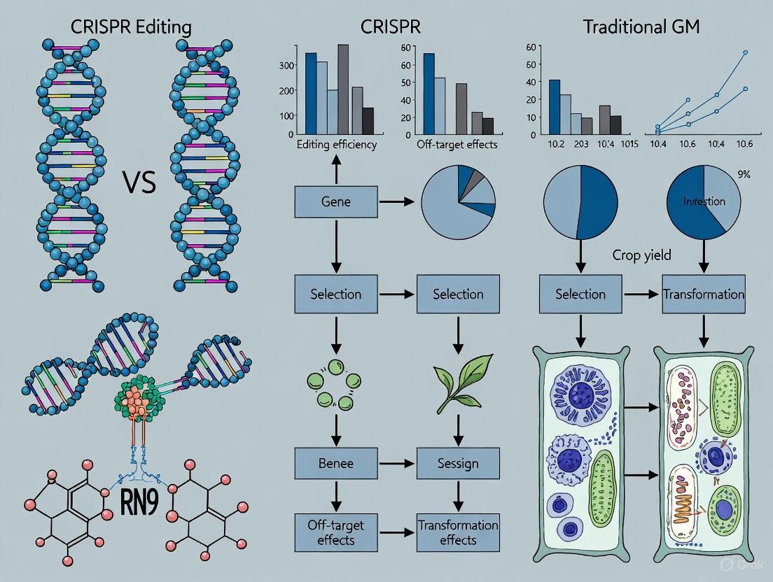
Figure 1: Comparative Workflows of Traditional GM and CRISPR Genome Editing
Technical Comparison: Efficiency, Precision, and Applications
Specificity and Unintended Effects
The precision of genetic interventions varies substantially between traditional and contemporary approaches. Traditional GM techniques are characterized by random integration events, which can lead to unpredictable disruption of native genes, alteration of regulatory elements, or positional effects that influence transgene expression [1]. In contrast, CRISPR-based editing offers targeted specificity through complementary base pairing between the gRNA and the target DNA sequence. However, CRISPR is not without its limitations regarding off-target effects, where the Cas9 nuclease may cleave sites with sequence similarity to the intended target [4] [3]. Studies have reported off-target activity frequencies of ≥50% for certain CRISPR constructs [3], though this varies significantly based on guide RNA design, Cas9 variant, and cell type.
Multiple strategies have been developed to mitigate CRISPR off-target effects:
- Computational guide design: Bioinformatic tools identify unique target sequences with minimal off-target potential [4].
- Engineered Cas variants: High-fidelity Cas9 proteins (e.g., SpCas9-HF1, eSpCas9) with reduced off-target activity [4].
- Modified guide RNAs: Truncated or chemically modified gRNAs with improved specificity [4].
- Alternative delivery methods: Ribonucleoprotein (RNP) complexes instead of plasmid DNA reduce temporal exposure [4].
- Advanced detection methods: Genome-wide approaches like GUIDE-seq and CIRCLE-seq identify off-target sites [4].
Table 2: Analytical Comparison of Editing Platforms
| Parameter | Traditional GM | ZFNs/TALENs | CRISPR-Cas9 |
|---|---|---|---|
| Targeting Specificity | Random integration | Protein-DNA recognition | RNA-DNA complementarity |
| Off-Target Risk | Position effects, disruption | Moderate | Moderate to High (guide-dependent) [3] |
| Efficiency | Variable, depends on transformation | Moderate | High [2] |
| Multiplexing Capacity | Limited | Difficult | High (multiple gRNAs) [2] |
| Development Timeline | Months to years | Months | Weeks [2] |
| Relative Cost | High | High | Low [2] |
Applications in Research and Therapeutics
The applications of traditional GM versus contemporary editing reflect their fundamental mechanistic differences. Traditional GM has been predominantly used in agriculture for introducing traits like herbicide tolerance (e.g., Roundup Ready system) [5] and insect resistance (Bt crops) [1], and in biopharmaceutical production (e.g., recombinant insulin). While effective, these applications typically involve adding new genetic functions rather than correcting existing sequences.
CRISPR editing has expanded these possibilities dramatically:
- Functional genomics: High-throughput knockout screens to identify essential genes and synthetic lethal interactions [2].
- Disease modeling: Precise introduction of disease-associated mutations in cell lines and animal models [2] [6].
- Gene therapy: Direct correction of pathogenic mutations in monogenic disorders like β-thalassemia and sickle cell anemia [2] [3].
- Agricultural improvements: Non-browning mushrooms, gluten-free wheat, disease-resistant citrus and cacao [7].
- Multiplexed editing: Simultaneous modification of multiple genetic loci [2].
Experimental Approaches and Methodologies
Delivery Methods
Efficient delivery of genetic material into target cells remains a critical challenge for both traditional and contemporary approaches. Traditional GM predominantly relies on:
- Agrobacterium-mediated transformation: Particularly for plants [1].
- Biolistic methods: Particle bombardment for organisms recalcitrant to Agrobacterium transformation [1].
- Viral vectors: Retroviruses, lentiviruses, and adenoviruses for animal cells [3].
Contemporary editing platforms utilize both viral and non-viral delivery systems:
- Viral vectors: Adeno-associated viruses (AAV) commonly used for in vivo therapeutic delivery [3].
- Physical methods: Electroporation, microinjection, and nanoparticle-mediated delivery [2].
- Chemical methods: Lipid-based transfection and polymer-based reagents [2].
- Ribonucleoprotein (RNP) complexes: Direct delivery of preassembled Cas9-gRNA complexes offering rapid activity and reduced off-target effects [4].
Analytical and Validation Methods
Rigorous validation of genetic modifications is essential for both research integrity and therapeutic safety. Traditional GM validation typically includes:
- Southern blotting: To confirm transgene integration and copy number.
- PCR analysis: To detect presence of the inserted sequence.
- Expression analysis: RT-qPCR, Western blot, or immunohistochemistry to confirm transgene expression.
CRISPR editing requires additional specificity assessments:
- Targeted deep sequencing: Of both on-target and predicted off-target sites [4].
- GUIDE-seq: Genome-wide, unbiased identification of DSBs enabled by sequencing [4].
- BLESS: Direct in situ breaks labeling, enrichment on streptavidin, and next-generation sequencing [4].
- Digenome-seq: In vitro nuclease-digested whole genome sequencing [4].
- T7E1 assay or Tracking assay: To detect nuclease-induced mutations [4].
The Research Toolkit: Essential Reagents and Methods
Table 3: Essential Research Reagents for Genome Editing Experiments
| Reagent/Method | Function | Considerations |
|---|---|---|
| Guide RNA (gRNA) | Targets Cas nuclease to specific DNA sequence | Design impacts efficiency and specificity; avoid off-target sites [4] |
| Cas9 Nuclease | Creates double-strand breaks at target sites | Multiple variants available with different PAM requirements and fidelity [8] |
| Repair Templates | Provides homology for HDR-mediated precise editing | Single-stranded or double-stranded DNA donors; design depends on edit type |
| Delivery Vehicles | Introduces editing components into cells | Viral (AAV, lentivirus) vs. non-viral (electroporation, lipofection) [3] |
| Selectable Markers | Enriches for successfully modified cells | Antibiotic resistance, fluorescence; can be excised after selection [1] |
| Off-Target Detection Assays | Identifies unintended edits | GUIDE-seq, CIRCLE-seq, targeted sequencing [4] |
Regulatory and Safety Considerations
The regulatory landscape for genetically modified organisms varies significantly across jurisdictions and reflects the technical distinctions between traditional GM and genome editing. The fundamental regulatory question centers on whether gene-edited organisms should be classified and regulated similarly to traditional GMOs [1] [9].
- United States: USDA has deregulated certain CRISPR-edited crops, especially those without foreign DNA (SDN-1 and SDN-2 modifications) [1].
- European Union: Court of Justice ruling classified gene-edited organisms as GMOs, subject to existing strict regulations [1].
- Argentina and Brazil: Implemented product-based regulatory approaches focusing on the presence of "novel combinations of genetic material" [9].
- Global Governance: The Cartagena Protocol on Biosafety provides an international framework, though key countries like the U.S. and Canada are not parties [9].
Safety considerations also differ between the technologies. Traditional GM raises concerns about:
- Allergenicity: Potential for novel proteins to trigger allergic responses [5].
- Antibiotic resistance: Use of antibiotic resistance markers in selection [5].
- Environmental impact: Gene flow to wild relatives, effects on non-target organisms [5].
CRISPR editing introduces distinct considerations:
- Off-target effects: Unintended modifications at similar genomic sequences [3].
- Mosaicism: Incomplete editing in some cells of the target organism [6].
- Immunogenicity: Immune responses against bacterial-derived Cas proteins in therapeutic applications [3].
- Long-term stability: Persistence of edits through cell divisions and generations.
Future Directions and Emerging Technologies
The field of genome editing continues to evolve rapidly, with several promising technologies emerging beyond standard CRISPR-Cas9 systems:
- Base editing: Enables direct, irreversible conversion of one DNA base to another without double-strand breaks, reducing indel formation [2].
- Prime editing: Offers precise small edits, insertions, and deletions without donor templates [2].
- AI-designed editors: Machine learning approaches generating novel editors with optimized properties [8].
- Epigenome editing: Targeted modification of epigenetic marks without changing DNA sequence.
- RNA editing: Precise modification of RNA transcripts for reversible therapeutic effects.
These advancements continue to blur the lines between traditional genetic modification and precision editing, while simultaneously raising new technical and ethical considerations for the research community.
The landscape of genetic engineering has fundamentally shifted from the random integration of foreign DNA characteristic of traditional GM to the precision targeting of contemporary genome editing platforms like CRISPR-Cas9. This transition has enabled unprecedented control over genetic modifications, with applications spanning basic research, therapeutic development, and agricultural improvement. While both approaches continue to have relevance in specific contexts, the precision, efficiency, and versatility of contemporary editing platforms have positioned them as the dominant technology for most research applications. However, technical challenges remain, particularly regarding off-target effects, delivery efficiency, and tissue-specific editing. Furthermore, the evolving regulatory landscape continues to shape the application of these technologies across different domains and jurisdictions. As the field advances with emerging technologies like base editing and AI-designed editors, researchers must maintain a nuanced understanding of both the technical capabilities and the ethical implications of these powerful genetic tools.
Before the advent of CRISPR-Cas9, the field of genome engineering was revolutionized by two groundbreaking protein-based technologies: Zinc Finger Nucleases (ZFNs) and Transcription Activator-Like Effector Nucleases (TALENs). These engineered nucleases provided researchers with the first tools for making targeted, precise modifications to genomes in a wide range of cell types and organisms [10] [11]. Both technologies operate on a similar principle: they fuse a customizable, sequence-specific DNA-binding domain to a non-specific DNA cleavage domain, creating molecular scissors that can induce double-strand breaks at predetermined genomic locations [10]. The cellular repair of these breaks—either through error-prone non-homologous end joining (NHEJ) or homology-directed repair (HDR)—enables a broad range of genetic modifications, from gene knockouts to precise nucleotide changes [10] [11]. This guide provides a comprehensive comparison of ZFNs and TALENs, examining their mechanisms, efficiencies, specificities, and practical applications in modern biological research and therapeutic development.
Molecular Mechanisms and Architectural Designs
Zinc Finger Nucleases (ZFNs): Modular Protein Assembly
ZFNs are fusion proteins comprising an array of engineered Cys2-His2 zinc finger DNA-binding domains attached to the catalytic domain of the FokI restriction endonuclease [10] [11]. Each zinc finger domain recognizes approximately 3 base pairs (bps) in the DNA major groove, with multiple fingers (typically 3-6) assembled in tandem to recognize contiguous 9-18 bp sequences [10] [12]. The FokI nuclease domain requires dimerization for activation, necessitating the design of two ZFN subunits that bind opposite DNA strands at sequences flanking the target site, with the dimerization inducing a double-strand break in the intervening spacer region [12] [11].
The modular assembly of zinc finger arrays presented significant engineering challenges. While zinc finger domains have been developed to recognize most of the 64 possible nucleotide triplets, zinc fingers assembled in arrays can exhibit context-dependent effects where the specificity of neighboring fingers influences overall DNA-binding affinity [10] [12]. This complexity made the rational design of effective ZFNs challenging for nonspecialists, though modular assembly systems and open-source component libraries were developed to address these limitations [10] [11].
Transcription Activator-Like Effector Nucleases (TALENs): Deciphered Code
TALENs similarly fuse DNA-binding domains to the FokI nuclease domain but utilize a different architectural principle derived from transcription activator-like effector (TALE) proteins of Xanthomonas bacteria [10] [11]. The DNA-binding domain consists of a series of 33-35 amino acid repeats, each recognizing a single DNA base pair [10]. Specificity is determined by two hypervariable amino acids at positions 12 and 13, known as repeat-variable diresidues (RVDs), which follow a simple recognition code: NI for adenine, NG for thymine, HD for cytosine, and NN for guanine/adenine [10] [11].
Unlike ZFNs, TALE repeats operate independently without significant neighbor effects, making TALEN design more straightforward and predictable [10] [12]. Like ZFNs, TALENs function as pairs binding opposite DNA strands, with FokI dimerization creating a double-strand break in the spacer region (typically 14-20 bp) between the binding sites [12].
Figure 1: Molecular Architecture of ZFNs and TALENs. Both systems utilize FokI nuclease domains that require dimerization for activation, but differ in their DNA recognition mechanisms: ZFNs use zinc finger arrays that recognize 3bp per module, while TALENs use TALE repeats that recognize 1bp per module through a simple RVD code.
Performance Comparison: Efficiency, Specificity, and Experimental Data
Quantitative Comparison of Editing Performance
Direct comparative studies provide valuable insights into the relative performances of ZFNs, TALENs, and the contemporary benchmark CRISPR-Cas9. A systematic comparison using GUIDE-seq methodology to evaluate genome-wide specificity at human papillomavirus 16 (HPV16) target sites revealed substantial differences in off-target profiles [13] [14].
Table 1: Quantitative Comparison of Editing Efficiency and Specificity Across Platforms
| Platform | Target Gene | On-Target Efficiency | Off-Target Count | Key Advantages | Key Limitations |
|---|---|---|---|---|---|
| ZFN | HPV16 URR | High | 287-1,856 [14] | High specificity when properly designed [15] | Context-dependent finger effects [12] |
| TALEN | HPV16 E7 | High | 36 [14] | Simple design rules, modular assembly [10] | Large repeat size challenges delivery [2] |
| CRISPR-Cas9 | HPV16 E7 | High | 4 [14] | Easy design, multiplexing capability [2] | Off-target concerns with standard Cas9 [2] |
In bovine and dairy goat fetal fibroblasts, CRISPR-Cas9 demonstrated significantly higher knock-in efficiency compared to both ZFNs and TALENs. For enhanced green fluorescent protein (eGFP) knock-in, CRISPR-Cas9 achieved 77.02% efficiency compared to 13.68% for ZFNs—approximately 5.6-fold higher [16]. Similarly, for humanized Fat-1 (hFat-1) knock-in, CRISPR-Cas9 achieved 79.01% efficiency while ZFNs failed to produce any knock-in events [16]. In direct comparison with TALENs, CRISPR-Cas9 showed more than double the knock-in efficiency for both eGFP (70.37% vs. 32.35%) and hFat-1 (74.29% vs. 26.47%) [16].
Experimental Protocols for Efficiency and Specificity Assessment
Gene Knock-in Efficiency Protocol
The comparative study of knock-in efficiencies in bovine and dairy goat fetal fibroblasts followed this methodology [16]:
Nuclease Design: ZFNs and CRISPR/Cas9 plasmids were designed to target exon 2 of the bovine myostatin (MSTN) gene; TALENs and CRISPR/Cas9 targeted exon 2 of the dairy goat β-casein gene.
Donor Construction: Donor plasmids were constructed in the GFP-PGK-NeoR plasmid backbone containing 5' and 3' homologous arms flanking either hFat-1 or eGFP transgenes.
Cell Transfection: ZFNs, TALENs, or CRISPR/Cas9 plasmids were co-transfected with donor plasmids into fetal fibroblasts via electroporation.
Selection and Cloning: After G418 (Geneticin) selection, single cells were isolated using mouth pipetting, flow cytometry, or a cell shove.
Analysis: Gene knock-in events were screened by PCR across homologous arms, with efficiencies calculated as the percentage of successfully edited clones.
GUIDE-seq Off-Target Detection Protocol
The genome-wide unbiased identification of double-stranded breaks enabled by sequencing (GUIDE-seq) method was adapted for ZFNs and TALENs as follows [13] [14]:
dsODN Tag Integration: Double-stranded oligodeoxynucleotides (dsODNs) were introduced into cells expressing the nucleases, where they integrated into nuclease-induced double-strand break sites.
Library Preparation and Sequencing: Genomic DNA was extracted, fragmented, and libraries prepared for next-generation sequencing with primers specific to the integrated dsODN tags.
Bioinformatic Analysis: Sequencing reads were mapped to the reference genome to identify off-target sites containing integrated dsODN tags.
Validation: Potential off-target sites were validated using targeted sequencing or T7 endonuclease I (T7E1) mismatch detection assays.
Research Reagent Solutions: Essential Materials for Gene Editing Studies
Table 2: Key Research Reagents for ZFN and TALEN Experiments
| Reagent Category | Specific Examples | Function and Application | Considerations |
|---|---|---|---|
| Nuclease Assembly Systems | Zinc Finger Modular Assembly [10], Golden Gate TALEN Assembly [10] | Construction of custom DNA-binding domains | TALEN assembly is more straightforward than ZFNs [10] |
| Delivery Vectors | Plasmid DNA, mRNA, Viral Vectors (Lentivirus, Adenovirus) [2] | Introduction of nuclease constructs into cells | Protein delivery reduces off-target effects [11] |
| Validation Assays | T7E1 Mismatch Detection [14], GUIDE-seq [13] [14] | Detection of nuclease activity and off-target effects | GUIDE-seq provides genome-wide off-target profiling [14] |
| Cell Culture Resources | G418 (Geneticin) [16], Fetal Fibroblasts [16] | Selection and maintenance of edited cells | Antibiotic selection enriches for successfully edited cells [16] |
| Repair Templates | ssODNs [11], Donor Vectors with Homology Arms [16] | Introduction of specific mutations via HDR | Homology arm length affects knock-in efficiency [16] |
Applications and Therapeutic Implementation
ZFNs and TALENs have enabled diverse applications across basic research, biotechnology, and clinical therapy. In biomedical research, they facilitate the creation of genetically modified cell lines and animal models for studying gene function and disease mechanisms [10] [17]. Therapeutically, ZFNs have advanced to clinical trials for HIV treatment through CCR5 disruption in CD4+ T cells, demonstrating safety and efficacy in reducing HIV DNA copies in patients [14]. Similarly, TALEN-engineered universal chimeric antigen receptor T-cells (UCART19) have induced molecular remission in B-cell acute lymphoblastic leukemia patients [14].
The choice between ZFNs and TALENs involves careful consideration of project requirements. ZFNs may be preferable for applications where smaller size is advantageous for delivery, while TALENs offer simpler design rules for laboratories with limited protein engineering expertise [10] [15]. Both platforms benefit from obligate heterodimer FokI variants that reduce off-target effects by preventing homodimer formation [11].
While CRISPR-Cas9 has largely superseded ZFNs and TALENs for many applications due to its simpler design, higher efficiency, and easier multiplexing capability [2] [16] [14], the protein-based platforms retain importance in specific contexts. ZFNs have established regulatory approval pathways and demonstrated clinical success, while TALENs offer high precision with potentially lower off-target risks in certain genomic contexts [2] [15]. The development of these technologies represented crucial milestones in genome engineering, providing the conceptual and technical foundations for programmable nuclease platforms and expanding the boundaries of biological research and therapeutic development [10] [11]. Their continued evolution and specialized applications ensure that ZFNs and TALENs remain valuable components of the modern genome editing toolkit, particularly for clinical applications where their long development history and characterized specificity profiles provide distinct advantages.
The landscape of genetic engineering has been fundamentally reshaped by the advent of CRISPR-Cas systems, which represent a significant departure from traditional gene-editing approaches. Unlike earlier protein-based methods, CRISPR technology utilizes an RNA-guided mechanism for precise DNA targeting, offering unprecedented simplicity, efficiency, and versatility. This revolutionary system, derived from a natural bacterial immune defense mechanism, has democratized genome editing by making precise genetic modifications accessible to researchers across diverse fields including therapeutics, agriculture, and basic biological research. As CRISPR technologies continue to evolve at a rapid pace, understanding their comparative advantages, limitations, and appropriate applications becomes essential for researchers, scientists, and drug development professionals seeking to harness their full potential.
Understanding Gene Editing Platforms: From Traditional Methods to CRISPR
The evolution of gene editing technologies has progressed through distinct generations, beginning with early methods that required complex protein engineering and culminating in the RNA-guided precision of CRISPR systems. Traditional gene editing platforms including Zinc Finger Nucleases (ZFNs) and Transcription Activator-Like Effector Nucleases (TALENs) pioneered targeted genetic modifications but presented significant technical challenges that limited their widespread adoption [2] [18].
ZFNs, developed first among programmable nucleases, utilize engineered zinc finger proteins that typically recognize 3-base pair DNA sequences. These domains are fused to the FokI nuclease domain, which requires dimerization to become active, necessitating the design of two separate ZFNs that bind opposite DNA strands in precise orientation and spacing [2] [18]. This requirement for paired proteins makes ZFN design complex and often unsuccessful. Similarly, TALENs employ transcription activator-like effector (TALE) proteins in which each repeat domain recognizes a single specific nucleotide, also fused to FokI nuclease domains requiring dimerization for activity [2]. While TALENs offer more straightforward design rules than ZFNs, their assembly remains labor-intensive due to the highly repetitive nature of TALE proteins and the substantial molecular cloning required [2] [18].
The fundamental distinction between these traditional methods and CRISPR systems lies in their targeting mechanisms: ZFNs and TALENs rely on protein-DNA interactions for sequence recognition, whereas CRISPR systems utilize RNA-DNA complementarity through a guide RNA molecule that directs Cas nucleases to specific genomic loci [18]. This fundamental difference in targeting mechanism explains much of CRISPR's advantage in design simplicity and versatility, as modifying RNA sequences is substantially easier than engineering custom DNA-binding proteins for each new target [2].
Mechanism of Action: RNA-Guided DNA Targeting
The CRISPR-Cas Molecular Machinery
The CRISPR-Cas system operates through an elegantly simple yet highly effective mechanism centered on RNA-guided DNA recognition. The core components include the Cas nuclease (most commonly Cas9) and a guide RNA (gRNA) molecule that combines the functions of CRISPR RNA (crRNA) and trans-activating crRNA (tracrRNA) into a single chimeric molecule [18] [19]. This gRNA contains a 20-nucleotide sequence that determines targeting specificity through complementary base pairing with the target DNA [20].
The process initiates when the Cas nuclease-gRNA complex scans DNA for sequences complementary to the gRNA that are adjacent to a short DNA sequence known as the protospacer adjacent motif (PAM) [18]. For the most widely used Cas9 from Streptococcus pyogenes (SpCas9), the PAM sequence is 5'-NGG-3', where "N" represents any nucleotide [18] [20]. PAM recognition triggers local DNA melting, followed by RNA-DNA hybridization between the gRNA spacer and the target DNA strand. Successful complementarity leads to activation of the Cas nuclease, which generates a double-strand break (DSB) approximately 3-4 nucleotides upstream of the PAM sequence [18] [19].
The following diagram illustrates the fundamental mechanism of RNA-guided DNA targeting by CRISPR-Cas systems:
DNA Repair Pathways and Editing Outcomes
Following the creation of a DSB, cellular repair mechanisms are activated that determine the ultimate editing outcome. The primary repair pathways include Non-Homologous End Joining (NHEJ) and Homology-Directed Repair (HDR) [2] [18]. NHEJ is an error-prone process that directly ligates broken DNA ends, often resulting in small insertions or deletions (indels) that can disrupt gene function by causing frameshift mutations or premature stop codons [2]. This pathway is predominant in most cell types and is highly efficient for gene knockout applications.
In contrast, HDR utilizes a DNA repair template to enable precise genetic modifications, including specific nucleotide changes, gene insertions, or reporter knock-ins [2] [21]. While HDR offers greater precision, its efficiency is generally lower than NHEJ and is restricted to specific cell cycle phases (S/G2 phases) [21]. The development of base editing and prime editing technologies has addressed some limitations of HDR by enabling precise nucleotide changes without requiring DSBs or donor templates, thereby expanding the toolkit for precise genome editing [22].
Comparative Analysis: CRISPR vs. Traditional Gene Editing Methods
Technical Performance Metrics
Direct comparison of CRISPR systems with traditional gene editing methods reveals distinct advantages and limitations across multiple performance parameters. The following table summarizes key comparative metrics based on current experimental data:
| Performance Parameter | CRISPR-Cas Systems | Zinc Finger Nucleases (ZFNs) | Transcription Activator-Like Effector Nucleases (TALENs) |
|---|---|---|---|
| Targeting Efficiency | 0-81% (High) [18] | 0-12% (Low) [18] | 0-76% (Moderate) [18] |
| Target Specificity | Moderate to high; predictable off-target effects [18] | High; less predictable off-target effects [2] | High; less predictable off-target effects [2] |
| Ease of Design | Simple; only gRNA requires redesign [2] [18] | Difficult; requires protein engineering [2] [18] | Difficult; requires protein engineering [2] [18] |
| Multiplexing Capacity | High; enables simultaneous editing of multiple genes [2] [18] | Limited; challenging to implement [18] | Limited; challenging to implement [18] |
| Development Timeline | Days (gRNA design and synthesis) [2] | Months (extensive protein engineering) [2] | Weeks to months (labor-intensive assembly) [2] |
| Cost Considerations | Low [2] [18] | High [2] [18] | High [2] [18] |
| Primary Applications | Broad range including functional genomics, therapeutics, agriculture, diagnostics [2] [23] [24] | Niche applications requiring high precision; stable cell line generation [2] | Niche applications requiring high precision; small-scale precision edits [2] |
Experimental Validation: Case Studies
The practical differences between editing platforms are exemplified in several experimental case studies. In one comparative analysis targeting the CCR5 gene (a co-receptor for HIV), TALENs demonstrated high specificity with minimal off-target effects, while CRISPR achieved superior editing efficiency and significantly reduced development time [2]. This efficiency advantage makes CRISPR particularly suitable for large-scale functional genomics screens, where thousands of genetic perturbations need to be tested in parallel [2].
For therapeutic applications, CRISPR-based therapies have demonstrated remarkable clinical success. In clinical trials for hereditary transthyretin amyloidosis (hATTR), CRISPR-Cas9 delivered via lipid nanoparticles achieved approximately 90% reduction in disease-related protein levels, with sustained response maintained over two years in all participants [24]. Similarly, in hereditary angioedema (HAE), CRISPR treatment resulted in an 86% reduction in kallikrein protein levels and significantly reduced inflammatory attacks [24]. These results highlight the therapeutic potential of CRISPR systems, which benefit from simplified design processes and efficient delivery mechanisms.
Advanced CRISPR Technologies and Methodologies
Expanding the CRISPR Toolkit
Beyond standard CRISPR-Cas9 systems, numerous advanced variants have been developed to address specific experimental needs and overcome limitations of first-generation tools. Base editing systems enable direct, irreversible conversion of one DNA base pair to another without requiring DSBs, thereby minimizing indel formation [22]. These systems combine a catalytically impaired Cas nuclease (nickase) with a deaminase enzyme, enabling precise C•G to T•A or A•T to G•C conversions [22].
Prime editing represents a further advancement, capable of installing all possible nucleotide transitions and transversions, as well as small insertions and deletions, without requiring DSBs or donor templates [21]. This technology uses a catalytically impaired Cas9 fused to a reverse transcriptase and a prime editing guide RNA (pegRNA) that both specifies the target site and encodes the desired edit [21].
The discovery and engineering of novel Cas variants continues to expand the targeting range and specificity of CRISPR systems. CRISPR-Cas12a (formerly Cpf1) differs from Cas9 in creating staggered DNA cuts rather than blunt ends, requiring a T-rich PAM sequence, and utilizing a single RNA molecule for processing and targeting [25]. Recent advances in metagenomic mining and machine learning approaches have identified numerous novel Cas12a subtypes with diverse properties, including distinct PAM preferences and editing windows [25].
Experimental Protocols for CRISPR Applications
Protocol 1: CRISPR-Cas9 Mediated Gene Knockout
This standard protocol enables efficient gene disruption through NHEJ-mediated repair:
gRNA Design: Select 20-nucleotide target sequences adjacent to 5'-NGG-3' PAM sites using established tools (e.g., CRISPOR, CHOPCHOP) [20]. Prioritize targets with minimal off-target potential based on specificity scores.
Vector Construction: Clone gRNA sequence into appropriate CRISPR plasmid backbone (e.g., pSpCas9(BB)) using Golden Gate assembly or other standardized methods.
Delivery System: Transfert target cells using appropriate method (lipofection, electroporation, or viral delivery) with 1:1 mass ratio of Cas9 and gRNA vectors [2].
Validation: Assess editing efficiency 48-72 hours post-transfection using T7E1 assay, TIDE analysis, or next-generation sequencing. Confirm phenotypic effects through functional assays.
For difficult-to-transfect cells, ribonucleoprotein (RNP) delivery of precomplexed Cas9 protein and gRNA can improve efficiency while reducing off-target effects [23].
Protocol 2: Base Editing for Precise Nucleotide Conversion
This protocol enables precise nucleotide changes without creating double-strand breaks:
Base Editor Selection: Choose appropriate base editor (e.g., BE4-Gam for C•G to T•A conversions; ABE7.10 or ABE8e for A•T to G•C conversions) [22].
gRNA Design: Design gRNAs positioning the target nucleotide within the editing window (typically positions 4-8 for ABE7.10 and BE4) [22]. Use prediction tools (e.g., CRISPRon-ABE, CRISPRon-CBE) to optimize efficiency and minimize bystander edits [22].
Delivery: Transfert base editor plasmid or deliver as RNP complex. For plasmid transfection, use 2:1 mass ratio of base editor to gRNA vector.
Analysis: Evaluate editing efficiency 72-96 hours post-transfection using Sanger sequencing or next-generation sequencing. Screen for potential off-target effects at predicted sites.
Recent advances in machine learning have significantly improved base editing outcome predictions. Deep learning models trained simultaneously on multiple experimental datasets now enable more accurate prediction of both editing efficiency and specific outcomes, addressing the challenge of data heterogeneity across different experimental conditions [22].
Research Reagent Solutions
Successful implementation of CRISPR technologies requires specific reagents and tools. The following table outlines essential research reagents and their applications:
| Research Reagent | Function | Example Applications |
|---|---|---|
| Cas9 Nuclease | RNA-guided DNA endonuclease creating double-strand breaks | Gene knockout, knock-in, functional screening [2] [18] |
| Base Editors | Fusion proteins enabling precise single nucleotide changes without DSBs | Disease modeling, therapeutic correction of point mutations [22] |
| Guide RNA (gRNA) | Synthetic RNA molecule directing Cas nuclease to specific DNA target | All CRISPR applications; determines targeting specificity [2] [20] |
| Lipid Nanoparticles (LNPs) | Delivery vehicles for CRISPR components; particularly efficient for liver targeting | Therapeutic applications (e.g., hATTR, HAE) [24] |
| CRISPR Screening Libraries | Pooled collections of gRNAs targeting multiple genes | Genome-wide functional screens, drug target identification [2] |
| Anti-CRISPR Proteins | Naturally occurring inhibitors of Cas nucleases | Control of editing timing, reduction of off-target effects [20] |
Emerging Trends and Future Directions
The CRISPR technology landscape continues to evolve rapidly, with several emerging trends shaping future applications. The discovery of novel Cas nucleases through metagenomic mining and machine learning approaches is expanding the available toolkit beyond the well-characterized Cas9 and Cas12a [26] [25]. These efforts have identified compact Cas variants such as Cas12f (approximately one-third the size of SpCas9) that enable delivery via size-constrained vectors [23] [25].
The application of artificial intelligence and deep learning represents another significant advancement, improving the prediction of editing outcomes and gRNA efficiency [22] [25]. For example, the development of "dataset-aware" training approaches allows models to effectively learn from multiple heterogeneous datasets while accounting for systematic differences between experimental conditions [22]. These AI-driven tools are becoming increasingly important for optimizing editing precision and reducing off-target effects.
In therapeutic development, personalized CRISPR treatments are emerging as a promising approach for rare genetic disorders. The landmark case of an infant with CPS1 deficiency who received a bespoke in vivo CRISPR therapy developed and delivered in just six months demonstrates the potential for rapid customization of CRISPR therapies for individual patients [24]. Additionally, the ability to administer multiple doses of LNP-delivered CRISPR treatments (as demonstrated in both the CPS1 deficiency and hATTR trials) represents a significant advantage over viral vector delivery, which typically precludes redosing due to immune responses [24].
The following diagram illustrates the workflow for developing and implementing CRISPR-based therapeutic interventions:
The CRISPR revolution has fundamentally transformed the landscape of genetic engineering, establishing RNA-guided DNA targeting as the predominant approach for precise genome manipulation. While traditional methods like ZFNs and TALENs continue to have value for specific applications requiring exceptional precision, CRISPR technologies offer superior versatility, efficiency, and accessibility for most research and therapeutic applications. The rapid advancement of base editing, prime editing, and novel Cas variants continues to expand the capabilities of CRISPR systems, addressing initial limitations and opening new possibilities for both basic research and clinical applications. As the field progresses, the integration of machine learning and computational approaches with experimental methods will further enhance the precision and predictability of CRISPR-mediated genome editing, solidifying its role as an indispensable tool in modern biological research and therapeutic development.
The field of genetic engineering has undergone a revolutionary transformation, evolving from the broad, imprecise techniques of recombinant DNA technology to the unparalleled precision of modern programmable nucleases. This journey represents a fundamental shift in our ability to interact with the genetic code, moving from the transfer of large DNA segments between species to the precise rewriting of nucleotides at single-base resolution. For researchers and drug development professionals, understanding this evolution is not merely an academic exercise but a critical framework for selecting appropriate experimental strategies, navigating regulatory landscapes, and developing novel therapeutic interventions. The comparative analysis between traditional genetically modified organism (GMO) approaches and contemporary gene-editing platforms reveals distinct paradigms in experimental design, risk assessment, and application potential. This guide provides a comprehensive technical comparison of these technologies, supported by experimental data and detailed methodologies, to inform strategic decision-making in research and therapeutic development.
Historical Trajectory and Technological Paradigms
The history of genetic manipulation is characterized by increasing precision and control over genetic outcomes, marked by several key technological breakthroughs that have fundamentally reshaped experimental possibilities.
Table 1: Key Historical Milestones in Genetic Engineering
| Time Period | Technology | Key Innovation | Primary Application |
|---|---|---|---|
| 1970s-1980s | Recombinant DNA Technology | Gene transfer between species | Production of therapeutic proteins (e.g., insulin) |
| 2000s | Zinc Finger Nucleases (ZFNs) | First programmable nucleases | Gene knockout in model organisms |
| 2011 | TALENs | Modular DNA-binding domain | Gene therapy development |
| 2012 | CRISPR-Cas9 | RNA-guided DNA cleavage | High-throughput functional genomics |
Recombinant DNA Technology: The Foundation Era
Recombinant DNA technology, emerging in the 1970s, enabled the transfer of genes between unrelated species, creating transgenic organisms with novel traits. This approach relied on random integration of foreign DNA into host genomes, typically using Agrobacterium-mediated transformation or biolistic methods for plants [1]. A classic example includes Bt crops, where genes from Bacillus thuringiensis were inserted into cotton and corn to confer insect resistance [1]. While revolutionary, this technology faced limitations including random integration sites, unpredictable expression levels, and significant public controversy regarding environmental and health impacts.
The Rise of Programmable Nucleases
The development of engineered nucleases marked a critical transition toward precision genetic manipulation. These technologies enabled targeted double-strand breaks (DSBs) at specific genomic locations, harnessing cellular repair mechanisms to generate desired modifications.
Zinc Finger Nucleases (ZFNs), the first programmable nucleases, combined zinc finger DNA-binding domains with the FokI nuclease domain [27]. Each zinc finger module recognizes a 3-base pair DNA sequence, and multiple fingers are assembled to target longer sequences (typically 18-24 bp) [2]. The requirement for FokI dimerization ensures high specificity, as two ZFN molecules must bind opposite DNA strands to enable cleavage [27].
TALENs (Transcription Activator-Like Effector Nucleases) improved upon ZFNs by utilizing TALE proteins from Xanthomonas bacteria, where each repeat domain recognizes a single nucleotide [2] [27]. This simpler recognition code provided greater design flexibility and targeting range compared to ZFNs, though protein engineering remained labor-intensive [2].
The CRISPR-Cas System represented a paradigm shift from protein-based to RNA-guided targeting. Originally identified as a bacterial adaptive immune system, CRISPR-Cas9 was adapted for genome editing in 2012 [28]. The system utilizes a Cas nuclease (e.g., Cas9) directed by a guide RNA (gRNA) complementary to the target DNA sequence, requiring only the redesign of the gRNA to redirect targeting [1] [28]. This simplicity, versatility, and efficiency led to its rapid adoption across biological research and therapeutic development.
Figure 1: Evolution of genetic engineering technologies showing increasing precision and accessibility.
Comparative Technical Analysis of Editing Platforms
Mechanism of Action and Experimental Workflows
Understanding the distinct molecular mechanisms of each editing platform is essential for appropriate experimental design and interpretation of results.
Recombinant DNA Technology relies on the insertion of foreign DNA fragments into host genomes, typically using vector systems (e.g., plasmids) containing the gene of interest along with regulatory elements and selectable markers. The random nature of integration leads to variable expression and potential disruption of endogenous genes [1].
Programmable Nuclease Platforms share a common mechanism involving the induction of targeted double-strand breaks (DSBs) followed by cellular repair, but differ in their targeting approaches:
ZFNs and TALENs: Both utilize protein-based DNA recognition domains fused to the FokI nuclease domain. The FokI domain must dimerize to become active, requiring two engineered nuclease proteins to bind opposite DNA strands in close proximity and correct orientation [27]. ZFNs recognize nucleotides in triplets via zinc finger modules, while TALENs use repeat-variable diresidues (RVDs) that each recognize a single nucleotide [2] [27].
CRISPR-Cas Systems: Utilize a complex of Cas nuclease with a guide RNA (gRNA) that hybridizes to the target DNA sequence via complementary base pairing. The Cas9 nuclease requires a protospacer adjacent motif (PAM) sequence adjacent to the target site for recognition and cleavage [1] [28]. Upon binding, Cas9 introduces a blunt-ended DSB approximately 3-4 nucleotides upstream of the PAM site [29].
Figure 2: Comparative mechanisms of programmable nuclease platforms.
Quantitative Performance Metrics
Direct comparison of technical parameters reveals the distinct advantages and limitations of each platform for specific research applications.
Table 2: Performance Comparison of Gene Editing Platforms
| Parameter | ZFNs | TALENs | CRISPR-Cas9 | Traditional Recombinant DNA |
|---|---|---|---|---|
| Targeting Specificity | High | High | Moderate to High | N/A (Random integration) |
| Off-Target Effects | Low | Low | Moderate to High [2] | N/A |
| Editing Efficiency | Moderate | Moderate | High | N/A |
| Multiplexing Capacity | Low | Low | High [2] | Limited |
| Design Complexity | High (Protein engineering) | High (Protein engineering) | Low (gRNA design) | Moderate (Vector construction) |
| Development Timeline | Weeks to months | Weeks to months | Days [2] | Weeks |
| Cost | High | High | Low | Moderate |
The data reveal CRISPR-Cas9's superior efficiency and multiplexing capabilities, while ZFNs and TALENs maintain advantages in specificity with lower off-target effects [2]. Traditional recombinant DNA operates in a fundamentally different paradigm without targeted specificity.
Experimental Design and Methodological Considerations
Protocol for CRISPR-Cas9 Genome Editing
The following detailed protocol outlines a standard workflow for CRISPR-Cas9 mediated gene editing in mammalian cells, incorporating critical optimization steps for high efficiency and specificity:
Target Selection and gRNA Design: Identify target sequence with 5'-NGG-3' PAM motif. Design gRNA with 20-nucleotide complementarity region. Utilize computational tools (e.g., CRISPR-GPT) to predict on-target efficiency and nominate potential off-target sites [30]. Select multiple gRNAs for comparison.
Vector Construction or RNP Preparation:
- Plasmid-based: Clone gRNA sequence into CRISPR expression vector (e.g., pX330) containing human codon-optimized Cas9 and gRNA scaffold.
- RNP-based: Complex purified Cas9 protein with in vitro transcribed or synthetic gRNA at 3:1 molar ratio (Cas9:gRNA) in nuclease-free buffer, incubate 10 minutes at room temperature for RNP formation [29].
Delivery Optimization:
- For plasmid DNA: Use lipid-based transfection (e.g., Lipofectamine 3000) or viral delivery (lentiviral, adenoviral).
- For RNP: Utilize electroporation (e.g., Neon System) for high efficiency delivery, particularly in primary cells.
- For in vivo applications: Employ lipid nanoparticles (LNPs) optimized for tissue targeting [24].
Editing Validation:
- 48-72 hours post-delivery, harvest cells for genomic DNA extraction.
- Assess editing efficiency using T7 Endonuclease I or TIDE assay for indel quantification.
- For precise edits, include donor DNA template (single-stranded oligodeoxynucleotide or double-stranded DNA) with 30-50 nt homology arms.
Off-Target Assessment:
- Perform BreakTag sequencing for unbiased nomination of off-target sites and characterization of nuclease activity [29].
- Utilize targeted amplicon sequencing of predicted off-target loci.
- Employ functional assays relevant to the specific experimental context.
Protocol for ZFNs/TALENs Genome Editing
The protein-based nuclease workflow shares similarities with CRISPR but involves distinct design and validation steps:
Target Site Selection: Identify pairs of binding sites in forward and reverse orientation with 16-20 bp spacing for FokI dimerization. Avoid sites with high DNA methylation or nucleosome occupancy.
Nuclease Assembly:
- For ZFNs: Assemble zinc finger arrays using modular assembly or selection-based methods (e.g., OPEN) to target 9-18 bp per monomer.
- For TALENs: Construct repeat arrays using golden gate cloning with specific RVDs (NI=A, HD=C, NN=G/A, NG=T) for target recognition.
Expression Vector Construction: Clone engineered nuclease sequences into mammalian expression vectors with nuclear localization signals. Co-express both monomers from separate promoters or vectors.
Delivery and Validation: Follow similar delivery and validation approaches as CRISPR-Cas9, with particular attention to balancing expression of both monomers for optimal cleavage efficiency.
Research Reagent Solutions Toolkit
Table 3: Essential Research Reagents for Genome Editing Experiments
| Reagent Category | Specific Examples | Function | Considerations |
|---|---|---|---|
| Nuclease Systems | SpCas9, AsCas12a, OpenCRISPR-1 [8] | DNA cleavage at target sites | PAM requirements, size constraints for delivery |
| Editing Enhancers | Base editors, Prime editors | Enable precise edits without DSBs | Editing window, product purity |
| Delivery Tools | Lipid nanoparticles (LNPs) [24], AAV vectors, Electroporation systems | Deliver editing components to cells | Packaging capacity, cell toxicity, tropism |
| gRNA Design Tools | CRISPR-GPT [30], BreakInspectoR [29] | Predict efficiency and specificity | Incorporates off-target predictions |
| Validation Assays | BreakTag [29], T7E1, NGS-based methods | Assess on-target editing and off-target effects | Sensitivity, throughput, cost |
| Cell Culture Reagents | Cytokines, Small molecules | Enhance HDR efficiency, support cell viability | Optimization required for cell type |
Regulatory and Safety Considerations
The regulatory landscape for gene-edited products varies significantly based on the technology used and the nature of the genetic modification. Traditional recombinant DNA techniques, which involve introducing foreign DNA, are typically subject to strict GMO regulations worldwide [1]. In contrast, the regulatory status of CRISPR-edited organisms remains complex and varies by jurisdiction:
- United States: The USDA has deregulated certain CRISPR-edited crops, particularly those lacking foreign DNA (transgenes), considering them equivalent to conventionally bred plants in some instances [1].
- European Union: Maintains a more precautionary approach, generally considering all plants modified through gene editing as GMOs, even if transgene-free [1] [27].
- Global Variation: Countries including Argentina, Brazil, and Japan have established differentiated approaches that may classify certain CRISPR edits as non-GM, particularly for SDN1 and SDN2 modifications that involve small changes without introducing foreign DNA [1].
For therapeutic applications, regulatory agencies like the FDA and EMA evaluate CRISPR-based therapies through existing drug approval pathways, with particular attention to off-target effects, delivery safety, and long-term consequences [24]. The first FDA-approved CRISPR therapy, Casgevy for sickle cell disease and beta thalassemia, has established important regulatory precedents for future applications [24].
Future Directions and Emerging Technologies
The field of genetic engineering continues to evolve rapidly, with several emerging technologies poised to address current limitations and expand application possibilities:
AI-Designed Editors: Large language models trained on CRISPR sequence diversity are now generating novel editors with optimized properties. OpenCRISPR-1, an AI-designed editor, demonstrates comparable activity to SpCas9 while being 400 mutations distant from natural sequences [8]. These approaches enable tailoring of editors for specific properties including size, PAM preferences, and editing efficiency.
Advanced Delivery Platforms: Extracellular vesicle-based delivery systems show promise for tissue-specific targeting while avoiding immune responses associated with viral vectors [31]. Lipid nanoparticles (LNPs) continue to be optimized for different tissue tropisms and redosing capability [24].
Epigenetic Modulators: Technologies combining TALE and dCas9 platforms achieve durable gene silencing without permanent DNA changes, offering potential therapeutic alternatives for conditions where temporary modulation is desirable [31].
Multiplexed Editing Approaches: Strategies simultaneously targeting multiple genomic loci address limitations of single-target approaches, such as variable therapeutic response in sickle cell disease treatment [31].
The integration of artificial intelligence throughout the experimental workflow—from editor design to gRNA optimization and experimental planning—is dramatically accelerating the pace of discovery and therapeutic development while potentially increasing accessibility to researchers across experience levels [30] [8].
Transforming Research: Methodological Applications in Drug Discovery and Therapy
High-throughput functional genomics has been revolutionized by the advent of CRISPR-based technologies, which enable systematic interrogation of gene function at unprecedented scale and precision. These approaches have largely superseded traditional gene modification methods, including zinc finger nucleases (ZFNs) and transcription activator-like effector nucleases (TALENs), which required intricate protein engineering and offered limited scalability [2]. CRISPR screening technologies now serve as cornerstone methodologies for identifying gene functions, validating drug targets, and understanding complex genetic networks in biomedical research [32] [33].
The fundamental advantage of CRISPR systems lies in their RNA-programmable nature, which allows researchers to rapidly redesign targeting specificity simply by modifying guide RNA (gRNA) sequences, dramatically reducing the time and cost associated with large-scale genetic screens [2]. This programmability has enabled the development of three principal screening modalities: CRISPR knockout (CRISPRko) for complete gene disruption, CRISPR interference (CRISPRi) for transcriptional repression, and CRISPR activation (CRISPRa) for gene upregulation [34]. Each approach offers distinct mechanistic advantages and is optimally suited for specific research applications in functional genomics and drug discovery.
Comparative Analysis of CRISPR Screening Modalities
Molecular Mechanisms and Applications
CRISPR knockout (CRISPRko) utilizes the wild-type Cas9 nuclease to create double-strand breaks in DNA, which are repaired by non-homologous end joining (NHEJ), often resulting in insertions or deletions (indels) that disrupt the coding sequence of the target gene [35] [33]. This approach permanently inactivates genes and is particularly valuable for identifying essential genes and investigating loss-of-function phenotypes. CRISPRko screens have proven highly effective for negative selection screens where the loss of essential genes leads to depletion of corresponding guide RNAs from the cell population [34].
CRISPR interference (CRISPRi) employs a catalytically dead Cas9 (dCas9) fused to repressive domains such as the KRAB (Krüppel-associated box) domain of Kox1 [36] [34]. Unlike CRISPRko, CRISPRi does not cleave DNA but instead sterically hinders transcription initiation or elongation when targeted to transcription start sites, resulting in reversible gene suppression without altering the underlying DNA sequence [36] [34]. This system is particularly advantageous when studying essential genes, as it avoids the potential toxicity associated with double-strand breaks in high-copy number genes and enables tunable, reversible suppression [34].
CRISPR activation (CRISPRa) also uses dCas9 but fused to transcriptional activation domains such as VP64, VP64-p65-Rta (VPR), or SunTag systems [36] [34]. By targeting specific regions upstream of transcription start sites (typically -75 to -150 nucleotides), CRISPRa recruits transcriptional machinery to drive gene expression [34]. This gain-of-function approach allows researchers to identify genes whose overexpression confers selective advantages, such as drug resistance, or to study the functions of lowly expressed genes that might be missed in loss-of-function screens [34].
Table 1: Comparison of Key Features of CRISPRko, CRISPRi, and CRISPRa Screening Approaches
| Feature | CRISPRko | CRISPRi | CRISPRa |
|---|---|---|---|
| Cas9 Type | Wild-type Cas9 nuclease | dCas9-KRAB fusion | dCas9-activator fusion |
| Mechanism | DNA cleavage → indels → gene disruption | Steric blockade of transcription | Recruitment of transcriptional machinery |
| Permanence | Permanent knockout | Reversible suppression | Reversible activation |
| Optimal Targeting | Early exons | Near transcription start site | -75 to -150 bp upstream of TSS |
| Primary Applications | Essential gene identification, loss-of-function studies | Fine-tuned repression, essential gene studies | Gain-of-function, drug resistance studies |
| Toxicity Concerns | Higher (DNA damage response) | Lower | Lower |
| Example Libraries | Brunello (77,441 sgRNAs) [34] | Dolcetto (3-6 sgRNAs/gene) [34] | Calabrese (6 sgRNAs/gene) [34] |
Performance Comparison with Traditional Methods
When compared to preceding gene perturbation technologies, CRISPR screens demonstrate significant advantages in specificity, scalability, and reproducibility. RNA interference (RNAi), while valuable for gene knockdown, operates at the post-transcriptional level and suffers from substantial off-target effects due to seed sequence matches in 3'UTRs [35]. Multiple studies have confirmed that CRISPR-based screens provide more consistent results with fewer off-target effects than RNAi screens [35]. Furthermore, because CRISPRko permanently disrupts gene function rather than transiently knocking down mRNA, it often produces stronger phenotypic signals and allows for analysis over extended timeframes [35].
The comparative efficiency of CRISPR systems becomes particularly evident in practical screening applications. In benchmark studies, optimized CRISPRko libraries such as Brunello with only 4 sgRNAs per gene have demonstrated superior performance in negative selection screens compared to libraries with more guides per gene, highlighting the impact of improved guide RNA design [34]. Similarly, the compact Dolcetto and Calabrese libraries (with only 3-6 sgRNAs per gene) for CRISPRi and CRISPRa respectively have shown enhanced performance in genome-wide screens, enabling more efficient screening in primary cells and complex model systems [34].
Screening Methodologies: Pooled vs. Arrayed Approaches
Experimental Workflows and Applications
CRISPR screens are implemented in two primary formats: pooled and arrayed screens, each with distinct experimental workflows and applications. Pooled screens involve introducing a complex mixture of lentiviral vectors, each encoding a specific sgRNA, into a single population of cells [35] [37]. After transduction, cells are cultured together under selective pressure or subjected to fluorescence-activated cell sorting (FACS) based on a desired phenotype. The relative abundance of each sgRNA before and after selection is determined by next-generation sequencing, revealing genes that confer selective advantages or disadvantages [37].
In contrast, arrayed screens involve distributing individual sgRNAs or gene targets across separate wells of multiwell plates, enabling researchers to directly link specific genetic perturbations to complex phenotypic readouts [35] [37]. This format is compatible with a wider range of assay types, including high-content imaging and multiparametric analysis of cell morphology and function [35] [37].
Table 2: Comparison of Pooled vs. Arrayed Screening Approaches
| Consideration | Pooled Screens | Arrayed Screens |
|---|---|---|
| Throughput | High (entire genome in one tube) | Moderate (one gene per well) |
| Phenotype Compatibility | Binary assays (viability, FACS) | Multiparametric assays (imaging, kinetics) |
| Cell Model Requirements | Dividing cells (for sgRNA integration) | Primary cells, neurons, non-dividing cells |
| Data Analysis | Complex (requires NGS and deconvolution) | Straightforward (direct genotype-phenotype link) |
| Equipment Needs | Standard lab equipment | Automated liquid handling, high-content imaging |
| Cost Structure | Lower upfront cost | Higher upfront cost |
| Optimal Use Cases | Primary screens, simple phenotypes | Secondary validation, complex phenotypes |
Implementation Considerations
The choice between pooled and arrayed screening formats depends on multiple factors, including the biological question, available resources, and desired readouts. Pooled screens excel in discovery-phase research where the goal is to identify hits from thousands of candidates using simple binary readouts like cell viability [37]. However, they require the generation of stable cell lines, extensive sequencing, and complex bioinformatic analysis [35] [37].
Arrayed screens, while more resource-intensive initially, provide immediate assignment of phenotypes to specific perturbations without requiring sequencing [37]. This format is particularly valuable for validating hits from primary screens in more physiologically relevant models, such as primary cells or complex co-culture systems [37]. The compatibility of arrayed screens with high-content imaging also enables deep phenotypic profiling, including analysis of subcellular morphology, protein localization, and dynamic processes [35].
Many research programs employ a hybrid approach, using pooled screens for primary discovery followed by arrayed screens for secondary validation and mechanistic follow-up studies [37]. This combined strategy leverages the strengths of both methodologies to maximize the robustness and biological relevance of screening outcomes.
Advanced Applications and Emerging Technologies
Inducible and Bidirectional Systems
Recent advancements in CRISPR screening technologies have focused on enhancing temporal control and enabling more complex genetic interactions. Inducible CRISPR systems, such as the iCRISPRa/i system, fuse CRISPR components with mutated human estrogen receptor (ERT2) domains that respond to 4-hydroxy-tamoxifen (4OHT) [36]. Upon 4OHT treatment, these systems rapidly translocate from the cytoplasm to the nucleus, enabling precise temporal control of gene perturbation with lower background activity and faster response times compared to earlier inducible systems [36]. This temporal precision is particularly valuable for studying genes involved in dynamic processes like development and cellular differentiation, where constitutive perturbation might cause embryonic lethality or mask transient phenotypes [36].
Bidirectional epigenetic editing systems, such as CRISPRai, represent another significant innovation, enabling simultaneous activation and repression of two distinct genomic loci in the same cell [38]. This approach combines orthogonal dCas9 proteins from different bacterial species (e.g., VPR-dSaCas9 for activation and dSpCas9-KRAB for repression) to enable independent targeting of two genetic elements [38]. When coupled with single-cell RNA sequencing (Perturb-seq), CRISPRai allows researchers to investigate genetic interactions and epistasis at unprecedented resolution, revealing hierarchical relationships in gene regulatory networks [38].
Specialized Screening Applications
The application scope of CRISPR screens has expanded beyond conventional cell line models to encompass more physiologically relevant systems. In vivo CRISPR screens conducted in animal models like zebrafish and mice enable functional genetic analysis in the context of intact tissues and whole-organism physiology [33]. For example, large-scale CRISPR screens in zebrafish have successfully identified genes essential for hair cell regeneration and retinal development, demonstrating the power of this approach for uncovering novel biological mechanisms [33].
CRISPR screens in primary human cells are also becoming more feasible with the development of compact, highly efficient guide RNA libraries that require fewer cells [34]. These advances are particularly valuable for immunology and cancer research, where primary immune cells and patient-derived samples offer more relevant models than established cell lines. Additionally, the integration of single-cell readouts with CRISPR screening (Perturb-seq) enables high-resolution mapping of genetic networks and their effects on transcriptional states [38].
Experimental Design and Protocols
Essential Research Reagents and Tools
Table 3: Key Research Reagent Solutions for CRISPR Screens
| Reagent Type | Specific Examples | Function and Application |
|---|---|---|
| CRISPR Libraries | Brunello (CRISPRko), Dolcetto (CRISPRi), Calabrese (CRISPRa) [34] | Genome-wide sgRNA collections optimized for specific screening modalities |
| Cas9 Variants | Wild-type SpCas9, dCas9-KRAB, dCas9-VPR [36] [34] | Effector proteins for gene knockout, interference, or activation |
| Delivery Systems | Lentiviral vectors, lipid nanoparticles, electroporation | Introduction of CRISPR components into target cells |
| Cell Models | Immortalized cell lines, primary cells, iPSCs, organoids | Cellular systems for conducting screens |
| Selection Markers | Antibiotic resistance, fluorescent proteins | Enrichment for successfully transduced cells |
| Sequencing Tools | Next-generation sequencing platforms | Detection and quantification of sgRNA abundance |
Workflow for a Typical Pooled CRISPRko Screen
The implementation of a genome-wide pooled CRISPRko screen involves multiple critical steps [35] [37]. First, a pooled sgRNA library is constructed or obtained from commercial sources. Libraries such as Brunello contain approximately 77,441 sgRNAs targeting ~19,000 human genes with 4 sgRNAs per gene, along with 1,000 non-targeting control guides [34]. The library is packaged into lentiviral particles at low multiplicity of infection (MOI ~0.3) to ensure most cells receive a single sgRNA [37].
Cells expressing Cas9 are transduced with the viral library and selected with antibiotics to generate a representative mutant pool. This pool is then divided and subjected to experimental conditions (e.g., drug treatment) alongside control conditions. After a sufficient period for phenotypic manifestation (typically 10-14 population doublings), genomic DNA is extracted from both experimental and control populations [37]. The integrated sgRNA sequences are amplified by PCR and quantified by next-generation sequencing. Bioinformatic analysis identifies sgRNAs significantly enriched or depleted in experimental versus control conditions, revealing genes that confer sensitivity or resistance to the treatment [35] [37].
Protocol for CRISPRi/a Screens with Inducible Systems
Inducible CRISPRi/a screens provide temporal control over gene perturbation, which is particularly valuable for studying essential genes or dynamic processes [36]. The protocol begins with generating stable cell lines expressing the inducible CRISPR machinery. For the iCRISPRa/i system, this involves expressing dCas9 fused to ERT2 domains that remain sequestered in the cytoplasm by HSP90 until induction [36]. These cells are then transduced with the sgRNA library (Dolcetto for CRISPRi or Calabrese for CRISPRa) at low multiplicity of infection to ensure single guide integration [34].
After allowing time for stable integration and expansion, gene perturbation is induced by adding 4OHT (for iCRISPRa/i) or doxycycline (for Tet-On systems), which triggers nuclear translocation of the CRISPR machinery [36] [38]. The induced cells are cultured for an appropriate duration to allow gene expression changes and phenotypic manifestation. For bidirectional CRISPRai screens, cells express both activator-fused dSaCas9 and repressor-fused dSpCas9, enabling simultaneous activation and repression of different target genes in the same cell [38].
Phenotypic analysis varies based on the screening format. Pooled screens typically use selection pressures or FACS sorting followed by NGS quantification of sgRNA abundance [37]. For arrayed screens, high-content imaging or other multiparametric assays directly measure phenotypic consequences in each well [35]. Advanced approaches like Perturb-seq combine single-cell RNA sequencing with gRNA detection to provide comprehensive transcriptional profiles for each genetic perturbation [38].
CRISPRko, CRISPRi, and CRISPRa technologies have established themselves as powerful, complementary tools for high-throughput functional genomics. CRISPRko remains the gold standard for complete gene inactivation and essential gene identification, while CRISPRi offers reversible, tunable suppression with reduced toxicity. CRISPRa enables gain-of-function studies that reveal genes whose overexpression drives specific phenotypes. The continuing evolution of these technologies—including inducible systems, bidirectional editing, and advanced readout methods—promises to further enhance their precision, scalability, and biological relevance.
When designing CRISPR screens, researchers should carefully consider their biological questions, available resources, and desired outcomes to select the most appropriate perturbation modality and screening format. As these technologies continue to mature and integrate with other emerging methodologies, they will undoubtedly remain at the forefront of functional genomics, drug discovery, and biomedical research.
Target identification (ID) and validation represent the critical foundational stages in the drug discovery pipeline, where potential therapeutic targets are pinpointed and their causal role in disease is confirmed. For decades, traditional genetic modification methods have served as the primary tools for probing gene function. However, the emergence of CRISPR-based editing systems has revolutionized this landscape by offering unprecedented precision, scalability, and efficiency. The comparative analysis between CRISPR editing and traditional GM approaches is not merely academic; it directly impacts the speed, cost, and success rate of early drug discovery [2] [39].
Traditional methods like Zinc Finger Nucleases (ZFNs) and Transcription Activator-Like Effector Nucleases (TALENs) provided early breakthroughs in targeted genetic modifications but required intricate protein engineering for each new DNA target sequence. This process was often time-consuming, expensive, and limited in its ability to scale for high-throughput functional genomics studies [2] [39]. The development of CRISPR-Cas systems has fundamentally altered this paradigm by introducing a programmable, RNA-guided platform that significantly simplifies the design and implementation of targeted genetic modifications, thereby accelerating the entire process from hit finding to mechanism-of-action (MOA) studies [2] [40].
This guide provides a comprehensive comparative analysis of these platforms, focusing on their application in target ID and validation workflows. By objectively evaluating performance metrics, experimental protocols, and practical implementation considerations, we aim to equip researchers with the information necessary to select the optimal technological approach for their specific drug discovery challenges.
Traditional Gene Editing Platforms
Traditional gene editing platforms are characterized by their protein-based targeting systems. ZFNs are engineered proteins that combine a zinc finger DNA-binding domain, where each finger recognizes a specific DNA triplet, with the FokI nuclease domain that creates double-strand breaks (DSBs). Similarly, TALENs utilize transcription activator-like effector (TALE) proteins, where each repeat domain recognizes a single DNA base pair, fused to the FokI nuclease [2] [39]. Both systems operate as dimers, requiring two constructs to bind opposite DNA strands for successful nuclease activity. Prior to these programmable nucleases, homologous recombination and RNA interference (RNAi) were widely used for gene targeting and silencing, respectively, though these methods often lacked the precision and efficiency of modern techniques [2].
CRISPR-Cas Systems
CRISPR (Clustered Regularly Interspaced Short Palindromic Repeats) systems originated as adaptive immune mechanisms in bacteria and archaea. The most commonly used CRISPR-Cas9 system has been adapted for precise genome editing through a simplified two-component system: a guide RNA (gRNA) that directs the Cas9 nuclease to a specific DNA sequence via complementary base pairing, and the Cas9 nuclease itself that induces a DSB [2] [40]. The cell's subsequent repair of this break—through error-prone Non-Homologous End Joining (NHEJ) or precise Homology-Directed Repair (HDR)—enables specific genetic modifications. Unlike protein-based platforms, CRISPR's programmability resides in its easily synthesized gRNA, fundamentally changing the economics and accessibility of precision gene editing [2].
Fundamental Mechanistic Differences
The core distinction between these platforms lies in their targeting mechanisms: protein-based (ZFNs, TALENs) versus RNA-based (CRISPR) recognition. This fundamental difference drives significant variations in design simplicity, multiplexing capability, and overall efficiency. CRISPR's RNA-guided system allows for rapid retargeting by simply modifying the gRNA sequence, whereas traditional methods require laborious protein re-engineering for each new target [2] [39]. Furthermore, CRISPR systems naturally lend themselves to multiplexed editing, where multiple gRNAs can be employed simultaneously to target several genes or genomic loci in a single experiment, a capability that is considerably more challenging and costly to implement with traditional platforms [2].
Performance Comparison: Quantitative Analysis
The following tables summarize key performance metrics and application-specific considerations for CRISPR and traditional gene editing platforms, based on current literature and experimental data.
Table 1: Overall Platform Performance Metrics for Target ID and Validation
| Feature | CRISPR Systems | Zinc Finger Nucleases (ZFNs) | TALENs |
|---|---|---|---|
| Targeting Precision | Moderate to High (subject to off-target effects; improved with HiFi variants) | High (better validation reduces risks) | High (better validation reduces risks) |
| Ease of Design & Use | Simple (gRNA design requires days) | Complex (protein engineering requires weeks/months) | Complex (protein engineering requires weeks/months) |
| Design Cost | Low | High | High |
| Scalability & Multiplexing | High (ideal for high-throughput, genome-wide screens) | Limited | Limited |
| Editing Efficiency | High across various cell types | Variable, context-dependent | Variable, context-dependent |
| Primary Applications in Drug Discovery | Functional genomics, CRISPR screening, novel drug target ID, MOA studies | Niche applications, stable cell line generation | Niche applications, small-scale precision edits |
| Delivery Methods | Compatible with viral vectors, nanoparticles, RNP complexes | Primarily relies on plasmid vectors | Primarily relies on plasmid vectors [2] |
Table 2: Application-Specific Performance in Validation Workflows
| Application | CRISPR Performance | Traditional Methods Performance | Supporting Experimental Evidence |
|---|---|---|---|
| Functional Genomics Screens | Excellent (enables genome-scale loss-of-function and gain-of-function screens) | Poor (limited by scalability and cost) | Crown Bioscience utilizes CRISPR screening to identify essential genes and uncover novel drug targets at unprecedented scale [2]. |
| Creating Disease Models | High efficiency in replicating genetic diseases in animal and cell models | Moderate efficiency (successful but slower) | Used to create precise disease models for drug testing and understanding disease mechanisms [2]. |
| Validation of Candidate Targets | High (rapid knockout/knockin for functional validation) | Moderate (slower turnaround for validation studies) | Systematic knocking out or activating genes helps uncover critical pathways and validate therapeutic targets [2]. |
| Multiplexed Gene Editing | High (simultaneous editing of multiple genes) | Very Limited | Enables studying complex diseases with polygenic backgrounds and gene networks [2]. |
| Therapeutic Development | Growing clinical traction (e.g., β-thalassemia, sickle cell anemia) | Established clinical use (e.g., HIV therapy) | TALENs achieved high specificity in CCR5 knockout for HIV, but CRISPR's efficiency favored for clinical trials [2]. |
Experimental Protocols for Target Validation
Protocol 1: CRISPR-Cas9 Knockout for Gene Validation
This protocol describes a standard workflow for validating gene function using CRISPR-Cas9-mediated knockout, suitable for both single genes and multiplexed screens.
Step 1: Guide RNA Design and Synthesis
- Design: Select 3-5 target-specific gRNAs per gene locus using established algorithms (e.g., CRISPRscan, ChopChop). Prioritize gRNAs with high on-target efficiency and minimal off-target potential by scanning against the reference genome.
- Synthesis: Chemically synthesize gRNAs with appropriate modifications for enhanced stability, or clone gRNA sequences into appropriate delivery vectors (e.g., lentiviral, all-in-one Cas9/gRNA plasmids) [2] [39].
Step 2: Delivery of CRISPR Components
- Method Selection: Choose delivery method based on cell type:
- Lipofection/Electroporation: For plasmid DNA or ribonucleoprotein (RNP) complexes in immortalized cell lines.
- Lentiviral Transduction: For hard-to-transfect cells (e.g., primary cells) requiring high efficiency and stable integration.
- RNP Complex Delivery: For minimal off-target effects and transient activity; pre-complex purified Cas9 protein with sgRNAs and deliver via electroporation [39] [40].
- Controls: Include non-targeting gRNA controls and untreated cells.
Step 3: Validation of Editing Efficiency
- Timeframe: Analyze cells 48-96 hours post-delivery.
- Methods:
- T7 Endonuclease I Assay or Surveyor Assay: Detect indel formation at target site.
- Next-Generation Sequencing: Quantify precise editing efficiency and profile indel spectra.
- Western Blot/Flow Cytometry: Confirm protein-level knockdown for functional assays [39].
Step 4: Phenotypic Screening
- Functional Assays: Perform assays relevant to target biology (e.g., proliferation, apoptosis, migration, specific signaling reporters) 5-7 days post-editing.
- Analysis: Compare phenotypes between target-gRNA and control-gRNA conditions to establish gene function [2] [39].
Protocol 2: TALEN-Mediated Knock-in for Functional Studies
This protocol outlines the use of TALENs for precise gene knock-in, useful for inserting tags, creating point mutations, or introducing reporter constructs.
Step 1: TALEN Pair Design and Assembly
- Target Selection: Identify a genomic locus with appropriate flanking sequences for TALEN binding (12-20 bp binding sites separated by a 12-20 bp spacer).
- Assembly: Construct TALEN repeats using modular assembly methods (e.g., Golden Gate cloning) to match the target sequence. Each repeat domain is specific for a single nucleotide (A, T, G, or C) [39].
Step 2: Donor Template Construction
- Design: Create a donor DNA template containing the desired insertion/modification flanked by homology arms (500-1000 bp each) corresponding to the sequences adjacent to the TALEN cut site.
- Delivery Format: Clone donor template into a plasmid or generate as a single-stranded DNA oligonucleotide for HDR [39].
Step 3: Co-delivery of TALENs and Donor Template
- Method: Co-transfect TALEN expression plasmids (or mRNA) with the donor template using appropriate methods (e.g., nucleofection).
- Optimization: Titrate TALEN:donor ratios to maximize HDR efficiency while minimizing cytotoxicity [39].
Step 4: Screening and Validation
- Timeframe: Allow 7-10 days for recovery and expression.
- Methods:
- PCR Screening: Use junction PCR to detect successful integration.
- Antibiotic Selection: If using a selection marker, apply appropriate selection pressure.
- Single-Cell Cloning: Isolate clonal populations for homogeneous populations.
- Sequencing: Confirm precise integration and sequence fidelity [39].
Workflow Visualization
The following diagram illustrates the key decision points and experimental workflows for selecting between CRISPR and traditional editing methods in target validation:
Research Reagent Solutions
Successful implementation of gene editing technologies requires access to high-quality reagents and tools. The following table outlines essential solutions for designing and executing target validation experiments.
Table 3: Essential Research Reagents for Gene Editing workflows
| Reagent Category | Specific Examples | Function in Target Validation | Considerations for Selection |
|---|---|---|---|
| Nucleases | Cas9 (Wild-type, HiFi), Cas12a, Base Editors, Prime Editors | Induce targeted DNA breaks or precise nucleotide changes | Select based on desired edit type (knockout vs. precise edit) and specificity requirements [2] [40] |
| Guide RNAs | Synthetic sgRNAs, crRNA-tracrRNA complexes, Pooled libraries | Direct nuclease to specific genomic loci | Chemical modification enhances stability; pre-validated libraries increase screen reliability [2] |
| Delivery Tools | Lentiviral particles, Electroporation systems, Nanoparticles, RNP complexes | Introduce editing components into cells | Choice affects efficiency, toxicity, and transient vs. stable editing; RNPs reduce off-targets [39] [40] |
| Design Software | CRISPRscan, ChopChop, Benchling | Predict gRNA efficiency and specificity | Algorithm performance varies by organism and genomic context [39] |
| Detection & Validation | T7E1/Surveyor assays, NGS platforms, Antibodies for protein detection | Confirm editing efficiency and functional knockout | Orthogonal validation methods recommended; NGS provides most comprehensive analysis [39] |
| Cell Culture Models | Immortalized lines, iPSCs, Primary cells, Organoids | Provide physiological context for target validation | Choose models with highest relevance to disease biology; editing efficiency varies considerably [2] |
Advanced Applications and Future Directions
Advanced CRISPR Technologies for MOA Studies
Beyond simple gene knockouts, advanced CRISPR systems are enabling more sophisticated MOA studies. CRISPR activation (CRISPRa) and interference (CRISPRi) allow for precise transcriptional control without altering DNA sequence, enabling gain-of-function and partial loss-of-function studies that more closely mimic therapeutic effects [2]. Base editing and prime editing technologies enable precise single-nucleotide changes without creating double-strand breaks, reducing indel formation and enabling modeling of specific disease-associated SNPs for more accurate target validation [2] [40]. Multiplexed editing approaches allow for studying genetic interactions and synthetic lethality, particularly valuable in oncology drug discovery for identifying combination therapy targets and understanding resistance mechanisms [2].
Emerging Delivery Technologies
Efficient delivery remains a critical challenge, particularly for therapeutic applications. Recent advancements include viral vectors (AAV, lentivirus) engineered for enhanced tissue tropism and packaging capacity, lipid nanoparticles (LNPs) optimized for nucleic acid delivery that gained prominence through COVID-19 vaccines, and virus-like particles (VLPs) that offer transient editing with reduced immunogenicity [40]. The development of cell-type-specific targeting modalities through engineered capsids or receptor ligands further enhances the precision of editing applications in complex tissues [40].
Integration with Functional Genomics
The integration of CRISPR screening with multi-omics technologies represents a powerful approach for comprehensive MOA elucidation. Single-cell CRISPR screens combine genetic perturbations with transcriptomic readouts, enabling high-resolution mapping of gene regulatory networks. CRISPR-based imaging technologies allow for visualizing genomic loci in live cells, facilitating studies of chromatin organization and dynamics. Furthermore, the combination of CRISPR screening with proteomics provides insights into post-translational mechanisms and protein complex alterations following target modulation [2].
The comparative analysis of gene editing platforms reveals a transformed landscape for target ID and validation. CRISPR technologies generally offer superior performance in most drug discovery applications, particularly where speed, scalability, and cost-effectiveness are paramount. Traditional methods like ZFNs and TALENs maintain relevance for niche applications requiring their proven high specificity or in contexts where CRISPR intellectual property presents barriers [2].
The selection between these platforms should be guided by specific project requirements rather than technological preference. For genome-wide functional genomics screens and rapid hit validation, CRISPR-based approaches are typically optimal. For precision editing in well-characterized systems where minimal off-target effects are critical, traditional methods may still be considered. As CRISPR technology continues to evolve with improved specificity, novel editing capabilities, and more efficient delivery systems, its dominance in accelerating the transition from hit finding to comprehensive MOA studies is likely to strengthen further [2] [40].
The ongoing integration of CRISPR with other emerging technologies—including single-cell analysis, artificial intelligence for guide design, and advanced delivery systems—promises to further accelerate target validation and deepen our understanding of disease mechanisms, ultimately enabling the development of more effective therapeutics.
The field of cellular therapy is undergoing a transformative shift, moving from first-generation cell products to precisely engineered living medicines. This evolution is largely driven by the convergence of chimeric antigen receptor (CAR) T-cell technology and CRISPR-based gene editing. Traditional genetic modification approaches, such as viral vector-based gene addition, are increasingly being compared with and supplemented by the precision of modern gene editors. While conventional methods established the foundational success of CAR-T cells for hematologic malignancies, they face limitations in scalability, precision, and applicability to solid tumors and non-cancerous diseases. CRISPR technology offers the potential to overcome these hurdles through its ability to make targeted genomic changes efficiently and multiplexibly. This guide provides a comparative analysis of these technological platforms, focusing on their performance in engineering cellular therapies, supported by current experimental data and detailed methodologies to inform research and development strategies.
Technical Comparison: CRISPR vs. Traditional Genetic Modification
The choice between gene editing platforms involves significant trade-offs in precision, efficiency, complexity, and cost. The table below provides a structured comparison of CRISPR-Cas9 against traditional gene editing methods like Zinc Finger Nucleases (ZFNs) and Transcription Activator-Like Effector Nucleases (TALENs), as well as conventional viral gene addition [2].
Table 1: Comparison of Genetic Modification Platforms for Cellular Therapies
| Feature | CRISPR-Cas9 | Zinc Finger Nucleases (ZFNs) | TALENs | Viral Gene Addition (Non-editing) |
|---|---|---|---|---|
| Targeting Mechanism | RNA-guided (gRNA) | Protein-based (Zinc Finger domains) | Protein-based (TALE repeats) | Random integration via viral vector |
| Precision | Moderate to High (subject to off-target effects) | High (well-validated) | High (well-validated) | Low (random integration) |
| Ease of Use & Design | Simple, rapid gRNA design | Complex, extensive protein engineering | Complex, labor-intensive protein assembly | Standardized vector production |
| Development Time & Cost | Low cost, rapid (days) | High cost, slow (weeks/months) | High cost, slow (weeks/months) | Moderate cost and time |
| Scalability & Multiplexing | High (ideal for high-throughput and multi-gene editing) | Limited | Limited | Limited to payload capacity |
| Primary Applications | Therapeutic knockouts, gene correction, functional genomics | Niche applications, stable cell line generation | Niche applications, high-specificity edits | CAR-T generation, gene replacement |
| Key Challenges | Off-target effects, immune responses to Cas9 | High cost, complexity, limited targeting sites | High cost, challenging scale-up | Insertional mutagenesis, transgene silencing |
While traditional methods like ZFNs and TALENs offer high specificity and have a longer history of validation, their complexity and cost have limited widespread adoption [2]. CRISPR's primary advantage lies in its user-friendly design and unparalleled scalability. Designing a new CRISPR target requires only the synthesis of a short guide RNA (gRNA), whereas ZFNs and TALENs demand the engineering of new custom proteins for each target, a process that is both time-consuming and expensive [2]. Furthermore, CRISPR is uniquely suited for multiplexing—simultaneously editing multiple genes in a single experiment—which is crucial for engineering complex cellular functions, such as knocking out multiple immune checkpoints in CAR-T cells [2] [41].
Performance Data: Efficacy and Safety in Clinical Applications
Quantitative data from preclinical and clinical studies highlight the tangible impact of CRISPR engineering on the safety and efficacy profiles of cellular therapies. The performance of edited cells is assessed through metrics such as editing efficiency, persistence, tumor clearance, and the incidence of adverse events.
Table 2: Performance Comparison of Select CRISPR-Engineered Cellular Therapies in Clinical Trials
| Therapy / Target | Editing Platform | Key Efficacy Metric | Result | Key Safety Findings | Source (Trial/Study) |
|---|---|---|---|---|---|
| CD19 CAR-T (Allogeneic) | CRISPR (CD7 & TRAC knockout) | Complete Remission (CR) Rate | 91.7% (11/12 patients) | Grade ≥3 CRS: 66.7%; No ICANS or GVHD | GC027 Phase I [42] |
| hATTR Amyloidosis | CRISPR-Cas9 (LNP delivery) | TTR Protein Reduction | ~90% sustained reduction at 24 months | Mild/Moderate infusion-related reactions | Intellia Phase I [24] |
| RASA2-silenced CAR-T | CRISPRoff (Epigenetic silencing) | Tumor Control & Survival | Significantly better vs control | Minimal toxicity with multiplexing (3-5 genes) | UCSF Preclinical [41] |
| CD7 CAR-T (Autologous) | PEBL (Protein Expression Blocker) | MRD-negative CR | 94.1% (16/17 patients) | CRS: 76.5% (all Grade 1-2) | Phase I T-ALL Trial [42] |
The data demonstrates that CRISPR-enhanced therapies can achieve high efficacy. For instance, allogeneic CD7 CAR-T cells for T-cell acute lymphoblastic leukemia (T-ALL), engineered with CRISPR to knock out the CD7 gene and the T-cell receptor alpha constant (TRAC) locus to prevent fratricide and graft-versus-host disease (GvHD), induced complete remission in 91.7% of patients [42]. In a different approach, a CRISPR-based in vivo therapy for hereditary transthyretin amyloidosis (hATTR) using lipid nanoparticles (LNPs) achieved a sustained ~90% reduction in disease-causing protein, showcasing the platform's potential beyond ex vivo cell therapy [24].
Regarding safety, next-generation CRISPR tools are addressing critical concerns. A 2025 study reported an RNA-based platform using CRISPRoff, an epigenetic editor that silences genes by adding methyl groups without cutting DNA [41]. When simultaneously silencing 3-5 genes in T cells (e.g., immune checkpoints like PD-1, FAS), CRISPRoff achieved high combined silencing efficiency (93.5% for 3 genes) with minimal cellular toxicity, whereas multiplexed editing with nuclease-active Cas9 caused substantial toxicity due to multiple double-strand breaks [41]. This illustrates a path toward safer, more complex cellular engineering.
Experimental Protocols: Methodologies for CRISPR-Enhanced Cellular Therapies
To implement these advanced engineering strategies, researchers require robust and reproducible protocols. The following section details key methodologies for creating CRISPR-enhanced therapies, from editing to validation.
Protocol 1: CRISPRko for Allogeneic CAR-T Cell Generation
This protocol outlines the creation of universal, off-the-shelf CAR-T cells by knocking out endogenous T-cell receptors (TCR) and surface antigens to prevent GvHD and fratricide, as used in clinical trials for T-ALL [42].
- T Cell Isolation and Activation: Isolate T cells from a healthy donor via leukapheresis and isolate using density gradient centrifugation. Activate T cells using anti-CD3/CD28 magnetic beads.
- CRISPR RNP Electroporation: For knockout of TRAC (to prevent GvHD) and CD7 (to prevent fratricide in CD7-targeting CAR-T cells), design gRNAs against these loci. Complex each gRNA with high-fidelity Cas9 protein to form a ribonucleoprotein (RNP). Electroporate the RNPs into the activated T cells using a specialized system (e.g., Lonza 4D-Nucleofector).
- CAR Transduction: 24 hours post-electroporation, transduce the T cells with a lentiviral vector encoding the desired CAR (e.g., anti-CD7 CAR).
- Expansion and Harvest: Culture the cells in media containing IL-2 for 10-14 days to expand the edited CAR-T population. Remove activation beads and harvest cells for cryopreservation.
- Quality Control: Validate editing efficiency via flow cytometry (for CD7 and TCR surface expression loss) and next-generation sequencing (on-target and genome-wide off-target analysis).
Protocol 2: Epigenetic Silencing with CRISPRoff for Multiplexed Engineering
This protocol, based on a 2025 Nature Biotechnology study, uses CRISPRoff for durable, multiplexed gene silencing without double-strand breaks, enhancing CAR-T cell function [41].
- mRNA Synthesis: Engineer a plasmid encoding the CRISPRoff construct (dCas9 fused to DNMT3A/DNMT3L methyltransferases and KRAB domain). In vitro transcribe the mRNA using a co-transcriptional capping system (e.g., CleanCap) with 100% 1-methylpseudouridine base substitution to reduce immunogenicity and enhance stability.
- T Cell Electroporation: Isolate and activate primary human T cells as in Protocol 1. Electroporate the optimized CRISPRoff mRNA together with synthetic gRNAs targeting genes of interest (e.g., RASA2, PTPN2, FAS) into the activated T cells.
- CAR-T Cell Manufacturing: Following electroporation, transduce the cells with a CAR-encoding lentiviral vector or simultaneously integrate the CAR cassette using a CRISPR-Cas12a RNP system for targeted knock-in at the TRAC locus.
- Validation of Silencing: At day 7-10 post-editing, assess silencing efficiency by measuring target gene expression (RT-qPCR) and protein levels (flow cytometry). Evaluate the durability of silencing over 28 days and through multiple rounds of antigen-specific stimulation.
- Functional Assays: Test the enhanced function of epigenetically edited CAR-T cells in vitro using repetitive tumor cell killing assays and in vivo using immunodeficient mouse models bearing human tumors.
Signaling Pathways and Workflows in CRISPR-Enhanced CAR-T Cell Engineering
The following diagrams illustrate the core logical workflow for engineering CRISPR-enhanced CAR-T cells and the critical signaling pathways modulated by these edits to enhance anti-tumor activity.
Diagram 1: CRISPR-CAR-T Engineering Workflow. This flowchart outlines the core process for creating CRISPR-enhanced CAR-T cells, involving isolation, genetic/epigenetic modification, and CAR introduction.
Diagram 2: Signaling Pathways in Enhanced CAR-T Cells. This diagram shows key signaling nodes (e.g., RASA2, PTPN2, immune checkpoints) targeted by CRISPR to improve CAR-T cell function and persistence.
The Scientist's Toolkit: Essential Reagents and Solutions
Successfully implementing these protocols requires a suite of specialized reagents and tools. The following table details essential components for CRISPR-based cellular engineering.
Table 3: Research Reagent Solutions for CRISPR-Enhanced Cellular Therapy
| Reagent / Solution | Function | Key Considerations & Examples |
|---|---|---|
| CRISPR Nuclease Systems | Creates double-strand breaks for gene knockout. | HiFi Cas9: Reduces off-target effects. Cas12a (CpF1): Enables simpler multiplexed gRNA expression and targeted CAR integration [41]. |
| CRISPR Epigenetic Editors | Silences or activates genes without DNA breaks. | CRISPRoff: Catalytically dead Cas9 (dCas9) fused to DNMT3A/DNMT3L/KRAB for durable gene silencing [41]. CRISPRon: dCas9-TET1 for gene activation. |
| Delivery Vehicles | Introduces editing components into cells. | Electroporation: For RNP delivery. Viral Vectors (LV/AAV): For stable expression. Lipid Nanoparticles (LNPs): For in vivo delivery, tropism for liver [24]. |
| gRNA Design & Synthesis | Directs Cas protein to specific DNA sequence. | Chemically modified gRNAs: Enhance stability and reduce immunogenicity. In vitro transcription or solid-phase synthesis. |
| Cell Culture Media & Cytokines | Supports T cell growth and expansion. | Serum-free media: For manufacturing consistency. IL-2/IL-7/IL-15: Promote T cell expansion and persistence. |
| Analytical Tools | Validates editing efficiency and function. | NGS: For on-target and off-target analysis. Flow Cytometry: For protein expression and phenotyping. Functional Cytotoxicity Assays. |
The comparative analysis clearly indicates that CRISPR-based engineering is surpassing traditional methods as the preferred platform for next-generation cellular therapies. Its simplicity, multiplexing capability, and versatility have enabled the development of more potent allogeneic products and sophisticated strategies to overcome the immunosuppressive tumor microenvironment. While traditional viral transduction remains the standard for CAR transgene delivery, CRISPR is now used to optimize the host cell genome prior to CAR insertion.
The future of the field lies in combining the strengths of these platforms and advancing toward greater precision. The emergence of epigenetic editing (CRISPRoff) and single-base editors promises to enhance safety by avoiding double-strand breaks altogether [41]. Furthermore, the application of these tools is expanding beyond oncology into autoimmune diseases and neurodegenerative disorders [43] [44]. As the toolkit evolves, the engineering of cellular therapies will become increasingly precise, safe, and effective, ultimately broadening the reach of these transformative medicines.
The development of isogenic cell lines—genetically identical cell populations that differ only at a specific, defined locus—has become a cornerstone of modern biomedical research. These precision models allow scientists to isolate the functional impact of genetic variations, study disease mechanisms, and screen potential therapeutics without the confounding effects of heterogeneous genetic backgrounds. The journey to create such perfectly matched cellular models has evolved significantly, transitioning from traditional methods reliant on homologous recombination in embryonic stem cells to more sophisticated genome engineering technologies. This shift has dramatically accelerated our ability to model human diseases with unprecedented precision, enabling researchers to establish direct causal relationships between genetic mutations and phenotypic outcomes.
The emergence of programmable nucleases has particularly revolutionized this field, with CRISPR-Cas systems leading this transformation by offering unprecedented simplicity, efficiency, and versatility. As we navigate the landscape of genetic engineering technologies, understanding the comparative advantages, limitations, and appropriate applications of each platform becomes essential for researchers aiming to develop robust disease models. This comparative analysis examines CRISPR-based editing alongside traditional genetic modification approaches, providing a framework for selecting optimal strategies for isogenic cell line development across diverse research contexts.
Comparative Analysis of Genome Engineering Platforms
Technical Specifications and Performance Metrics
The selection of an appropriate genome engineering platform requires careful consideration of multiple technical parameters. The table below provides a systematic comparison of CRISPR-Cas9 with traditional methods across key performance metrics:
Table 1: Comparison of Genome Engineering Platforms for Isogenic Cell Line Development
| Feature | CRISPR-Cas9 | Zinc Finger Nucleases (ZFNs) | TALENs | Classical Homologous Recombination |
|---|---|---|---|---|
| Targeting Mechanism | RNA-guided (gRNA) DNA recognition [2] | Protein-based DNA recognition (zinc finger domains) [2] | Protein-based DNA recognition (TALE repeats) [2] | Random integration with selection [45] |
| Ease of Design | Simple (gRNA design in days) [2] | Complex (protein engineering requiring months) [2] | Complex (protein engineering requiring months) [2] | Moderate (vector construction required) |
| Development Timeline | 5-8 weeks for complete workflow [46] | 6-12 months [2] | 6-12 months [2] | 9-15 months [45] |
| Cost Efficiency | Low (minimal reagent costs) [2] | High (custom protein engineering) [2] | High (custom protein engineering) [2] | Moderate (selection and screening intensive) |
| Editing Efficiency | High (often >50% in optimized systems) [46] | Moderate to High (protocol-dependent) [2] | Moderate to High (protocol-dependent) [2] | Very Low (relies on random integration) |
| Multiplexing Capacity | High (multiple gRNAs simultaneously) [2] | Limited (complex protein engineering) [2] | Limited (complex protein engineering) [2] | Very Limited |
| Scalability | Excellent for high-throughput studies [2] | Limited (cost and labor prohibitive) [2] | Limited (cost and labor prohibitive) [2] | Poor |
| Primary Applications | Functional genomics, therapeutic development, agricultural biotechnology [2] | Niche applications requiring validated high-specificity edits [2] | Niche applications requiring validated high-specificity edits [2] | Early-generation transgenic models |
Experimental Performance Data
When implementing these technologies, actual performance metrics provide critical insights for experimental planning. The following table summarizes quantitative outcomes from comparative studies:
Table 2: Experimental Performance Metrics Across Engineering Platforms
| Parameter | CRISPR-Cas9 | Zinc Finger Nucleases (ZFNs) | TALENs | Classical Methods |
|---|---|---|---|---|
| Success Rate in Mouse Embryos | High (>80% for constitutive knockouts) [6] | Moderate (protocol-dependent) | Moderate (protocol-dependent) | Low (dependent on ES cell viability) |
| Off-Target Effect Frequency | Moderate (improving with high-fidelity variants) [47] | Low [2] | Low [2] | High (random integration concerns) |
| HDR Efficiency | Moderate (enhanced with small molecules) | Moderate | Moderate | Very Low |
| Mosaicism in Founders | Common (challenge in embryo editing) [6] | Less common | Less common | Not applicable |
| Complex Allele Generation | Challenging in embryos [6] | Feasible with extensive screening | Feasible with extensive screening | Standard approach for complex modifications |
Recent advances in CRISPR technology have specifically addressed several limitations observed in earlier implementations. For instance, the development of high-fidelity Cas9 variants and anti-CRISPR protein systems has significantly reduced off-target effects, with one study demonstrating a 40% improvement in genome-editing specificity using the LFN-Acr/PA system to rapidly deactivate Cas9 after editing [47]. Furthermore, modified screening approaches such as "anchor screening" that leverage orthogonal Cas enzymes from S. pyogenes and S. aureus enable genetic interaction studies without the need for laborious single-cell cloning, dramatically reducing the timeline for isogenic cell line generation [46].
Methodologies for Isogenic Cell Line Development
CRISPR-Cas9 Workflow for Isogenic Line Generation
The development of isogenic cell lines using CRISPR-Cas9 follows a structured workflow that balances efficiency with comprehensive validation. The following diagram illustrates the key steps in this process:
Diagram: CRISPR-Cas9 workflow for isogenic cell line generation
A critical innovation in this domain is the "anchor screening" approach, which enables genetic interaction studies without single-cell cloning. This methodology utilizes orthogonal Cas enzymes from S. pyogenes and S. aureus to simultaneously target genes of interest alongside genome-wide libraries [46]. In practice, researchers first introduce the "anchor vector" containing SpyoCas9 and a Saur-guide into cells, followed by expansion and subsequent delivery of the "library vector" containing SaurCas9 and Spyo-guides. This sequential delivery ensures coordinated knockout of both anchor and library targets, enabling genetic interaction mapping in a pooled format that bypasses the need for laborious clonal isolation [46].
Advanced Applications: Stem Cell Engineering
The combination of CRISPR-Cas9 with induced pluripotent stem cell (iPSC) technology has proven particularly powerful for disease modeling. A recent study demonstrated this approach by generating two isogenic CEP290-mutated iPSC lines to model ciliopathies, including Leber congenital amaurosis and Joubert syndrome [48]. The methodology involved:
- Targeted mutation introduction using CRISPR-Cas9 in the control iPSC line HMGU1
- Comprehensive characterization of the resulting lines at both mRNA and protein levels
- Validation of differentiation potential into diverse cell types and organoids
This approach provides a robust resource for studying disease mechanisms while maintaining an otherwise identical genetic background, effectively controlling for confounding genetic factors [48]. Similarly, Japanese researchers have employed CRISPR-based epigenome editing to demethylate the Prader-Willi syndrome imprinting control region in patient-derived iPSCs, successfully reactivating silenced maternal genes—an achievement that demonstrates the expanding utility of CRISPR beyond simple gene knockout approaches [49].
Essential Research Reagents and Tools
Core Reagent Solutions
Successful development of isogenic cell lines requires access to specialized reagents and tools. The following table catalogues essential research solutions and their applications:
Table 3: Essential Research Reagents for Isogenic Cell Line Development
| Reagent Category | Specific Examples | Function & Application |
|---|---|---|
| Nuclease Systems | SpCas9, SaCas9, Cas12f [49] | DNA cleavage at target sequences; Cas12f offers compact size for viral delivery |
| Editing Enhancers | HDR enhancers, Anti-CRISPR proteins [47] | Improve homologous-directed repair efficiency; terminate Cas9 activity to reduce off-target effects |
| Delivery Vehicles | Lentiviral vectors, Lipid nanoparticles (LNPs), Adenoviral vectors [24] | Transport editing components into cells; LNPs excel at liver targeting and enable redosing |
| Selection Markers | Puromycin resistance, Neomycin resistance, Fluorescent proteins | Enumerate successfully transfected cells and isolate clonal populations |
| Stem Cell Resources | iPSCs, Differentiation kits, Organoid culture systems [50] | Provide pluripotent starting material for disease modeling; enable tissue-specific differentiation |
| Validation Tools | Sanger sequencing, NGS panels, Western blot, Flow cytometry | Confirm intended edits at DNA, RNA, and protein levels; assess functional consequences |
| Bioinformatic Tools | gRNA design software, Off-target prediction algorithms [49] | Identify optimal target sites with minimal off-target potential; analyze sequencing data |
Specialized Engineering Systems
Beyond core reagents, several specialized systems have been developed to address specific technical challenges in isogenic cell line generation. Stable Cas9-expressing cell lines eliminate the need for Cas9 delivery in each experiment, significantly streamlining the editing workflow [45]. For therapeutic applications, compact editing systems such as enhanced Cas12f1 variants (Cas12f1Super and TnpBSuper) offer full editing capability within the size constraints of viral delivery vectors, demonstrating up to 11-fold improved DNA editing efficiency in human cells [49]. Additionally, epigenetic editing tools including CRISPR-dCas9-based systems enable precise modification of chromatin states without altering DNA sequence, facilitating the study of epigenetic mechanisms in disease contexts [49].
Applications in Disease Modeling and Drug Development
Disease Mechanism Elucidation
Isogenic cell lines have become indispensable tools for unraveling disease pathogenesis, particularly for monogenic disorders. The isogenic CEP290-mutated iPSC lines exemplify this application, enabling detailed investigation of ciliopathy mechanisms without genetic background noise [48]. Similarly, cardiovascular organoids derived from pluripotent stem cells provide human-relevant models for studying cardiac development, congenital heart disease, and drug-induced cardiotoxicity [50]. These systems recapitulate aspects of human tissue architecture and function that are impossible to achieve with traditional two-dimensional cultures, offering unprecedented insight into disease processes.
The utility of isogenic models extends to complex diseases as well, including cancer. Genome-wide CRISPR-Cas9 screens have identified SETDB1 as essential for metastatic uveal melanoma cell survival, revealing that SETDB1 knockout induces DNA damage, senescence, and proliferation arrest through downregulation of replication and cell cycle genes [49]. Similarly, CRISPR screens in acute myeloid leukemia have uncovered the XPO7-NPAT pathway as a critical vulnerability in TP53-mutated cases, suggesting new therapeutic avenues for this treatment-resistant cancer [49]. These findings demonstrate how isogenic models can reveal disease-specific dependencies that may be exploited therapeutically.
Therapeutic Development and Screening
Isogenic cell lines provide robust platforms for both target validation and compound screening. In neurodegenerative disease research, iPSC-derived neural progenitor cells have been applied to model Parkinson's disease, allowing efficacy and toxicity assessment of potential therapeutics in human-relevant systems [45]. For cancer therapy development, CRISPR-engineered isogenic lines enable systematic evaluation of drug sensitivity specific to defined genetic alterations, as demonstrated by the creation of EML4-ALK fusion oncogene lines for ALK inhibitor testing [45].
The application of isogenic models extends to immunology and infectious disease through the development of specialized reporter lines. ATCC scientists have used ZFN engineering to introduce NanoLuc and HaloTag reporter genes into neural markers, creating validated systems for neurotoxicity screening [45]. In virology, CRISPR-Cas9 surface protein screens have identified LRP4 as a key entry receptor for yellow fever virus, enabling the development of soluble decoy receptors that block infection in vitro and protect mice in vivo [49]. These diverse applications underscore the broad utility of isogenic cell lines across therapeutic domains.
Future Perspectives and Emerging Technologies
The field of isogenic cell line development continues to evolve rapidly, with several emerging technologies poised to address current limitations. Prime editing systems that combine reverse transcriptase with Cas9 nickase offer potential for precise gene correction without double-strand breaks, as demonstrated by up to 60% editing efficiency in patient keratinocytes for junctional epidermolysis bullosa [49]. Base editing technologies enable direct chemical conversion of one DNA base to another, with recent studies showing superior performance compared to CRISPR-Cas9 in reducing red cell sickling in sickle cell disease models [49]. These precise editing approaches may overcome current challenges associated with traditional CRISPR editing, particularly unwanted indels and complex recombination events.
Delivery technologies continue to advance in parallel with editing systems. Lipid nanoparticles (LNPs) have emerged as particularly promising vehicles, enabling in vivo editing without the immunogenic concerns associated with viral vectors [24]. The clinical success of LNPs in delivering CRISPR components for hereditary transthyretin amyloidosis treatment, achieving ~90% reduction in disease-related protein levels, highlights the therapeutic potential of this delivery approach [24]. Additionally, the ability to safely administer multiple LNP doses—as demonstrated in both hATTR trials and the landmark case of an infant with CPS1 deficiency—represents a significant advantage over viral delivery methods [24].
As these technologies mature, their integration with advanced cellular models promises to further enhance their research utility. The combination of CRISPR engineering with complex organoid systems and assembloids that model inter-organ interactions will enable more comprehensive investigation of disease mechanisms and therapeutic effects in human-relevant contexts [50]. Meanwhile, ongoing efforts to address standardization, reproducibility, and scalability challenges will be essential for maximizing the impact of isogenic cell lines across basic research and translational applications [50].
The advent of Clustered Regularly Interspaced Short Palindromic Repeats (CRISPR)-based diagnostics represents a paradigm shift in molecular detection, offering a radical alternative to both traditional genetic modification (GM) approaches and conventional nucleic acid amplification methods. While traditional GM techniques often rely on lengthy processes to incorporate and express foreign genetic material, CRISPR diagnostics leverage programmable Cas enzymes for precise nucleic acid recognition without permanent genetic alteration. Unlike polymerase chain reaction (PCR)-based methods that require sophisticated thermal cycling equipment and centralized laboratories, platforms like SHERLOCK (Specific High-sensitivity Enzymatic Reporter unLOCKing) and DETECTR (DNA Endonuclease Targeted CRISPR Trans Reporter) utilize isothermal amplification and collateral cleavage activities to achieve comparable sensitivity and specificity with minimal instrumentation [51] [52]. This comparative analysis examines the technical foundations, performance characteristics, and practical applications of these two pioneering platforms within the broader landscape of molecular diagnostics.
Platform Origins and Molecular Mechanisms
SHERLOCK and DETECTR emerged from distinct research groups and utilize different CRISPR-associated enzymes with unique mechanistic properties. Understanding their fundamental operating principles is essential for appropriate platform selection.
SHERLOCK: RNA-Targeted Detection
Developed by researchers from the Broad Institute of MIT and Harvard, SHERLOCK utilizes Cas13a, an RNA-guided ribonuclease that targets single-stranded RNA (ssRNA) sequences [53] [54]. Upon recognition and binding to its target RNA sequence via a complementary CRISPR RNA (crRNA), the Cas13a enzyme undergoes a conformational change that activates its collateral cleavage activity, leading to non-specific degradation of nearby reporter RNA molecules [55] [56]. This collateral activity enables signal amplification through the cleavage of quenched fluorescent RNA reporters, which generates a detectable signal indicating the presence of the target pathogen [54]. A key advantage of certain Cas13 orthologs like Leptotrichia wadei Cas13a (LwaCas13a) is their lack of a protospacer flanking site (PFS) requirement, enabling them to target virtually any RNA sequence [51] [55].
Figure 1: SHERLOCK Workflow. The process involves sample amplification, transcription to RNA if needed, and Cas13-mediated target recognition triggering collateral cleavage and signal generation.
DETECTR: DNA-Targeted Detection
Pioneered by Jennifer Doudna's group at UC Berkeley, DETECTR employs Cas12a (formerly Cpf1), a DNA-guided deoxyribonuclease that targets specific DNA sequences [51] [54]. Similar to Cas13, Cas12a exhibits indiscriminate single-stranded DNA (ssDNA) cleavage activity after binding to its target sequence through a crRNA guide [51]. This trans-cleavage activity results in the degradation of fluorescently quenched ssDNA reporters, producing a detectable signal that indicates target presence [54]. Cas12a requires a protospacer adjacent motif (PAM) sequence for target recognition, which restricts its targeting range but enhances specificity [51]. The platform is particularly effective for detecting DNA viruses and bacterial pathogens, as demonstrated by its ability to distinguish between high-risk human papillomavirus (HPV) types 16 and 18 with single-base resolution [51] [54].
Figure 2: DETECTR Workflow. The process involves DNA sample amplification followed by Cas12a-mediated target recognition triggering collateral cleavage of ssDNA reporters and signal generation.
Performance Comparison and Experimental Data
Direct comparison of the technical specifications and performance metrics reveals distinct advantages and optimal use cases for each platform. The following table summarizes key characteristics based on published experimental data.
Table 1: Performance Comparison of SHERLOCK and DETECTR Platforms
| Parameter | SHERLOCK | DETECTR | Experimental Support |
|---|---|---|---|
| CRISPR Enzyme | Cas13 (LwaCas13a, CcaCas13b, PsmCas13b) | Cas12a (LbCas12a, AsCas12a) | [51] [55] |
| Nucleic Acid Target | RNA (with DNA detection via T7 transcription) | DNA | [51] [54] |
| Detection Mechanism | Collateral cleavage of ssRNA reporters | Collateral cleavage of ssDNA reporters | [51] [54] |
| Amplification Method | RPA with T7 transcription | RPA | [51] [57] |
| Reported Sensitivity | 2 × 10⁻¹⁸ M (original), 8 × 10⁻²¹ M (SHERLOCKv2) | 10−18 M | [51] |
| Single-Base Specificity | Yes (discrimination of single nucleotide polymorphisms) | Yes (distinguishes HPV16/18 with 6 nucleotide differences) | [51] [54] |
| Multiplexing Capacity | Up to 4 targets simultaneously | Limited multiplexing demonstrated | [53] |
| Detection Time | ~2-5 hours (original), ~0.5-43 hours (SHERLOCKv2) | ~2 hours (can be reduced to ~30 min) | [51] [57] |
| Readout Options | Fluorescence, lateral flow, colorimetric | Fluorescence, lateral flow | [55] [53] |
Sensitivity and Specificity Validation
Experimental data demonstrate that both platforms achieve exceptional sensitivity when combined with pre-amplification steps. SHERLOCK's sensitivity was validated through detection of Zika and Dengue virus in patient samples at atomolar concentrations (2 × 10⁻¹⁸ M), capable of distinguishing between closely related viral strains with single-base resolution [51] [54]. The enhanced SHERLOCKv2 platform incorporates additional CRISPR enzymes such as Csm6 to achieve 100-fold greater sensitivity, enabling detection of ultra-rare mutations in cell-free tumor DNA from lung cancer patients [53]. DETECTR similarly demonstrated robust performance in clinical validation, accurately differentiating between HPV16 and HPV18 in human cell samples with attomolar sensitivity (10−18 M) [51] [54]. This level of sensitivity compares favorably with traditional PCR methods while offering significantly faster turnaround times and reduced equipment requirements.
Experimental Protocols and Methodologies
Standardized protocols have been established for both platforms, enabling researchers to implement these diagnostic methods in laboratory settings. The following sections outline core methodological details.
SHERLOCK Implementation Protocol
The SHERLOCK procedure involves four main stages: reagent preparation, sample extraction, isothermal amplification, and CRISPR detection [55]. For reagent preparation, recombinant LwaCas13a protein is expressed and purified, while crRNAs are designed with 28-nucleotide spacer sequences and transcribed in vitro [55]. Sample processing involves nucleic acid extraction using commercial kits, with RNA typically extracted for viral detection applications. The amplification stage utilizes recombinase polymerase amplification (RPA) with primers designed to incorporate T7 promoter sequences; this enables transcription of amplified DNA to RNA for Cas13 detection [55]. The detection reaction combines amplified nucleic acids with Cas13-crRNA complexes and quenched fluorescent RNA reporters (e.g., 5'-6-FAM-UUUUUU-BHQ-1-3'), with fluorescence measured in real-time or endpoint formats [55]. For point-of-care applications, lateral flow detection employs biotin- and FAM-labeled reporters visualized on commercial dipsticks [53].
DETECTR Implementation Protocol
The DETECTR protocol similarly begins with sample preparation and DNA extraction, followed by isothermal amplification using RPA with target-specific primers [51] [57]. The CRISPR detection step combines LbCas12a protein with species-specific crRNAs and quenched single-stranded DNA reporters (e.g., 5'-6-FAM-TTATTATT-BHQ1-3') [51] [57]. Recent advancements have led to the development of one-tube DETECTR systems (termed OR-DETECTR for SARS-CoV-2 detection), which combine RT-RPA amplification with Cas12a detection in a single closed-tube format, reducing contamination risk and simplifying the workflow [57]. This integrated approach completes detection in approximately 50 minutes with a reported limit of detection of 2.5 copies/μl for SARS-CoV-2 RNA standards [57]. The system has been validated on clinical samples, showing 100% concordance with rRT-PCR results from patient pharyngeal swabs [57].
Research Reagent Solutions
Successful implementation of SHERLOCK and DETECTR platforms requires specific reagent systems. The following table outlines essential components and their functions.
Table 2: Essential Research Reagents for CRISPR Diagnostic Platforms
| Reagent Category | Specific Examples | Function | Platform |
|---|---|---|---|
| CRISPR Enzymes | LwaCas13a, CcaCas13b, PsmCas13b | Target-specific nucleic acid recognition and collateral cleavage | SHERLOCK |
| CRISPR Enzymes | LbCas12a, AsCas12a | DNA target recognition and ssDNA collateral cleavage | DETECTR |
| Auxiliary Enzymes | Csm6 | Signal amplification through secondary cleavage activity | SHERLOCKv2 |
| Amplification Systems | Recombinase Polymerase Amplification (RPA) kits | Isothermal nucleic acid amplification | Both |
| Reverse Transcriptases | RT-RPA enzymes | RNA reverse transcription for DNA amplification | Both (RNA targets) |
| Reporter Molecules | 5'-6-FAM-UUUUUU-BHQ1-3' (RNA) | Fluorescent signal generation upon cleavage | SHERLOCK |
| Reporter Molecules | 5'-6-FAM-TTATTATT-BHQ1-3' (DNA) | Fluorescent signal generation upon cleavage | DETECTR |
| Lateral Flow Components | Commercial dipsticks with anti-FAM and control antibodies | Visual readout without instrumentation | Both |
Comparative Advantages and Implementation Considerations
When evaluated against traditional diagnostic approaches, both SHERLOCK and DETECTR platforms offer significant advantages in specific application contexts. SHERLOCK's RNA-targeting capability makes it particularly suitable for RNA virus detection (e.g., Zika, Dengue, SARS-CoV-2) without reverse transcription, while its capacity for multiplexed detection enables parallel screening for multiple pathogens in co-infection scenarios [58] [53]. The platform's compatibility with Csm6-mediated signal amplification provides enhanced sensitivity for detecting low-abundance targets like cancer mutations in liquid biopsies [53] [54]. DETECTR excels in DNA virus and bacterial detection applications, with demonstrated efficacy in HPV genotyping and bacterial pathogen identification [51] [54]. The simpler detection mechanism without required transcription steps potentially reduces assay complexity and cost. Both platforms offer portable, instrument-free detection through lateral flow readouts, making them suitable for resource-limited settings where traditional PCR instrumentation is unavailable [53] [59].
Technical limitations persist for both platforms. SHERLOCK requires careful crRNA design to avoid off-target activation, while DETECTR's PAM sequence requirement constrains targetable genomic regions [51] [55]. Both systems may exhibit reduced sensitivity in complex clinical samples without adequate nucleic acid purification, and enzymatic inhibition in direct sample applications remains a challenge [60]. Nevertheless, ongoing optimization and commercial development (through companies like Sherlock Biosciences and Mammoth Biosciences) are addressing these limitations, paving the way for broader adoption in clinical and field settings [56] [59].
SHERLOCK and DETECTR represent transformative approaches to nucleic acid detection that fundamentally differ from both traditional GM methodologies and conventional PCR-based diagnostics. By leveraging the programmable precision of CRISPR systems with the collateral cleavage activities of Cas13 and Cas12a, these platforms achieve PCR-comparable sensitivity and specificity with minimal instrumentation requirements. The selection between platforms primarily depends on target nucleic acid type (RNA versus DNA), with SHERLOCK offering superior multiplexing capabilities and DETECTR providing streamlined DNA detection. As these technologies continue to evolve through integration with microfluidics, signal amplification strategies, and point-of-care form factors, they hold significant potential to democratize molecular diagnostics while providing researchers with powerful tools for pathogen detection, genotyping, and biomarker discovery. Their development represents a compelling case study in how CRISPR systems, initially discovered as bacterial defense mechanisms, can be repurposed to address critical challenges in human health and diagnostics.
Navigating Technical Hurdles: Off-Target Effects, Delivery, and Specificity
Gene editing has become a cornerstone of modern molecular biology, with applications spanning from basic research to clinical therapies. The field is dominated by several programmable nuclease platforms, primarily Clustered Regularly Interspaced Short Palindromic Repeats (CRISPR-Cas) systems and traditional methods such as Zinc Finger Nucleases (ZFNs) and Transcription Activator-Like Effector Nucleases (TALENs). While all these technologies enable targeted genetic modifications, they differ significantly in their mechanisms, ease of use, and particularly in their off-target profiles [2] [61].
Off-target effects refer to unintended genetic modifications at sites other than the intended target sequence. These inaccuracies pose substantial risks, particularly in therapeutic contexts, where they can lead to genotoxic side effects, including the activation of oncogenes or inactivation of tumor suppressor genes [62] [63]. A comprehensive understanding of the off-target potential of each platform is therefore essential for selecting the appropriate tool for any application, from functional genomics to clinical drug development. This guide provides a detailed, objective comparison of off-target effects between CRISPR and traditional gene editing methods, supported by current experimental data and analytical methodologies.
Fundamental Mechanisms and Editing Profiles
The fundamental mechanisms by which CRISPR, ZFNs, and TALENs recognize and edit DNA are the primary determinants of their specificity and off-target potential.
CRISPR-Cas9 Systems: This system functions as a simple two-component complex. The Cas9 nuclease is directed to its target DNA by a guide RNA (gRNA) that binds to complementary DNA sequences via Watson-Crick base pairing. A critical requirement for cleavage is the presence of a short Protospacer Adjacent Motif (PAM) immediately downstream of the target site. Once bound, the Cas9 protein induces a double-strand break (DSB) [3] [61]. The relative simplicity of programming CRISPR by designing a new gRNA is a key advantage. However, the gRNA can tolerate a certain number of mismatches with the genomic DNA, leading to potential cleavage at off-target sites with similar sequences [62].
Zinc Finger Nucleases (ZFNs): ZFNs are engineered fusion proteins. The DNA-binding domain consists of multiple zinc finger motifs, each recognizing a specific 3-base pair DNA triplet. This domain is fused to the FokI nuclease domain. A functional nuclease requires the dimerization of two ZFN monomers, each binding to opposite DNA strands, to activate the FokI cleavage domain [2] [61]. This dimerization requirement inherently increases specificity by effectively lengthening the total recognition sequence.
Transcription Activator-Like Effector Nucleases (TALENs): Similar to ZFNs, TALENs are fusion proteins that use the FokI nuclease domain. Their DNA-binding domain is derived from TALE proteins, where each repeat in the domain recognizes a single specific nucleotide. This one-to-one recognition code makes TALEN design more straightforward than ZFN design. Like ZFNs, TALENs also require dimerization of the FokI domain for activity, which constrains off-target cleavage [2] [61].
The table below summarizes the core mechanistic differences that influence off-target rates:
Table 1: Fundamental Mechanisms of Major Gene Editing Platforms
| Feature | CRISPR-Cas9 | Zinc Finger Nucleases (ZFNs) | TALENs |
|---|---|---|---|
| Targeting Mechanism | RNA-guided (gRNA) | Protein-based (Zinc Finger domains) | Protein-based (TALE repeats) |
| Recognition Code | ~20 nt gRNA sequence | Each finger recognizes 3 bp | Each repeat recognizes 1 bp |
| Nuclease Component | Cas9 | FokI (requires dimerization) | FokI (requires dimerization) |
| Key Specificity Constraint | PAM sequence & gRNA complementarity | Dimerization requirement & finger specificity | Dimerization requirement & repeat specificity |
| Primary Off-Target Risk | gRNA binding to sites with sequence homology, especially with bulges or mismatches | Mispairing of zinc finger arrays or non-specific FokI dimerization | Mispairing of TALE repeats or non-specific FokI dimerization |
The following diagram illustrates the fundamental mechanisms and off-target risks associated with each platform:
Comparative Analysis of Off-Target Effects
A critical evaluation of off-target effects reveals distinct profiles for each editing platform, influenced by their underlying biology and the maturity of their design tools.
CRISPR-Cas9 Off-Target Profile
CRISPR-Cas9's off-target effects are predominantly caused by the gRNA's ability to bind and cleave DNA sequences with partial complementarity. The widely used SpCas9 nuclease can tolerate up to 3-5 base pair mismatches between the gRNA and the target DNA, particularly if these mismatches are located distal from the PAM sequence [62]. The primary risks include:
- Mismatch Tolerance: Off-target editing at genomic loci with high sequence similarity to the intended gRNA.
- Structural Variations: Beyond small insertions and deletions (indels), CRISPR can induce large structural variations (SVs), including kilobase- to megabase-scale deletions, chromosomal truncations, and translocations. These are particularly concerning for therapeutic applications and may be exacerbated by strategies that inhibit DNA repair proteins like DNA-PKcs to enhance editing efficiency [63].
- On-Target Aberrations: Even at the intended target site, CRISPR editing can result in large, unintended deletions that may remove critical regulatory elements or entire genes [63].
ZFN and TALEN Off-Target Profiles
Traditional methods exhibit different off-target characteristics:
- ZFN Off-Targets: The main risks for ZFNs stem from the potential non-specific binding of individual zinc finger domains and non-specific dimerization of the FokI nuclease domains. This can lead to cleavage at non-target sites, though the dimerization requirement provides a natural barrier to widespread off-target activity [2] [61].
- TALEN Off-Targets: Due to their more straightforward one-repeat-to-one-base-pair recognition code, TALENs are generally considered to have higher specificity than ZFNs. Their off-target effects are typically low, as the requirement for two TALEN monomers to bind in correct orientation and spacing for FokI dimerization creates a high bar for cleavage [2].
Quantitative Comparison of Specificity
Table 2: Comparative Off-Target Profiles of Gene Editing Platforms
| Characteristic | CRISPR-Cas9 | ZFN | TALEN |
|---|---|---|---|
| Relative Off-Target Frequency | Moderate to High | Moderate | Low |
| Primary Cause of Off-Targets | gRNA homology & mismatch tolerance | Zinc finger mispairing & non-specific FokI dimerization | TALE repeat mispairing & non-specific FokI dimerization |
| Potential for Large Structural Variations | High (kilobase-scale deletions, translocations) | Moderate | Moderate |
| Design Maturity & Predictive Algorithms | Highly advanced (multiple algorithms & databases) | Moderate | Moderate |
| Ease of Multiplexing | High (multiple gRNAs simultaneously) | Low | Low |
| Typical Editing Efficiency | High | Variable | High |
Recent studies highlight that the risk of large structural variations is not unique to CRISPR but is a consequence of inducing double-strand breaks and is therefore shared by ZFNs and TALENs, though the frequency and patterns may differ [63]. For all platforms, the biological context—such as chromatin accessibility, DNA repair pathway activity, and cell type—plays a significant role in determining the actual off-target landscape.
Methodologies for Off-Target Detection and Analysis
Accurately assessing off-target activity is a critical step in the development of any gene editing application. The FDA now recommends using multiple methods, including genome-wide analysis, to characterize off-target editing [64]. The available assays can be broadly categorized into two groups: biased (hypothesis-driven) and unbiased (discovery-based) methods.
In Silico Prediction Tools (Biased Methods)
These computational tools are the first line of defense and are used during the gRNA design phase.
- Function: They scan the reference genome to identify sites with high sequence similarity to the intended target, predicting potential off-target loci based on factors like the number of mismatches and genomic location [62] [64].
- Common Tools: CRISPOR, Cas-OFFinder, and CCTop.
- Advantages: Fast, inexpensive, and require no wet-lab work.
- Limitations: They are purely predictive and do not account for biological variables like chromatin structure or nuclease activity in a cellular environment [64].
Biochemical and Cellular Assays (Unbiased Methods)
These experimental methods are essential for empirically identifying off-target sites.
Table 3: Experimental Methods for Genome-Wide Off-Target Detection
| Method | Category | Input Material | Key Principle | Strengths | Limitations |
|---|---|---|---|---|---|
| GUIDE-seq [64] | Cellular | Living cells | Integrates a double-stranded oligo tag at DSB sites, followed by sequencing. | High sensitivity; captures edits in a native cellular context. | Requires efficient delivery of the oligo tag into cells. |
| CIRCLE-seq [64] | Biochemical | Purified genomic DNA | Uses circularized DNA and exonuclease digestion to enrich for nuclease-induced breaks. | Ultra-sensitive; comprehensive; works without cells. | May overestimate cleavage due to lack of cellular context (e.g., chromatin). |
| DISCOVER-seq [64] | Cellular | Living cells | Identifies DSB sites by ChIP-seq of the DNA repair protein MRE11. | Reflects real nuclease activity in cells; does not require special tags. | Lower sensitivity; may miss rare off-target sites. |
| CHANGE-seq [64] | Biochemical | Purified genomic DNA | An improved version of CIRCLE-seq using tagmentation for library prep. | Very high sensitivity with reduced bias and false negatives. | Still lacks the full biological context of living cells. |
| UDiTaS [64] | Cellular | Genomic DNA from edited cells | An amplicon-based NGS assay to quantify indels and translocations at targeted loci. | High sensitivity for specific types of edits; works with low input. | Targeted approach; not fully genome-wide. |
The following workflow diagram outlines the process for a comprehensive off-target assessment, integrating both biased and unbiased methods:
Strategies for Mitigating Off-Target Effects
Substantial progress has been made in developing strategies to minimize off-target editing across all platforms. The chosen strategy often involves a trade-off between specificity, efficiency, and the desired type of edit.
CRISPR-Specific Mitigation Strategies
- High-Fidelity Cas Variants: Engineered Cas9 nucleases, such as HiFi Cas9 and eSpCas9, have been developed to reduce mismatch tolerance while maintaining robust on-target activity [62] [63].
- Alternative Cas Enzymes: Using nucleases with different PAM requirements (e.g., Cas12a, Cas12f) or inherently higher fidelity (e.g., Cas12fSuper) can expand the targetable space and reduce off-target risks [49].
- Modified gRNA Designs: Truncated or extended gRNAs, as well as gRNAs with specific chemical modifications (e.g., 2'-O-methyl analogs and phosphorothioate bonds), can increase specificity and stability [62].
- Cas9 Nickases and Base Editing: Using a pair of Cas9 nickases (nCas9) that create single-strand breaks instead of DSBs can significantly reduce off-target effects. Furthermore, base editors (which convert one base to another without a DSB) and prime editors offer even greater precision with lower associated off-target profiles [61] [49].
- Controlled Delivery and Expression: Transient delivery of CRISPR components (e.g., as ribonucleoprotein complexes, RNP) rather than prolonged plasmid-based expression limits the window for off-target activity [62].
General Mitigation Approaches
- Careful Target Site Selection: Choosing genomic targets with minimal sequence homology to other regions of the genome is a universal and critical step for all platforms [65].
- Avoiding DNA Repair Pathway Interference: As noted in recent studies, using small molecules to inhibit NHEJ pathway components (like DNA-PKcs) to favor HDR can dramatically increase the frequency of large, on-target structural variations and chromosomal translocations. Therefore, such strategies should be employed with caution [63].
The Scientist's Toolkit: Essential Reagents for Off-Target Analysis
A successful off-target assessment requires a suite of specialized reagents and tools. The table below details key solutions for researchers designing these experiments.
Table 4: Research Reagent Solutions for Off-Target Analysis
| Reagent / Tool | Primary Function | Application Context |
|---|---|---|
| Synthetic gRNA (Chemically Modified) | Guides Cas nuclease to target DNA; chemical modifications enhance stability and specificity. | All CRISPR editing experiments; essential for therapeutic development [62]. |
| High-Fidelity Cas9 Nuclease (e.g., HiFi Cas9) | Engineered nuclease variant with reduced off-target activity while maintaining on-target efficiency. | Critical for applications requiring high specificity, such as gene therapy [63]. |
| Tagmented Oligonucleotide (for GUIDE-seq) | Double-stranded oligo that integrates into double-strand breaks, enabling genome-wide mapping of editing sites. | Cellular off-target discovery using the GUIDE-seq protocol [64]. |
| MRE11 Antibody (for DISCOVER-seq) | Immunoprecipitation of the MRE11 DNA repair protein to identify native DSB sites in cells. | Cellular off-target discovery without the need for exogenous tags [64]. |
| MLE Restriction Enzyme & Linkers (for CIRCLE-seq) | Enzymatic processing of circularized genomic DNA to enrich and sequence nuclease cleavage sites. | Ultra-sensitive, cell-free biochemical off-target discovery [64]. |
| CAST-seq & LAM-HTGTS Kits | Specialized reagents for detecting chromosomal translocations and large structural variations. | Safety assessment for therapeutic editing, as required by regulatory agencies [63]. |
| ICE (Inference of CRISPR Edits) Software | Computational tool for analyzing Sanger or NGS data to determine on-target and off-target editing efficiencies. | Accessible analysis of editing outcomes from sequencing data [62]. |
The comparative analysis of off-target effects reveals that no gene editing platform is without risk. CRISPR-Cas9 offers unparalleled ease of use and multiplexing capability but has a demonstrated potential for off-target editing and, notably, for inducing large structural variations. Traditional methods (ZFNs and TALENs), while more complex and costly to design, can offer high specificity due to their protein-based targeting and dimerization requirements, though they are not immune to genotoxic risks [2] [63].
The choice of platform must be guided by the specific application. For large-scale functional genomics screens where throughput is key, CRISPR remains the superior tool. For therapeutic applications requiring the highest possible certainty of specificity, TALENs or high-fidelity CRISPR variants coupled with rigorous off-target assessment may be preferable.
The future of safe gene editing lies in continued technological refinement. The emergence of base editing, prime editing, and other DSB-free editing technologies promises to significantly reduce off-target risks [61] [49]. Furthermore, the development of standardized, sensitive, and comprehensive off-target detection assays, endorsed by regulatory bodies, will be crucial for the clinical translation of all gene editing platforms. As the field matures, a nuanced understanding of the comparative strengths and weaknesses of each system will empower researchers and clinicians to harness the power of gene editing safely and effectively.
The transformative potential of CRISPR-based genome editing for treating genetic disorders is fundamentally constrained by a single, critical factor: the safe and efficient delivery of editing components to target cells. While the comparison between CRISPR systems and traditional gene-editing methods like Zinc Finger Nucleases (ZFNs) and Transcription Activator-Like Effector Nucleases (TALENs) often highlights CRISPR's superior simplicity and cost-effectiveness, the delivery challenge remains a significant common hurdle [2]. For in vivo applications—where editing occurs directly inside the patient's body—the choice of delivery vector dictates the therapy's efficacy, specificity, and safety. Currently, recombinant Adeno-Associated Viruses (rAAVs) and Lipid Nanoparticles (LNPs) represent the two most prominent delivery platforms, each with distinct advantages and limitations for translational research and drug development [66] [67]. This guide provides a comparative analysis of these systems, supported by experimental data and protocols, to inform strategic decisions in therapeutic development.
Comparative Analysis of Delivery Platforms
The selection of a delivery vector involves careful trade-offs between packaging capacity, immunogenicity, editing durability, and manufacturing scalability. The table below summarizes the core characteristics of rAAV vectors and LNPs.
Table 1: Key Characteristics of Viral Vector and LNP Delivery Systems
| Feature | rAAV Vectors | Lipid Nanoparticles (LNPs) |
|---|---|---|
| Packaging Capacity | Limited (<4.7 kb) [67] | Larger capacity, suitable for Cas9 mRNA and sgRNA [66] |
| Immunogenicity | Pre-existing immunity can limit efficacy; high immunogenicity prevents re-dosing [66] [67] | Lower immunogenicity profile; enables safe administration of multiple doses [66] |
| Editing Duration | Long-term, stable expression [67] | High but transient expression, reducing off-target risks [66] |
| Manufacturing & Scalability | Complex, time-consuming process (several weeks) [66] | Streamlined process, completed within days [66] |
| Primary Tropism | Broad tissue tropism (serotype-dependent) [67] | Natural affinity for liver cells; targeting other organs requires formulation engineering [24] [66] |
| Key Advantage | High tissue specificity and sustained transgene expression [67] | Low immunogenicity, transient nature, and dosing flexibility [66] |
| Key Challenge | Overcoming packaging size limitations [67] | Expanding biodistribution beyond the liver [66] |
rAAV Vector Strategies to Overcome Packaging Constraints
The limited payload capacity of rAAVs has spurred the development of innovative solutions, which are detailed in the table below.
Table 2: Strategies for rAAV-Mediated Delivery of CRISPR Components
| Strategy | Mechanism | Experimental Evidence |
|---|---|---|
| Compact Cas Orthologs | Using smaller Cas proteins (e.g., SaCas9, CjCas9) that fit with sgRNA in a single vector [67] | rAAV8 encoding CasMINI_v3.1 achieved >70% transduction in mouse retinal cells and improved photoreceptor function [67]. |
| Dual rAAV Vectors | Splitting Cas9 and sgRNA into two separate vectors that reconstitute in the target cell [67] | Enables delivery of full-length SpCas9; widely used in preclinical models but can lead to lower co-transduction efficiency. |
| Trans-Splicing Vectors | Utilizing intein-mediated protein splicing to reassemble a large Cas protein from two separate vectors [67] | A technique to deliver oversized transgenes, though editing efficiency can be variable. |
Experimental Protocols and Workflows
Protocol for LNP-Mediated In Vivo CRISPR Delivery
The following workflow is adapted from landmark studies, including the first personalized in vivo CRISPR therapy for CPS1 deficiency [24] [66].
- Component Preparation: Synthesize Cas9 messenger RNA (mRNA) and target-specific single-guide RNA (sgRNA). The use of chemically modified nucleotides can enhance stability and reduce immunogenicity.
- LNP Formulation: Prepare LNP components—ionizable lipid (e.g., ALC-0315, ALC-0307), phospholipid, cholesterol, and PEG-lipid (e.g., ALC-0159) [66]. Combine these lipids in a defined molar ratio with the Cas9 mRNA and sgRNA in an acidic aqueous buffer. The ionizable lipid's positive charge facilitates efficient encapsulation of the nucleic acids [68] [66].
- Nanoparticle Formation: Use microfluidic mixing to rapidly combine the lipid solution with the nucleic acid payload. This process results in the self-assembly of homogeneous LNPs with a mean diameter of 50-120 nm, encapsulating the CRISPR machinery [66].
- Purification and Characterization: Purify the formulated LNPs via dialysis or tangential flow filtration to remove organic solvents and unencapsulated components. Characterize the final product for particle size, polydispersity, encapsulation efficiency, and endotoxin levels.
- In Vivo Administration: Administer the LNP formulation to the patient via systemic intravenous infusion. The LNPs naturally accumulate in the liver. Monitor the patient for infusion-related reactions.
- Dosing and Efficacy Assessment: As demonstrated in clinical trials, multiple doses can be administered to increase the percentage of edited cells [24]. Efficacy is assessed through blood tests to measure reduction in disease-related proteins, functional assessments, and next-generation sequencing to confirm on-target editing [24].
Protocol for rAAV-Mediated In Vivo CRISPR Delivery
This protocol outlines the strategy for delivering CRISPR components using a single rAAV vector, applicable to tissues like the retina or liver [67].
- Vector Design: Select a compact Cas ortholog (e.g., SaCas9) or a base editor. Clone the Cas gene and the sgRNA expression cassette into a single AAV plasmid backbone, ensuring the total size does not exceed the packaging limit.
- Vector Production and Purification: Produce the rAAV vectors (e.g., serotype AAV5, AAV8, or AAV9) using a triple-transfection system in HEK293 cells. Harvest and purify the viral particles using density gradient centrifugation or chromatography to remove empty capsids and cellular debris.
- Titration and Quality Control: Determine the viral genome titer using quantitative PCR (qPCR) or droplet digital PCR (ddPCR). Assess vector potency and confirm the absence of replication-competent AAV.
- In Vivo Administration: Administer the rAAV vector based on the target tissue. For retinal diseases (e.g., in the EDIT-101 trial for LCA10), this involves a subretinal injection [67]. For systemic diseases like those affecting the liver, intravenous infusion is used.
- Safety and Efficacy Monitoring: Monitor for immune responses and off-target effects. Evaluate editing efficiency longitudinally through biopsies (e.g., liver) or functional assays (e.g., electroretinography for retina), as the effects are designed to be long-lasting.
The following diagram illustrates the logical workflow and key decision points for selecting and applying these two delivery methods in a therapeutic pipeline.
The Scientist's Toolkit: Essential Research Reagents
Successful implementation of the protocols above relies on a suite of specialized reagents and tools.
Table 3: Key Research Reagent Solutions for CRISPR Delivery
| Reagent / Tool | Function | Application Example |
|---|---|---|
| Ionizable Cationic Lipids | Drive nucleic acid encapsulation, cellular uptake, and endosomal release within LNP formulations [68] [66]. | ALC-0315 (COMIRNATY vaccine) and ALC-0307 (personalized CRISPR therapy) are proprietary lipids critical for LNP function [66]. |
| Compact Cas Orthologs | Smaller Cas proteins (e.g., SaCas9, CjCas9) that fit into a single rAAV vector alongside promoters and gRNA [67]. | Essential for all-in-one rAAV therapies targeting tissues like the retina and liver [67]. |
| PEG-Lipids | Control the physical stability, circulation time, and biodistribution of LNPs during storage and in vivo [68] [66]. | ALC-0159 is a PEG-lipid used in both licensed vaccines and investigational CRISPR therapies [66]. |
| rAAV Serotypes | Engineered viral capsids with distinct tissue tropisms (e.g., AAV5 for retina, AAV8/9 for liver) [67]. | AAV5 was used for subretinal delivery in the EDIT-101 trial for LCA10 [67]. |
| Designed Ankyrin Repeat Proteins (DARPins) | Antibody-mimetic proteins conjugated to LNPs to redirect their tropism to specific cell types beyond the liver [66]. | DARPin-conjugated LNPs achieved ~90% expression in human CD8⁺ T cells in preclinical models [66]. |
The journey from a validated genetic target to an effective in vivo CRISPR therapy is paved with delivery challenges. rAAV vectors offer the benefit of durable expression and high tissue specificity but are constrained by packaging limits and immunogenicity that preclude re-dosing. LNPs provide a transient, flexible, and scalable platform that supports multiple administrations, though their natural hepatotropism requires further engineering to unlock their full potential for extra-hepatic diseases. The choice between these systems is not a matter of superiority, but of strategic alignment with the therapeutic goal—whether it requires lifelong expression from a single treatment or a titratable, transient editing effect. As exemplified by recent clinical breakthroughs, innovations in vector engineering and LNP targeting are continuously expanding the frontiers of treatable diseases, solidifying the central role of sophisticated delivery systems in the future of genomic medicine.
The emergence of CRISPR-Cas systems has fundamentally transformed genome engineering, providing a simpler, cost-effective, and highly adaptable platform that overshadows traditional methods like Zinc Finger Nucleases (ZFNs) and Transcription Activator-Like Effector Nucleases (TALENs) [2]. While these traditional techniques provided early breakthroughs in targeted genetic modifications, they required intricate protein engineering and significant expertise, limiting their widespread adoption and scalability [2]. The core innovation of CRISPR technology lies in its reliance on a guide RNA (gRNA) for target recognition, which can be reprogrammed with relative ease compared to the protein re-engineering required by ZFNs and TALENs [2] [69].
However, the initial CRISPR systems faced significant challenges, primarily off-target effects where non-specific DNA sequences are cleaved, and limited targeting scope due to stringent Protospacer Adjacent Motif (PAM) requirements [70] [71]. These limitations are particularly concerning for therapeutic applications in humans, where precision is paramount [70]. This review performs a comparative analysis of two pivotal solutions to these challenges: high-fidelity Cas variants engineered for enhanced specificity and DNA base-editing systems that enable precise nucleotide conversion without creating double-strand breaks (DSBs) [72] [73]. By examining their performance data, underlying mechanisms, and experimental applications, we provide researchers with a framework for selecting the optimal genome-editing tool for their specific needs.
High-Fidelity Cas9 Variants: Engineering Specificity
The widely used Streptococcus pyogenes Cas9 (SpCas9), while powerful, can tolerate mismatches between the gRNA and DNA target, leading to off-target editing [71]. To address this, researchers have developed high-fidelity variants through structure-guided engineering. The general strategy involves mutating specific amino acid residues that interact with the DNA phosphate backbone, thereby reducing non-specific DNA contacts and increasing the energy requirement for DNA cleavage. This makes the nuclease more sensitive to perfect complementarity between the gRNA and its target site [71].
Comparative Performance of Key High-Fidelity Variants
Extensive studies have quantified the performance of these engineered variants in human cells. The table below summarizes the editing efficiency and specificity of several prominent high-fidelity Cas9 variants.
Table 1: Performance Comparison of High-Fidelity Cas9 Variants
| Cas9 Variant | Key Mutations | On-Target Efficiency (Relative to Wild-Type SpCas9) | Reduction in Off-Target Effects | PAM Requirement |
|---|---|---|---|---|
| SpCas9-HF1 [71] | N497A, R661A, Q695A, Q926A | >70% for 32/37 sgRNAs tested; comparable for majority | Undetectable or nearly undetectable for 7/8 sgRNAs in GUIDE-seq | NGG |
| eSpCas9(1.1) [72] [74] | M495A, Y515A, K526E, K662A | Varies by site; outperformed other HiFi variants in ABE context | Up to 54.5-fold improvement in specificity ratio in ABE context [72] | NGG |
| HypaCas9 [72] [74] | K848A, K1003A, R1060A | Substantial reduction at tested sites (e.g., ~4% vs ~9% for WT) [72] | Reduced off-target editing to background levels [72] | NGG |
| evoCas9 [72] [74] | Derived from directed evolution | Low on-target activity (e.g., ~1.7% at HEK4 site) [72] | Reduced off-target editing to background levels [72] | NGG |
| Sniper-Cas9 [74] | Not specified | Retains robust on-target activity | Less off-target activity; compatible with truncated gRNAs | NGG |
The application of these high-fidelity variants is context-dependent. For instance, when integrated into an Adenine Base Editor (ABE), the eSpCas9(1.1) variant (forming e-ABE7.10) demonstrated superior on-target efficiency compared to HF1-, Hypa-, and evo-Cas9, while simultaneously reducing off-target editing to background levels across multiple genomic sites (HEK4, VEGFA3) [72]. This highlights the importance of variant selection not just for nuclease applications, but also for more precise base-editing platforms.
Base Editing: Precision Without Double-Strand Breaks
Base editing represents a paradigm shift in genome engineering by enabling direct, irreversible chemical conversion of one DNA base pair to another without inducing a DSB [73] [69]. This approach avoids the predominantly error-prone Non-Homologous End Joining (NHEJ) repair pathway, significantly reducing the formation of uncontrolled indels.
Mechanisms of Cytosine and Adenine Base Editors
There are two primary classes of DNA base editors:
- Cytosine Base Editors (CBEs) convert a C•G base pair to T•A. They typically consist of a catalytically impaired Cas9 (nCas9) that nicks the non-edited strand, fused to a cytidine deaminase enzyme [73].
- Adenine Base Editors (ABEs) convert an A•T base pair to G•C. These are created by fusing nCas9 to an engineered adenosine deaminase enzyme [73].
Both systems operate within a defined "editing window" of approximately 4-8 nucleotides within the protospacer, and their efficiency can be influenced by the sequence context of the target base [75].
Table 2: Comparison of Base Editing Systems with CRISPR-Cas9 Nuclease
| Feature | CRISPR-Cas9 Nuclease | Cytosine Base Editor (CBE) | Adenine Base Editor (ABE) |
|---|---|---|---|
| Core Mechanism | Creates double-strand breaks (DSBs) | Direct chemical conversion of C to T | Direct chemical conversion of A to G |
| DNA Repair Pathway | NHEJ or HDR | Base excision repair (BER) | Base excision repair (BER) |
| Primary Outcome | Insertions/Deletions (Indels) | C•G to T•A conversion | A•T to G•C conversion |
| Indel Formation | High (primary outcome) | Low (byproduct) | Low (byproduct) |
| Theoretical Correction | All mutation types | ~15% of known pathogenic SNPs [73] | ~10% of known pathogenic SNPs [73] |
| Key Limitation | Off-target indels | Bystander edits (other C's or A's in the window) [72] | Bystander edits (other A's in the window) [72] |
Bystander editing is a significant consideration for base editors, as multiple editable bases within the active window can lead to heterogeneous outcomes [72]. Strategies to mitigate this include the use of truncated sgRNAs, which can narrow the editing window, and the development of next-generation editors with more constrained activity profiles [72].
Experimental Protocols and Workflows
To ensure reproducibility and provide a practical guide for researchers, this section outlines standard protocols for assessing the specificity of high-fidelity Cas variants and the efficacy of base editors.
Protocol for Assessing Nuclease Specificity Using GUIDE-seq
The GUIDE-seq (Genome-wide Unbiased Identification of DSBs Enabled by sequencing) method is a robust, genome-wide approach for detecting off-target sites [71].
- dsODN Tag Transfection: Co-transfect human cells (e.g., HEK293T) with plasmids encoding the Cas9 nuclease (wild-type or high-fidelity variant) and a sgRNA of interest, along with a double-stranded oligodeoxynucleotide (dsODN) tag.
- Tag Integration: The dsODN tag is incorporated into CRISPR-induced DSBs via the NHEJ pathway.
- Genomic DNA Extraction and Processing: Harvest cells 2-3 days post-transfection. Extract genomic DNA and shear it by sonication.
- Library Preparation and Sequencing: Use primers specific to the dsODN tag to enrich and amplify tag-integrated genomic loci. Prepare a sequencing library and perform high-throughput sequencing.
- Data Analysis: Map the sequenced reads to the reference genome to identify all DSB sites. Compare the number and sequence homology of off-target sites identified for wild-type versus high-fidelity Cas9.
Protocol for Evaluating Base Editing Efficiency and Outcomes
This protocol quantifies base editing efficiency and identifies bystander edits using targeted amplicon sequencing [75] [72].
- Cell Transfection: Deliver the base editor (e.g., ABE7.10 or CBE) and target sgRNA into mammalian cells via an appropriate method (e.g., plasmid transfection, RNP delivery).
- Genomic DNA Extraction: Harvest cells 3-5 days post-transfection to allow for editing and repair. Extract high-quality genomic DNA.
- PCR Amplification: Design primers flanking the target locus and perform PCR to amplify the region of interest.
- Next-Generation Sequencing (NGS) Library Prep: Prepare an NGS library from the purified PCR amplicons.
- Data Analysis: Sequence the library and analyze the data using computational pipelines (e.g., CRISPResso2) to calculate:
- Base Editing Efficiency: The percentage of sequencing reads with the desired base conversion.
- Product Purity: The percentage of edited reads that contain only the desired edit.
- Bystander Editing Rate: The frequency of unintended base conversions within the editing window.
- Indel Frequency: The percentage of reads with insertions or deletions at the target site.
Visualization of Workflows and Relationships
The following diagrams illustrate the core mechanisms and experimental workflows discussed in this guide.
Base Editor Mechanism and Outcomes
High-Fidelity Cas9 Engineering Strategy
The Scientist's Toolkit: Essential Research Reagents
Successful implementation of high-fidelity editing requires a suite of reliable reagents. The following table details key solutions and their functions.
Table 3: Essential Reagents for High-Fidelity CRISPR and Base Editing Research
| Research Reagent / Tool | Function and Importance in the Workflow |
|---|---|
| High-Fidelity Cas9 Expression Plasmid | Plasmid encoding a high-specificity variant (e.g., SpCas9-HF1, eSpCas9(1.1)). Serves as the source of the nuclease in the editing complex [71] [74]. |
| Base Editor Expression Plasmid | Plasmid encoding a full base editor system (e.g., ABE7.10, BE4). Critical for delivering both the targeting (nCas9) and deamination functions [72] [73]. |
| sgRNA Expression Construct | Vector for expressing the target-specific guide RNA. Can be a single sgRNA or a multiplexed vector for multiple targets. Design is critical for efficiency and specificity [75] [74]. |
| GMP-Grade Cas9 Protein | Recombinant, clinical-grade Cas9 protein for Ribonucleoprotein (RNP) delivery. Offers high editing efficiency and reduced off-target effects due to transient activity [69]. |
| Adeno-Associated Virus (AAV) Vector | Viral delivery vehicle for CRISPR components in vitro and in vivo. Preferred for its broad tropism and reduced immunogenicity; size constraints favor smaller editors like SaCas9 or ABEs [70] [73]. |
| dsODN GUIDE-seq Tag | A short, double-stranded oligonucleotide tag that is incorporated into DSBs during repair, enabling genome-wide identification of off-target sites via sequencing [71]. |
| NGS Library Prep Kit | Commercial kit for preparing sequencing libraries from PCR amplicons of target sites. Essential for quantifying on-target efficiency, bystander edits, and indel rates [75] [72]. |
The journey toward perfect precision in genome editing is well underway. High-fidelity Cas variants and base-editing technologies represent two powerful, complementary solutions to the limitations of first-generation CRISPR systems and traditional gene-editing methods. The choice between a high-fidelity nuclease and a base editor is dictated by the experimental or therapeutic goal: high-fidelity nucleases are ideal for complete gene knockouts, while base editors offer a safer, more precise path for correcting point mutations. As the field advances, the integration of machine learning and artificial intelligence for the de novo design of novel editors—such as the recently reported OpenCRISPR-1—promises to further expand the capabilities and specificity of these transformative tools [8]. This ongoing innovation ensures that genome editing will continue to increase in precision, efficacy, and therapeutic relevance.
The revolutionary potential of CRISPR-Cas9 gene editing in therapeutic and research applications is fundamentally constrained by how cells respond to its introduction. These cellular responses operate on two critical fronts: the immediate DNA repair machinery that processes the CRISPR-induced DNA double-strand breaks (DSBs), and the innate immune system's capacity to recognize the bacterial-derived Cas9 protein as foreign. Unlike traditional Genetic Modification (GM) approaches—such as Zinc Finger Nucleases (ZFNs) and Transcription Activator-Like Effector Nucleases (TALENs)—which also create DSBs but use protein-based targeting, CRISPR-Cas9 employs a guide RNA (gRNA) for DNA recognition [2] [76]. This key difference influences both the spectrum of DNA repair pathway engagement and the potential for immune activation. A comparative understanding of these responses is essential for developing safer, more efficient genome-editing tools and strategies, particularly for clinical applications [76].
DNA Repair Pathways in CRISPR-Cas9 Mediated Editing
Mechanism of CRISPR-Cas9 Induced Double-Strand Breaks
The journey of CRISPR-Cas9 editing begins when the Cas9 endonuclease, guided by a synthetic gRNA, induces a site-specific DSB in the genomic DNA. The Cas9 protein is a large multidomain enzyme comprising a recognition lobe (REC) and a nuclease lobe (NUC). The NUC lobe contains the RuvC-like and HNH nuclease domains, which are responsible for cleaving the non-target and target DNA strands, respectively [77] [76]. This cleavage typically results in blunt or staggered ends 3-5 base pairs upstream of the Protospacer Adjacent Motif (PAM) sequence, a short, guanine-rich site essential for target recognition [77].
The cell perceives this DSB as a genotoxic lesion, triggering an immediate and complex DNA damage response. The ultimate editing outcome is not determined by the Cas9 enzyme itself, but by the cell's choice of pathway to repair this break [77]. The competition between various, often mutually exclusive, repair pathways is a major determinant of the precision and fidelity of genome editing.
Figure 1: Cellular DNA Repair Pathways Activated by CRISPR-Cas9-Induced Double-Strand Breaks (DSBs). The diagram illustrates the primary repair pathways—c-NHEJ, HDR, and MMEJ—competing to process DSBs, leading to distinct genomic outcomes.
Comparative Analysis of DNA Repair Pathways
The table below summarizes the key characteristics of the major DNA repair pathways engaged after a CRISPR-Cas9-induced DSB.
Table 1: Key Characteristics of DNA Repair Pathways in CRISPR-Cas9 Genome Editing
| Repair Pathway | Mechanism | Template Required | Fidelity | Primary Outcome | Relative Frequency in Mammalian Cells |
|---|---|---|---|---|---|
| Classical Non-Homologous End Joining (c-NHEJ) | Direct re-ligation of broken ends | No | Error-prone | Small insertions or deletions (indels); gene knockout [77] | High (Dominant in G0/G1 phase) [77] |
| Homology-Directed Repair (HDR) | Uses homologous sequence as a template for precise repair | Yes (e.g., donor DNA template) | High-fidelity | Precise gene insertion, correction, or replacement [77] [78] | Low (Active in S/G2 phases) [77] [78] |
| Microhomology-Mediated End Joining (MMEJ) | Uses microhomologous sequences (5-25 bp) for end alignment and repair | No | Error-prone | Deletions flanked by microhomology regions; gene knockout [77] | Variable (Considered part of "Alt-EJ") |
Experimental Protocols for Assessing Repair Pathway Engagement
Protocol 1: Quantifying HDR vs. NHEJ Efficiency Using Fluorescent Reporters
- Objective: To quantitatively compare the efficiency of HDR and NHEJ repair pathways in a given cell type following CRISPR-Cas9 editing.
- Key Reagents:
- Fluorescent Reporter Cell Line: A stable cell line containing an integrated, dysfunctional fluorescent protein (e.g., GFP) gene, which can be restored via HDR using a co-delivered repair template [78].
- CRISPR-Cas9 Components: Cas9 nuclease (as mRNA, protein, or plasmid) and a gRNA targeting the site within the reporter gene.
- HDR Donor Template: A single-stranded or double-stranded DNA oligonucleotide containing the corrective sequence with homologies to the region flanking the DSB.
- Methodology:
- Co-delivery: Transfect the reporter cell line with the CRISPR-Cas9 components and the HDR donor template.
- Flow Cytometry Analysis: 48-72 hours post-transfection, analyze cells by flow cytometry to quantify the percentage of GFP-positive cells (successful HDR events).
- NHEJ Quantification: Use T7 Endonuclease I assays or next-generation sequencing (NGS) of the target locus to quantify the frequency of indels resulting from NHEJ.
- Data Calculation: The HDR efficiency is calculated as (% GFP+ cells). The NHEJ efficiency is calculated as (% of sequenced reads with indels). The HDR:NHEJ ratio serves as a critical metric for evaluating the balance between precise and error-prone repair [78].
Protocol 2: DNA End Resection Assay to Probe Repair Pathway Choice
- Objective: To detect early biomarkers of DNA end resection, a process that commits a DSB to the HDR pathway and differentiates it from c-NHEJ.
- Key Reagents:
- Phospho-Specific Antibodies: Antibodies against resection-associated markers like RPA32 (phosphorylated) or CtIP.
- Cell Fixation and Permeabilization Reagents.
- Methodology:
- Induce DSBs: Introduce CRISPR-Cas9 into the target cells.
- Immunofluorescence (IF): At specific time points post-editing, fix and permeabilize cells. Stain with antibodies against resection markers and a DSB marker like γH2AX.
- Imaging and Analysis: Use confocal microscopy to detect co-localization of γH2AX foci with resection markers. The presence of these foci indicates active end resection and pathway commitment towards HDR/MMEJ, providing early insight into repair dynamics before final mutagenic outcomes are fixed [77].
Immune Recognition of Cas9: A Barrier to Therapeutic Applications
Mechanisms of Innate and Adaptive Immune Recognition
The bacterial origin of the commonly used Streptococcus pyogenes Cas9 (SpCas9) makes it a potential target for the human immune system. Immune recognition can occur through two primary mechanisms:
- Pre-Existing Humoral Immunity: Studies have detected pre-existing antibodies against SpCas9 in a significant proportion of the human population, likely stemming from previous exposure to the Streptococcus bacteria [76]. The presence of these antibodies can neutralize Cas9-based therapies upon administration.
- Cell-Mediated Immunity: The presence of pre-existing memory T-cells that recognize SpCas9 peptides can lead to the rapid elimination of cells that have been successfully edited, thereby undermining the persistence of the therapeutic effect [76].
- gRNA-Mediated Innate Immune Activation: Beyond the protein component, the in vitro transcribed gRNA can be recognized by the host's innate immune system. Specifically, gRNAs can be sensed by Toll-like receptor 7 (TLR7) within endosomes, triggering a pro-inflammatory cytokine response, including the production of interferon-α (IFN-α) [77]. This not only causes cytotoxicity but can also reduce editing efficiency by altering the cellular state.
Figure 2: Immune Recognition Pathways of CRISPR-Cas9 Components. The diagram shows how the bacterial Cas9 protein can trigger adaptive immunity, while the guide RNA can activate the innate immune system via TLR7.
Comparative Immune Profiling: CRISPR-Cas9 vs. Traditional GM Nucleases
The table below compares the immune and cellular recognition profiles of CRISPR-Cas9 with traditional gene-editing platforms.
Table 2: Comparison of Immune Recognition and Other Cellular Responses Between Gene-Editing Platforms
| Feature | CRISPR-Cas9 | Zinc Finger Nucleases (ZFNs) | Transcription Activator-Like Effector Nucleases (TALENs) |
|---|---|---|---|
| Foreign Component | Bacterial Cas9 protein and potentially gRNA [76] | Engineered human-derived zinc finger proteins fused to FokI nuclease [2] | Engineered TALE proteins (derived from plant bacteria) fused to FokI nuclease [2] |
| Pre-Existing Immunity (Clinical Concern) | Yes, documented for SpCas9 [76] | Lower likelihood, due to human-derived protein scaffolds | Potential, due to bacterial origin of TALE domains |
| Innate Immune Activation via Nucleic Acids | Yes, via gRNA and TLR7 [77] | No guide RNA involved | No guide RNA involved |
| Delivery Vehicle Impact | High (Viral vectors like AAV can themselves be immunogenic) [76] | Similar constraints with viral vectors | Similar constraints with viral vectors |
| Key Mitigation Strategies | 1. Use of Cas homologs from non-pathogenic bacteria.2. Chemical modification of gRNA.3. Use of RNP delivery to shorten exposure [77] [76] | Protein engineering to minimize immunogenicity | Protein engineering to minimize immunogenicity |
Experimental Protocol: Assessing Cas9 Immunogenicity
- Objective: To evaluate the potential for adaptive immune activation against a specific Cas9 variant.
- Key Reagents:
- Human Serum Samples: From healthy donors or the target patient population.
- Purified Cas9 Proteins: The therapeutic Cas9 variant (e.g., SpCas9) and, for comparison, a related bacterial protein as a control.
- Enzyme-Linked Immunosorbent Assay (ELISA) Kits: For detecting human IgG/IgM antibodies.
- Methodology:
- ELISA Plate Coating: Coat an ELISA plate with the purified Cas9 protein.
- Serum Incubation: Incubate the plate with diluted human serum samples. Antibodies against Cas9, if present, will bind.
- Detection: Use enzyme-conjugated secondary antibodies against human IgG/IgM for colorimetric detection. The signal intensity correlates with the titer of pre-existing anti-Cas9 antibodies in the serum [76].
- T-cell Activation Assays: Isolate T-cells from donor blood and stimulate them with Cas9-derived peptides. The proliferation (e.g., via CFSE dilution) and cytokine production (e.g., IFN-γ ELISpot) can be measured to assess cell-mediated immunity.
The Scientist's Toolkit: Key Reagents for Investigating Cellular Responses
Table 3: Essential Research Reagents for Studying DNA Repair and Immune Responses to CRISPR-Cas9
| Reagent / Tool | Function / Application | Key Considerations for Experimental Design |
|---|---|---|
| HDR Donor Templates (ssODN, dsDNA) | Provides homologous template for precise HDR-mediated edits [78] | Optimization of homology arm length (e.g., 80-100 bp for ssODNs) and format (single-stranded vs. double-stranded) is critical for efficiency. |
| NHEJ Inhibitors (e.g., SCR7) | Small molecule inhibitors that transiently suppress the c-NHEJ pathway to favor HDR [78] | Can increase HDR efficiency but may also promote error-prone MMEJ; requires careful titration to minimize cytotoxicity. |
| Chemically Modified gRNAs (e.g., 2'-O-methyl analogs) | Increases gRNA stability and reduces immune recognition by TLR7 [77] | Modifications at specific terminal positions are crucial to maintain high on-target editing efficiency while reducing off-target effects and immune activation. |
| Cas9 Ribonucleoprotein (RNP) | Pre-complexed Cas9 protein and gRNA; delivered directly to cells. | Reduces time of exposure to nucleic acids, potentially lowering immune activation and off-target effects compared to plasmid delivery [76]. |
| Anti-Cas9 Antibodies | For detection of pre-existing humoral immunity in patient sera via ELISA [76] | The source and specificity of the antibody (e.g., anti-SpCas9) must match the Cas9 variant used in the experimental or therapeutic context. |
The efficacy of CRISPR-Cas9 gene editing is inextricably linked to the cellular environment it operates within. The competing DNA repair pathways—primarily NHEJ versus HDR—dictate the precision of genomic modifications, while the inherent immunogenicity of the bacterial Cas9 protein and its gRNA presents a significant hurdle for in vivo therapies. A direct comparison with traditional GM technologies like ZFNs and TALENs reveals a distinct profile of challenges and advantages for CRISPR; its simplicity and scalability are counterbalanced by unique concerns regarding RNA-guided off-target effects and immune recognition. Future progress hinges on developing sophisticated strategies to manipulate DNA repair pathways in favor of HDR and to engineer less immunogenic Cas9 variants and formulations, thereby fully realizing the therapeutic potential of this transformative technology.
The ability to simultaneously modify multiple genomic loci, known as multiplexed editing, represents a transformative capability in modern genetic engineering. While traditional gene editing methods enabled targeted modifications one site at a time, many biological questions and therapeutic applications require coordinated manipulation of gene networks, polygenic traits, or complex metabolic pathways. The emergence of CRISPR-Cas systems has fundamentally changed the landscape of multiplexed genome engineering, offering unprecedented simplicity and scalability for multi-gene modifications compared to previous technologies. Where earlier methods like Zinc Finger Nucleases (ZFNs) and Transcription Activator-Like Effector Nucleases (TALENs) required extensive protein engineering for each target site, CRISPR systems achieve targeting through easily programmable guide RNAs, making simultaneous targeting of multiple loci dramatically more accessible [2] [79]. This comparative analysis examines the technical strategies, applications, and experimental considerations for achieving efficient multiplexed editing, with particular focus on how CRISPR-based approaches have overcome limitations of traditional genetic modification methods.
The fundamental advantage of CRISPR systems for multiplexing lies in their modular RNA-guided architecture. As noted in a 2025 review, "Unlike its predecessors, CRISPR-Cas9 requires less effort for target design. That is, its amino acids need not be engineered to bind at different target sites. Rather, one can modify any genomic region by simply swapping guide RNAs (gRNAs) complementary to the target site" [79]. This mechanistic difference enables researchers to target multiple sites by simply expressing multiple guide RNAs alongside a single Cas nuclease, bypassing the laborious protein engineering previously required for each target. The following sections explore how this core advantage has been leveraged across diverse applications from functional genomics to therapeutic development.
Comparative Platform Analysis: CRISPR vs. Traditional Editing Methods
Technical Mechanisms and Historical Context
Traditional genome editing platforms, including ZFNs and TALENs, provided the first breakthroughs in targeted genetic modification but faced significant limitations for multiplexed applications. Both systems operate through protein-DNA recognition: ZFNs use engineered zinc finger domains that each recognize approximately 3-base pair DNA sequences, while TALENs utilize transcription activator-like effector repeats that each recognize a single nucleotide [2]. Creating nucleases targeting new sequences required extensive protein engineering, and combining multiple nucleases in a single cell often resulted in toxicity and low efficiency [2] [80]. Additionally, both platforms suffered from context-dependent effects where the activity of individual DNA-binding domains could be influenced by neighboring domains, making reliable targeting unpredictable [80].
In contrast, CRISPR-Cas systems employ a RNA-guided mechanism where the Cas nuclease is directed to target sites by guide RNAs complementary to the DNA sequence. The most widely used CRISPR system, Cas9, requires only the expression of a ~100 nucleotide guide RNA containing a 20-nucleotide targeting sequence [79]. This fundamental difference makes CRISPR inherently more suitable for multiplexing, as targeting new sites requires only the synthesis of new guide RNA sequences rather than protein engineering. As noted in a recent review, "Due to its simplicity, CRISPR-Cas is recognized as the best candidate for multiplexed genome editing" [79].
Performance Comparison for Multiplexed Applications
Table 1: Comparison of Major Genome Editing Platforms for Multiplexed Applications
| Feature | CRISPR-Cas Systems | Zinc Finger Nucleases (ZFNs) | TALENs |
|---|---|---|---|
| Targeting Mechanism | RNA-guided (guide RNA) | Protein-DNA recognition | Protein-DNA recognition |
| Engineering Requirement | Simple guide RNA design | Complex protein engineering for each target | Complex protein engineering for each target |
| Multiplexing Capacity | High (demonstrated up to 10+ targets) | Limited (typically 1-2 targets) | Limited (typically 1-2 targets) |
| Development Timeline | Days for new targets | Weeks to months for new targets | Weeks to months for new targets |
| Cost Efficiency | Low cost for guide synthesis | High protein engineering costs | High protein engineering costs |
| Scalability | Excellent for high-throughput studies | Limited scalability | Limited scalability |
| Primary Applications | Broad (functional genomics, therapeutics, agriculture) | Niche applications (e.g., stable cell lines) | Niche applications (e.g., high-specificity edits) |
The performance advantages of CRISPR systems are particularly evident in large-scale functional genomics applications. CRISPR's simplicity enables the creation of comprehensive guide RNA libraries targeting thousands of genes simultaneously, facilitating genome-wide knockout screens that would be impractical with protein-based editors [2] [79]. The first demonstration of multiplexed CRISPR editing was reported by Cong et al. from the Zhang group, who simultaneously targeted two different genes (EMX1 and PVALB) and two sites on the same gene, observing efficient modification at all target sites [79]. This pioneering work established that "multiplex genome editing using CRISPR-Cas is capable of targeted gene knockouts" and opened the door to systematic genetic interaction mapping.
Implementation Strategies for CRISPR Multiplexing
Guide RNA Expression and Processing Systems
Effective multiplexed editing requires coordinated expression and processing of multiple guide RNAs. Several strategic approaches have been developed to achieve this:
Dual gRNA Systems: The simplest multiplexing approach utilizes two separate guide RNAs expressed from individual promoters. This strategy has been successfully implemented in CRISPR-based double-knockout (CDKO) libraries, such as one developed by the Bassik group that used human U6 and mouse U6 promoters to express paired guides, avoiding recombination between identical repeated sequences [79]. This library enabled screening of 490,000 gRNA pairs to identify synthetic lethal interactions in human cells [79].
crRNA Arrays: For higher-order multiplexing, CRISPR arrays containing multiple guide sequences can be expressed as single transcripts and processed into individual guides. Natural CRISPR systems employ this strategy for adaptive immunity, and it has been successfully adapted for eukaryotic editing using various processing mechanisms:
- tRNA-based processing: Incorporates tRNA sequences between gRNAs that are recognized and cleaved by endogenous tRNA processing enzymes [81].
- Ribozyme-mediated processing: Uses self-cleaving ribozymes flanking each gRNA to liberate individual guides [81].
- Csy4-based processing: Employs the bacterial Csy4 ribonuclease, which cleaves at specific recognition sites within a polycistronic gRNA transcript [80].
Golden Gate Assembly: This cloning strategy enables modular assembly of multiple gRNA expression cassettes. Sakuma et al. applied Golden Gate assembly to construct a single CRISPR-Cas9 cassette with seven gRNAs [79]. Further advancing this approach, Zuckermann et al. developed a 'PCR-on-ligation' method that enabled modular assembly of multiple gRNAs and demonstrated successful 10-plex gene editing in HEK293T cells [79]. Notably, "multiplexed targets were modified at levels similar to those of individual targeting" in these experiments [79].
Advanced CRISPR Systems for Enhanced Multiplexing
Beyond standard nuclease editing, several engineered CRISPR systems offer enhanced capabilities for specific multiplexing applications:
Base Editing: Base editors combine catalytically impaired Cas nucleases with deaminase enzymes to enable direct conversion of one nucleotide to another without creating double-strand breaks [2] [82]. This approach is particularly valuable for multiplexed installation of single-nucleotide variants while minimizing genotoxic stress associated with multiple double-strand breaks. A 2024 study established a prime editing platform capable of high-efficiency substitution editing suitable for functional interrogation of small genetic variants, demonstrating precise editing outcomes with minimal unwanted byproducts [82].
Prime Editing: Prime editing systems use Cas9 nickase fused to reverse transcriptase and specialized prime editing guide RNAs (pegRNAs) to directly write new genetic information into target sites [82]. This system enables precise installation of all possible base-to-base conversions, small insertions, and small deletions without double-strand breaks. Recent work has optimized prime editing efficiency through engineered pegRNAs and stabilization of editor expression, achieving near-perfect editing efficiency (approximately 95%) for specific targets in optimized systems [82].
Epigenetic Editing: Catalytically dead Cas (dCas9) fused to epigenetic modifiers enables multiplexed regulation of gene expression without altering DNA sequence. As noted in a 2025 review, "by utilizing engineered CRISPR-Cas proteins specialized for direct repression or activation of gene expression, one can perform multiplexed epigenetic editing" [79]. A recent study demonstrated optimized epigenetic regulators achieving 98% efficiency in mice and over 90% long-lasting gene silencing in macaques, highlighting the therapeutic potential of this approach [31].
Experimental Design and Workflow Considerations
Delivery Strategies for Multiplex Editing Components
Efficient delivery of editing components remains a critical challenge in multiplexed genome engineering. The following table summarizes key delivery platforms and their applications:
Table 2: Delivery Platforms for Multiplexed Genome Editing
| Delivery Method | Mechanism | Advantages | Limitations | Best Applications |
|---|---|---|---|---|
| Lentiviral Vectors | RNA virus-based integration | Stable integration, high efficiency, broad tropism | Limited packaging capacity, insertional mutagenesis risk | Genome-wide screens, stable cell line generation |
| AAV Vectors | Single-stranded DNA virus | High transduction efficiency, low immunogenicity | Very limited packaging capacity (<4.7kb) | Therapeutic applications, in vivo editing |
| Lipid Nanoparticles (LNPs) | Lipid-encapsulated nucleic acids | High efficiency for RNA delivery, low immunogenicity, clinical validation | Transient expression, potential cytotoxicity | Therapeutic applications, primary cells |
| Electroporation | Electrical field-mediated membrane permeabilization | High efficiency for RNP delivery, applicable to diverse macromolecules | Cell toxicity, specialized equipment required | Clinical cell therapy (e.g., CAR-T, Treg) |
| Extracellular Vesicles | Natural membrane vesicle delivery | Low immunogenicity, natural trafficking, engineerable | Variable loading efficiency, production complexity | Therapeutic delivery, hard-to-transfect cells |
The choice of delivery method significantly impacts editing outcomes. For example, a 2023 study by Rawlings and colleagues demonstrated dual-locus, dual-HDR editing in primary human T cells using electroporation for Cas9 RNP delivery followed by recombinant AAV6 for HDR template delivery [83]. This approach achieved efficient simultaneous editing at both the FOXP3 and TRAC loci to generate antigen-specific engineered regulatory T cells, highlighting the potential of combined delivery strategies for complex multiplexing applications.
Experimental Protocols for Dual-Target Editing
Protocol: Dual-Locus HDR Editing for Engineered T Cell Generation [83]
This protocol demonstrates a sophisticated approach for simultaneously introducing two precise edits using homology-directed repair:
Guide RNA Design and RNP Complex Formation:
- Design sgRNAs targeting desired loci (e.g., TRAC and FOXP3)
- Complex purified Cas9 protein with sgRNAs to form ribonucleoproteins (RNPs)
HDR Donor Template Design:
- Create recombinant AAV6 vectors containing homology arms (approximately 300-500bp) flanking the desired insertion cassette
- Incorporate heterodimeric selection systems (e.g., Split-CISC) across different donors to enable selection of dual-edited cells
Cell Electroporation and Transduction:
- Electroporate primary T cells with RNP complexes
- Transduce with rAAV6 donor vectors at appropriate MOI (e.g., 10,000 vg/cell)
Selection and Expansion:
- Culture cells with selective agents (e.g., rapamycin for CISC system)
- Expand dual-positive population for 7-14 days
Validation:
- Assess editing efficiency at both loci via flow cytometry for surface markers
- Confirm precise integration by genomic PCR and sequencing
- Evaluate functional outcomes through suppression assays
This protocol achieved efficient generation of enriched, antigen-specific engineered Treg cells with robust immunosuppressive capacity, demonstrating the power of combinatorial editing for cell therapy applications [83].
Visualization of Multiplexed Editing Strategies
Conceptual Framework for Multiplexed Editing
Diagram 1: Strategic Framework for Multiplexed Editing Workflows. This diagram illustrates the decision-making process for designing multiplexed editing experiments based on research objectives and available tools.
Experimental Workflow for Dual-Target Editing
Diagram 2: Experimental Workflow for Dual-Target HDR Editing. This workflow illustrates the key steps in implementing a dual-target editing strategy with integrated selection for edited cells, adapted from published protocols [83].
Research Reagent Solutions for Multiplexed Editing
Table 3: Essential Reagents for Multiplexed Genome Editing Experiments
| Reagent Category | Specific Examples | Function | Considerations |
|---|---|---|---|
| CRISPR Nucleases | Cas9, Cas12a, CasMINI | DNA recognition and cleavage | PAM requirements, size constraints, specificity |
| Editing Enhancers | PEmax, epegRNAs | Increase editing efficiency | Optimization required for specific targets |
| Delivery Vectors | Lentivirus, AAV6, LNPs | Component delivery to cells | Packaging capacity, tropism, toxicity |
| HDR Donor Templates | rAAV donors, ssODNs | Template for precise edits | Design affects HDR efficiency |
| Selection Systems | Split-CISC, fluorescence markers | Enrichment of edited cells | Minimal impact on cell function |
| gRNA Expression Systems | tRNA-gRNA arrays, Pol II/III systems | Express multiple guides | Processing efficiency, stability |
| MMR Inhibitors | MLH1 knockout, small molecules | Enhance editing efficiency (especially for base/prime editors) | Potential mutagenesis concerns |
Recent advances in reagent development have significantly improved multiplexed editing capabilities. For example, the development of engineered prime editing systems (PEmax) combined with engineered pegRNAs (epegRNAs) in MLH1-deficient cell lines has achieved remarkably high editing efficiencies of approximately 95% for specific targets [82]. Similarly, the creation of self-targeting "sensor" libraries that link epegRNA expression to target sequences has enabled high-throughput evaluation of editing efficiency across thousands of guide RNA-target pairs [82].
The Split-CISC (chemically inducible signaling complex) selection system represents an innovative approach for enriching dual-edited cells without fluorescence sorting or drug resistance markers. This system separates components of an inducible interleukin-2 signaling receptor across two different HDR donors, ensuring that only cells with successful integration at both loci can proliferate in response to rapamycin stimulation [83]. This strategy has enabled efficient generation of purified, dual-edited T cell populations for therapeutic applications.
Applications and Future Directions
Multiplexed editing technologies have enabled sophisticated applications across basic research and therapeutic development:
Functional Genomics: High-order combinatorial screening approaches like the CRISPR-based double-knockout (CDKO) library enable systematic mapping of genetic interactions and synthetic lethal relationships [79]. Similarly, dual gRNA libraries have been used to target noncoding elements, identifying previously unknown regulatory elements in the human genome [79].
Therapeutic Development: Multiplexed editing enables sophisticated cell engineering for therapeutic applications. For example, a 2023 study demonstrated dual-locus editing to generate enriched, antigen-specific engineered regulatory T cells by simultaneously introducing a pancreatic islet antigen-specific TCR at the TRAC locus and stable FOXP3 expression at its endogenous locus [83]. This approach produced cells with robust immunosuppressive capacity relevant for treating autoimmune diseases like type 1 diabetes.
Agricultural Biotechnology: Multiplex editing enables engineering of complex polygenic traits in crops. As noted in a 2025 review, "Multiplex CRISPR editing enables simultaneous manipulation of multiple loci, offering a powerful approach to dissect gene families and overcome genetic redundancy" [81]. For example, simultaneous knockout of three clade V MLO genes in cucumber was necessary to achieve full powdery mildew resistance, demonstrating how multiplexing can address genetic redundancy in crop improvement [81].
Future directions in multiplexed editing include the development of more sophisticated systems for spatial and temporal control of editing activity, enhanced methods for detecting and quantifying complex editing outcomes, and continued improvement in the efficiency and specificity of editing, particularly for precise modifications like base and prime editing. As these technologies mature, multiplexed genome engineering is poised to become increasingly central to both basic research and translational applications across diverse fields.
A Rigorous Comparison: Precision, Efficiency, and Regulatory Positioning
Gene editing has become a cornerstone of modern molecular biology, with applications ranging from basic research to clinical therapies and agricultural innovation. This guide provides a comparative analysis of modern gene-editing platforms, focusing primarily on the revolutionary CRISPR-Cas systems against traditional methods like Zinc Finger Nucleases (ZFNs) and Transcription Activator-Like Effector Nucleases (TALENs). While traditional methods provided early breakthroughs in targeted genetic modifications, the emergence of CRISPR-Cas systems has dramatically transformed the landscape of genetic engineering. This objective comparison examines key performance parameters including precision, cost, scalability, and ease of use to assist researchers, scientists, and drug development professionals in selecting the most appropriate technology for their specific applications [2].
Traditional Gene-Editing Platforms
Traditional gene-editing methods, primarily ZFNs and TALENs, function as engineered proteins that bind to specific DNA sequences and induce double-strand breaks (DSBs).
- Zinc Finger Nucleases (ZFNs): These are chimeric proteins composed of a zinc finger DNA-binding domain and a FokI restriction endonuclease domain [84]. Each zinc finger motif recognizes a specific DNA triplet, and multiple fingers must be assembled to target a unique sequence. The FokI nuclease domain requires dimerization to become active, necessitating pairs of ZFNs to bind opposite DNA strands with a specific spacer sequence [2] [84].
- Transcription Activator-Like Effector Nucleases (TALENs): Similar to ZFNs, TALENs utilize a DNA-binding domain derived from TALE proteins of the plant pathogen Xanthomonas, fused to the FokI nuclease domain [2] [84]. Each TALE repeat, comprising 33-35 amino acids, recognizes a single nucleotide, offering greater design flexibility compared to ZFNs [2].
Both ZFNs and TALENs introduce DSBs at target sites, triggering the cell's endogenous DNA repair mechanisms—either error-prone Non-Homologous End Joining (NHEJ) or precise Homology-Directed Repair (HDR) [2] [84].
CRISPR-Cas Systems
CRISPR-Cas systems originated as a bacterial adaptive immune defense and have been repurposed for precise genome editing. The most commonly used system, CRISPR-Cas9, consists of two fundamental components [2]:
- Guide RNA (gRNA): A synthetic RNA molecule that directs the Cas nuclease to a complementary DNA sequence.
- Cas9 Nuclease: An enzyme that cuts both strands of the target DNA, creating a DSB.
Upon DSB formation, cellular repair pathways (NHEJ or HDR) are activated, enabling gene knockouts or precise modifications [2]. The system's simplicity stems from its RNA-based targeting, which can be reprogrammed for different genomic loci by simply redesigning the gRNA sequence.
Comparative Performance Analysis
Quantitative Comparison Table
The following table summarizes the head-to-head comparison of key performance metrics between CRISPR and traditional gene-editing methods.
| Feature | CRISPR | ZFNs | TALENs |
|---|---|---|---|
| Precision | Moderate to high; subject to off-target effects [2] | High specificity; better validation reduces risks [2] | High specificity; lower off-target effects than CRISPR [84] |
| Cost | Low [2] | High [2] | Medium [84] |
| Ease of Use & Design Time | Simple gRNA design (within a week) [84] | Complex protein engineering (∼1 month) [84] | Complex protein engineering (∼1 month) [84] |
| Scalability | High; ideal for high-throughput experiments and multiplexing [2] | Limited scalability [2] | Limited; challenging to scale due to labor-intensive assembly [2] |
| Targeting Mechanism | RNA-guided (gRNA) [2] | Protein-based (Zinc Finger domains) [2] | Protein-based (TALE repeats) [2] |
| Multiplexing Capacity | High; can edit multiple genes simultaneously with different gRNAs [2] | Low; designing multiple protein-based nucleases is labor-intensive and costly [2] | Low; similar challenges to ZFNs for multiplexing [2] |
| Primary Applications | Broad (therapeutics, agriculture, functional genomics, drug discovery) [2] [85] | Niche (e.g., stable cell line generation, some clinical therapies) [2] | Niche (projects requiring validated high-specificity edits) [2] |
Key Experimental Data Supporting the Comparison
- Precision and Off-Target Effects: A study targeting the CCR5 gene for HIV therapy found that while TALENs achieved high specificity, CRISPR's efficiency and scalability made it preferable for clinical trials despite closer monitoring for off-target effects [2]. Protein-based platforms (ZFNs/TALENs) are generally less prone to off-target effects than RNA-guided CRISPR systems [2] [84].
- Efficiency in Agricultural Applications: Research on tomatoes demonstrated that CRISPR could domesticate wild varieties and improve traits like fruit size and vitamin content within approximately 3 years, compared to an estimated 10 years with previous approaches [7].
- Therapeutic Success: The first CRISPR-based therapy, CASGEVY, was approved in 2023 in the UK and U.S. for treating sickle cell disease and transfusion-dependent beta-thalassemia, marking a significant milestone for the technology [85].
Experimental Workflows and Protocols
Core Workflow for CRISPR-Cas9 Gene Editing
The experimental protocol for implementing CRISPR-Cas9 editing involves several key stages, as visualized below.
Diagram Title: CRISPR-Cas9 Experimental Workflow
Detailed Protocol Steps:
- Target Selection and gRNA Design: Identify the specific genomic locus for modification. Design a 20-nucleotide guide RNA (gRNA) sequence with high complementarity to the target DNA and minimal potential for off-target binding. Computational tools are used to predict efficiency and specificity [2] [84].
- Complex Formation: The synthesized gRNA is complexed with the Cas9 nuclease protein in vitro to form a ribonucleoprotein (RNP) complex, which is highly active and can reduce off-target effects.
- Delivery into Target Cells: The CRISPR components (as plasmid DNA, mRNA, or RNP) are delivered into the target cells or tissues. Common delivery methods include:
- Cellular Uptake and Nuclear Import: The delivered components are internalized by cells, and the Cas9-gRNA complex traffics to the nucleus.
- DNA Binding and Cleavage: The gRNA guides the Cas9 nuclease to the complementary DNA sequence. Upon binding, Cas9 induces a double-strand break (DSB) 3-4 nucleotides upstream of the Protospacer Adjacent Motif (PAM) [84].
- DNA Repair and Outcome Analysis:
- NHEJ Pathway: The dominant, error-prone repair pathway often results in small insertions or deletions (indels) at the cut site, leading to gene knockouts [2] [84].
- HDR Pathway: In the presence of a donor DNA template, precise genetic modifications (e.g., gene correction or knock-in) can be achieved. HDR is less frequent than NHEJ [2] [84].
- Validation: Edited cells are screened using techniques like Sanger sequencing, next-generation sequencing (NGS) to confirm on-target editing, and specialized assays (e.g., GUIDE-seq) to detect potential off-target effects.
Core Workflow for Protein-Based Editors (ZFNs/TALENs)
The general workflow for ZFNs and TALENs shares similarities with CRISPR but differs significantly in the design and assembly phase.
Diagram Title: ZFN/TALEN Experimental Workflow
Key Divergences from CRISPR Protocol:
- Step 1: Protein Engineering: This is the most complex and time-consuming part.
- For ZFNs, researchers must design and assemble zinc finger protein domains, where each domain recognizes a 3-base pair DNA triplet. Creating arrays that bind to unique genomic sequences with high specificity is challenging and requires specialized expertise [2] [84].
- For TALENs, the process involves designing and assembling TALE repeat domains, where each repeat binds to a single nucleotide. While the design code (e.g., NI for A, HD for C) is simpler than for ZFNs, the assembly of highly repetitive sequences can be laborious [2] [84].
- Step 2: Dimerization Requirement: Both ZFNs and TALENs utilize the FokI nuclease domain, which requires dimerization to become active. This necessitates designing two separate protein monomers that bind to the DNA sense and anti-sense strands with a specific spacer sequence in between. This constraint can complicate design but also increases specificity [84].
Essential Research Reagents and Materials
The following table details key reagent solutions required for executing gene-editing experiments.
| Research Reagent | Function in Experiment | Platform Applicability |
|---|---|---|
| Guide RNA (gRNA) | Synthetic RNA that specifies the target DNA sequence for Cas9. | CRISPR |
| Cas9 Nuclease | Bacterial-derived enzyme (e.g., SpCas9) that cuts DNA at the site specified by the gRNA. | CRISPR |
| Zinc Finger Array | Engineered protein domain that binds to a specific DNA triplet sequence. | ZFNs |
| TALE Repeat Array | Engineered protein domain that binds to a specific single nucleotide. | TALENs |
| FokI Nuclease Domain | Enzyme domain used in ZFNs and TALENs that cuts DNA upon dimerization. | ZFNs, TALENs |
| Delivery Vector (Viral/Non-Viral) | Vehicle to introduce editing components into cells (e.g., Lentivirus, AAV, lipid nanoparticles). | All Platforms |
| Donor DNA Template | A piece of DNA with homologous arms that serves as a repair template for the HDR pathway to introduce precise edits. | All Platforms (for HDR) |
| Selection Marker | A gene (e.g., antibiotic resistance) used to identify and select cells that have successfully incorporated the editing machinery. | All Platforms |
The choice between CRISPR and traditional gene-editing platforms involves a clear trade-off. CRISPR-Cas systems offer unparalleled advantages in ease of use, cost-effectiveness, and scalability for high-throughput applications, making them the default choice for most research and therapeutic development pipelines [2] [85]. However, traditional methods like ZFNs and TALENs retain their value for niche applications where the highest possible specificity is required and the challenges of protein engineering can be managed [2] [84]. As CRISPR technology continues to evolve with the advent of base editing, prime editing, and high-fidelity Cas variants, its precision and safety profile are expected to improve further, solidifying its role as the transformative technology in modern genetic engineering.
The field of genetic engineering has undergone a paradigm shift with the advent of clustered regularly interspaced short palindromic repeats (CRISPR)-based technologies, moving from traditional methods to a new era of precision genome editing. This transition is fundamentally reshaping therapeutic development and basic research. Traditional genetically modified (GM) approaches, such as those using zinc finger nucleases (ZFNs) and transcription activator-like effector nucleases (TALENs), rely on complex protein engineering to facilitate targeted DNA double-strand breaks (DSBs). In contrast, CRISPR-Cas systems utilize a guide RNA (gRNA) molecule to direct a nuclease to a specific DNA sequence, greatly simplifying the design process and expanding potential applications [2] [61].
The efficiency of any gene-editing platform is not absolute but is profoundly influenced by the biological context in which it operates. Factors such as cell type, delivery method, division status, and species-specific characteristics collectively determine the success rate of genomic modifications. For researchers and drug development professionals, understanding these variables is crucial for experimental design and therapeutic development. This analysis provides a comparative evaluation of editing success rates across diverse biological systems, detailing the experimental protocols that yield optimal results, and contextualizing these findings within the broader landscape of genetic engineering technologies [86] [61].
Comparative Analysis of Editing Platforms
Fundamental Mechanisms and Development Trajectories
Traditional gene-editing platforms ZFNs and TALENs function as engineered proteins that combine DNA-binding domains with a nuclease domain (typically FokI) that cleaves DNA. A ZFN's DNA-binding domain consists of C2H2 zinc finger arrays, where each finger recognizes a 3-base pair DNA sequence. TALENs utilize transcription activator-like effector (TALE) repeats, with each repeat binding to a single base pair, offering greater design flexibility than ZFNs. Both systems require the challenging engineering of specific proteins for each genomic target, a process that is both time-consuming and expensive [2] [61].
The CRISPR-Cas9 system, derived from a bacterial adaptive immune system, represents a significant departure from this protein-centric approach. Its core components are a Cas9 nuclease and a single-guide RNA (sgRNA) that combines the functions of CRISPR RNA (crRNA) and trans-activating CRISPR RNA (tracrRNA). The sgRNA, easily programmable to match any DNA sequence, directs Cas9 to the target site adjacent to a protospacer adjacent motif (PAM). This RNA-guided mechanism dramatically simplifies the design process, reducing the timeline for developing new editing constructs from weeks to days [2] [61].
Table 1: Fundamental Characteristics of Major Gene-Editing Platforms
| Feature | CRISPR-Cas9 | Zinc Finger Nucleases (ZFNs) | TALENs |
|---|---|---|---|
| Targeting Molecule | RNA (guide RNA) | Protein (Zinc Finger domains) | Protein (TALE repeats) |
| Protein Engineering | Not required; single Cas9 protein for multiple targets | Required for each new target; complex | Required for each new target; complex |
| Design Simplicity | High; simple guide RNA design | Low; challenging protein design | Moderate; modular but repetitive assembly |
| Typical Development Time | Days | Weeks to months | Weeks |
| Cost | Low | High | High |
| Multiplexing Capacity | High (multiple gRNAs) | Low | Low |
| Primary Advantage | Simplicity, versatility, and low cost | High specificity, well-characterized | High specificity, flexible targeting |
Clinical Development Success Rates
The clinical translation of gene-editing technologies carries immense financial and therapeutic implications. A large-scale analysis of cell and gene therapy (CGT) products, including those based on gene editing, revealed an overall likelihood of regulatory approval of 5.3% across 1,961 development programs. This probability, however, varies significantly based on product type and indication. Programs with an orphan designation showed a higher likelihood of approval (9.4%) compared to those without (3.2%). Furthermore, development programs for oncology indications had a lower likelihood of approval (3.2%) compared to non-oncology indications (8.0%) [87].
When analyzing specific technology classes, chimeric antigen receptor (CAR) T-cell therapies and adeno-associated viral (AAV) vector-based gene therapies demonstrated a similar overall likelihood of approval of approximately 13.6%. This suggests that well-defined therapeutic platforms can achieve greater success in navigating the clinical development pathway [87]. These statistics highlight the high-risk nature of the field but also point to strategic approaches that can increase the probability of success.
Editing Efficiency Across Biological Contexts
Cell Type-Dependent Variation in Editing Outcomes
The efficiency of CRISPR-mediated editing is highly dependent on the cellular context. Success rates can vary dramatically between cell types due to differences in delivery efficiency, intracellular trafficking, nuclease expression levels, DNA repair pathway activity, and chromatin accessibility.
Primary cells, which are freshly isolated from tissues and maintain their original biological characteristics, are often more difficult to edit than immortalized cell lines. A key challenge is that they are highly sensitive to manipulation and have a limited lifespan in culture. However, they are considered the "gold standard" for preclinical studies because their responses more closely mirror those of cells in a living organism [86]. For instance, editing resting primary human CD4+ T cells is intrinsically challenging due to limited viability and poor editing efficiency. The use of optimized protocols, including ribonucleoprotein (RNP) complexes and specialized nucleofection systems, has enabled researchers to achieve high-efficiency knockouts in these cells, which is crucial for developing therapies for cancer and viral infections [86].
Table 2: CRISPR-Cas9 Editing Efficiency in Different Cell Types and Organisms
| Cell Type / Organism | Efficiency Range | Key Influencing Factors | Primary Application |
|---|---|---|---|
| Human Primary T Cells | High efficiency knockout achieved with optimized RNP delivery [86] | Delivery method (RNP vs. plasmid), cell activation state, guide RNA design | CAR-T cell immunotherapies, HIV treatment |
| Human Fibroblasts | Successfully edited; first reports in 2014 [86] | Delivery method, cell cycle stage | Disease modeling, iPSC generation |
| Zebrafish/Mice | Up to 93% knockout efficiency [86] | Microinjection technique, sgRNA design | Functional genetics, disease modeling |
| Human Bone Marrow Stem Cells | High efficiency with LNP-SNA delivery [88] | Advanced delivery systems (LNPs, SNAs) | Hematopoietic disorders (e.g., Sickle Cell) |
| Human Kidney Cells | High efficiency with LNP-SNA delivery [88] | Advanced delivery systems (LNPs, SNAs) | Renal disease modeling |
| Liver Cells (in vivo) | ~90% protein reduction (hATTR trial) [24] | Lipid Nanoparticle (LNP) delivery, target accessibility | Treatment of hereditary diseases (hATTR, HAE) |
Organism-Level Editing Success
In vivo editing success is contingent upon effective delivery to the target tissue. The liver has emerged as a primary success story for early CRISPR therapies, largely because lipid nanoparticles (LNPs) naturally accumulate there after intravenous administration. Clinical trials for hereditary transthyretin amyloidosis (hATTR), a disease driven by the TTR protein produced in the liver, have demonstrated the dramatic potential of in vivo editing. A single dose of Intellia Therapeutics' CRISPR-LNP therapy resulted in an average 90% reduction in serum TTR protein levels, an effect that was sustained over two years [24]. Similarly, a therapy for hereditary angioedema (HAE) led to an 86% reduction in the disease-related kallikrein protein and a significant reduction in attacks, with 8 of 11 participants in the high-dose group being attack-free [24].
The first personalized in vivo CRISPR treatment was successfully administered to an infant with CPS1 deficiency. The therapy, delivered via LNP, was so well-tolerated that the patient safely received three successive doses, with each dose leading to further clinical improvement. This case establishes a precedent for re-dosing with LNP-based CRISPR therapies, an option that is generally not feasible with viral vectors due to immune reactions [24].
Advanced Delivery Systems and Experimental Protocols
Cutting-Edge Delivery Technologies
Delivery remains one of the most significant challenges in CRISPR medicine. Current systems are broadly divided into viral vectors (e.g., lentivirus, adenovirus) and non-viral methods (e.g., electroporation, nanoparticles). Viral vectors are efficient but can trigger immune responses and have limited cargo capacity. Standard lipid nanoparticles (LNPs), while safer, are often inefficient as they can become trapped in cellular endosomes [24] [88].
A groundbreaking development is the creation of lipid nanoparticle spherical nucleic acids (LNP-SNAs). This novel nanostructure wraps the full CRISPR machinery (Cas9, gRNA, repair template) in a protective shell of densely packed DNA. This DNA shell actively facilitates cellular uptake by interacting with cell surface receptors. In laboratory tests, LNP-SNAs entered cells three times more effectively than standard LNPs, tripled gene-editing efficiency, and dramatically reduced toxicity. They also improved the success rate of precise DNA repairs by over 60% [88]. This platform exemplifies the principle of structural nanomedicine, where the architecture of the delivery vehicle is as critical as its payload.
Diagram 1: Optimized CRISPR experimental workflow for high efficiency across cell types.
Detailed Methodologies for High-Efficiency Editing
Protocol 1: CRISPR Editing of Primary T Cells using RNP Complexes
This protocol is optimized for achieving high knockout efficiency in sensitive primary human T cells, a critical step for developing CAR-T cell therapies [86].
- sgRNA Design and Synthesis: Design a sgRNA with high on-target and low off-target potential. Use chemically modified synthetic sgRNAs (e.g., with 2'-O-methyl, 3' phosphorothioate, or thioPACE modifications at the terminal nucleotides) to enhance stability and reduce immune recognition.
- RNP Complex Formation: Complex the purified Cas9 protein with the synthetic sgRNA at a molar ratio of 1:2 (e.g., 10 µg Cas9: 4 µg sgRNA) in a nuclease-free buffer. Incubate at room temperature for 10-20 minutes to allow the RNP to fully assemble.
- T Cell Isolation and Activation: Isolate CD4+ or CD8+ T cells from human peripheral blood mononuclear cells (PBMCs) using magnetic bead-based separation. Activate the T cells for 24-48 hours using anti-CD3/CD28 antibodies in a medium containing IL-2.
- Electroporation: Use a specialized nucleofection system (e.g., Lonza's 4D-Nucleofector). Resuspend 1-2 million activated T cells in the appropriate nucleofection solution. Add the pre-formed RNP complexes (and, for knock-ins, a single-stranded DNA homology-directed repair template). Electroporate using a pre-optimized program for human T cells.
- Post-Transfection Culture: Immediately transfer the cells to pre-warmed culture medium supplemented with IL-2. Culture the cells for 48-72 hours before assessing editing efficiency.
- Efficiency Analysis: Analyze editing efficiency by extracting genomic DNA and using a mismatch detection assay (e.g., T7E1 or TIDE) or by next-generation sequencing of the target locus.
Protocol 2: In Vivo Liver Editing via LNP Delivery
This protocol outlines the strategy used in successful clinical trials for systemic in vivo genome editing [24].
- CRISPR Payload Formulation: Formulate LNPs containing mRNA encoding the Cas9 nuclease and a synthetic gRNA targeting the gene of interest (e.g., TTR for hATTR, kallikrein for HAE). The LNP composition is optimized for stability, liver tropism, and endosomal escape.
- Administration: Administer the LNP formulation via a single intravenous infusion. The dosage is calculated based on the patient's body weight.
- Monitoring and Re-dosing: Monitor serum levels of the target protein (e.g., TTR) as a biomarker for editing efficacy. In some cases, based on tolerability and initial response, additional doses may be administered to increase the percentage of edited cells, as demonstrated in the personalized CPS1 deficiency treatment [24].
Research Reagent Solutions for CRISPR Workflows
A successful gene-editing experiment relies on a suite of specialized reagents. The selection of these tools depends on the experimental goals, target cell type, and desired edit type (knockout vs. precise knock-in).
Table 3: Essential Research Reagents for CRISPR Genome Editing
| Reagent / Tool | Function | Application Notes |
|---|---|---|
| Cas9 Nuclease | Cuts DNA at the target site. | Available as protein (for RNP), mRNA, or encoded in plasmid. HiFi variants reduce off-target effects. |
| Synthetic sgRNA | Guides Cas9 to the specific DNA target sequence. | Chemically modified versions (e.g., 2'-O-methyl) enhance stability and editing efficiency in primary cells [86]. |
| Ribonucleoprotein (RNP) Complex | Pre-assembled complex of Cas9 protein and sgRNA. | Gold standard for primary cell editing; reduces off-target effects and cellular toxicity [86]. |
| Lipid Nanoparticles (LNPs) | Non-viral delivery vehicle for in vivo administration. | Naturally targets liver cells; used in clinical trials for hATTR and HAE [24]. |
| LNP-SNAs | Advanced nanoparticle with a spherical nucleic acid shell. | Boosts cellular uptake and editing efficiency 3-fold; reduces toxicity [88]. |
| Nucleofection System | Electroporation-based technology for hard-to-transfect cells. | Essential for efficient delivery of RNPs into primary T cells and hematopoietic stem cells [86]. |
| HDR Donor Template | Single-stranded or double-stranded DNA template for precise edits. | Used with RNP delivery to introduce specific sequences via homology-directed repair. |
The comparative analysis of editing success rates reveals a complex interplay between technological platform and biological context. While CRISPR-Cas9 has democratized gene editing through its simplicity and versatility, its efficiency is not universal. Success rates can range from over 90% in readily editable model organisms to more modest levels in challenging primary human cells, though advanced delivery methods are rapidly closing this gap.
The choice between CRISPR and traditional GM approaches is no longer a simple binary. For most applications, particularly those requiring high throughput, multiplexing, or broad accessibility, CRISPR is the unequivocal leader. However, for niche applications where the absolute minimal risk of off-target effects is paramount and cost is less prohibitive, ZFNs and TALENs may still be considered. The clinical success of in vivo liver editing and the rapid development of personalized therapies underscore that the future of the field lies not only in improving the editors themselves but also in solving the fundamental challenge of delivery. Innovations like LNP-SNAs and optimized RNP protocols for primary cells are therefore as critical as the discovery of new CRISPR systems, collectively paving the way for a new generation of genetic medicines.
The emergence of CRISPR gene editing has fundamentally challenged long-standing regulatory paradigms for biotechnology. Globally, regulators are grappling with a central question: should products altered using Precision Gene Editing be subject to the same stringent regulations developed for Traditional Transgenic GMOs? The core of this debate hinges on the fundamental differences in the techniques themselves. Traditional genetic engineering typically involves the random insertion of foreign DNA, often from distantly related species, into a plant genome to confer new traits [89]. In contrast, CRISPR-Cas9 functions as "genetic scissors" that introduce precise, targeted modifications in the genome, often without integrating any foreign DNA (transgenes) [89]. This key distinction has led to a global patchwork of regulatory approaches, creating significant challenges for researchers, developers, and international trade [90].
This guide provides a comparative analysis of these regulatory frameworks, offering scientists and drug development professionals a clear overview of the current global status, key classifying concepts, and the experimental data underpinning these distinctions.
Technical Comparison: Underlying Mechanisms and Applications
The divergence in regulatory treatment is rooted in the distinct molecular mechanisms of CRISPR versus traditional GM techniques.
Molecular Mechanisms and Workflows
The following diagram illustrates the fundamental differences in the processes of traditional genetic engineering and CRISPR-based gene editing.
Comparative Analysis of Key Features
Table 1: A direct comparison of the fundamental characteristics of Traditional GMOs and CRISPR-edited products.
| Feature | Traditional GMOs | CRISPR-Edited Products |
|---|---|---|
| Genetic Material | Often introduces foreign DNA (transgenes) from different species [89]. | Typically alters existing genes; can be transgene-free [89]. |
| Precision & Control | Random insertion of DNA into the genome; unpredictable position effects [89]. | Targeted, precise modifications at predetermined genomic loci [2]. |
| Development Timeline | Lengthy and complex, partly due to random integration and complex regulatory oversight. | Faster and more efficient due to precision and potentially simpler regulation [2]. |
| Typical Applications | Introduction of novel traits (e.g., Bt insect resistance, herbicide tolerance) [89]. | Gene knockouts, precise point mutations, tweaking endogenous traits [91]. |
| Public Perception | Often high public skepticism and resistance [92]. | Generally lower public resistance, though debate continues [92]. |
The Global Regulatory Framework Spectrum
Globally, nations have adopted divergent approaches to regulating CRISPR-edited products, primarily based on how they interpret the product's molecular characteristics.
Classification Based on Technical Process
A critical framework for regulatory decision-making is the Site-Directed Nuclease (SDN) classification, which categorizes the type of genetic modification:
- SDN-1: Introduces small, targeted breaks repaired by the cell's own mechanisms to create small insertions or deletions (indels). Often results in gene knockouts.
- SDN-2: Uses a DNA template to introduce a specific, precise change at the target site, such as a single nucleotide swap.
- SDN-3: Inserts a larger DNA sequence or entire gene at a specific genomic location.
The regulatory treatment of these categories varies significantly, with SDN-1 and SDN-2 edits often facing less stringent regulation if they are transgene-free and could theoretically occur through natural mutations or conventional breeding [89].
Regional Regulatory Models
Table 2: Overview of how major regulatory jurisdictions classify and govern CRISPR-edited crops and therapies.
| Region/Country | Regulatory Approach | Status of Transgene-Free CRISPR Edits | Governing Principle |
|---|---|---|---|
| United States (USDA) | Product-based [90] | Largely deregulated (e.g., CRISPR mushroom, non-browning apple) [89]. | Focuses on the final product's characteristics, not the process. |
| European Union | Process-based [93] [89] | Regulated as GMOs (subject to strict risk assessment and labeling) [93]. | Precautionary Principle (PP); regulation triggered by the use of biotechnology [90]. |
| Argentina, Brazil, Chile | Hybrid (Product/Process) | Often exempt from GMO regulations if no novel combination of genetic material [90]. | Case-by-case assessment; focuses on the presence of foreign DNA. |
| China, India | Evolving Framework | Moving towards differential treatment for SDN-1 vs. SDN-3 modifications [90]. | Aims to balance precaution with innovation. |
The European Commission has proposed a new framework that would exempt certain "category 1 NGT plants" (New Genomic Techniques) from strict GMO rules if they meet specific criteria, such as containing no more than 20 genetic modifications [93]. This proposal, however, has been criticized for oversimplifying genetic complexity and creating a regulatory gap [93].
Experimental Data and Methodologies Informing Regulation
The distinction between CRISPR edits and traditional GMOs is not merely theoretical; it is supported by empirical data from molecular analyses.
Key Analytical Workflow for Regulatory Compliance
The following chart outlines a generalized experimental workflow used to characterize and validate gene-edited products for regulatory submissions.
Essential Research Reagent Solutions
Table 3: Key reagents and tools required for the development and analysis of CRISPR-edited products, crucial for generating regulatory compliance data.
| Research Reagent / Tool | Primary Function | Application in Regulatory Context |
|---|---|---|
| CRISPR-Cas9 System (gRNA, Cas enzyme) | Creates targeted double-strand breaks in DNA. | Core editing machinery; source of intended on-target effects. |
| Delivery Tools (LNPs, Viral Vectors, Agrobacterium) | Introduces CRISPR components into target cells [24]. | Influences editing efficiency and potential for off-target effects. |
| DNA Sequencing Kits (NGS, Sanger) | Confirms the precise DNA sequence at the target locus and potential off-target sites. | Critical for proving precision and absence of unintended edits. |
| PCR & qPCR Assays | Detects the presence or absence of vector backbone sequences or transgenes. | Essential for demonstrating the "transgene-free" status of a final product [89]. |
| Off-Target Prediction Software (e.g., CRISPRon) | Computational tools to predict and identify potential off-target editing sites [22]. | Proactively addresses a key safety concern raised by regulators. |
Advanced tools like CRISPRon-ABE and CRISPRon-CBE are deep learning models trained on multiple datasets to predict base-editing outcomes with high accuracy, helping researchers select gRNAs that maximize on-target efficiency and minimize bystander edits [22].
The global regulatory landscape for CRISPR-edited products remains fragmented. The scientific consensus increasingly differentiates between transgenic GMOs and precision-edited products, especially those free of foreign DNA. This is driving a global trend towards Principle-Based Approaches (PBA) and product-based regulation that focus on the specific characteristics of the final product rather than the process used to create it [90].
For researchers and drug developers, this evolving landscape necessitates careful planning. Regulatory strategy must be considered early in the R&D pipeline. Choosing the right reagents and rigorous analytical methods to demonstrate precision and the absence of foreign DNA will be key to navigating the regulatory pathways of different countries. As one analysis concludes, a future-proof framework requires a hybrid model that integrates precaution with principle-based flexibility to keep pace with scientific innovation [90].
The emergence of clustered regularly interspaced short palindromic repeats (CRISPR) technology has revolutionized genetic engineering, prompting a reassessment of its public and professional perception compared to traditional genetically modified organisms (GMOs). Within scientific and drug development communities, acceptance is influenced by technical precision, regulatory pathways, and potential applications. This guide provides an objective comparison between CRISPR-based editing and traditional GM approaches, focusing on the mechanistic differences, regulatory classifications, and experimental data that shape professional perspectives. Understanding these distinctions is crucial for researchers navigating the evolving landscape of genetic engineering technologies and their application in therapeutics and agriculture.
Fundamental Technological Differences
CRISPR and traditional genetic modification represent distinct eras of biotechnology. The core distinction lies in the precision and origin of the genetic changes introduced.
Mechanism of Action
- Traditional GMOs: Rely on transgenic technology, which involves transferring a gene from one species into another to confer a new trait. For example, Bt corn incorporates genes from the soil bacterium Bacillus thuringiensis to provide insect resistance [94]. This process results in the introduction of foreign DNA into the host genome [89].
- CRISPR Gene Editing: Functions as a precise editing tool within the same species. It uses a guide RNA and a Cas enzyme (e.g., Cas9) to create targeted double-strand breaks in the DNA. The cell's natural repair mechanisms are then harnessed to introduce specific genetic changes—such as disabling a gene, correcting a mutation, or inserting a helpful sequence—without necessarily adding foreign DNA [95] [96]. It is often described as a pair of "molecular scissors" that can accelerate improvements that could occur naturally over long periods [95] [94].
Precision and Scope of Genetic Changes
The following table summarizes the key technical distinctions that influence professional acceptance and application choices.
Table 1: Fundamental Technical Comparison Between GMO and CRISPR Techniques
| Feature | Traditional GMO Technology | CRISPR Gene Editing |
|---|---|---|
| Genetic Material | Introduces foreign DNA (transgenes) from different species [95] [89]. | Typically makes precise changes to the organism's own DNA; no foreign DNA required [95] [96]. |
| Precision | Random insertion of genetic material into the host genome, which can be unpredictable [89]. | Highly precise targeting of specific DNA sequences for modification [95] [89]. |
| Development Timeline | Can be a lengthy process [7]. | Significantly faster trait development; e.g., tomato domestication traits in 3 years vs. 10 with prior methods [7]. |
| Typical Outcome | Confers entirely new traits (e.g., pest resistance, herbicide tolerance) [94]. | Can knock out, modify, or fine-tune existing genes to enhance or suppress traits [95] [7]. |
Regulatory and Perception Landscape
The differential perception of CRISPR and GMOs is rooted in their technical mechanisms, which in turn drives distinct regulatory frameworks.
Basis for Public and Professional Perception
- GMOs: Public concerns often center on the introduction of foreign DNA, which critics believe may have unintended consequences for health or the environment [95] [89]. This has led to significant public controversy and strict legal challenges in many regions [95].
- CRISPR: Because it can make changes without inserting foreign genes and mimics natural genetic variation, it is often viewed as a more precise and "natural" technology by both professionals and the public [95] [94]. This has generally resulted in less public resistance compared to first-generation GMOs [97].
Global Regulatory Classification
The regulatory landscape for CRISPR-edited products is complex and varies globally, directly impacting their commercial pathway and acceptance by industries.
Table 2: Comparative Regulatory Status for CRISPR-Edited Organisms
| Region/Country | Regulatory Stance on CRISPR (Transgene-Free) | Key Rationale |
|---|---|---|
| United States | Considered non-GMO in many instances (e.g., USDA ruling on non-browning mushroom) [89] [96]. | Modifications are equivalent to what could be achieved through conventional breeding and contain no foreign DNA [89]. |
| European Union (EU) | Largely considered GMO under the existing GMO Directive [89] [96]. | The regulatory definition focuses on the use of biotechnology techniques to alter genetic material, regardless of the presence of foreign DNA [89]. |
| Argentina & Brazil | Considered non-GMO (for SDN1/SDN2 edits) [89]. | Focus on the final product; if no transgene is present, it is not subject to strict GMO regulations [89]. |
This regulatory mosaicism means that a CRISPR-edited crop approved in the U.S. may not be automatically approved in the E.U., creating a significant consideration for research, development, and commercialization [89].
Experimental Data and Case Studies in Agriculture
Empirical data from agricultural research provides a direct comparison of the outcomes and efficiencies of both technologies.
Quantitative Trait Improvements
The table below summarizes experimental results from various crop studies, highlighting the efficacy of CRISPR editing.
Table 3: Experimental Data from CRISPR-Edited Crops
| Crop | Trait Modified | Experimental Outcome | Research Stage |
|---|---|---|---|
| Rice | Grain Yield | 25-31% increased grain yield in field tests in Shanghai & Hainan Island [7]. | Research ongoing [7]. |
| Wheat | Gluten Content | 85% reduction in immunoreactivity; 35 out of 45 genes successfully mutated [7]. | Research complete - in trial stage [7]. |
| Tomato (Wild) | Fruit Size & Nutrition | 3x increase in size, 500% improvement in fruit lycopene accumulation [7]. | Research complete - in trial stage [7]. |
| Mushroom | Browning (Shelf Life) | Knockout of PPO genes reduced browning activity by 30% [7]. | Ready for market; not regulated by USDA [7]. |
Experimental Protocol for CRISPR Crop Development
A typical research workflow for developing a CRISPR-edited plant involves the following key stages [95] [7] [89]:
- Target Identification: Extensive knowledge of the genome sequence and gene function is required to select a target gene responsible for a specific trait (e.g., the PPO gene for browning in mushrooms) [95] [7].
- gRNA Design and Complex Formation: A guide RNA (gRNA) is designed to be complementary to the target DNA sequence. This gRNA is then complexed with a Cas9 enzyme to form the functional editing machinery [96].
- Delivery into Plant Cells: The CRISPR-Cas9 complex is introduced into plant cells using methods such as Agrobacterium-mediated transformation or biolistic delivery (gene gun) [89].
- DNA Cleavage and Repair: Inside the cell nucleus, the Cas9 enzyme creates a double-strand break at the precise target location. The cell's innate DNA repair mechanisms, primarily Non-Homologous End Joining (NHEJ), are harnessed to repair the break. This repair often results in small insertions or deletions (indels) that can disrupt or "knock out" the target gene [95] [89].
- Regeneration and Selection: Plant cells that have successfully undergone the desired edit are regenerated into whole plants. Selectable marker genes may be used initially to identify transformed cells, but these can be removed through crossing in subsequent generations to obtain transgene-free plants [89].
- Molecular Analysis: Edited plants are rigorously analyzed using techniques like DNA sequencing to confirm the presence and specificity of the intended edit and to check for potential off-target effects [89].
The Scientist's Toolkit: Research Reagent Solutions
Successful implementation of CRISPR technology relies on a suite of specialized reagents and tools.
Table 4: Essential Reagents for CRISPR-Based Genetic Engineering
| Research Reagent / Tool | Function in Experimentation |
|---|---|
| Cas9 Enzyme | The "scissors" that creates a double-strand break at the target DNA sequence [95]. |
| Guide RNA (gRNA) | A short RNA sequence that directs the Cas9 enzyme to the specific target locus in the genome [89]. |
| Delivery Vectors (e.g., LNPs) | Vehicles for introducing CRISPR components into cells. Lipid Nanoparticles (LNPs) are a non-viral delivery method showing enhanced cellular uptake and reduced toxicity [88]. |
| Repair Template | A DNA template provided to the cell to guide precise homology-directed repair (HDR) for specific gene corrections or insertions [88]. |
| Selectable Markers | Genes (e.g., for antibiotic resistance) used to identify and select cells that have successfully incorporated the CRISPR constructs in initial stages [89]. |
The comparative analysis reveals that CRISPR gene editing and traditional genetic modification are distinct technologies with different mechanisms, outcomes, and pathways to public and professional acceptance. For researchers and drug development professionals, the choice between these technologies involves strategic consideration of the desired trait, the required precision, and the target regulatory environment. CRISPR's ability to make precise, transgene-free edits positions it as a technology with distinct advantages in both public perception and regulatory approval in key markets like the United States. However, the evolving and fragmented global regulatory framework remains a critical factor for the commercialization of CRISPR-edited products. Future research will likely focus on enhancing delivery systems, improving specificity, and navigating the international policy landscape to fully realize the potential of this transformative technology.
Gene editing has revolutionized biomedical research and therapy, providing powerful tools for precise genomic modifications. This field is primarily dominated by traditional platforms like Zinc-Finger Nucleases (ZFNs) and Transcription Activator-Like Effector Nucleases (TALENs), and the more recent CRISPR-Cas systems [2] [61]. While traditional methods provided the first breakthroughs in targeted gene modification, the advent of CRISPR has democratized access to gene editing due to its simplicity, cost-effectiveness, and versatility [2]. These technologies now enable the investigation of gene function, development of therapeutic interventions for genetic disorders, and creation of genetically modified organisms for various applications [2].
The evolution of gene editing has progressed from early homologous recombination experiments in the 1980s to the development of programmable ZFNs and TALENs in the 2000s [2]. The discovery of CRISPR in 2012 marked a significant turning point, accelerating advancements across scientific disciplines and paving the way for novel therapeutic applications [2]. More recently, next-generation CRISPR technologies including base editors and prime editors have emerged, enabling precise nucleotide changes without creating double-strand breaks (DSBs), thus expanding the therapeutic potential of gene editing [98] [61].
This guide provides a comparative analysis of CRISPR and traditional gene editing platforms, focusing on their mechanisms, clinical successes, and applications in therapy. By examining experimental data and case studies, we aim to offer researchers, scientists, and drug development professionals a comprehensive resource for selecting appropriate gene editing tools for specific therapeutic applications.
Platform Mechanisms and Methodologies
Traditional Editing Platforms: ZFNs and TALENs
Zinc-Finger Nucleases (ZFNs)
ZFNs are engineered proteins that combine a DNA-binding zinc-finger protein (ZFP) domain with the FokI restriction enzyme nuclease domain [61]. The ZFP domain consists of consecutive arrays of C2H2 zinc fingers, with each finger recognizing a specific 3-base pair DNA sequence [61]. Typically, 3 to 6 zinc fingers are assembled to create an individual ZFN subunit capable of binding to 9-18 base pair DNA sequences [61]. DNA cleavage requires dimerization of the FokI nuclease domain, which involves collaboration between two ZFN monomers to create an active nuclease [61]. This dimerization requirement extends recognition site length, improving precision, though off-target effects still occur [61].
Transcription Activator-Like Effector Nucleases (TALENs)
TALENs share structural organization with ZFNs, featuring the FokI nuclease domain at their carboxyl termini [61]. However, TALENs employ distinct DNA-binding domains derived from transcription activator-like effectors (TALEs) from Xanthomonas bacteria [61]. TALEs consist of consecutive arrays of 33-35 amino acid repeats, with each repeat recognizing a single base pair [61]. Nucleotide specificity is determined by repeat variable diresidues (RVDs) at positions 12 and 13, with common RVD modules including Asn-Asn for guanine, Asn-Ile for adenine, His-Asp for cytosine, and Asn-Gly for thymine recognition [61]. Constructing TALE arrays is challenging due to potential complexity and recombination risks between highly homologous sequences [61].
CRISPR-Cas Systems
CRISPR-Cas9 Mechanism
CRISPR-Cas systems utilize a natural bacterial defense mechanism for precise gene editing [61]. The system consists of the Cas9 protein guided by a small RNA molecule (guide RNA or gRNA) complementary to the target DNA sequence [61]. The gRNA is a chimeric single guide RNA (sgRNA) that combines CRISPR RNA (crRNA) for target recognition and trans-activating RNA (tracrRNA) for crRNA maturation and Cas9 association [61]. Target recognition requires a Protospacer Adjacent Motif (PAM) sequence adjacent to the target site [61]. Upon PAM binding and DNA melting, the sgRNA invades the DNA to test for complementarity [61]. With sufficient complementarity, Cas9 cleaves both DNA strands using its HNH and RuvC domains, creating a double-strand break [61].
DNA Repair Pathways
CRISPR-induced DSBs activate cellular DNA repair pathways [61]:
- Non-Homologous End Joining (NHEJ): An error-prone pathway that directly ligates broken DNA ends, often resulting in small insertions or deletions (indels) that disrupt gene function [61].
- Homology-Directed Repair (HDR): A precise repair pathway that uses a template DNA molecule to introduce specific genetic modifications at the target site [61]. HDR is less efficient than NHEJ and primarily occurs in late S and G2 cell cycle phases [61].
Next-Generation CRISPR Technologies
Base Editing
Base editors are fusion proteins combining a Cas9 nickase (nCas9) with a nucleobase deaminase enzyme, enabling single nucleotide changes without DSBs [98] [61]. Cytosine Base Editors (CBEs) convert cytosine (C) to uracil (U), resulting in C→T transitions [61]. Adenine Base Editors (ABEs) convert adenine (A) to inosine (I), resulting in A→G transitions [61]. The deaminase enzyme operates on single-stranded DNA within a narrow window of nucleotides exposed by Cas9 binding [98]. Cellular repair mechanisms then resolve the base mismatch, with nCas9 nicking the non-edited strand to enhance repair efficiency [98].
Prime Editing
Prime editors use a fusion of Cas9 nickase with a reverse transcriptase enzyme, directed by a prime editing guide RNA (pegRNA) that specifies both the target site and contains the desired edit template [98]. This system enables precise small insertions, deletions, and all 12 possible base-to-base conversions without DSBs [98]. Prime editing offers greater versatility but can have variable efficiency depending on the target sequence [98].
The following diagram illustrates the key mechanistic differences between these platforms:
Comparative Performance Analysis
Technical Parameter Comparison
Table 1: Technical comparison of gene editing platforms
| Feature | ZFNs | TALENs | CRISPR-Cas9 | Base Editors | Prime Editors |
|---|---|---|---|---|---|
| Targeting Mechanism | Protein-DNA binding [61] | Protein-DNA binding [61] | RNA-DNA complementarity [61] | RNA-DNA complementarity + deaminase [98] | RNA-DNA complementarity + reverse transcriptase [98] |
| Recognition Specificity | 3 bp per zinc finger [61] | 1 bp per TALE repeat [61] | 20 nt gRNA + PAM [61] | 20 nt gRNA + PAM [98] | pegRNA + PAM [98] |
| Editing Action | DSB [61] | DSB [61] | DSB [61] | Single-base change without DSB [98] | Precise edits without DSB [98] |
| Efficiency | Moderate to high [2] | High [2] | High [2] | Moderate to high [98] | Variable [98] |
| Multiplexing Capacity | Limited [2] | Limited [2] | High [2] | Moderate [98] | Moderate [98] |
| Primary Applications | Gene knockout, correction [2] | Gene knockout, correction [2] | Gene knockout, activation, repression [2] | Point mutation correction [98] | Precise small edits [98] |
Therapeutic Application Comparison
Table 2: Therapeutic application comparison with clinical trial examples
| Platform | Therapeutic Application | Disease Target | Development Stage | Key Results |
|---|---|---|---|---|
| ZFN | CCR5 knockout for HIV resistance [2] | HIV infection [2] | Clinical trials [2] | High specificity, clinical efficacy demonstrated [2] |
| TALEN | Stable cell line generation [2] | Various genetic disorders [2] | Preclinical & clinical [2] | High precision for validated edits [2] |
| CRISPR-Cas9 | BCL11A enhancer editing [24] | Sickle cell disease, β-thalassemia [24] | FDA approved (Casgevy) [24] | Elimination of vaso-occlusive crises [24] |
| CRISPR-Cas9 | TTR gene knockout [24] [99] | Transthyretin amyloidosis [24] [99] | Phase III trials [99] | ~90% reduction in TTR protein levels [24] |
| CRISPR-Cas9 | KLKB1 gene knockout [99] | Hereditary angioedema [99] | Phase I/II trials [99] | 86% reduction in kallikrein, reduced attacks [24] |
| Base Editor (ABE) | PCSK9 inactivation [99] | Familial hypercholesterolemia [99] | Phase Ib trials [99] | LDL cholesterol reduction, well-tolerated [99] |
| Prime Editor | NCF1 mutation correction [99] | Chronic granulomatous disease [99] | Phase I starting 2025 [99] | Preclinical correction of mutations [99] |
Clinical Case Studies
CRISPR-Cas9 Success: Sickle Cell Disease and β-Thalassemia
The most notable clinical success for CRISPR-based therapy is Casgevy (exagamglogene autotemcel), the first FDA-approved CRISPR medicine for treating sickle cell disease (SCD) and transfusion-dependent beta thalassemia (TBT) [24]. This ex vivo therapy uses CRISPR-Cas9 to edit autologous CD34+ hematopoietic stem cells to target the BCL11A gene enhancer, reducing expression of the BCL11A repressor and increasing fetal hemoglobin production [24]. Clinical trials demonstrated that this approach effectively eliminated vaso-occlusive crises in SCD patients and enabled TBT patients to become independent from transfusions [24]. The treatment involves myeloablative conditioning followed by infusion of CRISPR-edited cells, with patients showing sustained therapeutic effects [24].
CRISPR-Cas9 Success: Transthyretin Amyloidosis
Intellia Therapeutics' NTLA-2001 represents a breakthrough as the first systemically administered CRISPR therapy [24] [99]. This in vivo treatment uses lipid nanoparticles (LNPs) to deliver CRISPR-Cas9 components targeting the TTR gene in the liver, reducing production of the disease-causing transthyretin protein [24]. Phase I results published in the New England Journal of Medicine showed rapid, deep, and long-lasting reductions of ~90% in TTR protein levels sustained throughout the trial [24]. All 27 participants who reached two-year follow-up maintained sustained response with no evidence of waning effect, and functional assessments showed disease stability or improvement [24]. The treatment is now in global Phase III trials [99].
Base Editing Success: Familial Hypercholesterolemia
Verve Therapeutics' VERVE-101 is the first base editing therapy to reach clinical trials, targeting the PCSK9 gene for patients with heterozygous familial hypercholesterolemia [99]. This in vivo approach uses adenine base editing to introduce a single nucleotide change that permanently inactivates the PCSK9 gene in liver cells, reducing LDL cholesterol levels [99]. While preliminary results showed promising reductions in LDL cholesterol, the trial was paused due to laboratory abnormalities, leading Verve to focus on their next-generation candidate VERVE-102, which uses a different GalNAc-LNP delivery system [99]. Early results from VERVE-102 indicate good tolerability with no serious adverse events [99].
Traditional Platform Success: HIV Resistance
SB-728-T using ZFNs represents a successful application of traditional gene editing platforms [2]. This ex vivo approach involves engineering CD4+ T-cells to disrupt the CCR5 co-receptor, which HIV uses for cellular entry [2]. Clinical trials demonstrated that the treatment was well-tolerated and could lead to functional control of HIV replication, with some patients showing reduced viral loads and increased CD4+ counts [2]. While CRISPR approaches have since been developed for similar targets, this ZFN-based therapy demonstrated the potential of gene editing for infectious diseases and provided important early clinical validation for the entire field [2].
Experimental Workflows
Ex Vivo Therapeutic Editing Workflow
The following diagram illustrates a generalized workflow for ex vivo gene therapies, as used in Casgevy for sickle cell disease and SB-728-T for HIV:
In Vivo Therapeutic Editing Workflow
The following diagram illustrates a generalized workflow for in vivo gene therapies, as used in NTLA-2001 for transthyretin amyloidosis and VERVE-101 for familial hypercholesterolemia:
Research Reagent Solutions
Table 3: Essential research reagents for gene editing applications
| Reagent Category | Specific Examples | Function | Considerations |
|---|---|---|---|
| Nuclease Systems | ZFN modules, TALEN constructs, Cas9 nucleases, Base editors, Prime editors [2] [98] | Catalyze targeted DNA modifications | Select based on desired edit type: DSB (nucleases) vs. single-base (base editors) vs. precise (prime editors) [2] [98] |
| Guide RNAs | sgRNAs, pegRNAs [61] [98] | Target specificity through DNA complementarity | Design considerations: on-target efficiency, off-target potential, PAM requirements [61] |
| Delivery Vehicles | Lipid nanoparticles (LNPs), AAV vectors, lentiviral vectors, electroporation systems [24] [98] | Deliver editing components to cells | In vivo vs. ex vivo applications; payload size; tropism; immunogenicity [24] |
| Repair Templates | ssODNs, dsDNA donors with homology arms [61] | Provide template for HDR-mediated precise editing | Design homology arms (typically 800-1000 bp); optimize for HDR efficiency [61] |
| Cell Culture Systems | Primary cells, cell lines, organoids [2] | Provide cellular context for editing | Physiological relevance; editing efficiency; expansion capability [2] |
| Analysis Tools | NGS-based assays, T7E1 assay, tracking indels by decomposition (TIDE) [2] | Assess editing efficiency and specificity | Sensitivity; quantitative capability; off-target detection [2] |
The comparative analysis of CRISPR and traditional gene editing platforms reveals a dynamic and rapidly evolving therapeutic landscape. Traditional platforms (ZFNs and TALENs) demonstrated the initial clinical feasibility of gene editing, with proven precision and established regulatory pathways for specific applications [2]. However, CRISPR-based systems have dramatically expanded the scope and accessibility of gene editing therapeutics through their simplicity, cost-effectiveness, and versatility [2].
The clinical success of CRISPR-based therapies like Casgevy for sickle cell disease and β-thalassemia represents a watershed moment for the field [24]. Meanwhile, next-generation CRISPR technologies including base editors and prime editors show tremendous promise in addressing the limitations of traditional CRISPR approaches by enabling precise edits without double-strand breaks [98]. The first personalized in vivo CRISPR treatment developed for an infant with CPS1 deficiency in 2025 further demonstrates the rapidly advancing capabilities of this technology [24].
Platform selection depends on specific research or therapeutic requirements: ZFNs and TALENs remain valuable for applications requiring proven precision with established regulatory precedents [2], while CRISPR-Cas9 offers superior flexibility and efficiency for most research and therapeutic applications [2]. Base editors provide optimal solutions for specific point mutation corrections [98], and prime editors offer the highest precision for small edits without DSBs [98]. As delivery technologies continue to advance and safety profiles improve, gene editing platforms are poised to become increasingly transformative tools for treating genetic disorders, cancers, and infectious diseases.
Conclusion
The comparative analysis reveals that CRISPR-Cas systems and traditional GM methods are not simply replacements for one another but are complementary tools with distinct strengths. CRISPR offers unparalleled simplicity, scalability, and versatility for high-throughput drug discovery and precise genetic manipulations, accelerating everything from target identification to the development of advanced cell therapies. However, challenges with off-target effects, efficient in vivo delivery, and immune responses remain active areas of optimization. Traditional methods like ZFNs and TALENs, while more complex and costly, maintain relevance in applications requiring proven, high-specificity edits with a longer regulatory history. The future of biomedical research lies in a strategic, context-dependent selection of the appropriate editing platform. Emerging innovations like base editing, prime editing, and novel Cas variants will further blur the lines between editing and therapy, promising a new era of personalized medicine. Navigating the evolving regulatory and public perception landscapes will be as crucial as the technological advances themselves to fully realize the therapeutic potential of these powerful genome engineering tools.
References
- 1. GMOs or non-GMOs? The CRISPR Conundrum - PMC [https://pmc.ncbi.nlm.nih.gov/articles/PMC10591184/]
- 2. Comparing Gene Editing Platforms: CRISPR vs. Traditional ... [https://blog.crownbio.com/comparing-gene-editing-platforms-crispr-vs-traditional-methods]
- 3. CRISPR Gene Therapy: Applications, Limitations, and ... [https://pmc.ncbi.nlm.nih.gov/articles/PMC7427626/]
- 4. Methods for Optimizing CRISPR-Cas9 Genome Editing ... [https://pmc.ncbi.nlm.nih.gov/articles/PMC4976696/]
- 5. Genetically Modified Products, Perspectives and Challenges [https://pmc.ncbi.nlm.nih.gov/articles/PMC7164548/]
- 6. CRISPR Genome Engineering: Advantages and Limitations [https://www.taconic.com/resources/crispr-genome-engineering-advantages-limitations]
- 7. CRISPR in Agriculture: An Era of Food Evolution [https://www.synthego.com/blog/crispr-agriculture-foods/]
- 8. Design of highly functional genome editors by modelling ... [https://www.nature.com/articles/s41586-025-09298-z]
- 9. Revisiting Risk Governance of GM Plants: The Need to ... [https://www.frontiersin.org/journals/plant-science/articles/10.3389/fpls.2018.01874/full]
- 10. ZFN, TALEN and CRISPR/Cas-based methods for genome ... [https://pmc.ncbi.nlm.nih.gov/articles/PMC3694601/]
- 11. Expanding the genetic editing tool kit: ZFNs, TALENs, and ... [https://www.jci.org/articles/view/72992]
- 12. CRISPR-Cas9, TALENs and ZFNs - the battle in gene editing [https://www.ptglab.com/news/blog/crispr-cas9-talens-and-zfns-the-battle-in-gene-editing/?srsltid=AfmBOorGJAWpcFIciq3UY7tlQHLSQVIFI8g6OHtDA3OzgFcAhCdrRuWQ]
- 13. The comparison of ZFNs, TALENs, and SpCas9 by GUIDE- ... [https://www.sciencedirect.com/science/article/pii/S216225312100202X]
- 14. The comparison of ZFNs, TALENs, and SpCas9 by GUIDE ... [https://pmc.ncbi.nlm.nih.gov/articles/PMC8655392/]
- 15. CRISPR-Cas9 vs TALEN vs ZFN: Precision Gene Editing ... [https://synapse.patsnap.com/article/crispr-cas9-vs-talen-vs-zfn-precision-gene-editing-compared]
- 16. Comparing successful gene knock-in efficiencies of ... [https://www.sciencedirect.com/science/article/pii/S2095311917617489]
- 17. Pros and cons of ZNFs, TALENs, and CRISPR/Cas [https://www.jax.org/news-and-insights/jax-blog/2014/march/pros-and-cons-of-znfs-talens-and-crispr-cas]
- 18. Exploring CRISPR-Cas: The transformative impact of gene ... [https://pmc.ncbi.nlm.nih.gov/articles/PMC12506487/]
- 19. Unlocking genetic potential: a review of the role of CRISPR ... [https://link.springer.com/article/10.1007/s44154-025-00229-6]
- 20. Unlocking the potential of CRISPR tools and databases for ... [https://www.frontiersin.org/journals/plant-science/articles/10.3389/fpls.2025.1563711/full]
- 21. Advances in large-scale DNA engineering with the ... [https://www.nature.com/articles/s12276-025-01530-0]
- 22. Breaking: Deep learning multi-dataset training improves ... [https://crisprmedicinenews.com/news/breaking-deep-learning-multi-dataset-training-improves-crispr-base-editing-predictions/]
- 23. Editorial: Engineering future crops through genome editing [https://pmc.ncbi.nlm.nih.gov/articles/PMC12605012/]
- 24. CRISPR Clinical Trials: A 2025 Update [https://innovativegenomics.org/news/crispr-clinical-trials-2025/]
- 25. Discovery of CRISPR-Cas12a clades using a large ... [https://www.nature.com/articles/s41467-025-63160-4]
- 26. Emerging nucleases in crop genome editing [https://www.maxapress.com/article/doi/10.48130/seedbio-0025-0007]
- 27. Genome-Editing Products Line up for the Market [https://pmc.ncbi.nlm.nih.gov/articles/PMC11353485/]
- 28. How Seek Labs is Redefining CRISPR-based ... [https://seeklabs.com/world-crispr-day-2025/]
- 29. Multilevel characterization of genome editor nuclease ... [https://pubmed.ncbi.nlm.nih.gov/41258015/?utm_source=FeedFetcher&utm_medium=rss&utm_campaign=None&utm_content=10MSL6XygFtCE0XpaQpog-pEt8342fXkY9lzrR7q6H0wVRyEa2&fc=None&ff=20251120105246&v=2.18.0.post22+67771e2]
- 30. AI-powered CRISPR could lead to faster gene therapies ... [https://med.stanford.edu/news/all-news/2025/09/ai-crispr-gene-therapy.html]
- 31. CMN Weekly (3 October 2025) [https://crisprmedicinenews.com/news/cmn-weekly-3-october-2025-your-weekly-crispr-medicine-news/]
- 32. High-throughput functional genomics using CRISPR–Cas9 [https://www.nature.com/articles/nrg3899]
- 33. CRISPR-based functional genomics tools in vertebrate ... [https://www.nature.com/articles/s12276-025-01514-0]
- 34. New Optimized Genome-wide CRISPRko, CRISPRi, and ... [https://blog.addgene.org/new-optimized-genome-wide-crisprko-crispri-and-crispra-libraries]
- 35. CRISPR Screens: Approaches, Strategies, and Workflow [https://www.synthego.com/guide/crispr-methods/crispr-screen/]
- 36. Novel drug-inducible CRISPRa/i systems for rapid and ... [https://pmc.ncbi.nlm.nih.gov/articles/PMC12185803/]
- 37. Pooled Versus Arrayed Screens: Considerations Before ... [https://www.editco.bio/blog/pooled-versus-arrayed-screens]
- 38. Bidirectional epigenetic editing reveals hierarchies in gene ... [https://www.nature.com/articles/s41587-024-02213-3]
- 39. Recent Advancements in the Use of CRISPR/Cas9 ... [https://pmc.ncbi.nlm.nih.gov/articles/PMC8929161/]
- 40. Recent advances of CRISPR-based genome editing for ... [https://www.frontiersin.org/journals/plant-science/articles/10.3389/fpls.2024.1478398/full]
- 41. CRISPR Epigenetic Editing Delivers Durable Multiplexed ... [https://crisprmedicinenews.com/news/crispr-epigenetic-editing-delivers-durable-multiplexed-gene-silencing-in-t-cells/]
- 42. CD7 CAR-T therapy: current developments, improvements, ... [https://pmc.ncbi.nlm.nih.gov/articles/PMC12327587/]
- 43. Stem cell and CRISPR/Cas9 gene editing technology in ... [https://pmc.ncbi.nlm.nih.gov/articles/PMC12417523/]
- 44. CAR T-cell Therapy Market Forecasts Indicate Strong ... [https://www.prnewswire.com/news-releases/car-t-cell-therapy-market-forecasts-indicate-strong-growth-trajectory-in-the-7mm-during-the-forecast-period-20252034--delveinsight-302596178.html]
- 45. Engineering | ATCC Cell Line [https://www.atcc.org/the-science/genetic-engineering/cell-line-engineering]
- 46. Genetic screens in isogenic mammalian cell without single lines ... cell [https://www.nature.com/articles/s41467-020-14620-6?error=cookies_not_supported&code=85245898-0159-496f-a29c-733a279a4235]
- 47. A boost for the precision of genome editing [https://news.mit.edu/2025/boost-precision-genome-editing-0820]
- 48. /Cas9-mediated generation of two CRISPR CEP290-mutated... isogenic [https://pubmed.ncbi.nlm.nih.gov/40701116/]
- 49. CMN Weekly (31 October 2025) [https://crisprmedicinenews.com/news/cmn-weekly-31-october-2025-your-weekly-crispr-medicine-news/]
- 50. Human Stem Cells in Disease Modelling and Treatment [https://pmc.ncbi.nlm.nih.gov/articles/PMC12467989/]
- 51. SHERLOCK and DETECTR: CRISPR-Cas Systems as ... [https://pmc.ncbi.nlm.nih.gov/articles/PMC8106734/]
- 52. CRISPR-based diagnostics | Nature Biomedical Engineering [https://www.nature.com/articles/s41551-021-00760-7]
- 53. SHERLOCK team advances its CRISPR-based diagnostic ... [https://www.broadinstitute.org/news/sherlock-team-advances-its-crispr-based-diagnostic-tool]
- 54. Finding nucleic acids with SHERLOCK and DETECTR [https://blog.addgene.org/finding-nucleic-acids-with-sherlock-and-detectr]
- 55. SHERLOCK: Nucleic acid detection with CRISPR nucleases [https://pmc.ncbi.nlm.nih.gov/articles/PMC6956564/]
- 56. SHERLOCK CRISPR Kit: The Next Generation of ... [https://www.synthego.com/blog/sherlock-crispr-diagnostics/]
- 57. One-tube SARS-CoV-2 detection platform based on RT ... [https://pmc.ncbi.nlm.nih.gov/articles/PMC7884969/]
- 58. CRISPR in Diagnostics: SHERLOCK vs. DETECTR for ... [https://synapse.patsnap.com/article/crispr-in-diagnostics-sherlock-vs-detectr-for-disease-detection]
- 59. CRISPR-Based Diagnostics: The Future of Disease Detection [https://www.news-medical.net/health/CRISPR-Based-Diagnostics-The-Future-of-Disease-Detection.aspx]
- 60. CRISPR‐driven diagnostics: Molecular mechanisms, clinical ... [https://pmc.ncbi.nlm.nih.gov/articles/PMC12464570/]
- 61. Advancing CRISPR genome editing into gene therapy clinical trials: progress and future prospects - PMC [https://pmc.ncbi.nlm.nih.gov/articles/PMC12094669/]
- 62. CRISPR Off-Target Editing: Prediction, Analysis, and More [https://www.synthego.com/blog/crispr-off-target-editing/]
- 63. The hidden risks of CRISPR/Cas: structural variations and ... [https://www.nature.com/articles/s41467-025-62606-z]
- 64. BLOG: Selecting the Right Gene Editing Off-Target Assay [https://seqwell.com/guide-to-selecting-right-gene-editing-off-target-assay/]
- 65. Off-target effects in CRISPR-Cas genome editing for ... [https://pubmed.ncbi.nlm.nih.gov/40777742/]
- 66. Advancing gene editing: the role of lipid nanoparticles in ... [https://www.drugtargetreview.com/article/188056/advancing-gene-editing-the-role-of-lipid-nanoparticles-in-crispr-delivery/]
- 67. innovations and challenges in rAAV vector-based CRISPR ... [https://www.nature.com/articles/s41434-025-00573-2]
- 68. Lipid-Nanoparticle-Based Delivery of CRISPR/Cas9 Genome ... [https://pmc.ncbi.nlm.nih.gov/articles/PMC9176214/]
- 69. Base Editing vs. CRISPR: Precision in Gene Therapy [https://kactusbio.com/blogs/gene-editing/base-editing-vs-crispr-navigating-precision-gene-therapy?srsltid=AfmBOoqFRsJkTnECcLnhvavd9NYnw9KIt0aaZ67wK_gYa0q9wEoJ03ix]
- 70. How to Choose the Right Cas Variant for Every CRISPR ... [https://www.synthego.com/guide/how-to-use-crispr/cas-nuclease-variants/]
- 71. High-fidelity CRISPR-Cas9 variants with undetectable ... - PMC [https://pmc.ncbi.nlm.nih.gov/articles/PMC4851738/]
- 72. Development of a high-fidelity Cas9-dependent adenine ... [https://pmc.ncbi.nlm.nih.gov/articles/PMC10308163/]
- 73. CRISPR-Cas9 DNA Base-Editing and Prime-Editing - PMC [https://pmc.ncbi.nlm.nih.gov/articles/PMC7503568/]
- 74. CRISPR Guide [https://www.addgene.org/guides/crispr/]
- 75. Deep learning models simultaneously trained on multiple ... [https://www.nature.com/articles/s41467-025-65200-5]
- 76. CRISPR/Cas9 therapeutics: progress and prospects [https://www.nature.com/articles/s41392-023-01309-7]
- 77. DNA repair pathway choices in CRISPR-Cas9 mediated ... [https://pmc.ncbi.nlm.nih.gov/articles/PMC8187289/]
- 78. CRISPR-Cas9-mediated homology-directed repair for ... [https://www.sciencedirect.com/science/article/pii/S2162253124002312]
- 79. Applications of multiplexed CRISPR–Cas for genome ... [https://www.nature.com/articles/s12276-025-01500-6]
- 80. Programmable Multiplex Genome Editing: Innovations in ... [https://www.sciepublish.com/article/pii/680]
- 81. harnessing multiplex editing for polygenic trait engineering ... [https://pmc.ncbi.nlm.nih.gov/articles/PMC12527382/]
- 82. A benchmarked, high-efficiency prime editing platform for ... [https://pmc.ncbi.nlm.nih.gov/articles/PMC11725502/]
- 83. Dual-locus, dual-HDR editing permits efficient generation ... [https://pmc.ncbi.nlm.nih.gov/articles/PMC10556186/]
- 84. Precision gene editing: The power of CRISPR-Cas in ... [https://pmc.ncbi.nlm.nih.gov/articles/PMC12590234/]
- 85. CRISPR Market Size Hits USD 3.27 Billion in 2025 [https://www.towardshealthcare.com/insights/crispr-market-sizing]
- 86. CRISPR Editing in Primary Cells [https://www.synthego.com/crispr-primary-cells/]
- 87. Mapping the Clinical Development Trajectory of Cell and ... [https://pmc.ncbi.nlm.nih.gov/articles/PMC12447590/]
- 88. Scientists just made CRISPR three times more effective [https://www.sciencedaily.com/releases/2025/09/250907024543.htm]
- 89. GMOs or non-GMOs? The CRISPR Conundrum [https://www.frontiersin.org/journals/plant-science/articles/10.3389/fpls.2023.1232938/full]
- 90. Rethinking gene-edited crop regulation: advancing a Principle ... [https://pmc.ncbi.nlm.nih.gov/articles/PMC12629328/]
- 91. State of CRISPR Gene Editing in the Drug Discovery Sector [https://www.synthego.com/blog/survey-report-crispr-therapeutics/]
- 92. A comparative review of CRISPR/Cas and GM technologies [https://ui.adsabs.harvard.edu/abs/2025HorPJ..11.1807H/abstract]
- 93. The European Commission's regulatory proposal on new ... [https://enveurope.springeropen.com/articles/10.1186/s12302-025-01199-2]
- 94. What's the difference between GMO and CRISPR [https://geneticliteracyproject.org/2025/10/01/whats-the-difference-between-gmo-and-crispr/]
- 95. How is CRISPR different than GMO foods? - CLEAR Center [https://clear.ucdavis.edu/explainers/how-crispr-different-gmo-foods]
- 96. The Future of Food? CRISPR-Edited Agriculture [https://www.fdli.org/2021/11/the-future-of-food-crispr-edited-agriculture/]
- 97. Genetic modification techniques in plant breeding [https://www.sciencedirect.com/science/article/pii/S2468014124001675]
- 98. Next-Generation CRISPR Technologies and their ... [https://pmc.ncbi.nlm.nih.gov/articles/PMC8166939/]
- 99. CRISPR Clinical Trials to Follow [https://www.synthego.com/blog/crispr-clinical-trials/]