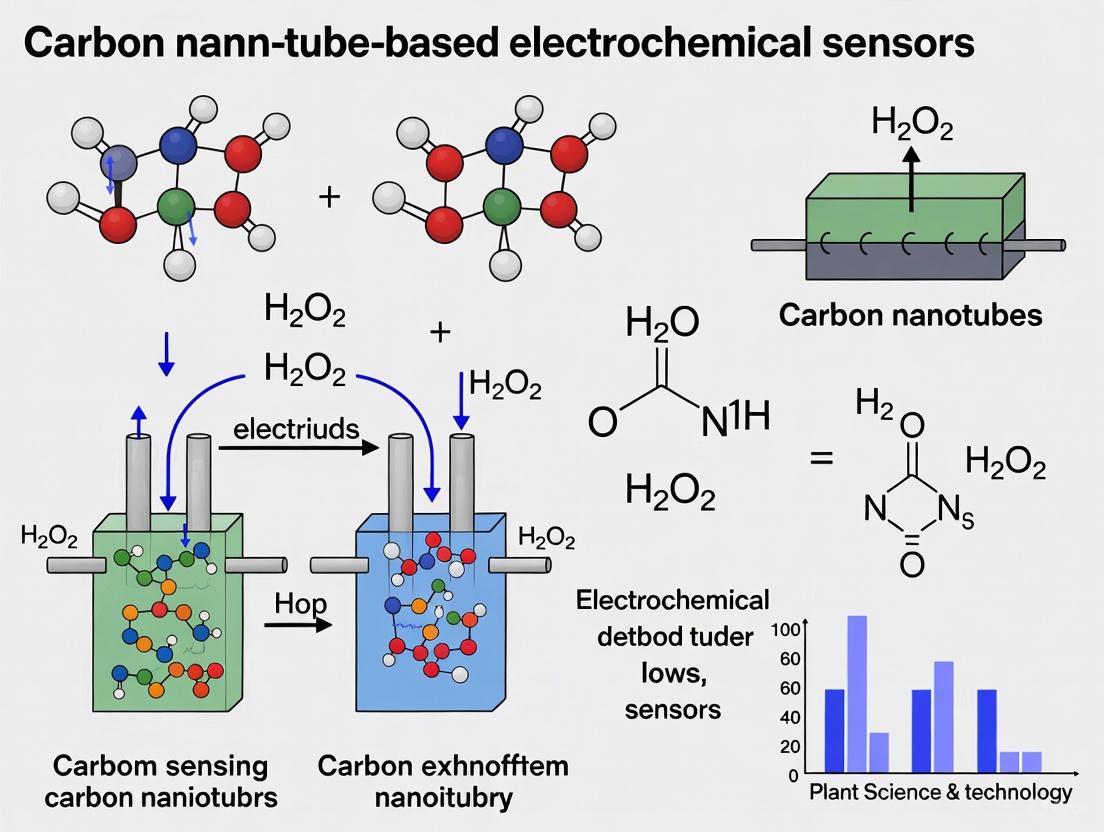
Carbon Nanotube-Based Electrochemical Sensors for H<sub>2</sub>O<sub>2</sub>: A Comprehensive Guide for Biomedical Research and Drug Development
This article provides a comprehensive analysis of carbon nanotube (CNT)-based electrochemical sensors for the detection of hydrogen peroxide (H<sub>2</sub>O<sub>2</sub>), a critical biomarker in cellular metabolism and disease pathogenesis.
Carbon Nanotube-Based Electrochemical Sensors for H2O2: A Comprehensive Guide for Biomedical Research and Drug Development
Abstract
This article provides a comprehensive analysis of carbon nanotube (CNT)-based electrochemical sensors for the detection of hydrogen peroxide (H2O2), a critical biomarker in cellular metabolism and disease pathogenesis. Tailored for researchers, scientists, and drug development professionals, it explores the foundational principles of CNT-enabled sensing, details cutting-edge fabrication methodologies and material integrations, addresses key challenges in sensor optimization and selectivity, and offers a comparative evaluation of sensor performance. By synthesizing recent scientific advances, this review serves as a vital resource for the development of highly sensitive, selective, and reliable biosensing platforms for biomedical diagnostics and therapeutic monitoring.
The Critical Role of H2O2 Sensing and the Foundational Advantages of Carbon Nanotubes
Hydrogen Peroxide as a Pivotal Biomarker in Cellular Homeostasis and Disease
Hydrogen peroxide (H₂O₂) is a key reactive oxygen species (ROS) with a dual role in biological systems. At physiological levels, it acts as a crucial signaling molecule involved in cellular processes such as membrane signal transduction, gene expression, cell differentiation, and growth factor-induced signaling cascades [1]. However, when produced in excess, H₂O₂ becomes a potent mediator of oxidative stress, leading to cellular damage and contributing to the pathogenesis of numerous diseases [1] [2] [3]. Its accumulation can disrupt redox homeostasis, trigger apoptosis, and cause oxidative damage to proteins, lipids, and DNA [2]. The detection and quantification of H₂O₂ are therefore critical for understanding cellular health and disease mechanisms, positioning it as a pivotal biomarker in biomedical research.
The pathological implications of H₂O₂ are diverse and depend on its site of accumulation. At a cellular level, the build-up of H₂O₂ can trigger apoptosis, a process implicated in autoimmune diseases like systemic lupus erythematosus (SLE) [1]. On a tissue level, excess H₂O₂ in the colonic epithelium leads to inflammation and ulcerative colitis (UC), while on a systemic level, toxic concentrations in the blood can cause the bioenergetic failure and multiorgan dysfunction characteristic of advanced sepsis [1]. Furthermore, elevated H₂O₂ is strongly associated with neurodegenerative disorders such as Parkinson's disease, where it modulates striatal dopamine signaling, leading to suppressed neurotransmitter release [4]. In osteoarthritis, H₂O₂ disrupts chondrocyte homeostasis, causing endoplasmic reticulum stress, cytoskeletal remodeling, and an altered secretome composition, which contributes to cartilage degradation [2].
Carbon Nanotube-Based Sensors for H₂O₂ Detection
Carbon nanotube (CNT)-based electrochemical sensors represent a cutting-edge technology for the sensitive and selective detection of H₂O₂. CNTs provide an excellent platform for sensing due to their high electrical conductivity, large specific surface area, and exceptional electrocatalytic properties [5] [3]. They can be functionalized or combined with other nanomaterials to create nanocomposites that enhance sensitivity, selectivity, and stability for non-enzymatic (enzyme-free) H₂O₂ sensing, thereby overcoming the limitations of traditional enzymatic sensors, such as poor stability and high cost [6] [3].
The following table summarizes the performance of various CNT-based nanocomposites developed for H₂O₂ sensing:
| Nanocomposite Material | Linear Detection Range (μM) | Detection Limit (μM) | Key Characteristics | Source |
|---|---|---|---|---|
| CNTs/Lithium Ferrite (LFO) | 0.1 - 500 | 0.005 | High stability, cost-effective, saturation magnetization of 25 emu g⁻¹ for 2% LFO | [6] |
| CNTs/Molybdenum Diselenide (MoSe₂) | 1 - 1000 | 0.29 | Vertically aligned CNT structure, exceptional mechanical robustness, pseudo-capacitive behavior | [3] |
| Flexible CNT Yarns (CNTYs) | 10 - 10000 | 0.65 | High flexibility, twistable, suitable for wearable medical devices, fast response (<5 s) | [5] |
Diagram 1: Workflow for CNT-based H₂O₂ Sensor Fabrication
Experimental Protocols
Protocol: Fabrication of a CNTs/Lithium Ferrite (CNTs/LFO) Nanocomposite Sensor
This protocol describes the synthesis of CNTs/LFO nanocomposites and their application in electrode modification for non-enzymatic H₂O₂ sensing, adapted from [6].
I. Synthesis of Lithium Ferrite (LFO) Nanoparticles
- Solution Preparation: Dissolve ferric nitrate (Fe(NO₃)₃·9H₂O) and lithium nitrate (LiNO₃·3H₂O) in 100 mL deionized water. Stir for 15 minutes.
- Chelation: Introduce citric acid as a chelating agent at a 1:1 molar ratio with respect to the total metal ions.
- pH Adjustment: Adjust the solution pH to 7.0 using drops of ammonia solution (33%).
- Gel Formation: Continuously stir and heat the solution at 130 °C until it transforms into a xerogel.
- Combustion and Sintering: Initiate combustion in an oven to form a burgundy-colored ferrite nano-powder. Sinter this powder in a furnace at 600 °C for 4 hours to obtain the final brown LFO product.
II. Preparation of CNTs/LFO Nanocomposites
- CNT Suspension: Prepare a suspension of carbon nanotubes (1 mg/mL) in double-distilled water and stir with a magnetic stirrer.
- Nanocomposite Formation: Add varying amounts of LFO powder (e.g., 0.5, 1.0, and 2.0 mg) to the CNT dispersion to create a series of nanocomposites with different ferrite concentrations.
- Reaction: Facilitate the reaction using a microwave at high power for 20 minutes to ensure homogeneity.
III. Electrode Modification
- Dispersion Preparation: Disperse 10 mg/mL of the CNTs/LFO nanocomposite in 1.0 mL of double-distilled water. Ultrasonicate for 30 minutes to obtain a homogeneous suspension.
- Drop-Casting: Drop-cast a 30 μL aliquot of the suspension onto the surface of a screen-printed electrode (SPE).
- Drying: Allow the modified electrode to dry at room temperature before use.
Protocol: Electrochemical Detection and Quantification of H₂O₂
This protocol outlines the standard procedure for characterizing the sensor and measuring H₂O₂, consolidated from [5] [6] [4].
I. Sensor Characterization via Cyclic Voltammetry (CV)
- Setup: Use a standard three-electrode system with the modified SPE (working electrode), a reference electrode (e.g., Ag/AgCl), and a counter electrode (e.g., Pt wire).
- Redox Probe: Prepare a 5.0 mM solution of potassium ferricyanide/ferrocyanide, [Fe(CN)₆]³⁻/⁴⁻, in 0.1 M KCl as the redox probe.
- Measurement: Perform CV measurements by scanning the potential, for example, from -0.4 V to 1.4 V, at various scan rates (e.g., 50-500 mV/s). A well-defined, reversible redox peak indicates successful electrode modification and efficient electron transfer.
II. H₂O₂ Sensing via Chronoamperometry
- Background: Place the characterized sensor in phosphate-buffered saline (PBS, pH 7.4) under continuous stirring.
- Calibration: Record the steady-state background current. Subsequently, successively add known concentrations of H₂O₂ standard solution into the PBS buffer.
- Data Recording: Monitor the change in current (typically at an applied potential of +0.5 V to +0.8 V vs. Ag/AgCl) after each addition. The current response is proportional to the H₂O₂ concentration.
- Analysis: Plot the current response against H₂O₂ concentration to obtain a calibration curve, which is used to determine the sensor's sensitivity, linear range, and limit of detection.
Protocol: Monitoring Endogenous H₂O₂ in Biological Systems
For real-time monitoring of dynamic H₂O₂ fluctuations in complex biological environments like live cells or tissue, Fast-Scan Cyclic Voltammetry (FSCV) at carbon-fiber microelectrodes is a powerful technique [4].
I. FSCV Measurement Setup
- Electrode: Use a bare carbon-fiber microelectrode.
- Waveform: Apply a triangular waveform, ramping from -0.4 V to +1.4 V and back every 100 ms at a scan rate of 400 V/s.
- Data Processing: Subtract the large, stable background current to reveal faradaic currents from electroactive analytes like H₂O₂, which shows a characteristic oxidation peak at ~1.2 V on the reverse scan.
II. In Situ Measurement in Brain Tissue
- Preparation: Position the carbon-fiber electrode in the region of interest (e.g., the dorsal striatum of an anesthetized rat).
- Baseline Recording: Record naturally occurring H₂O₂ fluctuations under basal conditions.
- Pharmacological Challenge: To elevate endogenous H₂O₂, microinfuse agents such as:
- Mercaptosuccinate (MCS, 100 mM): A glutathione peroxidase inhibitor that blocks H₂O₂ degradation.
- Rotenone (1 µM - 1 mM): A mitochondrial complex I inhibitor that increases ROS production.
- Validation: The analyte is confirmed as H₂O₂ by the high correlation (Pearson correlation factor >0.94) of its voltammogram with that of an authentic H₂O₂ standard [4].
The Scientist's Toolkit: Research Reagent Solutions
The table below lists key reagents and materials essential for experiments involving H₂O₂ biology and sensor development.
| Reagent/Material | Function/Application | Source/Example |
|---|---|---|
| Carbon Nanotubes (CNTs) | Core sensing element; provides high conductivity and surface area for electrocatalysis | Multi-walled CNTs [5] [6] |
| Lithium Ferrite (LFO) Nanoparticles | Catalytic nanomaterial in nanocomposites for enhanced H₂O₂ sensing | Synthesized via citrate-gel auto-combustion [6] |
| Molybdenum Diselenide (MoSe₂) | 2D material that enhances sensor performance when combined with CNTs | Deposited via Chemical Vapor Deposition (CVD) [3] |
| Screen-Printed Electrodes (SPEs) | Portable, disposable platform for easy sensor modification and electrochemical testing | Commercial three-electrode systems [6] |
| Phosphate Buffered Saline (PBS) | Physiological pH buffer for electrochemical measurements and cell culture | pH 7.4 tablets [6] |
| Mercaptosuccinate (MCS) | Inhibitor of glutathione peroxidase; used to elevate endogenous H₂O₂ levels in biological models | ~100 mM for microinfusion studies [4] |
| Rotenone | Mitochondrial complex I inhibitor; induces oxidative stress by increasing superoxide and H₂O₂ production | 1 µM - 1 mM for in vitro and in vivo studies [4] |
| Primary Human Chondrocytes | Cell model for studying H₂O₂-induced oxidative stress in cartilage and osteoarthritis | Normal Human Articular Knee Chondrocytes (NHAC-Kn) [2] |
Data Interpretation and Analysis
Accurate interpretation of data is crucial for validating sensor performance and drawing biological conclusions. The following table outlines key performance metrics to evaluate when developing or using a CNT-based H₂O₂ sensor.
| Performance Metric | Description & Significance | Ideal Outcome |
|---|---|---|
| Linear Detection Range | The range of H₂O₂ concentrations over which the sensor's response is linearly proportional. Determines the utility for a given application (e.g., physiological vs. pathological levels). | Wide range, covering relevant biological concentrations (e.g., µM to mM) [5] [6] [3]. |
| Detection Limit (LOD) | The lowest concentration of H₂O₂ that can be reliably distinguished from the background noise. Critical for detecting low-abundance biomarkers. | As low as possible (e.g., nanomolar range) for high sensitivity [6]. |
| Sensitivity | The slope of the calibration curve (current vs. concentration). Indicates how much the signal changes per unit change in concentration. | High slope value, indicating a large signal change for a small concentration change. |
| Selectivity | The sensor's ability to respond to H₂O₂ in the presence of other interfering substances (e.g., ascorbic acid, uric acid, dopamine). | Minimal interference from common biological electroactive species [5]. |
| Stability | The consistency of the sensor's response over time and/or after multiple uses. | High stability with minimal signal degradation, essential for long-term or continuous monitoring [5]. |
Diagram 2: Pathological Consequences of H₂O₂ Accumulation
The Limitations of Conventional H₂O₂ Detection Methods in Biological Settings
Hydrogen peroxide (H₂O₂) plays a dual role in biological systems, acting as a crucial signaling molecule at physiological concentrations while contributing to oxidative stress and disease pathogenesis at elevated levels [7]. Its accurate detection is therefore vital for understanding cellular processes and developing diagnostic tools. Conventional methods for H₂O₂ detection, particularly in complex biological environments, face significant challenges that can compromise their reliability and applicability. This application note details these limitations, provides experimental protocols for assessing detection methods, and highlights how emerging nanomaterial-based strategies, specifically carbon nanotube (CNT) electrochemical sensors, address these critical shortcomings to enable more accurate biological monitoring.
Critical Limitations of Conventional H₂O₂ Detection Methods
The table below summarizes the principal limitations associated with conventional H₂O₂ detection techniques when deployed in biological settings.
Table 1: Key Limitations of Conventional H₂O₂ Detection Methods in Biological Systems
| Method Category | Specific Limitations | Impact on Biological Detection |
|---|---|---|
| Fluorescence Methods | Strong autofluorescence from tissues/bodily fluids [7] | Severely affects signal-to-noise ratio of imaging [7] |
| Continuous excitation required [7] | Increases background interference, limits penetration depth | |
| Enzyme-Based Electrochemical Sensors | Enzyme denaturation at non-physiological temperatures/pH [5] [8] [9] | Limited operational stability and short sensor lifetime [5] |
| High cost and complex fabrication [8] [9] | Hinders widespread adoption and disposable use | |
| Chemiluminescence Methods | Susceptibility to interference from metal ions [7] | Reduces selectivity in complex biological matrices |
| Conventional Metal-Based Non-Enzymatic Sensors | Poor selectivity and high cost of noble metals (Au, Ag, Pd, Pt) [6] | Limited applicability for real-world bio-sensing [6] |
| Low electronic conductivity of metal oxides [8] | Compromised electrochemical performance and sensitivity |
Experimental Protocols for Method Validation
Protocol: Evaluating Autofluorescence Interference in Serum Samples
Purpose: To quantify the autofluorescence background in biological fluids and its impact on the signal-to-noise ratio (SNR) of a fluorescent H₂O₂ probe.
Materials:
- Persistent luminescent nanoprobes (e.g., ZGSCY@MON) [7]
- Human serum samples (diluted 1:10 in PBS)
- H₂O₂ standards (0-100 µM) prepared in PBS and diluted serum
- Microplate reader or fluorescence spectrometer
- Black 96-well plates
Procedure:
- Sample Preparation: Add 100 µL of each H₂O₂ standard (prepared in PBS and in diluted serum) to separate wells. Include serum blanks without H₂O₂.
- Probe Addition: Add 10 µL of the persistent luminescence nanoprobe stock solution to each well. Mix gently.
- Excitation and Measurement: Excite the plate using the appropriate UV wavelength. After stopping excitation, measure the afterglow intensity at the specified emission wavelength.
- Data Analysis:
- Calculate the SNR for each H₂O₂ concentration in both matrices:
SNR = (Signal_sample - Signal_blank) / Standard Deviation_blank. - Compare the limit of detection (LOD) in PBS versus serum. The LOD can be calculated as
LOD = 3.3 * (Standard Deviation of the blank) / Slope of the calibration curve.
- Calculate the SNR for each H₂O₂ concentration in both matrices:
Expected Outcome: The SNR and LOD will be significantly poorer in the serum matrix due to autofluorescence, demonstrating a key limitation of fluorescence methods [7].
Protocol: Assessing the Operational Stability of an Enzyme-Based H₂O₂ Sensor
Purpose: To evaluate the stability of an enzymatic sensor under varying pH and temperature conditions.
Materials:
- Enzyme-based H₂O₂ sensor (e.g., HRP-modified electrode) [5]
- Phosphate buffer saline (PBS) at pH 5.0, 7.4, and 9.0
- 50 µM H₂O₂ solution prepared in each PBS buffer
- Amperometric or potentiostatic setup
Procedure:
- Initial Response: At room temperature (25°C) and pH 7.4, record the amperometric response of the sensor to the 50 µM H₂O₂ standard.
- pH Stability Test: Measure the sensor's response to the same H₂O₂ concentration in buffers at pH 5.0 and 9.0. Return to pH 7.4 and re-measure to check for signal recovery.
- Temperature Stress Test: Place the sensor in a 50°C environment. At set intervals (0, 1, 2, 4 hours), cool to 25°C and measure its response to 50 µM H₂O₂ at pH 7.4.
- Data Analysis: Plot the normalized sensor response (% of initial response) versus time and versus pH.
Expected Outcome: A significant drop in sensor response will be observed at non-physiological pH and after thermal stress, illustrating the inherent instability of enzyme-based biosensors [8] [9].
Signaling Pathways and Experimental Workflows
The following diagram illustrates the core mechanism of a chemiresistive CNT-based sensor for H₂O₂ detection, highlighting its advantages for biological settings.
CNT Chemiresistive H₂O₂ Sensing Mechanism.
The Scientist's Toolkit: Research Reagent Solutions
Table 2: Essential Materials for CNT-Based H₂O₂ Sensor Development
| Reagent/Material | Function/Description | Key Utility |
|---|---|---|
| Functionalized CNTs (Carboxylated, Hydroxylated) | Transducer element; surface functional groups enhance electrocatalytic activity and biomolecule immobilization [5]. | Provides high conductivity and large surface area; foundation for sensor design. |
| Screen-Printed Electrodes (SPEs) | Disposable, miniaturized platform for sensor fabrication [6]. | Enables portable, low-cost sensor development suitable for field use. |
| Metal Nanoparticles/Nanocomposites (e.g., CNT/Lithium Ferrite) | Catalyze H₂O₂ redox reaction, improving sensitivity and selectivity for non-enzymatic detection [6]. | Replaces enzymes, enhancing sensor stability and shelf-life. |
| Persistent Luminescent Nanoparticles (PLNPs) | Luminescent probes that emit after excitation ceases [7]. | Enables autofluorescence-free bioimaging, drastically improving SNR in tissues. |
| Phosphate Buffered Saline (PBS), pH 7.4 | Standard physiological buffer for preparing standards and samples. | Maintains biological relevance during in vitro testing. |
Conventional H₂O₂ detection methods are hampered by fundamental issues including low signal-to-noise ratios in biological fluids, limited sensor stability due to enzyme dependency, and susceptibility to interferences. These limitations restrict their reliability for critical applications in biomedical research and clinical diagnostics. The emergence of nanomaterial-based platforms, particularly carbon nanotube electrochemical sensors, offers a promising path forward. These platforms address key shortcomings by providing high sensitivity, enzyme-free operation, and the potential for miniaturization, paving the way for the next generation of robust, reliable, and clinically viable H₂O₂ monitoring tools.
Carbon nanotubes (CNTs) have emerged as a cornerstone material in the development of advanced electrochemical sensors, particularly for the detection of hydrogen peroxide (H₂O₂). Their unique structural characteristics confer a trio of inherent properties—high electrical conductivity, large surface area, and exceptional mechanical strength—that are indispensable for enhancing sensor performance [10] [11]. The integration of CNTs into electrochemical sensing platforms directly addresses critical challenges in H₂O₂ detection, enabling devices with superior sensitivity, rapid response times, and excellent operational stability [9] [3]. This application note details how these fundamental properties of CNTs are harnessed, providing structured experimental protocols and analytical data to support their use in research and development of H₂O₂ sensors for biomedical, pharmaceutical, and environmental monitoring.
Inherent Properties and Sensing Mechanisms
The efficacy of CNT-based electrochemical sensors for H₂O₂ detection is rooted in the nanomaterial's intrinsic physical and electronic characteristics.
- High Electrical Conductivity: CNTs exhibit ballistic charge transport and excellent electrical conductivity, which significantly enhances electron transfer kinetics in electrochemical reactions [11]. This property is crucial for the amplification of the electrochemical signal generated during the reduction or oxidation of H₂O₂, leading to higher sensitivity [3].
- Large Surface Area: With specific surface areas often exceeding 1000 m²/g, CNTs provide a vast platform for the immobilization of catalytic nanoparticles or enzymes, and for the adsorption of analyte molecules [12] [10]. This large area increases the number of active sites for H₂O₂ reactions, thereby improving the sensor's response.
- Exceptional Mechanical Strength: Possessing a Young's modulus of approximately 1 TPa, CNTs contribute to the fabrication of robust and durable sensor architectures [10]. This mechanical resilience ensures the sensor's longevity and reliability under repeated use or in demanding environments.
The synergy of these properties in H₂O₂ sensing is illustrated below. The diagram shows how H₂O₂ molecules interact with a CNT-based electrode, where the high surface area allows for ample adsorption, the conductivity facilitates efficient electron transfer to the external circuit, and the mechanical strength ensures structural integrity.
Performance of CNT-Based H₂O₂ Sensors
CNTs can be functionalized or combined with other nanomaterials to create composite sensors with enhanced performance. The following table summarizes the electrochemical performance of various CNT-based configurations for H₂O₂ sensing, as reported in recent literature.
Table 1: Performance Metrics of Recent CNT-Based H₂O₂ Sensors
| Sensing Material | Sensitivity (µA mM⁻¹ cm⁻²) | Linear Range (mM) | Detection Limit (µM) | Key Characteristics | Reference |
|---|---|---|---|---|---|
| 3DGH/NiO25 Nanocomposite | 117.26 | 0.01 – 33.58 | 5.3 | Non-enzymatic; good selectivity & stability | [9] |
| MoSe₂/CNT Electrode | 133.8 | 1 – 11 | 1.36 | Vertically aligned CNTs; non-enzymatic | [3] |
| PMWCNT/ChOx Bioplatform | 26.15 | 0.4 – 4.0 | 0.43 | Enzymatic; ChOx enhances sensitivity 21x | [13] |
The selection of a sensing material depends on the application requirements. Non-enzymatic sensors offer greater stability, while enzymatic platforms can provide exceptional selectivity for specific bio-analytes.
Experimental Protocols
Protocol: Fabrication of a Non-enzymatic 3DGH/NiO Nanocomposite H₂O₂ Sensor
This protocol describes the synthesis of a three-dimensional graphene hydrogel (3DGH) decorated with nickel oxide (NiO) octahedrons for non-enzymatic H₂O₂ detection [9].
Research Reagent Solutions
- Graphene Oxide (GO) Dispersion: Synthesized via a modified Hummers method and dispersed in deionized water (1.5 mg/mL).
- NiO Octahedrons: Synthesized using a hard template (SBA-15 silica) and nickel nitrate hexahydrate precursor.
- Phosphate Buffer Saline (PBS): 0.1 M, pH 7.4, used as the electrolyte.
- H₂O₂ Stock Solution: Diluted from 30% v/v aqueous solution to required concentrations in PBS daily.
Methodology
- Preparation of NiO Octahedrons:
- Dissolve 10 mg of silica (SBA-15) and 10 mg of nickel nitrate hexahydrate in 100 ml of anhydrous ethanol.
- Stir the mixture for 24 hours at room temperature.
- Dry the solution at 80°C for 48 hours, then grind the resulting powder.
- Calcinate the powder in a muffle furnace at 550°C for 3 hours with a heating rate of 2°C/min.
- Remove the silica template by treating the product with 2 M NaOH at 60°C, followed by repeated washing with ethanol and water. Dry the final NiO octahedrons in a vacuum oven at 70°C for 12 hours.
Self-Assembly of 3DGH/NiO Nanocomposite:
- Disperse 48 mg of GO and 12 mg of the as-prepared NiO octahedrons in 32 mL of deionized water.
- Sonicate the mixture for 2 hours in a bath sonicator, followed by 1.5 hours of probe sonication to achieve a homogeneous dispersion.
- Transfer the mixture into a 45 mL Teflon-lined autoclave and maintain it at 180°C for 12 hours for the hydrothermal reaction.
- After natural cooling, wash the resulting 3DGH/NiO25 hydrogel repeatedly with deionized water and freeze-dry to obtain the final nanocomposite.
Electrode Modification and Electrochemical Measurement:
- Prepare an ink by dispersing the 3DGH/NiO25 nanocomposite in a mixture of water and Nafion.
- Drop-cast a measured volume of the ink onto a polished glassy carbon electrode (GCE) and allow it to dry.
- Perform electrochemical measurements (Cyclic Voltammetry and Chronoamperometry) in PBS with successive additions of H₂O₂ stock solution.
The experimental workflow from synthesis to testing is outlined below.
Protocol: Fabrication of an Enzymatic PMWCNT/ChOx H₂O₂ Biosensor
This protocol outlines the development of a biosensing platform using a multi-walled carbon nanotube paste (PMWCNT) immobilized with the enzyme Cholesterol Oxidase (ChOx) for the electrochemical reduction of H₂O₂ [13].
Research Reagent Solutions
- Activated MWCNTs: Outer diameter 6–13 nm, length 2.5–20 μm, purified with nitric and sulfuric acid.
- Mineral Oil: Binder for the carbon paste electrode.
- Cholesterol Oxidase (ChOx) Solution: 20 U/mL, prepared daily in 0.050 M phosphate buffer (PB), pH 7.4.
- Phosphate Buffer (PB): 0.050 M, pH 7.4, used as supporting electrolyte and solvent.
Methodology
- MWCNT Activation:
- Place MWCNTs in 1 M nitric acid and sonicate for 30 minutes.
- Filter and transfer the MWCNTs to 1 M sulfuric acid, followed by sonication for another 30 minutes.
- Repeat this acid-washing process twice.
- Finally, filter the MWCNTs and wash extensively with ethanol and acetone until the washing residues reach a neutral pH.
Paste Electrode (PMWCNT) Preparation:
- Thoroughly mix the activated MWCNTs with mineral oil in a 70:30 (w/w) ratio to form a homogeneous paste.
Sensor Assembly and Bioplatform Preparation:
- Polish a glassy carbon (GC) cylinder with alumina slurry (1 µm and 0.5 µm), rinse with deionized water, and dry under a nitrogen stream.
- Pack the PMWCNT paste onto the GC contact to form the working electrode.
- Drop-cast 10 µL of the ChOx solution (20 U/mL) onto the PMWCNT surface.
- Allow the modified electrode (PMWCNT/ChOx) to dry for 10 minutes at room temperature before use.
Electrochemical Characterization and H₂O₂ Quantification:
- Characterize the platform using Cyclic Voltammetry (CV) and Electrochemical Impedance Spectroscopy (EIS) in PB.
- For H₂O₂ detection, use amperometry by applying a constant reduction potential and making successive additions of H₂O₂ stock solution.
The Scientist's Toolkit
Table 2: Essential Research Reagent Solutions for CNT-based H₂O₂ Sensor Development
| Reagent/Material | Typical Specification/Function |
|---|---|
| Single-Walled CNTs (SWCNTs) | Diameter: 1-2 nm, Purity: >99%. Used for high-sensitivity FET sensors due to defined semiconducting properties [11] [14]. |
| Multi-Walled CNTs (MWCNTs) | Diameter: 10-20 nm, Purity: >95%. Common for composite electrodes and paste due to higher mechanical strength [14] [13]. |
| Functionalized CNTs (e.g., -COOH) | Enhanced dispersibility in solvents and improved compatibility with polymers or biomolecules for immobilization [12] [11]. |
| Transition Metal Catalysts (e.g., Fe, Co) | Catalyze CNT growth via Chemical Vapor Deposition (CVD); critical for controlling CNT diameter and structure [15] [3]. |
| Molybdenum Diselenide (MoSe₂) | A transition metal dichalcogenide (TMD) that forms synergistic composites with CNTs to enhance electrocatalytic activity for H₂O₂ reduction [3]. |
| Nickel Oxide (NiO) | A transition metal oxide with excellent electrochemical activity; used to decorate CNTs for non-enzymatic H₂O₂ sensing [9]. |
| Cholesterol Oxidase (ChOx) | An oxidoreductase enzyme; immobilization on CNTs creates a highly specific and sensitive biosensing platform for H₂O₂ [13]. |
| Nafion Perfluorinated Resin | A perfluorosulfonated ionomer; used as a binder in electrode inks and to provide selective permeability in biosensors. |
Within the advancing field of nanotechnology, carbon nanotubes (CNTs) have emerged as a cornerstone material for developing sophisticated electrochemical biosensors, particularly for the detection of hydrogen peroxide (H2O2). Their unique cylindrical nanostructure, characterized by a high surface-to-volume ratio, exceptional electrical conductivity, and remarkable mechanical strength, makes them ideal transducers and immobilization supports in biosensing platforms [16] [17]. However, the inherent hydrophobicity of pristine CNTs and their tendency to aggregate severely limit their application in biological environments. Furthermore, the effective integration of biorecognition elements, such as enzymes, antibodies, or DNA, requires a tailored interface on the CNT surface [18]. This is where functionalization strategies become paramount. By chemically modifying the CNT surface, researchers can significantly enhance the biocompatibility and dispersion stability of these nanomaterials in aqueous solutions, while also providing anchoring sites for biomolecules [16] [17]. The two principal approaches for this modification are covalent and non-covalent functionalization, each with distinct advantages and mechanisms. This application note details these strategies, framed within the context of developing high-performance CNT-based electrochemical sensors for H2O2 research, providing researchers with structured data, detailed protocols, and essential resources.
Covalent functionalization strategies
Covalent functionalization involves the formation of strong chemical bonds between functional groups and the carbon nanostructure of CNTs. This method permanently alters the surface chemistry, typically by introducing hydrophilic groups that improve solubility and provide reactive sites for subsequent bioconjugation [16] [18].
The most prevalent method involves the oxidative treatment of CNTs with strong acids to generate carboxyl (-COOH) groups on their sidewalls and ends. These carboxyl groups can then be activated by carbodiimide compounds, such as N-ethyl-N'-(3-dimethylaminopropyl) carbodiimide hydrochloride (EDC), which facilitates an amide bond formation with primary amine (-NH2) groups present on biomolecules. The addition of N-hydroxysuccinimide (NHS) stabilizes the intermediate and increases the efficiency of the coupling reaction [16]. This EDC/NHS chemistry is a workhorse for the covalent immobilization of proteins and enzymes.
An alternative covalent strategy involves the "grafting" of redox-active mediators onto the CNT surface. For instance, ferrocene can be covalently attached to amine-functionalized CNTs (MWCNT-NH2) to create a highly stable and sensitive non-enzymatic H2O2 sensor. The ferrocene acts as an effective redox mediator, enabling electron transfer at low operating potentials and thereby improving selectivity by minimizing interference from other electroactive species [19].
Table 1: Performance Comparison of H2O2 Sensors Based on Covalently Functionalized CNTs.
| Functionalization | Sensor Type | Linear Range (μM) | Detection Limit (μM) | Key Feature | Reference |
|---|---|---|---|---|---|
| Ferrocene Grafting | Non-enzymatic, Amperometric | 1 – 1,000 | 0.49 | Low-potential operation (-0.15 V), high selectivity | [19] |
| Oxidative Acid Treatment (for enzyme attachment) | Enzymatic | Varies by enzyme and design | Varies by enzyme and design | Stable amide bond, versatile for various biomolecules | [16] |
Experimental protocol: Covalent functionalization with ferrocene for H2O2 sensing
Objective: To synthesize ferrocene-grafted multi-walled carbon nanotubes (MWCNT-FeC) for the development of a sensitive and selective non-enzymatic H2O2 sensor [19].
Materials:
- Amine-functionalized multi-walled carbon nanotubes (MWCNT-NH2)
- Ferrocene carboxaldehyde (FeC-CHO)
- Sodium cyanoborohydride (NaCNBH3)
- Chitosan (CS) solution (0.5 mg/mL in 0.2 M acetic acid)
- Methanol, acetic acid, and double-distilled water
- Screen-printed carbon electrodes (SPCEs)
Procedure:
- Dispersion: Disperse 30 mg of MWCNT-NH2 in 2.5 mL of 0.1 M acetic acid solution.
- Reaction Mixture: Dissolve 35 mg of FeC-CHO in 5 mL of methanol. Add this solution to the MWCNT-NH2 dispersion.
- Sonication and Stirring: Sonicate the mixture for 10 minutes until fully dispersed, then stir for 4 hours at 35°C.
- Reduction: Add 50 mg of NaCNBH3 to the reaction mixture and continue stirring for another 24 hours.
- Purification: Extract the resulting MWCNT-FeC product by centrifugation. Rinse the pellet thoroughly with double-distilled water and methanol three times to remove unreacted precursors.
- Drying: Dry the final product under vacuum to obtain a powder for storage.
- Electrode Modification: Re-disperse 1 mg of the MWCNT-FeC powder in 1 mL of chitosan solution. Probe-sonicate for 20 minutes to achieve a homogeneous ink. Drop-cast 4 μL of this ink onto the working electrode of an SPCE and allow it to dry at 50°C for 30 minutes.
Non-covalent functionalization strategies
Non-covalent functionalization relies on physical interactions—such as π-π stacking, van der Waals forces, electrostatic interactions, and hydrophobic effects—to adsorb molecules onto the CNT surface. A key advantage of this approach is that it preserves the intrinsic electronic and structural properties of the CNTs, which are crucial for high-sensitivity electrochemical transduction [16] [20].
A wide range of molecules can be used for non-covalent functionalization:
- Aromatic Compounds: Molecules with aromatic rings can adsorb strongly onto the CNT surface via π-π interactions. A prime example is a rationally designed Schiff base containing boronic acid (SB-dBA), which exfoliates CNTs and provides a platform for specifically anchoring glycoproteins like horseradish peroxidase (HRP) through boronate ester formation [20].
- Polymers and Surfactants: Biopolymers like chitosan [19] and proteins such as avidin or concanavalin A (ConA) can wrap around CNTs, improving their hydrophilicity and stability. This method also facilitates the supramolecular assembly of complex biosensing architectures [20].
- Inorganic Nanoparticles: Decorating CNTs with metal oxide nanoparticles, such as TiO2-ZrO2 composites, enhances their electrocatalytic properties and provides a high-surface-area platform for immobilizing catalysts like Prussian blue (PB) for H2O2 reduction [21].
Table 2: Performance Comparison of H2O2 Sensors Based on Non-Covalently Functionalized CNTs.
| Functionalization | Sensor Type | Linear Range (μM) | Detection Limit (μM) | Key Feature | Reference |
|---|---|---|---|---|---|
| TiO2-ZrO2 Nanoparticles | Non-enzymatic, Prussian Blue-based | 100 – 1,000 | 17.93 | Enhanced immobilization of PB electrocatalyst | [21] |
| CNTs/Lithium Ferrite (LFO) | Non-enzymatic | 0.1 – 500 | 0.005 | Accelerated electron transfer, magnetic properties | [6] |
| Schiff Base Boronic Acid (SB-dBA) | Enzymatic (HRP-based) | Not Specified | Highly competitive | Specific anchoring of glycoproteins | [20] |
Experimental protocol: Non-covalent functionalization with TiO2-ZrO2 and Prussian blue
Objective: To modify a glassy carbon (GC) electrode with TiO2-ZrO2-doped functionalized CNTs (TiO2.ZrO2-fCNTs) and electrodeposit Prussian blue for H2O2 sensing [21].
Materials:
- Functionalized carbon nanotubes (fCNTs)
- Titanium and Zirconium precursors (e.g., nitrates or alkoxides)
- Prussian blue electrodeposition solution (containing FeCl3 and K3[Fe(CN)6] in KCl/HCl)
- Phosphate buffer saline (PBS, pH 7.4)
- Glassy carbon electrode (GCE)
Procedure:
- Synthesis of TiO2.ZrO2-fCNTs: Directly synthesize titania-zirconia nanoparticles on the walls of fCNTs using a sol-gel method. Age the synthesized nanostructured material for 20 days to achieve a well-dispersed distribution and high surface area.
- Electrode Modification: Deposit a homogeneous suspension of the TiO2.ZrO2-fCNTs nanostructured material onto the surface of a clean GCE and allow it to dry.
- Prussian Blue Electrodeposition: Immerse the modified electrode in an electrodeposition solution. Using cyclic voltammetry, cycle the potential (e.g., between -0.05 V and 0.35 V vs. Ag/AgCl) to electrochemically deposit Prussian blue onto the TiO2.ZrO2-fCNTs matrix.
- Activation and Stabilization: Condition the resulting PB/TiO2.ZrO2-fCNTs/GC electrode in an acidic KCl solution to activate and stabilize the Prussian blue film.
The following diagram illustrates the core decision-making workflow for selecting and implementing a functionalization strategy for H2O2 biosensor development.
The scientist's toolkit: Essential research reagents
This table catalogs key materials and their functions for implementing the functionalization strategies and sensor development discussed in this note.
Table 3: Essential Reagents for CNT Functionalization and H2O2 Sensor Fabrication.
| Reagent/Material | Function/Application | Examples & Notes |
|---|---|---|
| MWCNTs / SWCNTs | Core transducer material; provides high surface area and electrical conductivity. | Purity and dimensions (diameter, length) affect performance [16] [17]. |
| EDC / NHS | Cross-linking agents for covalent amide bond formation between -COOH and -NH2 groups. | Critical for stable immobilization of enzymes and antibodies [16]. |
| Amine-Functionalized CNTs (MWCNT-NH2) | Starting material for covalent grafting of molecules containing aldehydes or carboxyl groups. | Enables direct conjugation with redox mediators like ferrocene [19]. |
| Ferrocene Derivatives | Redox mediator for non-enzymatic H2O2 sensors; enables low-potential operation. | Ferrocene carboxaldehyde is used for grafting [19]. |
| Chitosan (CS) | Biopolymer for non-covalent dispersion of CNTs and forming biocompatible films on electrodes. | Improves uniformity and adhesion of modified layer [19]. |
| Schiff Base Boronic Acid (SB-dBA) | Non-covalent exfoliant and specific anchor for glycoproteins via boronate ester formation. | Used for immobilizing HRP in enzymatic biosensors [20]. |
| Titanium-Zirconia Oxide (TiO2.ZrO2) | Metal oxide nanocomposite for non-covalent CNT modification; enhances catalyst support. | Increases surface area and improves immobilization of Prussian blue [21]. |
| Prussian Blue (PB) | "Artificial peroxidase"; electrocatalyst for H2O2 reduction in non-enzymatic sensors. | Known for high selectivity and activity in neutral media [21]. |
| Lithium Ferrite (LFO) | Magnetic nanoparticle for nanocomposites; enhances electrocatalytic activity. | Combined with CNTs for highly sensitive non-enzymatic sensing [6]. |
| Screen-Printed Carbon Electrodes (SPCEs) | Low-cost, disposable, and miniaturizable platform for practical sensor deployment. | Ideal for point-of-use testing [19]. |
The strategic choice between covalent and non-covalent functionalization is fundamental to the success of carbon nanotube-based electrochemical biosensors for H2O2. Covalent strategies provide robust and stable interfaces for biomolecule attachment, while non-covalent methods maintain the superior electronic properties of CNTs and offer versatile supramolecular assembly routes. The experimental protocols and data summarized in this application note provide a foundation for researchers to optimize these strategies. The continued refinement of these functionalization approaches, guided by the provided frameworks and toolkit, is poised to yield the next generation of highly sensitive, selective, and reliable biosensors for healthcare, environmental monitoring, and the food industry.
Carbon nanotube (CNT)-based electrochemical sensors represent a frontier technology for the precise detection of hydrogen peroxide (H₂O₂), a critical biomarker of oxidative stress and cellular signaling. The interface between these nanoscale sensors and biological systems is governed by sophisticated cellular uptake mechanisms that enable intracellular sensing. Single-walled carbon nanotubes (SWCNTs) and multi-walled carbon nanotubes (MWCNTs) serve as exceptional transducer elements due to their unparalleled electrical conductivity, high surface-to-volume ratio, and versatile functionalization capabilities [11] [22]. For H₂O₂ research, this translates to sensors with enhanced sensitivity, rapid response times, and the ability to operate within complex biological milieus.
The strategic functionalization of CNTs is paramount for successful biological interfacing. By engineering their surface chemistry with specific molecular recognition elements, researchers can create sensors that not only detect H₂O₂ with high specificity but also navigate the biological barriers to reach their intracellular targets. This application note details the operational mechanisms, practical protocols, and key considerations for implementing CNT-based sensors in biological H₂O₂ detection, providing a framework for researchers and drug development professionals.
Cellular Uptake Mechanisms and Intracellular Sensing
The journey of a CNT-based sensor from the extracellular environment to its intracellular site of action is a critical process that determines the efficacy and reliability of the measurement. Understanding these pathways is essential for experimental design and data interpretation.
Primary Uptake Pathways
CNT-based sensors interface with cells primarily through endocytic pathways. The functionalization of the CNT surface directly influences the preferred route of internalization.
- Receptor-Mediated Endocytosis: This is the most common and efficient pathway for the cellular uptake of functionalized CNTs. By conjugating the CNT surface with specific ligands (e.g., peptides, antibodies, or folic acid), the sensor can target overexpressed receptors on the cell membrane. This binding triggers the invagination of the membrane, forming a vesicle that transports the sensor into the cell. This mechanism is highly targeted and can facilitate the delivery of sensors to specific organelles.
- Phagocytosis/Macropinocytosis: Larger CNT aggregates or sensors designed for immune cell studies may be internalized via these actin-dependent mechanisms, which involve the engulfment of larger particles.
- Direct Translocation: In some cases, ultra-small, highly functionalized CNTs can passively diffuse across the lipid membrane, though this is less common and highly dependent on surface charge and functional groups.
The following diagram illustrates the primary signaling pathways and workflows involved in the cellular uptake and sensing mechanism of CNT-based H₂O₂ sensors.
Post-Uptake Intracellular Fate and Sensing
Once internalized, the sensor is typically encapsulated within an endosome. Its ability to escape this compartment is crucial for accessing the cytosolic environment where many H₂O₂ signaling events occur. Certain functional polymers or cell-penetrating peptides can facilitate endosomal escape. The final location of the sensor—whether free in the cytosol or targeted to specific organelles like mitochondria (a major source of H₂O₂)—is a function of its surface design. The actual H₂O₂ detection then occurs via the established electrochemical mechanisms, with the resulting signal being transduced to an external electrode for measurement.
Experimental Protocols for Sensor Fabrication and Application
This section provides a detailed methodology for constructing a CNT-based electrochemical sensor and applying it for the detection of H₂O₂ in a biologically relevant context.
Protocol 1: Fabrication of a CNT/Lithium Ferrite (LFO) Nanocomposite Sensor for H₂O₂ Detection
This protocol outlines the synthesis of a highly sensitive non-enzymatic H₂O₂ sensor using a CNT/LFO nanocomposite, adapted from recent literature [6].
Principle: The composite leverages the high conductivity of CNTs and the electrocatalytic activity of lithium ferrite (LFO) nanoparticles for the reduction or oxidation of H₂O₂, avoiding the instability associated with enzymatic sensors.
Materials:
- Carbon Nanotubes (CNTs): MWCNTs (>95% purity, <7 nm diameter).
- Precursors: Ferric nitrate (Fe(NO₃)₃·9H₂O), Lithium nitrate (LiNO₃·3H₂O).
- Chemicals: Citric acid (chelating agent), Ammonia solution (33%, for pH adjustment).
- Electrochemical Cell: Phosphate-buffered saline (PBS, pH 7.4), Potassium ferricyanide/ferrocyanide (K₃[Fe(CN)₆]/K₄[Fe(CN)₆]).
- Equipment: Screen-printed electrodes (SPEs), Potentiostat (e.g., PalmSens 4), Ultrasonic bath, Muffle furnace, Magnetic stirrer.
Procedure:
- Synthesis of LFO Nanoparticles:
- Dissolve ferric nitrate and lithium nitrate in a 5:1 molar ratio in 100 mL deionized water. Stir for 15 minutes.
- Add citric acid (1:1 molar ratio to total metal ions) under continuous stirring.
- Adjust the pH of the solution to 7.0 using ammonia solution.
- Heat the solution at 130 °C with constant stirring until a viscous xerogel forms.
- Transfer the xerogel to a pre-heated oven (~300 °C) for auto-combustion. A self-sustaining exothermic reaction will yield a burgundy-colored powder.
- Sinter the powder in a muffle furnace at 600 °C for 4 hours to obtain crystalline, brown-colored LFO nanoparticles.
Preparation of CNTs/LFO Nanocomposite:
- Prepare a uniform suspension of CNTs (1 mg/mL) in deionized water using ultrasonication for 30 minutes.
- Add LFO powder to the CNT dispersion to achieve the desired mass ratio (e.g., 0.5%, 1%, 2% LFO). Designate these as CNTs/LFO (0.5%), CNTs/LFO (1%), and CNTs/LFO (2%).
- Subject the mixture to microwave irradiation at high power for 20 minutes to facilitate nanocomposite formation.
Electrode Modification:
- Prepare an ink by dispersing 10 mg of the CNTs/LFO nanocomposite in 1.0 mL of deionized water via ultrasonication for 30 minutes.
- Drop-cast 30 µL of the homogeneous suspension onto the working electrode surface of a screen-printed electrode (SPE).
- Allow the modified electrode (CNTs/LFO/SPE) to dry completely at room temperature before use.
The workflow for this fabrication protocol is summarized in the following diagram:
Protocol 2: Cell Culture and Intracellular H₂O₂ Monitoring
This protocol describes the application of a CNT-based sensor for monitoring H₂O₂ in a cellular model, such as a cisplatin-induced Acute Kidney Injury (AKI) model where oxidative stress is a key pathological factor [23].
Principle: The sensor is used to measure extracellular H₂O₂ released by cells under oxidative stress, providing a non-invasive means to monitor cellular state and drug efficacy.
Materials:
- Cell Line: Relevant cell line (e.g., human renal tubular epithelial cells).
- Culture Reagents: Dulbecco's Modified Eagle Medium (DMEM), Fetal Bovine Serum (FBS), Penicillin-Streptomycin.
- Inducers/Treatments: Cisplatin (DDP) to induce oxidative stress, Cimetidine (CMTD) as a protective agent.
- Equipment: Cell culture incubator (37°C, 5% CO₂), Sterile cultureware, Potentiostat.
Procedure:
- Cell Culture and Model Establishment:
- Culture cells in DMEM supplemented with 10% FBS and 1% penicillin-streptomycin at 37°C in a 5% CO₂ atmosphere.
- To establish an AKI model, treat cells with a clinically relevant concentration of cisplatin (e.g., 20 µM) for 24 hours.
- For the treatment group, pre-treat or co-treat cells with cimetidine.
- Electrochemical Measurement of Extracellular H₂O₂:
- Prior to measurement, calibrate the CNTs/LFO/SPE sensor in PBS using standard H₂O₂ solutions via chronoamperometry.
- At the end of the treatment period, collect the cell culture medium and centrifuge to remove any detached cells or debris.
- Transfer the clear supernatant to the electrochemical cell.
- Use the calibrated CNTs/LFO/SPE sensor to measure the H₂O₂ concentration in the supernatant via chronoamperometry at a fixed potential (e.g., -0.4 V vs. Ag/AgCl).
- Correlate the measured H₂O₂ levels with the cellular state, as validated by traditional markers like cell viability assays.
Performance Data and Research Reagents
A critical step in experimental planning is the selection of appropriate materials and an understanding of expected sensor performance. The table below summarizes quantitative data from recent studies on CNT-based H₂O₂ sensors.
Table 1: Performance Comparison of CNT-Based H₂O₂ Sensors
| Sensor Material | Detection Principle | Linear Range (μM) | Sensitivity | Limit of Detection (μM) | Reference |
|---|---|---|---|---|---|
| CNTs/Lithium Ferrite (2%) | Amperometry | 0.1 – 500 | Not Specified | 0.005 | [6] |
| Bi₂S₃@Cu₀.₁ Nanomaterial | Chronoamperometry | 0.5 – 1400 | 85.3 μA mM⁻¹ cm⁻² | 0.528 | [23] |
| Fenton-Activated CNTs (CNT_F24h) | Amperometry | Not Specified | Outperformed N-CNT (Cathodic) | Not Specified | [24] |
The Scientist's Toolkit: Essential Research Reagent Solutions
Table 2: Key Reagents for CNT-based H₂O₂ Sensor Development
| Reagent / Material | Function / Role | Example & Notes |
|---|---|---|
| Multi-Walled Carbon Nanotubes (MWCNTs) | Core transducer; provides high surface area and electrical conductivity. | Purity >95%, diameter <7 nm. Requires functionalization for dispersion and biocompatibility [24] [6]. |
| Lithium Ferrite (LFO) Nanoparticles | Electrocatalyst; enhances the electron transfer rate for H₂O₂ reduction/oxidation. | Synthesized via citrate-gel auto-combustion; reduces agglomeration when composited with CNTs [6]. |
| Screen-Printed Electrodes (SPEs) | Miniaturized, disposable platform for electrochemical measurement. | Ideal for rapid testing. Working electrode is typically modified with the CNT nanocomposite [6]. |
| Cisplatin (DDP) | Chemotherapeutic agent; induces oxidative stress and apoptosis in cells. | Used in vitro to establish a disease model (e.g., AKI) for validating sensor functionality [23]. |
| Cimetidine (CMTD) | Nephroprotective agent; reduces cisplatin-induced kidney damage. | Serves as a positive control treatment to demonstrate sensor ability to monitor therapeutic efficacy [23]. |
| Phosphate-Buffered Saline (PBS) | Physiological buffer; maintains stable pH and ionic strength during electrochemical testing. | pH 7.4 is standard for simulating biological conditions [23] [6]. |
CNT-based electrochemical sensors offer a powerful and versatile platform for H₂O₂ research within biological systems. Their success hinges on the rational design of the CNT interface, which must be engineered for both high electrochemical performance and effective, controlled interaction with living cells. The protocols and data provided here serve as a foundation for researchers to develop and apply these sensors in studies of oxidative stress, drug screening, and disease mechanisms.
Key Considerations for Implementation:
- Biocompatibility and Cytotoxicity: While functionalization improves biocompatibility, the potential toxicity of CNTs must be evaluated for each specific cell type and application [11] [22].
- Specificity: In complex biological fluids, the sensor's surface must be designed to minimize interference from common electroactive species like ascorbic acid, uric acid, and acetaminophen [23].
- Reproducibility and Scaling: Batch-to-batch consistency in CNT synthesis and functionalization remains a challenge for widespread commercialization. Standardized protocols are essential for reproducible sensor fabrication [11] [22].
Fabrication Techniques and Advanced Material Integrations for Enhanced H2O2 Sensing
The detection of hydrogen peroxide (H₂O₂) is critical across biomedical, industrial, and environmental fields. As a by-product of cellular metabolism, its concentration is a key biomarker for cellular homeostasis and oxidative stress, with abnormal accumulation linked to the formation and spread of cancer cells [5]. Similarly, in industrial processes and environmental monitoring, precise H₂O₂ tracking is essential [25] [6]. Electrochemical sensors have emerged as a paramount tool for this purpose, offering high sensitivity, portability, and capability for real-time analysis [5]. Within this domain, carbon nanotube (CNT)-based electrodes represent a significant evolution, breaking from the paradigm of planar static electrodes and enabling a new generation of sensing platforms, from flexible, wearable yarns to mass-producible, modified screen-printed devices [26] [27]. This article details the application notes and experimental protocols for these two principal design paradigms, contextualized within a broader thesis on advanced carbon nanotube-based electrochemical sensors.
Flexible Carbon Nanotube Yarn (CNTY) Based Sensors
Carbon nanotube yarns are macroscopic assemblies of CNT bundles, forming flexible, conductive, and strong filaments suitable for use as working electrodes without additional rigid supports [26] [5]. Their inherent flexibility and high surface area make them ideal for wearable and implantable sensing applications.
Application Notes
Flexible CNTY-based H₂O₂ sensors leverage the yarn's excellent electrocatalytic activity, which is further enhanced by functional groups like carboxyl (–COOH) and hydroxyl (–OH) on the CNT surface [5]. These sensors are designed to maintain electrochemical performance under mechanical deformation, a crucial requirement for wearable medical devices that conform to biological tissues.
Key Advantages:
- Flexibility and Durability: Can withstand substantial bending and stretching without performance degradation [5].
- Self-Sufficient Electrodes: Do not require additional flexible carriers or expensive metal nanoparticles, simplifying fabrication [5].
- High Performance: Exhibit wide linear range, low detection limit, and fast response for H₂O₂ detection [5].
- Biocompatibility Potential: Their flexibility and carbon-based composition are beneficial for in-vivo applications.
Experimental Protocol: Fabrication and Testing of a CNTY H₂O₂ Sensor
Principle: A flexible electrochemical sensor is constructed using a twisted CNTY as the working electrode, which electrocatalytically oxidizes H₂O₂. The electrical conductivity of the CNTYs can be enhanced prior to sensor fabrication through a chemical-free cyclic loading process, which aligns the CNT bundles and improves inter-tube contact [28].
Table 1: Research Reagent Solutions for Flexible CNTY Sensor
| Item | Function/Description |
|---|---|
| Multi-walled CNT Forest | Source material for drawing CNT yarns; purity > 99% [5]. |
| Chemical Vapor Deposition (CVD) System | For synthesis of the base MWCNT forest using acetylene as a carbon source [5] [28]. |
| Phosphate Buffered Saline (PBS) | Standard electrolyte solution for electrochemical testing, pH 7.4 [5]. |
| H₂O₂ Standard Solution (30%) | Primary analyte for calibration and sensitivity testing [5]. |
| Mechanical Tester (e.g., Instron) | For applying cyclic loading to enhance CNTY electrical conductivity [28]. |
| Two-Probe Multimeter (e.g., FLUKE 179) | For simultaneous measurement of electrical resistance during mechanical testing [28]. |
Procedure:
- CNTY Fabrication: Draw and twist a continuous yarn from a forest of multi-walled carbon nanotubes synthesized via CVD [5].
- Conductivity Enhancement (Optional but Recommended): Subject the as-received CNTY to uniaxial cyclic loading.
- Mount a CNTY sample on a mechanical tester with a gauge length of 70 mm.
- Apply 100 cycles of stretching and relaxation to a predetermined strain value at a rate of 5 mm/min [28].
- Simultaneously monitor the electrical resistance. An 80% reduction in resistance can be achieved, attributed to the orientation and compaction of CNT bundles [28].
- Sensor Assembly: Integrate a segment of the treated or untreated CNTY (typically 1-2 cm) as the working electrode in a standard three-electrode cell setup, using a platinum wire as the counter electrode and an Ag/AgCl reference electrode.
- Electrochemical Characterization:
- Perform Cyclic Voltammetry (CV) in a 0.1 M PBS solution both with and without additions of H₂O₂, scanning within a potential window from -0.2 V to 0.8 V (vs. Ag/AgCl) [5].
- The electrocatalytic oxidation of H₂O₂ will be observed as a significant increase in anodic current.
- Calibration and Sensing: Use Amperometry (i-t curve) at a fixed optimal potential (e.g., +0.5 V) while successively adding aliquots of H₂O₂ standard solution under stirred conditions. Plot the steady-state current response against H₂O₂ concentration to obtain a calibration curve.
The workflow for this protocol is summarized in the diagram below.
Modified Screen-Printed Electrode (SPE) Based Sensors
Screen-printed electrodes are mass-produced, planar devices typically consisting of a working electrode (WE), counter electrode (CE), and a pseudo-reference electrode (RE) printed on a ceramic or plastic substrate [29]. Their low cost, disposability, and ease of modification make them ideal for decentralized, point-of-care testing.
Application Notes
The performance of SPEs for specific analytes like H₂O₂ is drastically enhanced by modifying the working electrode surface with nanocomposites. A prominent example is the use of carbon nanotubes/lithium ferrite (CNTs/LFO) composites, which combine the high conductivity of CNTs with the catalytic properties of LFO for non-enzymatic H₂O₂ sensing [25] [6].
Key Advantages:
- Mass-Producible and Low-Cost: Ideal for single-use, disposable sensors [30] [29].
- Easily Modifiable: The WE surface can be readily customized with nanomaterials, enzymes, or polymers to enhance sensitivity and selectivity [27] [29].
- Portability: Compatible with compact, handheld potentiostats for on-site analysis [29].
- Enhanced Performance with Nanocomposites: CNTs/LFO composites overcome the low conductivity and agglomeration issues of pure LFO, leading to superior H₂O₂ sensing [6].
Experimental Protocol: CNTs/Lithium Ferrite Modified SPE for H₂O₂ Sensing
Principle: The working electrode of a commercial SPE is modified with a CNTs/LFO nanocomposite. The CNTs provide a high-surface-area, conductive scaffold that facilitates electron transfer, while the LFO nanoparticles act as the electrocatalyst for H₂O₂ reduction/oxidation, enabling sensitive and stable non-enzymatic detection [25] [6].
Table 2: Research Reagent Solutions for CNTs/LFO-modified SPE
| Item | Function/Description |
|---|---|
| Screen-Printed Electrodes (SPEs) | Disposable electrochemical platform (e.g., from Metrohm DropSens) [30] [6]. |
| Carbon Nanotubes (CNTs) | Conductive scaffold; procured as powder (e.g., from Nanoridge) [6]. |
| Lithium Nitrate & Ferric Nitrate | Precursors for lithium ferrite (LFO) synthesis [6]. |
| Citric Acid | Chelating agent for the sol-gel auto-combustion synthesis of LFO [6]. |
| Phosphate Buffered Saline (PBS) | Electrolyte for electrochemical testing, pH 7.4 [6]. |
| Potassium Ferricyanide/Ferrocyanide | Redox probe ([Fe(CN)₆]³⁻/⁴⁻) for electrode characterization via EIS and CV [6]. |
Procedure:
- Synthesis of LFO Nanoparticles:
- Use a citrate–gel auto-combustion method. Dissolve stoichiometric amounts of ferric nitrate (Fe(NO₃)₃·9H₂O) and lithium nitrate (LiNO₃·3H₂O) in deionized water.
- Add citric acid as a chelating agent at a 1:1 molar ratio to the metal ions.
- Adjust the pH to 7.0 using ammonia solution.
- Heat at 130 °C with continuous stirring to form a xerogel, which is then combusted in an oven to form LFO powder.
- Sinter the powder at 600 °C for 4 hours to crystallize the LFO [6].
- Preparation of CNTs/LFO Nanocomposite:
- Create a suspension of CNTs in distilled water (e.g., 1 mg/mL).
- Add varying amounts of LFO powder (e.g., 0.5%, 1%, and 2% by weight) to the CNT dispersion under sustained stirring.
- Treat the mixture in a microwave at high power for 20 minutes to facilitate integration [6].
- Modification of SPE:
- Prepare an ink by dispersing 10 mg of the CNTs/LFO nanocomposite in 1.0 mL of distilled water via ultrasonication for 30 minutes.
- Drop-cast a 30 µL aliquot of this homogeneous suspension onto the working electrode surface of the SPE.
- Allow the modified SPE to dry at room temperature [6].
- Electrochemical Characterization:
- Characterize the modified SPE using Electrochemical Impedance Spectroscopy (EIS) and CV in a 5.0 mM [Fe(CN)₆]³⁻/⁴⁻ solution with 0.1 M KCl. A decreased charge transfer resistance (Rₑₜ) indicates improved electron transfer kinetics [6].
- H₂O₂ Sensing:
- Perform CV in PBS (pH 7.4) with successive additions of H₂O₂ to identify the optimal sensing potential.
- Use Chronoamperometry (CA) at the determined fixed potential to measure the current response with successive H₂O₂ additions. The optimized CNTs/LFO (2%) electrode can achieve a low detection limit of 0.005 µM and a wide linear range of 0.1–500 µM [6].
The workflow for this protocol is summarized in the diagram below.
Performance Comparison and Data Presentation
The quantitative performance of the two sensor paradigms, as reported in the literature, is summarized below for direct comparison.
Table 3: Performance Comparison of CNT-Based H₂O₂ Sensors
| Sensor Type | Linear Range (µM) | Detection Limit (µM) | Key Characteristics | Reference |
|---|---|---|---|---|
| Flexible CNTY Sensor | Not explicitly stated, wide | Not explicitly stated, low | High flexibility, maintained performance after deformation, intrinsic catalysis from –COOH/–OH groups. | [5] |
| CNTs/LFO (2%) Modified SPE | 0.1 – 500 | 0.005 | Excellent stability, non-enzymatic, high sensitivity, requires nanocomposite synthesis. | [25] [6] |
The Scientist's Toolkit: Essential Materials for H₂O₂ Sensor Development
This table catalogs key reagents and materials central to the development of CNT-based H₂O₂ sensors, as featured in the discussed research.
Table 4: Essential Research Reagent Solutions for CNT-Based H₂O₂ Sensors
| Item | Function/Application |
|---|---|
| Carbon Nanotube Yarns (CNTYs) | Serve as flexible, self-standing working electrodes; provide high conductivity and large specific surface area for electrocatalysis [26] [5]. |
| CNTs/LFO Nanocomposite | Acts as an electroactive layer on SPEs; CNTs enhance electron transfer while LFO provides catalytic sites for non-enzymatic H₂O₂ detection [25] [6]. |
| Phosphate Buffered Saline (PBS) | A standard physiological buffer used as the electrolyte medium for electrochemical testing, crucial for simulating biological conditions [5] [6]. |
| Potassium Ferricyanide/Ferrocyanide | A common redox probe ([Fe(CN)₆]³⁻/⁴⁻) used in electrochemical impedance spectroscopy (EIS) and cyclic voltammetry (CV) to characterize the electron transfer properties of modified electrodes [6]. |
| Screen-Printed Electrodes (SPEs) | Provide a low-cost, disposable, and mass-producible platform for electrochemical sensor development, ideal for decentralized testing [30] [29]. |
The two design paradigms—flexible CNTY sensors and modified SPEs—cater to distinct but equally critical applications in modern electroanalysis. CNTYs offer a path toward conformable, implantable, and long-term monitoring devices for biomedical diagnostics, leveraging their intrinsic mechanical and electrochemical properties. In contrast, modified SPEs, particularly with advanced nanocomposites like CNTs/LFO, provide a route toward highly sensitive, mass-producible, and disposable point-of-care sensors. The choice between these paradigms depends on the specific application requirements, such as the need for flexibility versus ultra-low detection limits and cost-effectiveness. Together, they underscore the versatility and potential of carbon nanotube-based materials in advancing electrochemical sensing technology for H₂O₂ and beyond.
Hydrogen peroxide (H₂O₂) is a significant molecule in biological systems and various industrial processes. At elevated concentrations, it exhibits cytotoxicity and has been linked to diseases such as diabetes, cancer, and neurodegenerative disorders [31] [32]. Consequently, precise monitoring of H₂O₂ is crucial for both biomedical diagnostics and industrial applications [33]. Electrochemical sensing has emerged as a preferred technique due to its high sensitivity, rapid response, and cost-effectiveness [32] [34].
Traditional enzymatic sensors, while selective, suffer from drawbacks such as high cost, limited stability, and sensitivity to environmental conditions [33]. Non-enzymatic sensors based on metal oxides and carbon nanomaterials offer a robust alternative. Among these, composites of carbon nanotubes (CNTs) and spinel ferrites have demonstrated exceptional electrocatalytic performance for H₂O₂ detection, combining the high conductivity and surface area of CNTs with the unique catalytic properties of ferrites [31] [32] [34].
Performance Comparison of CNT-Spinel Ferrite Nanocomposites
The table below summarizes the electrochemical performance of various CNT-spinel ferrite nanocomposites for H₂O₂ detection, highlighting key metrics such as detection limit and linear range.
Table 1: Performance of CNT-Spinel Ferrite Nanocomposites for H₂O₂ Sensing
| Nanocomposite | Synthesis Method | Detection Limit (μM) | Linear Range (μM) | Reference/Key Finding |
|---|---|---|---|---|
| CNTs/Lithium Ferrite (LFO) | Citrate-gel auto-combustion & microwave-assisted reaction [31] [6] | 0.005 [31] | 0.1 - 500 [31] | Superior electron transfer, excellent stability [25] |
| CoFe₂O₄/CNTs | Hydrothermal method [32] | Information missing | Information missing | Prevents nanoparticle agglomeration, provides 3D conductive network [32] |
| Ni-doped ZnFe₂O₄(Zn₀.₇Ni₀.₃Fe₂O₄) | Hydrothermal method [34] | 5 [34] | 20 - 10,000 [34] | Modulates Fe²⁺/Fe³⁺ ratio to enhance Fenton reaction [34] |
| Fe₃O₄/CNTs | One-step catalytic chemical vapor deposition (CVD) [35] | Information missing | Information missing | In-situ synthesis on NaCl support, synergistic effect lowers electron transfer impedance [35] |
Detailed Experimental Protocols
Synthesis of CNTs/Lithium Ferrite (LFO) Nanocomposite
This protocol details the citrate-gel auto-combustion method for synthesizing CNTs/LFO nanocomposites, which demonstrated a low detection limit of 0.005 μM [31] [6].
Materials:
- Ferric nitrate (Fe(NO₃)₃·9H₂O)
- Lithium nitrate (LiNO₃·3H₂O)
- Citric acid (C₆H₈O₇)
- Ammonia solution (NH₄OH, 33%)
- Multi-walled carbon nanotubes (CNTs)
Procedure:
- Precursor Solution Preparation: Dissolve stoichiometric amounts of ferric nitrate and lithium nitrate in 100 mL of deionized water. Stir for 15 minutes to ensure complete dissolution [6].
- Chelation: Introduce citric acid as a chelating agent at a 1:1 molar ratio with respect to the total metal ions [6].
- pH Adjustment: Adjust the pH of the solution to 7.0 using drops of ammonia solution (33%) under continuous stirring [6].
- Gel Formation: Heat the solution at 130 °C with constant stirring until it transforms into a viscous xerogel [6].
- Auto-combustion: Transfer the xerogel to an oven preheated to ~300 °C to initiate a self-sustaining combustion reaction, resulting in a burgundy-colored LFO powder [6].
- Annealing: Sinter the as-combusted powder in a furnace at 600 °C for 4 hours to obtain crystalline LFO nanoparticles (final product is brown) [6].
- Nanocomposite Formation: Prepare a suspension of CNTs (1 mg/mL) in deionized water. Add varying amounts of the synthesized LFO powder (e.g., 0.5, 1.0, and 2.0 mg per mL of CNT suspension) to create different doping levels. Subject the mixture to microwave irradiation at high power for 20 minutes to form the final CNTs/LFO nanocomposite [6].
Electrode Modification and Electrochemical Characterization
This protocol describes the modification of screen-printed electrodes (SPEs) and the subsequent electrochemical evaluation of the nanocomposite for H₂O₂ sensing [31] [6].
Materials:
- Screen-printed electrodes (SPE)
- Phosphate buffered saline (PBS, pH 7.4)
- Potassium ferricyanide (K₃[Fe(CN)₆])
- Potassium chloride (KCl)
- Hydrogen peroxide (H₂O₂, 30%)
Procedure:
- Electrode Modification:
- Disperse 10 mg of the synthesized CNTs/LFO nanocomposite in 1.0 mL of double-distilled water.
- Sonicate the suspension for 30 minutes to achieve a homogeneous dispersion.
- Drop-cast 30 μL of the suspension onto the working electrode surface of the SPE.
- Allow the modified electrode to dry at room temperature [6].
Electrochemical Impedance Spectroscopy (EIS):
Cyclic Voltammetry (CV) and Chronoamperometry for H₂O₂ Sensing:
- Record CV curves in 0.1 M PBS (pH 7.4) both in the absence and presence of H₂O₂.
- The CNTs/LFO modified electrode will show a significant increase in reduction current upon addition of H₂O₂, confirming electrocatalytic activity [31].
- For analytical measurement, use chronoamperometry at a fixed potential.
- Successively add aliquots of H₂O₂ to the stirred PBS solution and record the current response.
- Plot the steady-state current versus H₂O₂ concentration to generate the calibration curve used to determine the linear range and detection limit [31] [6].
The Scientist's Toolkit: Essential Research Reagents
Table 2: Key Reagents for CNT-Ferrite Nanocomposite Synthesis and Sensing
| Reagent/Chemical | Function/Application | Example from Literature |
|---|---|---|
| Multi-walled Carbon Nanotubes (CNTs) | Conductive scaffold; enhances electron transfer and prevents nanoparticle agglomeration. | Used as a base nanomaterial in all composites discussed [31] [32] [35]. |
| Metal Nitrates(e.g., Fe(NO₃)₃, LiNO₃) | Precursor sources for metal ions in the spinel ferrite structure. | Ferric and lithium nitrates used for LFO synthesis [6]. |
| Citric Acid | Chelating agent in sol-gel processes; facilitates homogeneous mixing of metal ions. | Used in the citrate-gel auto-combustion synthesis of LFO [6]. |
| Phosphate Buffered Saline (PBS) | Electrolyte for electrochemical testing; maintains physiological pH (7.4). | Standard medium for H₂O₂ sensing experiments [31] [6]. |
| Hydrogen Peroxide (H₂O₂) | Primary analyte for calibration and sensitivity tests. | A 30% stock solution is typically diluted for experimental use [32] [6]. |
| Potassium Ferricyanide/Ferrocyanide | Redox probe for characterizing electrode kinetics via EIS and CV. | Used in a 5.0 mM solution with KCl to test electron transfer efficiency [6]. |
Signaling Pathways and Workflow Visualization
The enhanced sensing performance of CNT-ferrite nanocomposites can be understood through their electrocatalytic mechanism, particularly the Fenton-like reaction facilitated by transition metals like iron. The experimental workflow from synthesis to application is summarized below.
Diagram 1: Mechanism and workflow for CNT-ferrite H₂O₂ sensors. The Fenton reaction at the ferrite surface enhances the electrocatalytic signal, which is harnessed through a structured synthesis and sensor fabrication workflow.
The detection of hydrogen peroxide (H₂O₂) is critical across biomedical research, clinical diagnostics, and environmental monitoring. Electrochemical biosensors utilizing carbon nanotubes (CNTs) have emerged as powerful platforms for H₂O₂ quantification, primarily employing either enzymatic or non-enzymatic sensing mechanisms. This application note examines the fundamental trade-offs between these approaches, with particular focus on stability and selectivity parameters. Enzymatic sensors leverage biological recognition elements like catalase for exceptional specificity but suffer from limited operational lifetime under suboptimal conditions. Non-enzymatic alternatives employ direct electrocatalysis at nanomaterial-modified electrodes, offering enhanced stability and cost-effectiveness while grappling with selectivity challenges. Within this framework, CNT-based architectures provide unique advantages for both platforms, facilitating electron transfer and enabling novel hybrid designs. We present standardized protocols for fabricating both sensor types, performance comparison data, and implementation guidelines to assist researchers in selecting appropriate sensing methodologies for specific application requirements.
Hydrogen peroxide serves as a vital biomarker and messenger molecule in numerous physiological processes, with abnormal concentrations indicating oxidative stress and associated pathological conditions including neurodegenerative diseases, cancer, and inflammation [36] [37]. The accurate detection of H₂O₂ is equally crucial in food safety monitoring, environmental protection, and industrial process control [38] [39]. Electrochemical biosensors have gained significant traction for H₂O₂ monitoring due to their sensitivity, rapid response, and potential for miniaturization [40] [9].
The integration of carbon nanotubes (CNTs) as sensing substrates has revolutionized electrochemical biosensor design, leveraging their exceptional electrical conductivity, high surface-to-volume ratio, and functionalization capabilities [10]. CNT-enhanced electrodes demonstrate significantly improved electron transfer kinetics and lower detection limits compared to conventional electrodes [36] [10]. These nanomaterials serve as effective scaffolds for both enzyme immobilization and direct electrocatalysis, positioning them at the forefront of H₂O₂ sensor development.
A fundamental dichotomy exists in electrochemical biosensor design between enzymatic and non-enzymatic detection mechanisms, each presenting distinct trade-offs in the critical performance parameters of stability, selectivity, sensitivity, and cost-effectiveness. This application note provides a comprehensive technical comparison of these approaches within the context of carbon nanotube-based electrochemical sensors for H₂O₂ research, offering standardized protocols and implementation guidelines for the scientific community.
Sensing mechanisms and trade-offs
Enzymatic H₂O₂ sensing
Enzymatic biosensors utilize biological recognition elements, primarily catalase or horseradish peroxidase, immobilized on electrode surfaces to achieve highly specific H₂O₂ detection [41] [39]. These enzymes catalyze the conversion of H₂O₂ while facilitating electron transfer to the electrode transducer. The integration of CNTs within enzymatic sensors creates a favorable microenvironment that promotes direct electron transfer between the enzyme's active site and the electrode, often eliminating the need for mediators [39].
A representative enzymatic sensing architecture employs a hybrid nano-interface of iron oxide nanoparticles and carbon nanotubes to immobilize catalase. In this configuration, the CNT matrix provides high conductivity and large surface area for enzyme binding, while iron oxide nanoparticles enhance biocompatibility and further promote electron transfer kinetics [39]. This synergistic combination results in exceptional sensor performance, with demonstrated detection limits as low as 3.7 nM and rapid response times under 1 second in milk quality monitoring applications [39].
Table 1: Performance characteristics of enzymatic H₂O₂ sensors
| Sensor Architecture | Linear Range | Detection Limit | Response Time | Reference |
|---|---|---|---|---|
| Catalase/Fe₃O₄-CNT/Au | 1.2–21.6 μM | 3.7 nM | <1 s | [39] |
| Glycerol kinase/Glycerol-3-phosphate oxidase/Pt-Ir | N/A | N/A | Real-time (continuous) | [41] |
Figure 1: Enzymatic H₂O₂ sensing mechanism. Catalase or HRP immobilized on CNT surfaces enables specific H₂O₂ recognition and facilitated electron transfer to the electrode.
Non-enzymatic H₂O₂ sensing
Non-enzymatic sensors utilize direct electrocatalytic oxidation or reduction of H₂O₂ at electrode surfaces modified with catalytic nanomaterials. These sensors employ various nanostructured materials including metal nanoparticles, metal oxides, and carbon-based nanomaterials to enable enzyme-free detection [36] [37] [9]. CNTs serve as excellent supporting matrices in these architectures, preventing nanoparticle aggregation and enhancing electron transfer through their conductive networks [36] [10].
Notable non-enzymatic approaches include MWCNT-platinum nanoparticle nanohybrids, which demonstrate favorable catalytic activity toward H₂O₂ reduction with a detection limit of 0.3 μM and sensitivity of 205.80 μA mM⁻¹ cm⁻² at 0 mV working potential [36]. Similarly, Prussian blue and δ-FeOOH anchored on carbon felt electrodes achieve a linear detection range of 1.2 to 300 μM with excellent selectivity against common interferents like dopamine, uric acid, and ascorbic acid [37]. Recent innovations include NiO octahedron-decorated 3D graphene hydrogels, which provide wide linear ranges (10 μM–33.58 mM) and detection limits of 5.3 μM [9].
Table 2: Performance characteristics of non-enzymatic H₂O₂ sensors
| Sensor Architecture | Linear Range | Detection Limit | Sensitivity | Applied Potential | Reference |
|---|---|---|---|---|---|
| MWCNTs/Pt NPs/Pt | 0.01–2.0 mM | 0.3 μM | 205.80 μA mM⁻¹ cm⁻² | 0 mV | [36] |
| CF/PB-FeOOH | 1.2–300 μM | 0.36 μM | N/A | N/A | [37] |
| 3DGH/NiO25 | 10 μM–33.58 mM | 5.3 μM | 117.26 μA mM⁻¹ cm⁻² | N/A | [9] |
Figure 2: Non-enzymatic H₂O₂ sensing mechanism. Catalytic nanomaterials enable direct H₂O₂ electroanalysis, with CNTs facilitating electron transfer while potentially suffering from interference.
Critical performance trade-offs
The selection between enzymatic and non-enzymatic sensing approaches involves navigating fundamental trade-offs across multiple performance parameters:
Selectivity: Enzymatic sensors provide exceptional specificity due to the inherent molecular recognition capabilities of biological enzymes. Catalase exhibits high substrate specificity for H₂O₂, significantly minimizing interference from other electroactive species [41] [39]. Non-enzymatic sensors typically suffer from poorer selectivity due to the non-specific nature of electrocatalytic reactions, particularly in complex matrices containing ascorbic acid, uric acid, and acetaminophen [36] [38].
Stability and Lifetime: Non-enzymatic sensors demonstrate superior operational stability, shelf life, and tolerance to environmental variations (pH, temperature). Enzyme-based sensors are susceptible to denaturation under suboptimal conditions, resulting in significant signal drift and limited operational lifespan [37] [39]. CNT-based non-enzymatic sensors maintain performance after substantial mechanical deformation and extended storage [5].
Sensitivity and Detection Limits: Both approaches can achieve excellent sensitivity, though enzymatic sensors generally provide lower detection limits, reaching nanomolar concentrations [39] [42]. Non-enzymatic sensors typically operate in the micromolar range but offer wider linear detection ranges [36] [9].
Cost and Fabrication Complexity: Enzymatic sensors require complex enzyme purification and immobilization procedures, increasing production costs. Non-enzymatic sensors utilize more economical nanomaterials and simpler fabrication processes, enhancing their commercial viability [37] [9].
Experimental protocols
Protocol 1: Enzymatic H₂O₂ biosensor with CNT-Iron Oxide hybrid interface
This protocol describes the fabrication of a highly sensitive enzymatic H₂O₂ biosensor utilizing a hybrid nano-interface of carbon nanotubes and iron oxide nanoparticles for immobilization of catalase, adapted from established methodologies [39].
Reagents and materials
- Multi-walled carbon nanotubes (MWCNTs, purity >95%)
- Iron (II) chloride and Iron (III) chloride (precursors for Fe₃O₄ nanoparticles)
- Catalase enzyme (from bovine liver, ≥10,000 units/mg)
- Nafion solution (5 wt% in lower aliphatic alcohols)
- Phosphate buffer saline (PBS, 0.1 M, pH 7.4)
- Hydrogen peroxide (30% solution)
- Gold working electrode (2 mm diameter)
- Ethanol, deionized water
Procedure
Step 1: Preparation of Fe₃O₄-CNT nanocomposite
- Synthesize Fe₃O₄ nanoparticles via co-precipitation method: dissolve FeCl₂ and FeCl₃ in molar ratio 1:2 in deoxygenated deionized water at 80°C under nitrogen atmosphere.
- Add ammonium hydroxide solution dropwise with vigorous stirring until black precipitate forms.
- Separate nanoparticles magnetically, wash repeatedly with deionized water and ethanol, then redisperse in water.
- Functionalize MWCNTs by acid treatment (3:1 H₂SO₄:HNO₃) for 4 hours to introduce carboxylic groups.
- Mix functionalized MWCNTs with Fe₃O₄ nanoparticle suspension in 1:2 mass ratio, sonicate for 1 hour.
Step 2: Electrode modification and enzyme immobilization
- Polish gold working electrode with 0.3 μm and 0.05 μm alumina slurry, then clean in ethanol and water via sonication.
- Prepare catalytic ink by mixing 2 mg Fe₃O₄-CNT nanocomposite with 500 μL Nafion solution (0.5% in ethanol) and 50 μL catalase solution (10 mg/mL in PBS).
- Deposit 10 μL of the catalytic ink onto the gold electrode surface, allow to dry at room temperature for 2 hours.
- Store modified electrode at 4°C in PBS when not in use.
Step 3: Electrochemical measurement and calibration
- Perform amperometric measurements in stirred PBS (0.1 M, pH 7.4) at applied potential of -0.2 V vs. Ag/AgCl.
- Record baseline current until stable (approximately 5-10 minutes).
- Add successive aliquots of H₂O₂ stock solution to achieve desired concentrations in measurement cell.
- Record current response after each addition, noting steady-state values.
- Construct calibration curve plotting current versus H₂O₂ concentration.
Step 4: Interference testing
- Challenge sensor with potential interferents including ascorbic acid, uric acid, glucose, and dopamine at physiological concentrations.
- Verify minimal response compared to H₂O₂ signal.
Validation and quality control
- Verify sensor functionality daily using standard H₂O₂ solutions.
- Calculate sensor sensitivity from calibration curve slope.
- Determine detection limit as signal-to-noise ratio of 3.
- Assess reproducibility across multiple electrode preparations (n≥5).
Protocol 2: Non-enzymatic H₂O₂ sensor based on MWCNT-Platinum nanoparticle nanohybrids
This protocol details the fabrication of a non-enzymatic H₂O₂ sensor using in-situ synthesized platinum nanoparticles on multi-walled carbon nanotubes, providing excellent catalytic activity and stability [36].
Reagents and materials
- Multi-walled carbon nanotubes (MWCNTs with carboxylic acid groups)
- Potassium chloroplatinate (K₂PtCl₆)
- Phosphate buffer saline (PBS, 0.1 M, pH 7.0)
- Hydrogen peroxide (30% solution)
- Platinum wire counter electrode
- Ag/AgCl reference electrode
- Glassy carbon working electrode (3 mm diameter)
Procedure
Step 1: Synthesis of MWCNTs/Pt nanohybrids
- Disperse 50 mg of carboxylic acid-functionalized MWCNTs in 100 mL deionized water via sonication for 1 hour.
- Add 10 mL of 10 mM K₂PtCl₆ aqueous solution to the MWCNT dispersion with continuous stirring.
- Heat mixture to 80°C and maintain for 4 hours without additional reducing agents – Pt nanoparticles form in-situ on MWCNT surfaces.
- Collect resulting MWCNTs/Pt nanohybrids via centrifugation, wash thoroughly with deionized water, and dry at 60°C overnight.
Step 2: Electrode modification
- Polish glassy carbon electrode sequentially with 1.0, 0.3, and 0.05 μm alumina slurry, then rinse with deionized water.
- Prepare homogeneous ink by dispersing 5 mg MWCNTs/Pt nanohybrids in 1 mL ethanol with 20 minutes sonication.
- Deposit 8 μL of the ink onto glassy carbon electrode surface and dry under infrared lamp.
- As control, prepare MWCNTs-modified electrode without Pt nanoparticles following same procedure.
Step 3: Electrochemical characterization
- Perform cyclic voltammetry in 0.1 M PBS (pH 7.0) from -0.5 V to +0.5 V at scan rate of 50 mV/s.
- Conduct amperometric measurements at applied potential of 0 mV vs. Ag/AgCl in stirred PBS.
- Add successive H₂O₂ aliquots to construct calibration curve ranging from 0.01–2.0 mM.
Step 4: Interference and stability testing
- Test sensor response to ascorbic acid, uric acid, and acetaminophen at typical physiological concentrations.
- Evaluate long-term stability by measuring sensitivity retention over 30-day period with storage at 4°C.
Validation and quality control
- Characterize nanohybrid morphology by TEM to verify Pt nanoparticle distribution (1-2 nm particles).
- Calculate electroactive surface area using Randles-Sevcik equation.
- Determine sensitivity from amperometric calibration curve.
- Assess interference rejection ratio (signal for interferent vs. signal for H₂O₂).
Performance comparison and analysis
Table 3: Comprehensive comparison of enzymatic vs. non-enzymatic H₂O₂ sensors
| Parameter | Enzymatic Sensors | Non-enzymatic Sensors | Remarks |
|---|---|---|---|
| Selectivity | Excellent (due to specific enzyme-substrate recognition) | Moderate (requires optimization for specific applications) | Non-enzymatic sensors may need additional permselective membranes |
| Stability | Limited (enzyme denaturation over time) | Excellent (months of stability) | Enzymatic sensors sensitive to temperature, pH fluctuations |
| Sensitivity | High (nM detection limits) | High (μM detection limits) | Enzymatic superior for trace analysis |
| Response Time | <1 second to minutes | Typically 3-10 seconds | Varies with sensor design and diffusion barriers |
| Lifetime | Days to weeks | Months to years | Enzymatic sensors degrade with use |
| Cost | High (enzyme purification) | Low (nanomaterial synthesis) | Non-enzymatic more cost-effective for long-term use |
| Fabrication Complexity | High (enzyme immobilization) | Moderate (nanomaterial deposition) | Enzymatic requires careful handling of biological components |
| Environmental Tolerance | Narrow (optimal pH/temperature range) | Wide (robust across conditions) | Enzymatic performance degrades outside physiological conditions |
The scientist's toolkit: Research reagent solutions
Table 4: Essential materials for CNT-based H₂O₂ sensor development
| Reagent/Material | Function/Application | Examples/Specifications |
|---|---|---|
| Carbon Nanotubes | Electrode nanoscaffold | MWCNTs (carboxylic acid functionalized), SWCNTs for flexible sensors |
| Catalase Enzyme | Biological recognition element | From bovine liver, ≥10,000 units/mg, for enzymatic sensors |
| Metal Salts | Nanoparticle precursors | K₂PtCl₆, Ni(NO₃)₂·6H₂O, FeCl₂/FeCl₃ for nanohybrid synthesis |
| Electrode Materials | Sensor substrates | Glassy carbon, gold, carbon felt, flexible CNT yarns |
| Stabilizing Polymers | Enzyme/nanoparticle immobilization | Nafion, polyvinylpyrrolidone (PVP), chitosan |
| Buffer Systems | Electrochemical measurements | Phosphate buffer saline (PBS, 0.1 M, pH 7.4) |
| Reference Electrodes | Potential control | Ag/AgCl (3M KCl), saturated calomel electrode |
| Interference Compounds | Selectivity validation | Ascorbic acid, uric acid, dopamine, acetaminophen |
Implementation guidelines
Sensor selection criteria
Choosing between enzymatic and non-enzymatic H₂O₂ sensing approaches requires careful consideration of application requirements:
Select enzymatic sensors when: Maximizing specificity in complex biological matrices, detecting ultralow (nM) concentrations, and when environmental conditions can be maintained within physiological ranges.
Select non-enzymatic sensors when: Long-term stability, cost-effectiveness, and operation under variable environmental conditions are prioritized, and when sample matrix allows for adequate selectivity.
Consider hybrid approaches: Emerging research explores enzyme-mimetic nanomaterials and semi-artificial architectures that combine advantages of both approaches [38] [9].
Optimization recommendations
For enzymatic sensors: Focus on enzyme immobilization techniques that maximize retention of catalytic activity while maintaining enzyme stability. Cross-linking methods and hydrophilic polymer matrices often improve operational lifespan.
For non-enzymatic sensors: Prioritize nanocomposite design that enhances selectivity through molecular imprinting, selective membranes, or careful potential control. Combinatorial material approaches can address interference challenges.
CNT functionalization: Appropriate surface modification of CNTs is critical for both enzyme binding (enzymatic sensors) and nanoparticle dispersion (non-enzymatic sensors). Carboxylic acid functionalization provides versatile anchoring sites for subsequent modifications.
The strategic selection between enzymatic and non-enzymatic sensing mechanisms for H₂O₂ detection involves navigating fundamental trade-offs between biological specificity and operational robustness. Enzymatic sensors offer exceptional selectivity and sensitivity but suffer from limited stability under non-physiological conditions. Non-enzymatic alternatives provide enhanced longevity and reduced cost while requiring additional engineering to achieve adequate specificity. Carbon nanotubes serve as versatile platforms for both approaches, facilitating electron transfer and enabling novel sensor architectures. Researchers should base their selection on specific application requirements, considering the analytical performance needs against practical constraints including operational lifetime, environmental conditions, and production costs. Future developments will likely focus on hybrid approaches that combine the advantages of both biological recognition and synthetic nanomaterials.
Carbon nanotube (CNT)-based electrochemical sensors represent a powerful analytical tool for detecting hydrogen peroxide (H₂O₂), a critical molecule in biological systems, food safety, and environmental monitoring. The integration of CNTs with advanced nanomaterials and redox mediators has led to significant enhancements in sensor performance, pushing the boundaries of detection limits, linear dynamic range, and operational stability. This application note provides a detailed protocol and performance analysis for developing high-performance CNT-based electrochemical sensors for H₂O₂ detection, with a specific focus on achieving wide linear range, low detection limits, and fast response times. We present standardized methodologies and performance metrics for two distinct sensor architectures: a single-molecule detection system using single-walled carbon nanotube (SWNT) arrays and a highly sensitive non-enzymatic amperometric sensor employing ferrocene-functionalized multi-walled carbon nanotubes (MWCNTs).
Performance Metrics of CNT-Based H₂O₂ Sensors
The following table summarizes the key performance metrics achieved by recent CNT-based sensor configurations, providing benchmarks for researchers in the field.
Table 1: Performance comparison of CNT-based electrochemical sensors for H₂O₂ detection
| Sensor Architecture | Detection Limit | Linear Range | Response Time / Characteristics | Key Performance Features |
|---|---|---|---|---|
| SWNT Array [42] | Single-molecule detection | Quantification of 2 nmol H₂O₂ over 50 min from single cells | Real-time, discrete stochastic quenching events; 3000s observation window | High spatial-temporal resolution; selective for H₂O₂ over other ROS; infinite photoemission lifetime for continuous detection |
| Ferrocene-grafted MWCNT on SPCE [19] | 0.49 μM | 1 μM – 1 mM | Low operating potential (-0.15 V) | High selectivity (avoids interference from dopamine, glucose, ascorbic acid); reproducible and reliable |
| CNT/Lithium Ferrite (2% LFO) Nanocomposite [25] | 0.005 μM | 0.1 – 500 μM | Accelerated electron transfer | Excellent stability; wide linear response range; cost-effective synthesis |
Experimental Protocols
Protocol 1: Single-Molecule H₂O₂ Sensing with SWNT Arrays
This protocol enables the detection of discrete, stochastic H₂O₂ quenching events from individual stimulated cells, providing unparalleled spatial and temporal resolution for redox signaling studies [42].
Materials and Reagents
- Single-walled carbon nanotubes (SWNTs)
- Collagen matrix for SWNT array embedding
- A431 human epidermal carcinoma cells (or other relevant cell line)
- Epidermal Growth Factor (EGF), 500 ng/mL concentration
- Phosphate Buffered Saline (PBS), pH 7.4
- Manganese oxide (MnO₂) for H₂O₂ decomposition control experiments
Equipment and Instrumentation
- Fluorescence microscopy setup with appropriate excitation/emission filters for SWNT photoluminescence
- Hidden Markov Model algorithm software for single-molecule transition analysis
- Environmental control chamber for live-cell imaging (37°C, 5% CO₂)
- Data acquisition system capable of real-time intensity tracking
Detailed Procedure
SWNT Array Fabrication: Embed SWNTs in a thin film collagen matrix with controlled roughness (~2 nm) and open porosity (average pore size ~30 nm) to ensure selective access for H₂O₂ while excluding interference from short-lived reactive oxygen species [42].
Cell Plating and Culture: Plate A431 cells directly onto the collagen-SWNT array and culture under standard conditions (37°C, 5% CO₂) until reaching desired confluency.
Stimulation and Imaging:
- Replace culture medium with appropriate imaging buffer.
- Position the prepared sample on the fluorescence microscope stage.
- Acquire baseline photoluminescence for a minimum of 5 minutes.
- Stimulate cells by adding EGF to a final concentration of 500 ng/mL.
- Continuously record SWNT photoluminescence for the desired observation period (typically 3000s/50min).
Data Analysis:
- Process fluorescence traces using a Hidden Markov algorithm to identify discrete quenching and dequenching transitions.
- Map detection frequencies spatially to identify "hot spots" of H₂O₂ emission.
- Calculate quenching rates in real-time to analyze response dynamics to EGF stimulation.
Protocol 2: Non-enzymatic Amperometric H₂O₂ Sensor with Ferrocene-MWCNT
This protocol describes the fabrication of a highly sensitive, selective, and stable non-enzymatic H₂O₂ sensor ideal for point-of-use applications in food safety and biomedical monitoring [19].
Materials and Reagents
- Plasma-created amine-functionalized multi-walled carbon nanotubes (MWCNT-NH₂)
- Ferrocene carboxaldehyde (FeC-CHO)
- Sodium cyanoborohydride (NaCNBH₃)
- Chitosan (CS, 85% deacetylated)
- Screen-printed carbon electrodes (SPCEs) or alternative electrode systems
- Phosphate-buffered saline (PBS, 10X, pH 7.4)
- Hydrogen peroxide (30% solution) for standard curve preparation
- Interferents: dopamine, glucose, ascorbic acid for selectivity testing
Equipment and Instrumentation
- Electrochemical workstation with potentiostat
- Ultrasonic probe sonicator
- Centrifuge for product extraction
- Vacuum oven for drying nanocomposites
- Screen-printing equipment for electrode fabrication (optional)
Detailed Procedure
Synthesis of MWCNT-FeC Nanocomposite:
- Disperse 30 mg MWCNT-NH₂ in 2.5 mL 0.1 M acetic acid solution.
- Dissolve 35 mg FeC-CHO in 5 mL methanol and add to MWCNT-NH₂ dispersion.
- Sonicate the mixture for 10 minutes followed by stirring for 4 hours at 35°C.
- Add 50 mg NaCNBH₃ and continue the reaction for 24 hours.
- Extract products by centrifugation and rinse thoroughly with distilled water and methanol.
- Dry the final MWCNT-FeC powder under vacuum [19].
Electrode Modification:
- Prepare a homogeneous dispersion of 1 mg MWCNT-FeC in 1 mL chitosan solution (0.5 mg/mL in 0.2 M acetic acid) using probe sonication for 20 minutes.
- Drop-cast 4 μL of the MWCNT-FeC/CS solution onto the working electrode of SPCE.
- Bake at 50°C for 30 minutes to form a stable modified electrode.
Electrochemical Measurement:
- Perform amperometric measurements at a low operating potential of -0.15 V vs. Ag/AgCl.
- Record the current response upon successive additions of H₂O₂ standards or samples.
- Validate sensor performance in complex matrices (e.g., spiked milk, juice) with standard addition methods.
Critical Parameters for Optimization
Electrode Characteristics and Stability
The performance of electrochemical biosensors heavily depends on electrode characteristics. For gold film electrodes, increasing thickness from 0.5 μm to 3.0 μm significantly improves stability and response characteristics by decreasing sheet resistance [43]. Successive cyclic voltammetry scans should show constant anodic current values with a coefficient of variation <1% for optimal electrode stability [43].
Nanocomposite Structure and Reproducibility
The structure and composition of CNT nanocomposites directly impact sensor reproducibility. While ZnO NRs:reduced graphene oxide (RGO) composites show increased anodic current due to superior conductivity, ZnO NRs alone demonstrate better reproducibility (coefficient of variation 5.1% vs. 25% for composites) [43]. Proper seeding layer development with twelve nucleation layers (12GO12ZnAc) facilitates growth of ZnO NRs with higher density and perpendicular orientation to the substrate, which is critical for consistent performance [43].
Operational Potential for Selectivity
Operating amperometric sensors at low potentials dramatically improves selectivity by minimizing interference from common electroactive compounds. The MWCNT-FeC sensor operates at -0.15 V vs. Ag/AgCl, effectively eliminating cross-reactivity with dopamine, glucose, and ascorbic acid [19]. This strategic potential selection provides inherent specificity without requiring additional membranes or separation steps.
Research Reagent Solutions
Table 2: Essential research reagents for CNT-based H₂O₂ sensor development
| Reagent / Material | Function / Role | Application Notes |
|---|---|---|
| Single-walled Carbon Nanotubes (SWNTs) | Fluorescent transducer for single-molecule detection | Enable stochastic quenching detection; infinite photoemission lifetime allows continuous monitoring [42] |
| Amine-functionalized MWCNTs (MWCNT-NH₂) | Scaffold for ferrocene immobilization | Provides covalent attachment sites for redox mediators; large surface area enhances electron transfer [19] |
| Ferrocene carboxaldehyde (FeC-CHO) | Redox mediator | Enables low-potential H₂O₂ detection; covalently grafting prevents mediator leaching [19] |
| Chitosan (CS) | Biopolymer matrix | Improves nanocomposite uniformity on electrode surface; enhances stability of modified electrode [19] |
| Zinc Oxide Nanorods (ZnO NRs) | Nanostructured pathway for immobilization | Aids antibody immobilization; improves electron transfer between biomolecules and electrode [43] |
| Lithium Ferrite (LFO) Nanoparticles | Magnetic electrocatalyst | Enhances H₂O₂ sensing activity when composited with CNTs; accelerates electron transfer [25] |
| Screen-Printed Carbon Electrodes (SPCEs) | Disposable electrode platform | Enables low-cost, portable sensor production; suitable for mass production and point-of-use testing [19] |
The integration of carbon nanotubes with advanced nanomaterials and strategic redox mediators has enabled remarkable advancements in H₂O₂ sensor performance. The protocols and performance metrics detailed in this application note provide researchers with standardized methodologies for developing sensors capable of single-molecule detection, wide linear ranges from sub-micromolar to millimolar concentrations, and rapid response times. By optimizing critical parameters including electrode characteristics, nanocomposite structure, and operational potential, researchers can tailor CNT-based sensors for diverse applications ranging from fundamental studies of redox signaling in single cells to practical point-of-use monitoring in food safety and biomedical diagnostics. The continued refinement of these platforms promises to further enhance our ability to detect and quantify H₂O₂ across diverse chemical and biological environments.
Carbon nanotube (CNT)-based electrochemical sensors represent a transformative technology in biomedical research, enabling precise detection of hydrogen peroxide (H₂O₂) across diverse application landscapes. This application note details how these advanced sensors bridge the gap between laboratory research and clinical application, from facilitating real-time monitoring in wearable devices to providing critical insights in cancer cell studies. The integration of CNTs with various nanocomposites addresses long-standing challenges in sensitivity, selectivity, and form factor, creating new possibilities for diagnostic and research tools. We present specific application showcases, detailed protocols, and performance data to empower researchers and drug development professionals in implementing these cutting-edge technologies.
Application Showcase 1: Flexible Wearable Sensors
The convergence of flexible electronics and advanced nanomaterials has unlocked new frontiers in non-invasive health monitoring.
- Technology Overview: A flexible electrochemical H₂O₂ sensor was fabricated using twisted carbon nanotube yarns (CNTYs) pulled from a multi-walled carbon nanotube forest. The CNTYs themselves function as the working electrode, eliminating the need for additional flexible substrates or expensive metal nanoparticles [5]. The intrinsic flexibility and high conductivity of the CNTYs are the keys to this application.
- Key Advantages for Wearability: This sensor maintains excellent electrochemical performance after substantial mechanical deformation and possesses the capability for long-term stable detection [5]. Its robust nature, combined with a wide linear range and low detection limit, makes it suitable for integration into textiles or direct skin attachment for continuous biomarker monitoring.
- Broader Context: Wearable biosensors are increasingly deployed in the form of accessories, integrated clothing, and body attachments for real-time, non-invasive monitoring of physiological signals, a trend accelerated by developments in microelectronics and biocompatible materials [44].
Application Showcase 2: Real-Time Monitoring in Cancer Research
Understanding the role of H₂O₂ in cancer progression and treatment requires tools capable of real-time measurement within living cells.
- Technology Overview: A bismuth-based Metal-Organic Framework (Bi-MOF) incorporated with silver nanoparticles (Ag NPs) was used to modify a glassy carbon electrode (GCE) for the electrochemical quantification of H₂O₂ released by cancer cells [45]. The MOF's high surface area and porous nature provide accessible sites for H₂O₂ adsorption, while the Ag NPs enhance conductivity and catalytic performance.
- Experimental Implementation: Researchers successfully demonstrated the real-time sensing of H₂O₂ produced by mouse pituitary gland (AtT-20) and human (THP-1) cancer cells. The sensor tracked H₂O₂ secretion in response to stimulation, showcasing its utility for probing redox biology in oncological models [45].
- Biological Significance: H₂O₂ is a key redox signaling molecule, and its excessive production is a common pathological marker in various cell types, with abnormal accumulation closely related to the formation and spread of cancer cells [5] [3]. Monitoring these dynamics is crucial for understanding disease mechanisms and screening therapeutic compounds.
Signaling Pathway in Cancer Cell H₂O₂ Production
The following diagram illustrates a generalized signaling pathway that leads to H₂O₂ generation in cancer cells, a key process detectable with CNT-based sensors.
Diagram 1: Generalized signaling pathway for H₂O₂ production in cancer cells and its detection.
Performance Comparison of H₂O₂ Sensors
The table below summarizes the performance metrics of various CNT and nanocomposite-based H₂O₂ sensors, highlighting their suitability for different applications.
Table 1: Performance Metrics of Selected Non-Enzymatic H₂O₂ Sensors
| Sensor Material | Detection Limit | Linear Range | Sensitivity | Key Application Feature | Ref. |
|---|---|---|---|---|---|
| CNTs/Lithium Ferrite (LFO) | 0.005 µM | 0.1 – 500 µM | Not Specified | High sensitivity for low-concentration detection | [25] |
| Flexible CNT Yarns (CNTYs) | Low (µM range, value not specified) | Wide (values not specified) | Excellent | Mechanical flexibility and stability for wearables | [5] |
| 3D Graphene Hydrogel/NiO | 5.3 µM | 10 µM – 33.58 mM | 117.26 µA mM⁻¹ cm⁻² | Robust composite for analysis in complex media (e.g., milk) | [9] |
| Ag-Bi-MOF | 20.1 nM | 10 µM – 5 mM; 5 – 145 mM | Not Specified | Ultra-low detection limit for real-time tracking in cancer cells | [45] |
| MWCNTs/Platinum NPs | 0.3 µM | 0.01 – 2.0 mM | 205.80 µA mM⁻¹ cm⁻² | High catalytic activity and stability | [36] |
Detailed Experimental Protocols
Protocol 1: Fabrication of a Flexible CNTYs H₂O₂ Sensor
This protocol describes the self-assembly of a flexible sensor using carbon nanotube yarns [5].
Materials:
- Multi-walled carbon nanotube (MWCNT) forest (purity > 99%).
- Chemical Vapor Deposition (CVD) system.
- Phosphate Buffered Saline (PBS, 0.1 M, pH 7.4).
- Hydrogen Peroxide (H₂O₂, 30%).
Procedure:
- Synthesis of CNTYs: Draw and twist CNTYs directly from a vertically aligned MWCNT forest prepared by CVD using acetylene as a carbon source.
- Electrode Preparation: Use the twisted CNTYs directly as the working electrode without additional modification. The inherent carboxyl (–COOH) and hydroxyl (–OH) groups on the CNTYs enhance H₂O₂ detection performance.
- Electrochemical Measurement: Perform chronoamperometric measurements in a standard three-electrode system with the CNTYs as the working electrode, a Pt wire as the counter electrode, and an Ag/AgCl reference electrode in 0.1 M PBS (pH 7.4) under magnetic stirring.
- Calibration: Successively add aliquots of H₂O₂ standard solution into the cell and record the current response.
Validation: The sensor demonstrates excellent bendability, high sensitivity, wide linear range, good reproducibility, and acceptable selectivity. It maintains performance after substantial mechanical deformation [5].
Protocol 2: Real-Time H₂O₂ Detection in Cancer Cells
This protocol outlines the use of a Bi-MOF-based sensor for monitoring H₂O₂ efflux from live cancer cells [45].
Materials:
- Sensor: Ag-Bi-BDC(s) MOF modified Glassy Carbon Electrode (GCE).
- Cell Lines: THP-1 (human leukemia) or AtT-20 (mouse pituitary) cancer cells.
- Stimulant: Ascorbic acid solution (e.g., 800 µM).
- Buffer: Deaerated Phosphate Buffered Saline (PBS, pH 7.4).
Procedure:
- Cell Preparation: Culture and harvest the cancer cells. Wash the cells with PBS and resuspend in a suitable volume.
- Sensor Setup: Place the Ag-Bi-MOF modified GCE in an electrochemical cell containing 5 mL of deaerated PBS.
- Amperometric Recording: Begin amperometric measurement at a constant applied potential.
- Stimulation and Measurement: Introduce 40 µL of ascorbic acid (800 µM) into the PBS to stimulate the cells. Monitor the amperometric current in real-time, which corresponds to the instantaneous concentration of H₂O₂ released by the cells.
Validation: The sensor exhibits a low detection limit of 20.1 nM and two wide linear ranges (10 µM – 5 mM and 5 mM – 145 mM), making it suitable for tracking subtle changes in H₂O₂ flux from cells [45].
Experimental Workflow for Cancer Cell Monitoring
The workflow for real-time H₂O₂ monitoring in cancer research, from sensor preparation to data analysis, is summarized below.
Diagram 2: Workflow for real-time H₂O₂ monitoring in cancer cell cultures.
The Scientist's Toolkit: Research Reagent Solutions
This table lists key materials and their functions for developing and working with CNT-based H₂O₂ sensors.
Table 2: Essential Research Reagents and Materials for CNT-based H₂O₂ Sensing
| Item | Function / Role | Example / Specification |
|---|---|---|
| Carbon Nanotubes (CNTs) | Core sensing element; provides high conductivity, large surface area, and promotes electron transfer. | Multi-walled CNTs (MWCNTs), vertically aligned forests, or CNT yarns (CNTYs). |
| Metal/Metal Oxide Nanoparticles | Enhances electrocatalytic activity towards H₂O₂ reduction/oxidation. | Platinum NPs (Pt NPs), Silver NPs (Ag NPs), Nickel Oxide (NiO) octahedrons, Lithium Ferrite (LFO). |
| Support Matrices | Prevents nanomaterial agglomeration, provides structural integrity, and can enhance performance. | 3D Graphene Hydrogel, Metal-Organic Frameworks (MOFs e.g., Bi-BDC), screen-printed electrodes (SPEs). |
| Electrochemical Cell | Standardized platform for conducting electrochemical measurements. | Three-electrode system: CNT-based working electrode, Pt wire counter electrode, Ag/AgCl reference electrode. |
| Buffer Solution | Provides a stable ionic strength and pH environment for consistent electrochemical measurements. | 0.1 M Phosphate Buffered Saline (PBS), pH 7.4. |
Overcoming Key Challenges: Selectivity, Reproducibility, and Real-World Deployment
Electrochemical sensors based on carbon nanotubes (CNTs) offer a powerful platform for the sensitive detection of hydrogen peroxide (H₂O₂), a crucial analyte in cancer research and therapeutic drug monitoring [46]. However, their practical application is severely hampered by interference from electroactive species commonly present in biological fluids, primarily uric acid (UA), dopamine (DA), and glucose [47] [48]. These molecules oxidize at potentials similar to H₂O₂, generating non-specific signals that compromise sensor accuracy. UA and DA are particularly problematic interferents due to their high electroactivity and overlapping oxidation potentials with H₂O₂, while high glucose levels can also influence readings in certain sensor designs [48]. This application note details proven strategies and protocols to engineer CNT-based electrochemical sensors with the high selectivity required for reliable H₂O₂ quantification in physiologically relevant environments.
Understanding the Interferents: Properties and Interference Mechanisms
A targeted mitigation strategy requires a fundamental understanding of the interferents' properties and how they interact with the sensor surface.
- Uric Acid (UA): As the end product of purine metabolism, UA is an endogenous compound that can foul electrode surfaces through the accumulation of oxidation products, leading to signal drift and slow electron transfer kinetics [48]. Its oxidation at CNT-based electrodes occurs at approximately +0.45 V (vs. Ag/AgCl) at neutral pH [48].
- Dopamine (DA): This catecholamine neurotransmitter is a cation at physiological pH and readily adsorbs to carbon surfaces via electrostatic interactions [47] [49]. Its oxidation potential overlaps with that of H₂O₂ and other biomolecules, making its discrimination a common challenge in sensor development [47].
- Glucose: While not inherently electroactive at lower potentials, glucose can interfere with H₂O₂ detection in two primary ways: first, through its enzymatic conversion by endogenous glucose oxidase, which produces H₂O₂ and creates a false positive signal; and second, through direct interference on some continuous glucose monitoring (CGM) systems, especially at non-physiological concentrations [48].
Table 1: Key Characteristics of Major Interfering Substances
| Interferent | Chemical Classification | Oxidation Potential (approx.) | Primary Interference Mechanism | Typical Physiological Concentration |
|---|---|---|---|---|
| Uric Acid (UA) | Purine | ~0.45 V (vs. Ag/AgCl) [48] | Electrooxidation causing surface fouling | Upper level ~6 mg/dL [48] |
| Dopamine (DA) | Phenethylamine (Catecholamine) | Overlaps with H₂O₂ [47] | Electrooxidation & strong adsorption | 0.01–1 µM (in brain extracellular fluid) [47] |
| Glucose | Aldohexose (Monosaccharide) | Not directly electroactive | Enzymatic production of H₂O₂; direct sensor interference | 4.4-6.6 mM (fasting plasma) |
Strategic Approaches to Mitigate Interference
Overcoming interference requires a multi-faceted approach focused on material engineering and physical separation.
Material Engineering and Surface Functionalization
Tailoring the CNT surface chemistry is a highly effective strategy to enhance selectivity.
- Creation of Defect Sites: Intentional introduction of defect sites on the CNT lattice can enhance electrocatalytic activity for H₂O₂ while potentially passivating responses to interferents. For instance, treating multi-walled CNTs with Fenton reagent (H₂O₂ + Fe(II)) for 24 hours created defect-rich CNTs (CNTF₂₄h) that showed superior sensitivity for H₂O₂ detection. This was correlated with a high content of surface defects and oxygen [24].
- Surface Charge Modification: Applying permselective membranes that carry a specific charge can repel interferents based on their ionic state at physiological pH. Since UA is negatively charged, a negatively charged membrane (e.g., Nafion) can effectively block its access to the electrode surface through electrostatic repulsion [48].
- Composite Structures with Metal-Organic Frameworks (MOFs): Decorating CNTs with other nanomaterials can create synergistic effects. For example, composites with nickel-based Metal-Organic Frameworks (MOFs) decorated with gold nanoparticles (Au@Ni-MOF) have demonstrated the ability to simultaneously and independently quantify DA and UA, indicating a pathway for resolving overlapping signals from H₂O₂ [49].
Physical Separation via Permselective Membranes
The use of barrier membranes remains a cornerstone of commercial sensor design.
- Membrane Function: A permselective membrane acts as a physical and chemical filter, allowing the analyte of interest (H₂O₂) to diffuse to the transducer while excluding larger or differently charged molecules like UA, DA, and proteins [48].
- Implementation: As described in a patent for a Dexcom CGM system, a modified permselective membrane design was shown to reduce the signal from uric acid by leveraging charge repulsion properties, although a clinically significant response remained at physiological UA levels [48]. This highlights the need for continuous membrane optimization.
Experimental Protocol: Fabrication of a Selective CNT-Based H₂O₂ Sensor
The following protocol outlines the steps for creating a CNT-based electrode modified for selective H₂O₂ detection, incorporating defect engineering and a Nafion coating.
Materials and Equipment
Table 2: Research Reagent Solutions and Essential Materials
| Item | Function/Description | Supplier Example |
|---|---|---|
| Multiwalled CNTs (MWCNTs) | Electrode base material; provides high surface area and electrocatalysis. | US Research Nanomaterials [24] |
| Ferrous Sulfate (FeSO₄) | Component of Fenton reagent; generates hydroxyl radicals for defect creation. | Sigma-Aldrich |
| Hydrogen Peroxide (H₂O₂), 30% | Component of Fenton reagent; oxidant for defect creation. | Sigma-Aldrich |
| Nafion Perfluorinated Resin | Cation-exchange polymer coating; blocks anionic interferents like UA. | Sigma-Aldrich |
| Phosphate Buffered Saline (PBS), 0.01 M, pH 7.4 | Electrolyte for electrochemical testing and sample matrix. | Systerm, Malaysia [49] |
| Screen-Printed Carbon Electrode (SPCE) | Disposable electrochemical platform. | DS Dropsens [49] |
| Ultrasonic Cell Disruptor | Homogenizes and disperses CNT solutions. | NanoRuptor [50] |
| Potentiostat/Galvanostat | Instrument for performing electrochemical measurements. | Keysight Technologies [51] |
Step-by-Step Procedure
Step 1: Fenton-Reagent Activation of CNTs
- Disperse 10 mg of pristine MWCNTs in 10 mL of deionized water.
- Add 1 mL of 1 M FeSO₄ solution and 1 mL of 30% H₂O₂ to the CNT dispersion.
- Sonicate the mixture for 1 hour at room temperature to facilitate the Fenton reaction, which generates hydroxyl radicals (*OH) that create defect sites and oxygen-functional groups on the CNT surface [24].
- Centrifuge the resulting product (CNTF₂₄h) at 10,000 rpm for 15 minutes, discard the supernatant, and wash the residue with deionized water. Repeat the centrifugation and washing cycle three times.
- Dry the purified CNTF₂₄h in an oven at 60°C for 6 hours.
Step 2: Electrode Modification
- Prepare a 1 mg/mL dispersion of the activated CNTF₂₄h in a 1:1 water-ethanol mixture with 30 minutes of sonication.
- Drop-cast 5 µL of the CNT dispersion onto the working electrode of a clean SPCE and allow it to dry at room temperature.
- Subsequently, drop-cast 2 µL of a 0.5% Nafion solution in ethanol over the CNT-modified electrode and allow it to dry, forming a protective, charge-selective layer.
Step 3: Selectivity Validation Test
- Prepare solutions of the target analyte (H₂O₂) and the primary interferents (UA, DA, Glucose) in 0.01 M PBS (pH 7.4) at physiologically relevant concentrations (e.g., 100 µM H₂O₂, 500 µM UA, 1 µM DA, 5 mM Glucose).
- Using a potentiostat, perform amperometric i-t measurements at a fixed detection potential (e.g., -0.2 V vs. Ag/AgCl for H₂O₂ reduction or +0.5 V for oxidation).
- Record the current response upon successive additions of H₂O₂ to establish a calibration curve.
- Challenge the sensor by adding the interfering species one by one. A well-designed sensor will show a significant response to H₂O₂ but a minimal response (< 5% signal change) to the interferents.
Workflow Visualization
The following diagram illustrates the sequential protocol for sensor fabrication and validation.
Data Analysis and Interpretation
After performing the selectivity validation test, analyze the data to quantify sensor performance.
Table 3: Example Selectivity Performance Data
| Sensor Configuration | Sensitivity for H₂O₂ (µA/µM⁻¹cm⁻²) | Signal Change from 500 µM UA | Signal Change from 1 µM DA | Signal Change from 5 mM Glucose | Reference |
|---|---|---|---|---|---|
| Pristine CNT/SPCE | 0.15 | +250% | +80% | +3% | [48] [24] |
| CNTF₂₄h/SPCE | 0.42 | +45% | +25% | +2% | [24] |
| CNTF₂₄h/Nafion/SPCE | 0.38 | <+5% | <+10%* | +2% | Protocol Objective |
Note: A Nafion membrane is highly effective against anionic UA but less so against cationic DA. Further strategies, such as incorporating cellulose acetate, may be needed for complete DA rejection.
Achieving reliable selectivity against uric acid, dopamine, and glucose is a critical milestone in the development of CNT-based H₂O₂ sensors for drug development and clinical research. The combined strategy of chemical activation of CNTs to refine their electrocatalytic properties and the application of charge-selective membranes provides a robust and experimentally tractable path forward. The protocol outlined here serves as a foundational method that researchers can adapt and optimize further, for instance, by exploring novel composite materials like Au@Ni-MOF to tackle the most persistent interference challenges [49]. By systematically applying these strategies, scientists can enhance the translational potential of their electrochemical sensing platforms.
Ensuring Batch-to-Batch Reproducibility and Sensor-to-Sensor Uniformity
The integration of carbon nanotubes (CNTs) into electrochemical sensors for hydrogen peroxide (H₂O₂) detection has revolutionized sensing capabilities across biomedical, environmental, and industrial applications. CNT-based electrodes leverage the exceptional electrical conductivity, high surface-to-volume ratio, and electrocatalytic properties of carbon nanomaterials to achieve remarkable sensitivity, with detection limits reported from sub-micromolar to nanomolar ranges [52] [53]. However, the transition from laboratory prototypes to reliable, commercially viable sensing platforms is hindered by two interconnected critical challenges: batch-to-batch reproducibility and sensor-to-sensor uniformity. These challenges primarily stem from the inherent variability in CNT synthesis, functionalization, and electrode deposition processes [52]. This document provides detailed protocols and application notes to standardize fabrication workflows, ensuring consistent performance in CNT-based H₂O₂ electrochemical sensors.
Key Challenges and Fundamental Principles
Achieving consistency in CNT-based sensors requires a thorough understanding of the sources of variability. A major challenge is the agglomeration of CNTs due to strong van der Waals interactions, which leads to the formation of heterogeneous, non-uniform films on electrode surfaces [52]. Simple deposition methods like drop-casting often result in irregular coverage and "coffee-ring" effects, directly impacting electroactive surface area and causing significant performance variation between sensors [52].
Furthermore, the apparent electrocatalytic activity of CNTs can be inconsistent. Studies suggest that what is often attributed to intrinsic electrocatalysis may, in some cases, arise from mass-transport effects or variations in metal nanoparticle impurities or functional groups introduced during synthesis [52] [36]. Therefore, rigorous material characterization and standardized dispersion protocols are not merely recommended but are foundational to ensuring reproducibility.
The diagram below illustrates the primary factors influencing reproducibility and uniformity in CNT-based H₂O₂ sensors and their complex interrelationships.
Standardized Experimental Protocols
Protocol 1: Synthesis of MWCNT/Pt Nanohybrids
This protocol describes a reproducible method for preparing multi-wall carbon nanotube-platinum nanoparticle (MWCNT/Pt) nanohybrids, which form a highly sensitive catalytic platform for H₂O₂ detection [36].
- Principle: Carboxylic acid groups on functionalized MWCNTs serve as both a catalyst and a support for the in-situ reduction of Pt⁴⁺ ions to metallic Pt nanoparticles (Pt NPs) without requiring additional reducing agents [36].
- Reagents:
- Carboxyl-functionalized Multi-Wall Carbon Nanotubes (MWCNTs-COOH)
- Potassium chloroplatinate (K₂PtCl₆)
- Ultra-pure water (resistivity ≥18.2 MΩ·cm)
- Procedure:
- Disperse 20 mg of MWCNTs-COOH in 50 mL of ultra-pure water via probe sonication (500 W, 30 min, in an ice bath to prevent overheating).
- Add 10 mL of an aqueous K₂PtCl₆ solution (2 mg/mL) to the dispersed MWCNTs under constant magnetic stirring.
- Continue stirring the mixture at room temperature for 24 hours. The color of the solution will darken, indicating the reduction of Pt ions and the formation of Pt NPs on the MWCNT surfaces.
- Collect the resulting MWCNT/Pt nanohybrids by vacuum filtration through a 0.2 µm polycarbonate membrane.
- Wash thoroughly with copious amounts of ultra-pure water and ethanol to remove any unreacted precursors.
- Dry the final product in a vacuum oven at 60°C for 12 hours.
- Validation: Characterize the nanohybrids using Transmission Electron Microscopy (TEM) to confirm the uniform deposition of Pt NPs (typically 1-2 nm in size) on the MWCNT convex surfaces [36]. Energy Dispersive X-ray Spectroscopy (EDS) should be used to verify the presence and purity of Pt.
Protocol 2: Fabrication of a CNT-Based Modified Electrode
This protocol outlines the critical steps for fabricating a uniform and reproducible CNT-based working electrode.
- Principle: Creating a stable, homogeneous CNT ink is paramount to depositing a consistent film, which directly determines the electroactive surface area and the number of accessible catalytic sites [52].
- Reagents:
- MWCNT/Pt nanohybrids (from Protocol 1) or pristine functionalized CNTs
- Nafion perfluorinated resin solution (5 wt% in lower aliphatic alcohols)
- Anhydrous ethanol
- Phosphate Buffered Saline (PBS, 0.1 M, pH 7.0)
- Procedure:
- Prepare a 1 mg/mL dispersion of the CNT material (MWCNT/Pt nanohybrids or MWCNTs-COOH) in a solvent mixture of 3:1 v/v Ethanol:Water.
- Sonicate the dispersion using a bath sonicator for 60 minutes to achieve a homogeneous, agglomerate-free ink.
- Add Nafion solution to the dispersed ink to a final concentration of 0.05% w/v. Vortex for 30 seconds. Nafion acts as a stabilizing binder and enhances selectivity by repelling anionic interferents.
- Prior to modification, clean the bare Glassy Carbon Electrode (GCE) successively with 0.3 µm and 0.05 µm alumina slurry on a microcloth, followed by rinsing with water and ethanol in an ultrasonic bath.
- Deposit a precise volume (e.g., 5 µL) of the prepared CNT ink onto the mirror-like surface of the GCE.
- Allow the electrode to dry under ambient conditions for a standardized time (e.g., 30 minutes), or in a desiccator to control humidity.
- Validation: The modified electrode's electroactive surface area should be determined using Cyclic Voltammetry (CV) in a 5 mM K₃[Fe(CN)₆]/K₄[Fe(CN)₆] solution in 0.1 M KCl. Calculate the area using the Randles-Sevcik equation. Consistent peak currents (≤5% RSD) across multiple electrodes indicate successful standardization [36].
Performance Data and Reproducibility Metrics
The following tables summarize the analytical performance and key reproducibility metrics for different CNT-based H₂O₂ sensors reported in the literature, providing benchmarks for expected outcomes.
Table 1: Analytical Performance of CNT-Based H₂O₂ Sensors
| Electrode Modification | Linear Range (μM) | Limit of Detection (LOD, μM) | Sensitivity | Applied Potential (V) | Reference |
|---|---|---|---|---|---|
| MWCNTs/Pt Nanohybrids | 10 - 2000 | 0.3 | 205.80 μA mM⁻¹ cm⁻² | 0.00 (vs. Ag/AgCl) | [36] |
| Co-NC/PS@CC* | 1 - 17328 | 0.17 | Not specified | Not specified | [54] |
| Ag-CeO₂/Ag₂O/GCE | 0.01 - 500 | 6.34 | 2.728 μA cm⁻² μM⁻¹ | Not specified | [55] |
| Rh/GCE | 5 - 1000 | 1.2 | 172.24 μA mM⁻¹ cm⁻² | -0.10 (vs. Ag/AgCl) | [56] |
Cobalt single-atom catalyst on carbon cloth, included for reference of advanced carbon-based materials. *Non-CNT metal/metal-oxide sensors, included for performance comparison.
Table 2: Critical Reproducibility and Stability Metrics
| Metric | Target Value | Assessment Method | Significance |
|---|---|---|---|
| Inter-Electrode RSD | ≤ 5% | Amperometric response to a standard H₂O₂ concentration (e.g., 100 μM) across a batch of sensors (n≥5). | Direct measure of sensor-to-sensor uniformity [56]. |
| Long-Term Stability | > 90% signal retention over 4 weeks | Periodic measurement of response to a standard H₂O₂ concentration; storage in PBS at 4°C. | Indicates robustness and shelf-life [55]. |
| Selectivity | > 95% signal retention | Amperometric response in the presence of common interferents (e.g., Ascorbic Acid, Uric Acid, Dopamine, Glucose). | Ensures accuracy in complex matrices like biological fluids [36] [55]. |
The Scientist's Toolkit: Essential Research Reagents
Table 3: Key Reagent Solutions for CNT-based H₂O₂ Sensor Development
| Reagent / Material | Function / Role in Experiment | Specification / Quality Control |
|---|---|---|
| Carboxylated CNTs | The core sensing nanomaterial; provides a high surface area platform and facilitates electron transfer. | Specify diameter, length, and -COOH content from supplier. Validate functionalization via FT-IR. |
| Nafion Solution | Ionomer binder; disperses CNTs, forms a stable film on the electrode, and imparts selectivity. | Use a consistent lot from a reliable supplier (e.g., Sigma-Aldrich). Dilute to 0.05% w/v in ethanol. |
| Chloroplatinic Acid (H₂PtCl₆) | Precursor for in-situ synthesis of catalytic Platinum Nanoparticles (Pt NPs) on CNTs. | >99.9% purity. Prepare fresh solutions for nanohybrid synthesis [36]. |
| Phosphate Buffered Saline (PBS) | Standard electrolyte for electrochemical testing; mimics physiological pH. | 0.1 M, pH 7.4. Use high-purity salts and ultra-pure water to minimize contamination. |
| H₂O₂ Standard Solution | The target analyte; used for calibration and performance validation. | Dilute from a certified 30% stock solution daily. Standardize concentration via titration if needed. |
Troubleshooting and Quality Control Workflow
A standardized workflow for quality control is essential for identifying and rectifying issues during sensor fabrication and testing.
The performance of carbon nanotube (CNT)-based electrochemical sensors is not merely a function of the intrinsic properties of the nanotubes but is profoundly influenced by the architecture of the sensing interface. Dispersion quality, CNT alignment, and strategic electrode modification are critical assembly parameters that dictate electron transfer kinetics, active site availability, and overall sensor efficacy [57] [11]. This Application Note delineates protocols and provides quantitative data to guide the optimization of these parameters for the development of high-sensitivity, non-enzymatic hydrogen peroxide (H₂O₂) sensors, a crucial tool for biomedical research and drug development [25] [35].
Key Optimization Parameters and Quantitative Impact
The interplay between assembly parameters and sensor performance metrics can be quantified. The table below summarizes key findings from recent studies on CNT-based nanocomposites for H₂O₂ sensing.
Table 1: Quantitative Impact of CNT Nanocomposite Design on H₂O₂ Sensor Performance
| Nanocomposite | Key Optimization Parameter | Detection Limit (μM) | Linear Range (μM) | Sensitivity | Reference |
|---|---|---|---|---|---|
| CNT/Lithium Ferrite (LFO) | LFO doping level (2% optimal) | 0.005 | 0.1 – 500 | Superior electron transfer vs. pure LFO | [25] |
| Fe₃O₄/CNTs | In-situ synthesis vs. mechanical mixing | Not Specified | Up to 2000 (tested) | Higher current response, lower impedance | [35] |
| Vertically Aligned CNTs (VACNTs) | Array density & orientation consistency | Theoretically enhanced | Theoretically enhanced | High current driving ability, efficient electron transport | [58] |
Experimental Protocols
Protocol: Synthesis of CNT/Lithium Ferrite Nanocomposites via Citrate-Gel Auto-Combustion
This protocol details the synthesis of optimized CNT/LFO nanocomposites for enhanced H₂O₂ sensing [25].
1. Reagents and Materials:
- Multi-walled or single-walled carbon nanotubes
- Iron nitrate nonahydrate (Fe(NO₃)₃·9H₂O)
- Lithium nitrate (LiNO₃)
- Citric acid (C₆H₈O₇)
- Deionized water
2. Procedure:
- Step 1: Precursor Solution Preparation. Dissolve stoichiometric amounts of Fe(NO₃)₃·9H₂O and LiNO₃ in deionized water to achieve the desired LFO doping level (e.g., 0.5%, 1%, 2%).
- Step 2: Citrate Complexation. Add citric acid to the metal nitrate solution in a 1:1 molar ratio (metal ions to citric acid). Stir vigorously until a homogeneous mixture is obtained.
- Step 3: CNT Dispersion. Functionalize and disperse CNTs in deionized water via ultrasonication for 30-60 minutes. Integrate the CNT dispersion into the citrate-metal ion solution.
- Step 4: Gel Formation and Auto-Combustion. Heat the mixed solution on a hot plate at 80-100°C under constant stirring to evaporate water and form a viscous gel. Continue heating until the gel ignites and undergoes a self-propagating combustion reaction, yielding a fluffy powder.
- Step 5: Calcination. Anneal the as-synthesized powder in a muffle furnace at 400-600°C for 2-4 hours to crystallize the lithium ferrite phase.
3. Electrode Modification:
- Prepare an ink by dispersing the CNT/LFO nanocomposite in a suitable solvent (e.g., ethanol/water mixture) with a binder like Nafion.
- Deposit a precise volume of the ink onto a polished glassy carbon electrode.
- Allow the solvent to evaporate at room temperature to form a stable modified electrode.
Protocol: One-Step In-Situ Preparation of Fe₃O₄/CNTs Using a Water-Soluble Support
This protocol leverages a novel, one-step method to create a synergistic Fe₃O₄/CNT interface for H₂O₂ sensing [35].
1. Reagents and Materials:
- Sodium chloride (NaCl) particles (20-40 mesh)
- Iron(III) nitrate nonahydrate (Fe(NO₃)₃·9H₂O)
- Anhydrous ethanol
- Ethyl acetate (as carbon source)
- Hydrogen/Argon gas mixture
2. Procedure:
- Step 1: Catalyst Preparation. Dissolve Fe(NO₃)₃·9H₂O in anhydrous ethanol. Add this solution to NaCl particles within a PTFE-lined autoclave. Seal and heat in an oven at 120°C for 12 hours. Dry the resulting Fe₂O₃/NaCl catalyst particles at 60°C.
- Step 2: In-Situ CVD Growth. Place the Fe₂O₃/NaCl catalyst in a quartz tube reactor. Heat the reactor to 600°C under an Ar atmosphere. Introduce a H₂/Ar gas mixture, followed by vaporized ethyl acetate carried by Ar. Maintain the reaction for a set duration (e.g., 4 hours) to grow CNTs embedded with Fe₃O₄ nanoparticles.
- Step 3: Purification. After cooling, wash the resulting Fe₃O₄/CNTs/NaCl composite with water to dissolve the NaCl support, leaving behind the purified Fe₃O₄/CNTs nanocomposite.
Protocol: Magnetic Alignment of CNTs within an Epoxy Matrix
This protocol describes a method to control CNT alignment in a composite structure, which is critical for optimizing electrical and mechanical properties [59].
1. Reagents and Materials:
- Multi-walled CNTs
- Nickel (Ni) for sputtering or ferric salts for in-situ magnetization
- Epoxy resin and hardener (e.g., EPON 862/EPIKURE W system)
- Neodymium magnets
2. Procedure:
- Step 1: CNT Magnetization. Deposit a thin layer (e.g., 80 nm) of nickel onto a CNT forest using e-beam evaporation. Alternatively, functionalize CNTs with magnetic nanoparticles via chemical precipitation.
- Step 2: Dispersion and Mixing. Disperse the magnetized CNTs in a suitable solvent via ultrasonication. Mix the dispersion with the epoxy resin precursor thoroughly.
- Step 3: Magnetic Alignment. Pour the CNT-epoxy mixture into a mold. Place the mold between powerful permanent magnets (e.g., generating a field of 100-500 mT). The magnetic field will induce the alignment of CNTs along the field lines.
- Step 4: Curing. Cure the epoxy under the applied magnetic field according to the resin manufacturer's specifications (e.g., time and temperature). The aligned structure is permanently fixed upon complete curing.
Optimization Pathways for H₂O₂ Sensor Assembly
The following diagram illustrates the logical relationship between key assembly parameters, the resulting structural properties of the CNT-based electrode, and the final sensor performance metrics.
The Scientist's Toolkit: Research Reagent Solutions
Table 2: Essential Materials for CNT-based H₂O₂ Sensor Development
| Reagent/Material | Function in Sensor Assembly | Exemplary Use Case |
|---|---|---|
| Functionalized CNTs | Core conductive scaffold; provides high surface area and electron transfer pathways. | Base material for nanocomposite formation [25] [35]. |
| Transition Metal Salts (e.g., Fe, Co, Ni salts) | Precursors for catalytic metal oxides (Fe₃O₄, LFO) that provide redox activity for H₂O₂ electrocatalysis. | Active component in CNT/LFO and Fe₃O₄/CNT sensors [25] [35]. |
| Water-Soluble Supports (e.g., NaCl) | Catalyst carrier for in-situ CNT growth; easily removed post-synthesis to expose active sites. | Used in the one-pot synthesis of Fe₃O₄/CNTs [35]. |
| Magnetic Nanoparticles (e.g., Ni, Fe₃O₄) | Enable alignment of CNTs within a polymer matrix using an external magnetic field. | Creating aligned CNT-epoxy composites for optimized percolation [59]. |
| Carbon Sources for CVD (e.g., Ethyl Acetate, Biomass derivatives) | Feedstock for the in-situ growth of CNTs, influencing quality and structure. | Used with Fe₂O₃/NaCl catalyst to grow CNTs [35] [15]. |
| Bimetallic Catalyst Systems (e.g., ZIF-67, NiCo) | Enhance growth kinetics and structural uniformity of CNTs during synthesis. | Production of high-quality, uniform CNT fibers via FCCVD [15]. |
{article content start}
Enhancing long-term stability and operational lifetime under physiological conditions
For carbon nanotube (CNT)-based electrochemical sensors, achieving long-term stability under physiological conditions is a significant challenge crucial for reliable hydrogen peroxide (H2O2) detection in biomedical applications such as cancer diagnostics [46]. Physiological environments present complex challenges, including protein fouling, variable pH, and complex matricies that can degrade sensor performance. This protocol details a methodology that significantly enhances operational stability, drawing on advanced material preparation and rigorous testing protocols. The approach integrates a multi-walled carbon nanotube paste (PMWCNT) platform with the enzyme Cholesterol Oxidase (ChOx), which has demonstrated a 21-fold increase in sensitivity for H2O2 detection and maintains robust performance, making it highly suitable for prolonged use in research and clinical settings [13].
Experimental Protocols
Preparation of the PMWCNT/ChOx Sensing Bioplatform
Principle: The foundation of a stable sensor is a well-prepared electrode. Activating the carbon nanotubes increases their surface reactivity and ensures uniform dispersion, while proper immobilization of the enzyme preserves its bioactivity [13].
Materials:
- Multi-walled carbon nanotubes (MWCNTs) (outer diameter: 6–13 nm, length: 2.5–20 μm, purity > 98%)
- Cholesterol oxidase (ChOx) lyophilized powder
- Mineral oil
- Nitric acid (1 M) and Sulfuric acid (1 M)
- Sodium phosphate buffer (PB, 0.050 M, pH 7.4)
- Ethanol and Acetone
Procedure:
- MWCNT Activation: Place MWCNTs in 1 M nitric acid and sonicate for 30 minutes. Filter the MWCNTs and transfer them to 1 M sulfuric acid for another 30-minute sonication. Repeat this acid-washing cycle twice. Finally, filter the MWCNTs and wash extensively with ethanol and acetone until the washing residues reach a neutral pH [13].
- Paste Formation: Thoroughly mix the activated MWCNTs with mineral oil in a 70/30 (w/w) ratio to form a homogeneous paste (PMWCNT) [13].
- Electrode Assembly: Polish a glassy carbon (GC) electrode surface sequentially with 1 µm and 0.5 µm alumina slurry. Rinse with deionized water and sonicate for 1 minute to remove any residues. Dry the GC electrode with nitrogen gas. Pack the PMWCNT paste onto the cleaned GC surface [13].
- Enzyme Immobilization: Drop-cast 10 µL of Cholesterol oxidase solution (20 U/mL in phosphate buffer) onto the PMWCNT surface. Allow the platform to dry for 10 minutes at room temperature before use [13].
Electrochemical Characterization and Stability Assessment
Principle: Comprehensive electrochemical characterization validates the successful fabrication of the sensor and establishes a baseline for its performance and stability under simulated operational conditions [13].
Methods:
- Cyclic Voltammetry (CV): Record cycles from -0.80 V to 0.20 V (vs. Ag/AgCl) at a scan rate of 0.10 V/s in a 0.050 M phosphate buffer (pH 7.4). This assesses the electrochemical behavior and the effect of ChOx immobilization [13].
- Electrochemical Impedance Spectroscopy (EIS): Perform EIS in the same phosphate buffer to evaluate the charge transfer resistance at the electrode interface, which can indicate the quality of the immobilization and the presence of fouling layers [13].
- Amperometric H2O2 Quantification: Use the PMWCNT/ChOx platform as the working electrode in a standard three-electrode cell. Apply a constant potential suitable for H2O2 reduction and record the current while making successive additions of H2O2 stock solution to achieve concentrations from 0.4 to 4.0 mM [13].
Analysis of Operational Stability
Principle: Long-term stability is quantified by monitoring the sensor's sensitivity and response over time and through multiple use cycles, simulating physiological application scenarios.
Accelerated Aging and Stability Testing:
- Continuous Operation: Perform amperometric measurements over an extended duration (e.g., 2-4 hours) while monitoring the baseline drift and response to a standard H2O2 concentration.
- Storage Stability: Store the prepared PMWCNT/ChOx sensors in phosphate buffer (pH 7.4) at 4°C. Periodically test their sensitivity to a known H2O2 concentration over several days or weeks to determine the shelf-life.
- Real Matrix Testing: Challenge the sensor with complex biological fluids (e.g., cell culture media, diluted serum) to assess robustness against fouling and interference in physiologically relevant conditions [46].
Data Presentation and Analysis
The performance of electrochemical sensors is quantified through key analytical parameters. The table below summarizes the typical performance data for the PMWCNT/ChOx biosensing platform, demonstrating its enhanced sensitivity and stability for H2O2 detection.
Table 1: Analytical performance parameters of the PMWCNT/ChOx biosensing platform for H2O2 detection.
| Parameter | Value | Experimental Conditions |
|---|---|---|
| Linear Range | 0.4 - 4.0 mM | Phosphate Buffer (0.050 M, pH 7.4) |
| Sensitivity | 26.15 µA/mM | --- |
| Limit of Detection (LOD) | 0.43 µM | --- |
| Limit of Quantification (LOQ) | 1.31 µM | --- |
| Sensitivity Enhancement (vs. non-enzymatic PMWCNT) | 21-fold | --- |
| Stability Assessment | Performance maintained over specified period with X% signal loss | Continuous operation / storage stability test conditions would be detailed here. |
Visualization of Concepts and Workflows
Signaling Pathway of H2O2 in Cancer Cells
Experimental Workflow for Sensor Fabrication and Testing
The Scientist's Toolkit: Research Reagent Solutions
The following table lists the essential materials and their specific functions in creating and operating the stable PMWCNT/ChOx H2O2 sensing platform.
Table 2: Key research reagents and materials for the PMWCNT/ChOx H2O2 sensing platform.
| Reagent/Material | Function and Rationale |
|---|---|
| Multi-walled Carbon Nanotubes (MWCNTs) | Form the conductive backbone of the electrode paste; high surface area and excellent electron transfer properties enhance sensitivity [13]. |
| Cholesterol Oxidase (ChOx) | Enzyme acts as the biological recognition element; its flavin adenine dinucleotide (FAD) cofactor provides redox properties for enhanced H2O2 electrocatalytic reduction, boosting sensitivity [13]. |
| Mineral Oil | Serves as the non-conductive binder for the MWCNTs, creating a cohesive and packable paste electrode [13]. |
| Phosphate Buffer (pH 7.4) | Provides a physiologically relevant pH environment for testing and is used as the solvent for enzyme and H2O2 stock solutions [13]. |
| Acids (HNO₃, H₂SO₄) | Used for MWCNT activation; acid treatment removes metallic impurities and introduces functional groups on the CNT surface, improving hydrophilicity and catalyst dispersion [13]. |
{article content end}
Navigating Material Toxicity and Ensuring Biocompatibility for In Vivo Applications
The integration of carbon nanotube (CNT)-based electrochemical sensors into in vivo systems represents a frontier in biomedical diagnostics, particularly for the real-time monitoring of hydrogen peroxide (H₂O₂), a critical biomarker of oxidative stress and cellular signaling [36]. The transition from in vitro to in vivo applications, however, is contingent upon a rigorous evaluation of material toxicity and the implementation of robust strategies to ensure biocompatibility. This document provides detailed application notes and protocols to guide researchers in navigating these challenges, framed within the context of developing CNT-based H₂O₂ sensors for biomedical research.
Toxicity Profile of Carbon Nanomaterials
A comprehensive understanding of the potential adverse effects of carbon nanomaterials is the foundation of safe in vivo application. Toxicity is influenced by a multitude of factors including size, shape, surface chemistry, and functionalization [60] [61].
In Vitro Cytotoxicity Findings
Systematic in vitro studies are essential for preliminary toxicity screening. The table below summarizes comparative cell viability data for various carbon nanomaterials in two human adenocarcinoma cell lines.
Table 1: In Vitro Cytotoxicity of Carbon Nanomaterials in Human Adenocarcinoma Cell Lines [61]
| Carbon Nanomaterial | Cell Viability (Caco-2) | Cell Viability (MCF-7) | Key Observations |
|---|---|---|---|
| Carbon Nanoplates (CNP) | Lowest | Lowest | Remarkably high ROS production |
| Carbon Nanohorns (CNH) | Low | Low | - |
| Reduced Graphene Oxide (RGO) | Intermediate | Intermediate | - |
| Carbon Nanotubes (CNT) | Intermediate | Intermediate | - |
| Graphene Oxide (GO) | High | High | - |
| Nanodiamonds (ND) | Highest | Highest | - |
In Vivo Toxicity and Enzymatic Disruption
Oral exposure to multi-walled carbon nanotubes (MWCNTs) in Swiss albino mice has demonstrated dose- and time-dependent toxicity. A 14-day exposure to MWCNTs at doses of 0.45 µg (low) and 0.90 µg (high) resulted in significantly elevated levels of key enzymes compared to a control group, indicating organ stress and inflammatory response [62].
Table 2: In Vivo Enzymatic Markers in Mice After 14-Day Oral MWCNT Exposure [62]
| Enzyme Assay | Function & Pathological Significance | Change vs. Control (14-day exposure) |
|---|---|---|
| Angiotensin Converting Enzyme (ACE) | Blood pressure regulation; marker for lung toxicity | Significantly Elevated |
| NADPH Oxidase | Reactive Oxygen Species (ROS) generation | Significantly Elevated |
| Alanine Aminotransferase (ALT) | Marker for liver damage | Significantly Elevated |
| Aspartate Aminotransferase (AST) | Marker for liver and heart damage | Significantly Elevated |
Concurrent histopathological examination revealed substantial tissue damage in relevant organs, corroborating the enzymatic findings [62].
Biocompatibility Enhancement Strategies
The inherent toxicity of pristine CNTs can be mitigated through strategic functionalization and surface modification, which also enhances dispersibility and provides anchor points for further bioconjugation.
Covalent Functionalization
- Acid Treatment: Reflecting common pre-functionalization, MWCNTs can be refluxed in a mixture of 3 M HNO₃ and 1 M H₂SO₄ at 80°C for 6 hours. This process adds carboxylic acid (-COOH) and hydroxyl (-OH) groups to the nanotube surface, improving hydrophilicity and providing reactive sites [63]. The presence of these groups is crucial, as they have been shown to play an "essential role" in the subsequent synthesis and binding of catalytic nanoparticles like platinum [36].
- Polymer Coating (PEGylation): Non-covalent coating with amphiphilic polymers like Pluronic F-127 or covalent functionalization with polyethylene glycol (PEG) is a widely used strategy. PEGylation confers "stealth" properties, prolonging circulation half-life in vivo by reducing opsonization and subsequent clearance by the reticuloendothelial system (RES). This functionalization has been shown to remarkably reduce in vivo toxicity and avoid accumulation in the liver and spleen [61].
Purification
Synthesis procedures, especially those using metal catalysts, can introduce transition metal contaminants (e.g., Co, Ni, Fe) that contribute to cytotoxicity and catalytic interference. Purification steps, such as acid reflux or air oxidation, are necessary to remove these impurities and obtain CNTs of high purity (>95-98%) suitable for biomedical applications [60] [64] [61].
Experimental Protocols for Biocompatibility Assessment
Protocol: In Vitro Cytotoxicity Screening (MTT Assay)
This protocol assesses the metabolic activity of cells as an indicator of viability after exposure to CNT-based materials [61].
- Cell Culture: Culture adherent cell lines (e.g., Caco-2, MCF-7) in appropriate media under standard conditions (37°C, 5% CO₂) until 70-80% confluent.
- Material Preparation: Disperse the CNT-based sensor material in sterile saline or culture medium, often with a dispersant like 1% Pluronic F-127. Sonicate the suspension (e.g., 30 min at 4°C) to prevent aggregation.
- Cell Seeding and Exposure: Seed cells into a 96-well plate at a density of ~1x10⁴ cells/well and allow to adhere overnight. Replace the medium with fresh medium containing serial dilutions of the CNT dispersion. Include wells with medium only (blank) and cells with medium only (control).
- Incubation and MTT Addition: Incubate the plate for 24-72 hours. Then, add MTT (3-(4,5-dimethylthiazol-2-yl)-2,5-diphenyltetrazolium bromide) solution to each well to a final concentration of 0.5 mg/mL. Incubate for 2-4 hours to allow formazan crystal formation.
- Solubilization and Measurement: Carefully remove the medium and dissolve the formed formazan crystals in an organic solvent like DMSO. Measure the absorbance of the solution at 570 nm using a microplate reader.
- Data Analysis: Calculate cell viability as a percentage of the untreated control: `(Absorbance of treated sample / Absorbance of control) * 100%.
Protocol: In Vivo Oral Toxicity and Enzymatic Assessment
This protocol outlines the assessment of acute toxicity and enzymatic disruptions following oral exposure to MWCNTs in a murine model [62].
- Animal Grouping and Housing: Use adult male Swiss albino mice (e.g., 7-8 weeks old). House them under standard laboratory conditions (20-24°C) with free access to food and water. Randomly divide animals into groups (e.g., n=4): vehicle control (e.g., 1% Tween 20 in saline), low-dose, and high-dose MWCNT groups.
- Dose Preparation and Administration: Prepare MWCNT suspensions in a vehicle (e.g., sterile saline with 1% Tween 20). Sonicate immediately before administration to ensure dispersion. Administer the suspension via oral gavage daily for the exposure period (e.g., 7 or 14 days). Monitor animal weight and health daily.
- Sample Collection: At the endpoint, anesthetize animals and collect whole blood via cardiac puncture. Centrifuge blood at 3,000 rpm for 15 minutes to separate serum.
- Enzymatic Analysis: Use commercial Enzyme-Linked Immunosorbent Assay (ELISA) kits to measure serum levels of enzymes such as ALT, AST, ACE, and NADPH Oxidase, following the manufacturer's instructions.
- Histopathological Examination: Perfuse and harvest target organs (e.g., liver, lungs, testes). Fix tissues in 10% formalin, process, embed in paraffin, section, and stain with Hematoxylin and Eosin (H&E). Examine slides under a light microscope for structural damage, inflammation, and other pathological changes.
The Scientist's Toolkit: Research Reagent Solutions
Table 3: Essential Materials for CNT-based Sensor Development and Biocompatibility Testing
| Reagent/Material | Function/Application | Example & Notes |
|---|---|---|
| Functionalized CNTs | Sensor transducer; core platform for H₂O₂ detection. | NANOCYL NC3150 (short, purified MWCNTs) or acid-functionalized MWCNTs for nanoparticle attachment [36] [61]. |
| Pluronic F-127 | Non-covalent dispersant for CNTs in aqueous and biological media. | Enhances dispersibility and stability for in vitro and in vivo applications; reduces aggregation [61]. |
| Prussian Blue (PB) | "Artificial peroxidase"; electrocatalyst for H₂O₂ reduction. | Provides high selectivity and sensitivity for H₂O₂ detection at low operating potentials (~0 V) [21]. |
| Metal Oxide Nanoparticles | Enhance sensing interface and biocompatibility. | TiO₂ and ZrO₂ nanoparticles increase surface area, catalytic properties, and biocompatibility when doped onto CNTs [63] [21]. |
| ELISA Kits | Quantify enzymatic markers of toxicity in serum. | Used for measuring ALT, AST, ACE, and NADPH Oxidase levels for in vivo toxicity assessment [62]. |
| Horseradish Peroxidase (HRP) | Biological recognition element for enzymatic H₂O₂ biosensors. | Can be immobilized on nanocomposite films (e.g., TiO₂-fCNT) to create enzymatic biosensors [63]. |
Toxicity Assessment Workflow and Mechanism
The following diagram visualizes the logical workflow for assessing the toxicity of CNT-based sensors, integrating the protocols described above.
Toxicity Assessment Workflow
The primary mechanisms through which CNTs are known to exert toxicity include oxidative stress via reactive oxygen species (ROS) generation and direct physical damage to cellular structures. The diagram below illustrates these pathways.
CNT Toxicity Mechanisms
Benchmarking Performance: A Comparative Analysis of CNT-Based H2O2 Sensors
This document provides a detailed comparative analysis and experimental protocol for three prominent carbon nanotube (CNT)-based sensor architectures, contextualized within a broader thesis on electrochemical sensors for hydrogen peroxide (H₂O₂) research. Hydrogen peroxide is a critical analyte in industrial processes, environmental monitoring, and biomedical diagnostics, where its precise detection is essential [32]. Excessive concentration in biological systems is linked to conditions such as cancer, diabetes, and neurodegenerative diseases, making its reliable sensing a priority for drug development professionals and researchers [32] [65]. CNT-based sensors offer a promising pathway due to their high electrical conductivity, large surface area, and excellent mechanical properties [66] [67]. This application note directly compares three material platforms—CNT Yarns, CNT/Ferrite Nanocomposites, and CNT Buckypaper—evaluating their fabrication, operational principles, and performance metrics to guide sensor selection and development for H₂O₂ detection.
Comparative Performance Analysis
The quantitative performance metrics of the three sensor architectures are summarized in the table below for direct comparison.
Table 1: Performance Comparison of CNT-Based H₂O₂ Sensor Architectures
| Sensor Architecture | Detection Principle | Linear Range (mM) | Detection Limit (μM) | Key Advantages | Reported Applications |
|---|---|---|---|---|---|
| CNT Yarns [5] [68] | Electrocatalytic oxidation | 0.05 – 47.45 | 8.57 | High flexibility, stretchability, good stability over 2 months | Wearable medical detection, real-time sensing |
| CNT/Ferrite Nanocomposites [31] [32] [69] | Electrocatalytic oxidation/reduction | 0.0001 – 0.5 | 0.005 – 0.02 | High sensitivity, low detection limit, good selectivity | Commercial juice analysis, environmental and biomedical monitoring |
| CNT Buckypaper [66] | Piezoresistive response | N/A (Strain Sensor) | N/A (Strain Sensor) | Infer strain direction, isotropic/anisotropic electrical properties | Strain sensing for aerospace, microelectronics |
Table 2: Material Composition and Fabrication Complexity
| Sensor Architecture | Key Materials | CNT Alignment | Fabrication Process |
|---|---|---|---|
| CNT Yarns | MWCNT film, -COOH, -OH groups [5] | Aligned, twisted structure | Stretching, self-assembly, twisting from VA-CNT forest |
| CNT/Ferrite Nanocomposites | CoFe₂O₄, LiFeO (LFO), MWCNTs [31] [32] | Random in composite | Hydrothermal synthesis, drop-casting |
| CNT Buckypaper | MWCNTs, solvents (DMF, Ethanol) [66] | Random (or aligned if knocked-down) | Vacuum filtration, hot-press compression |
Detailed Experimental Protocols
Sensor Fabrication Protocols
Protocol 1: Fabrication of CNT Yarn-based Sensor
- Principle: CNT yarns (CNTYs) are pulled and twisted from a vertically aligned MWCNT forest, creating a flexible, self-standing working electrode. Surface functional groups provide active sites for H₂O₂ electrocatalysis [5] [68].
- Materials: Vertically aligned MWCNT forest, chemical vapor deposition (CVD) system.
- Procedure:
- Synthesis: Grow a forest of multi-walled carbon nanotubes via CVD using acetylene as a carbon source [5].
- Drawing and Twisting: Manually draw a thin film of CNTs from the forest. Twist the multi-layered CNT film into a tight, coherent yarn with a smooth surface and helical structure [5].
- Sensor Integration: Connect the CNT yarn directly as a working electrode in a two- or three-electrode electrochemical cell. No additional flexible substrate is required [5].
Protocol 2: Fabrication of CNT/Ferrite Nanocomposite-modified Electrode
- Principle: CNTs are combined with metal ferrites (e.g., CoFe₂O₄, LiFeO) to form nanocomposites. The CNTs enhance electron transfer and prevent nanoparticle agglomeration, while the ferrite provides catalytic activity for H₂O₂ reduction/oxidation [31] [32] [69].
- Materials: Multi-wall CNTs, Cobalt nitrate (Co(NO₃)₂), Ferric chloride (FeCl₃), LiNO₃, Fe(NO₃)₃·9H₂O, citric acid, Glassy Carbon Electrode (GCE) or screen-printed carbon electrode (SPCE).
- Procedure:
- Nanocomposite Synthesis: Use a one-pot hydrothermal method or a citrate–gel auto-combustion route.
- Hydrothermal Method: Disperse CNTs in water via sonication. Add metal precursors (e.g., Co²⁺ and Fe³⁺ salts). Adjust the pH to ~9-10 with ammonium hydroxide. Transfer the solution to a Teflon-lined autoclave and heat at 120-180°C for 6-12 hours. Cool, collect the precipitate via centrifugation, and dry [32] [69].
- Citrate–gel route: Mix metal nitrates with citric acid as a fuel in an aqueous solution. Heat the mixture to form a gel and initiate auto-combustion to form the crystalline ferrite/CNT powder [31].
- Electrode Modification: Prepare a stable dispersion of the nanocomposite powder in a solvent like ethanol or a chitosan solution. Drop-cast a measured volume (e.g., 4-8 µL) onto the polished surface of a GCE or SPCE. Allow the solvent to evaporate to form a stable film [19] [32].
- Nanocomposite Synthesis: Use a one-pot hydrothermal method or a citrate–gel auto-combustion route.
Protocol 3: Fabrication of CNT Buckypaper-based Sensor
- Principle: Buckypaper is a freestanding sheet of entangled CNTs, which can be fabricated with random or aligned microstructures. Its electrical resistivity changes with strain, making it suitable for physical sensing applications [66].
- Materials: Commercial MWCNTs (e.g., Nanocyl NC7000), solvents (N,N-Dimethylformamide - DMF, or Ethanol), porous nylon membrane (45 µm), polyimide (PI) film (e.g., Kapton).
- Procedure:
- CNT Dispersion: Disperse 0.025 g of MWCNTs in 100 mL of DMF or 50 mL of Ethanol. For DMF, stir for 5 hours followed by 2 hours in an ultrasonic bath. For Ethanol, use an ultrasonic tip at 50% speed for 30 minutes [66].
- Vacuum Filtration: Filter the dispersion through a porous nylon membrane under vacuum. Wash the resulting film with Millipore water to remove residual solvent [66].
- Drying and Transfer: Dry the freestanding Buckypaper (BP) at 60°C. Peel it off from the membrane, cut it into desired dimensions (e.g., 10 × 10 mm), and transfer it onto a polyimide film substrate [66].
- Electrode Attachment: Apply a silver conductive epoxy adhesive as electrodes for electrical connection and resistance measurements [66].
Electrochemical Detection Protocol
A standard procedure for evaluating H₂O₂ sensor performance using amperometry or square wave voltammetry is outlined below.
Diagram 1: H₂O₂ Sensor Testing Workflow
The Scientist's Toolkit: Essential Research Reagents & Materials
Table 3: Key Reagents and Materials for CNT-based H₂O₂ Sensor Development
| Item Name | Function / Role | Specific Examples & Notes |
|---|---|---|
| Multi-Walled Carbon Nanotubes (MWCNTs) | Conductive backbone; provides high surface area for catalysis and electron transfer. | Commercial sources (e.g., Nanocyl NC7000); purity >95% [66] [32]. |
| Metal Salt Precursors | Source of metal cations for ferrite nanoparticle synthesis. | Cobalt nitrate (Co(NO₃)₂), Ferric chloride (FeCl₃), Lithium nitrate (LiNO₃) [31] [32]. |
| Screen-Printed Carbon Electrodes (SPCEs) | Low-cost, disposable, and miniaturizable platform for sensor fabrication. | Homemade or commercial three-electrode systems printed on PET substrates [19]. |
| Glassy Carbon Electrode (GCE) | Polished, stable surface for drop-casting nanocomposites in lab-scale experiments. | Requires polishing before modification with nanocomposites [32]. |
| Chitosan (CS) | Biopolymer used to form a uniform, stable film of nanocomposites on the electrode surface. | Prepared in dilute acetic acid solution [19]. |
| Phosphate Buffered Saline (PBS) | Electrolyte solution for maintaining stable pH during electrochemical testing. | Typically used at 0.1 M concentration, pH 7.0-7.4 [5] [32]. |
| Ferrocene Derivatives | Redox mediator to lower operating potential and enhance selectivity. | Ferrocene carboxaldehyde grafted onto MWCNTs [19]. |
| N,N-Dimethylformamide (DMF) | Solvent for dispersing CNTs during Buckypaper fabrication. | Provides stable CNT dispersions for vacuum filtration [66]. |
Operational Principles and Signaling Pathways
The operational principles of electrochemical H₂O₂ sensors, particularly non-enzymatic ones, rely on the direct electrocatalytic oxidation or reduction of H₂O₂ at the modified electrode surface. The following diagram illustrates the signaling logic and electron transfer pathways.
Diagram 2: H₂O₂ Sensing Electron Transfer Pathway
- CNT Yarns: The –COOH and –OH groups on the yarns act as catalytic sites, facilitating the oxidation of H₂O₂. The twisted, aligned structure promotes efficient electron transfer along the yarn to the measurement circuit [5] [68].
- CNT/Ferrite Nanocomposites: The metal ferrite nanoparticles (e.g., Co²⁺/Co³⁺, Fe²⁺/Fe³⁺) undergo reversible redox reactions that catalyze the breakdown of H₂O₂. The CNT network acts as a highly conductive highway, shuttling electrons between the catalyst nanoparticles and the electrode surface, thereby amplifying the signal [31] [32].
- Mediated Systems (e.g., Ferrocene): In sensors like the MWCNT-Ferrocene, the ferrocene/ferrocenium (Fc/Fc⁺) redox couple acts as an electron shuttle. It is first reduced at the electrode, then re-oxidized by H₂O₂ in solution, generating a current proportional to H₂O₂ concentration at a low operating potential, which minimizes interference [19].
The development of non-enzymatic electrochemical sensors for hydrogen peroxide (H₂O₂) detection represents a significant focus in analytical chemistry, driven by applications in biomedical diagnostics, environmental monitoring, and industrial process control [70]. While carbon nanotubes (CNTs) offer promising platforms for such sensors, their performance must be evaluated against well-established conventional materials, including metal nanoparticles, graphene derivatives, and conducting polymers [71]. These material classes have been extensively researched and optimized, providing valuable benchmarks for sensitivity, selectivity, and stability. This application note provides a systematic comparison of these conventional materials for H₂O₂ sensing, along with detailed experimental protocols for sensor fabrication and evaluation, framed within the broader context of carbon nanotube-based electrochemical sensor research.
Performance Benchmarking of Conventional Materials
Extensive research has established performance baselines for H₂O₂ sensors based on metal nanoparticles, graphene, and conducting polymers. The table below summarizes key electrochemical performance metrics for representative sensors from each material category.
Table 1: Performance comparison of conventional materials for H₂O₂ electrochemical sensing
| Material Category | Specific Composition | Linear Range (μM) | Detection Limit (μM) | Sensitivity | Reference |
|---|---|---|---|---|---|
| Metal Nanoparticles | Au@Pt Hairy Nanorods | 0.5 - 50 | 0.189 | ~2x higher than Smooth NRs | [72] |
| Metal Nanoparticles | Au@Pt Smooth Nanorods | 1 - 50 | 0.370 | Baseline reference | [72] |
| Graphene Composites | rGO-PANI-PtNP | Not specified | Significantly lowered | Markedly enhanced | [73] |
| Graphene Composites | NanoPd@LIG | Not specified | Not specified | Enhanced current response | [74] |
| Conducting Polymers | POT-rGO | Not specified | Not specified | 3x improvement over pure POT | [75] |
| Carbon/Metal Hybrid | Highly defective CNTs with AuNPs | Wide range | 0.23 | 47.53 μA mM⁻¹ (low range) | [71] |
Table 2: Additional characteristics of conventional sensing materials
| Material Category | Response Time | Stability | Key Advantages | Fabrication Challenges |
|---|---|---|---|---|
| Metal Nanoparticles | <5 seconds stabilization | Good | High catalytic activity, rapid kinetics, biocompatibility | Cost of precious metals, controlled morphology synthesis |
| Graphene Composites | Varies by composite | Good to excellent | High surface area, excellent conductivity, synergistic effects | Control of reduction level, preventing re-stacking |
| Conducting Polymers | ~100 seconds (response) | Stable over 50 days | Room temperature operation, flexibility, tunable properties | Batch-to-batch reproducibility, humidity sensitivity |
| Carbon/Metal Hybrid | Not specified | Good | Defect-rich surface for enhanced activity, environmentally friendly production | Control of defect density, metal nanoparticle dispersion |
Material-Specific Sensing Mechanisms
Metal Nanoparticles
Metal nanoparticles, particularly platinum and gold, facilitate H₂O₂ detection through electrocatalytic reduction or oxidation mechanisms [72]. The surface oxidation state significantly influences catalytic performance; for instance, Pt(0) sites in "Hairy" Au@Pt nanorods demonstrate superior activity compared to Pt(II)-rich "Smooth" variants [72]. Core-shell structures optimize precious metal utilization while maintaining performance, with the rough, high-surface-area geometry of "Hairy" nanorods contributing to their enhanced sensitivity and lower detection limit (189 nM) compared to smooth morphologies (370 nM) [72].
Graphene and its Derivatives
Graphene-based sensors leverage the material's exceptional electrical conductivity and high specific surface area to enhance electron transfer and analyte adsorption [73] [76]. Composite formation with polymers or metal nanoparticles addresses graphene's tendency to restack while creating synergistic effects [73] [74]. For example, in rGO-PANI-PtNP composites, reduced graphene oxide provides a conductive scaffold, polyaniline offers redox activity, and platinum nanoparticles catalyze H₂O₂ reduction, collectively enhancing sensor performance [73]. Laser-induced graphene (LIG) with palladium nanoparticles represents an advanced fabrication approach, creating porous three-dimensional structures that increase active surface area and enhance electrochemical responses [74].
Conducting Polymers
Conducting polymers such as polyaniline (PANI), polypyrrole (PPY), and poly(o-toluidine) (POT) undergo reversible changes in electrical conductivity upon interaction with target analytes [75] [77]. These changes occur through charge transfer interactions where H₂O₂ acts as an electron acceptor or donor, modulating the polymer's charge carrier concentration [77]. Composites with carbon materials like reduced graphene oxide (rGO) significantly enhance performance; for instance, POT-rGO nanocomposites demonstrate threefold higher sensitivity compared to pure POT, attributed to improved charge transfer and increased active surface area [75]. These materials typically operate at room temperature with low power requirements, making them suitable for portable sensing applications [77].
Figure 1: Electrochemical H₂O₂ sensing mechanisms across material classes. Each material type operates through distinct mechanisms that ultimately generate measurable electrochemical signals.
Detailed Experimental Protocols
Protocol 1: Fabrication of Au@Pt Nanorod-Modified Electrodes
This protocol describes the synthesis of core-shell Au@Pt nanorods with controlled morphology and their application in H₂O₂ sensors, achieving detection limits as low as 189 nM [72].
Research Reagent Solutions:
- Cetyltrimethylammonium bromide (CTAB) solution: 0.1 M in Milli-Q water; structure-directing agent
- Gold(III) chloride trihydrate (HAuCl₄·3H₂O): 10 mM stock solution; gold precursor
- Silver nitrate (AgNO₃): 10 mM solution; controls nanorod aspect ratio
- L-ascorbic acid (AA): 0.1 M solution; mild reducing agent
- Potassium tetrachloroplatinate(II) (K₂PtCl₄): 5 mM solution; platinum precursor
- Phosphate buffered saline (PBS): 0.1 M, pH 7.4; electrochemical testing medium
Procedure:
- Seed-mediated Au nanorod synthesis:
- Prepare gold seed solution by adding 0.6 mL ice-cold sodium borohydride (10 mM) to 10 mL CTAB (0.1 M) containing 0.25 mL HAuCl₄ (10 mM) under vigorous stirring.
- For growth solution, combine 10 mL CTAB (0.1 M), 0.5 mL HAuCl₄ (10 mM), 0.1 mL AgNO₃ (10 mM), and 0.07 mL ascorbic acid (0.1 M).
- Add 0.024 mL of seed solution to growth solution and incubate at 27°C for 12 hours.
Pt shell deposition:
- For "Smooth" Au@Pt NRs: Mix 10 mL of purified Au NRs with 0.2 mL K₂PtCl₄ (5 mM) and 0.1 mL ascorbic acid (0.1 M). Stir gently for 30 minutes.
- For "Hairy" Au@Pt NRs: Use higher K₂PtCl₄ concentration (0.5 mL) with polyvinylpyrrolidone (PVP) as capping agent.
Electrode modification:
- Polish glassy carbon electrode (GCE, 3 mm diameter) with 1 μm diamond suspension.
- Deposit 5 μL of Au@Pt NR solution onto GCE surface.
- Dry for 2 hours in dark at room temperature.
Characterization:
- TEM analysis: Confirm nanorod dimensions (~40 nm length) and core-shell structure.
- XPS: Verify Pt oxidation state (higher Pt(0) content in "Hairy" NRs enhances performance).
- Cyclic voltammetry: Assess electrochemical active surface area in 0.1 M PBS.
Protocol 2: rGO-PANI-PtNP Nanocomposite Sensor Fabrication
This protocol outlines the synthesis of a ternary nanocomposite for non-enzymatic H₂O₂ detection, combining the advantages of conducting polymers, graphene, and metal nanoparticles [73].
Research Reagent Solutions:
- Graphene oxide (GO) dispersion: 1 mg/mL in water; prepared by modified Hummers' method
- Aniline monomer: 0.1 M in 0.1 M sulfuric acid; polymer precursor
- Chloroauric acid (HAuCl₄): 0.2% solution; for nanoparticle synthesis
- Ammonium persulfate (APS): 0.1 M solution; oxidizing agent for polymerization
- Phosphate buffer (PB): 0.1 M, pH 7.4; for electrochemical testing
Procedure:
- GO-PANI composite synthesis:
- Mix 5 mL GO dispersion (1 mg/mL) with 5 mL aniline solution (0.1 M in H₂SO₄).
- Add 5 mL APS solution (0.1 M) dropwise with constant stirring.
- Allow polymerization to proceed for 6 hours at 0-5°C.
- Centrifuge and wash the resulting GO-PANI composite.
Electrochemical reduction and PtNP deposition:
- Deposit 10 μL GO-PANI suspension onto polished GCE.
- Perform cyclic voltammetry (CV) in 0.1 M PBS (pH 7.4) from -1.5 to 0.6 V for 10 cycles to reduce GO to rGO.
- Transfer to 0.2% HAuCl₄ solution and electrodeposit PtNPs by CV (-0.9 to 0.3 V, 10 cycles).
Sensor characterization:
- Perform CV in 5 mM [Fe(CN)₆]³⁻/⁴⁻ to verify electrode activation.
- Test H₂O₂ detection in 0.1 M PB (pH 7.4) using amperometric i-t curve at -0.2 V.
Performance Validation:
- Calculate sensitivity from calibration curve of current vs. H₂O₂ concentration.
- Determine detection limit based on signal-to-noise ratio (S/N=3).
- Test interference resistance with common biological molecules (ascorbic acid, uric acid, glucose).
Protocol 3: Defective CNT-based H₂O₂ Sensor Preparation
This protocol describes an environmentally friendly approach to synthesizing highly defective carbon nanotubes for H₂O₂ sensing, achieving sensitivity of 47.53 μA mM⁻¹ [71].
Research Reagent Solutions:
- Calcium carbonate (CaCO₃) support: Commercial powder, used as substrate
- Nickel nitrate solution: 0.1 M; catalyst precursor
- Acetylene gas: Carbon source for CNT growth
- Carbon dioxide gas: Oxidizing agent for defect formation
- Gold nanoparticle solution: (PVP-DMAP capped), for hybrid material formation
Procedure:
- Catalyst preparation:
- Impregnate CaCO₃ support with Ni catalyst (1.25 wt%) using incipient wetness method.
- Dry at 110°C for 12 hours and calcine at 300°C for 2 hours.
Defective CNT synthesis:
- Place catalyst in CVD reactor and heat to 400°C under N₂ flow.
- Introduce C₂H₂:CO₂ mixture (1:4 ratio) for 30 minutes.
- Cool to room temperature under N₂ flow.
Acid treatment and electrode preparation:
- Remove CaCO₃ support by stirring in 2 M HCl for 6 hours.
- Filter and wash CNTs until neutral pH.
- Mix defective CNTs with Au-(PVP-DMAP) solution (1:1 mass ratio).
- Deposit 10 μL of hybrid material onto GCE and dry at room temperature.
Characterization:
- Raman spectroscopy: Evaluate defect density (ID/IG ratio >1.2).
- TEM: Confirm CNT morphology and AuNP attachment.
- Electrochemical impedance spectroscopy: Measure charge transfer resistance.
Figure 2: Experimental workflow for fabricating and characterizing H₂O₂ sensors. The diagram outlines the key steps common to all protocols, from electrode preparation to performance evaluation, with material-specific variations highlighted.
The Scientist's Toolkit: Essential Research Reagents
Table 3: Key research reagents for H₂O₂ sensor fabrication
| Reagent | Function/Application | Typical Concentration | Critical Parameters |
|---|---|---|---|
| Cetyltrimethylammonium bromide (CTAB) | Structure-directing surfactant for metal nanorod synthesis | 0.1 M | Purity >99%, fresh solution preparation |
| Chloroauric acid (HAuCl₄·3H₂O) | Gold precursor for nanoparticle synthesis | 10 mM stock solution | Light-sensitive, store in amber glass |
| Potassium tetrachloroplatinate(II) (K₂PtCl₄) | Platinum source for core-shell structures | 5 mM working solution | Moisture-sensitive, prepare fresh |
| Aniline monomer | Precursor for polyaniline synthesis | 0.1 M in acid solution | Must be distilled before use |
| Graphene oxide (GO) dispersion | 2D carbon scaffold for composites | 1 mg/mL in water | Degree of oxidation affects properties |
| Ammonium persulfate (APS) | Oxidizing agent for polymer synthesis | 0.1 M solution | Prepare fresh, thermal instability |
| L-ascorbic acid | Mild reducing agent for nanoparticle growth | 0.1 M solution | pH-dependent reducing power |
| Phosphate buffered saline (PBS) | Electrochemical testing medium | 0.1 M, pH 7.4 | Decoxygenate with N₂ before measurements |
This application note has established performance benchmarks for conventional materials in H₂O₂ sensing, providing researchers with standardized protocols for comparative evaluation of new carbon nanotube-based sensors. Metal nanoparticles offer exceptional catalytic activity and rapid response, graphene composites provide enhanced surface area and conductivity, while conducting polymers enable flexible, room-temperature operation [72] [73] [75]. These material systems continue to serve as valuable references for assessing advancements in carbon nanotube-based sensor technology, particularly in achieving the sensitivity, selectivity, and stability required for biomedical and environmental applications. The protocols outlined herein ensure reproducible fabrication and standardized performance assessment, enabling meaningful comparison between emerging CNT-based sensors and established conventional materials.
Carbon nanotube (CNT)-based electrochemical sensors represent a significant advancement in the detection of hydrogen peroxide (H2O2), an analyte of critical importance in biomedical, environmental, and industrial fields. The exceptional electrical conductivity, high surface-to-volume ratio, and tunable surface chemistry of CNTs make them ideal transducer materials for electrochemical sensing platforms. This document provides a systematic evaluation of the analytical performance of various CNT-based electrochemical sensors for H2O2 detection, focusing on key performance metrics including detection limits, sensitivity, and linear dynamic ranges. The protocols and data presented herein are designed to support researchers in the development and validation of high-performance H2O2 sensors for applications ranging from point-of-care diagnostics to environmental monitoring.
Performance comparison of CNT-based H2O2 sensors
The integration of CNTs with various functional materials, including metal oxides, polymers, and hybrid nanocomposites, has yielded sensors with enhanced electrocatalytic activity toward H2O2. Table 1 summarizes the quantitative analytical performance of recently developed CNT-based H2O2 sensors, providing a benchmark for comparison and sensor selection.
Table 1: Analytical performance of CNT-based electrochemical sensors for H2O2 detection
| Sensor Configuration | Detection Limit (μM) | Sensitivity | Linear Range (μM) | Reference |
|---|---|---|---|---|
| CNT Yarns (CNTYs) | Not specified | High sensitivity reported | Wide linear range reported | [5] |
| CNTs/Lithium Ferrite (LFO) Nanocomposite | 0.005 | Not specified | 0.1 – 500 | [6] |
| 3D Graphene Hydrogel/NiO Octahedrons | 5.3 | 117.26 µA mM⁻¹ cm⁻² | 10 – 33,580 | [9] |
| MoSe₂/CNTs | 0.26 | 486.4 μA mM⁻¹ cm⁻² | 1 – 1,000 | [3] |
| Prussian Blue/TiO₂.ZrO₂-fCNTs/GC Electrode | 17.93 | Not specified | 100 – 1,000 | [21] |
The data in Table 1 illustrates that nanocomposites, such as CNTs/LFO and MoSe₂/CNTs, achieve superior performance, particularly in achieving ultra-low detection limits down to 0.005 μM and wide linear ranges spanning several orders of magnitude [6] [3]. The high sensitivity of the 3DGH/NiO and MoSe₂/CNTs configurations underscores the benefit of combining CNTs with catalytic nanomaterials to enhance signal transduction [9] [3].
Detailed experimental protocols
Sensor fabrication and modification
Protocol: Synthesis of CNTs/Lithium Ferrite (LFO) Nanocomposite
This protocol describes the citrate–gel auto-combustion synthesis of a CNTs/LFO nanocomposite for non-enzymatic H2O2 sensing, achieving a low detection limit of 0.005 μM [6].
- Reagents: Carbon nanotubes (CNTs), Ferric nitrate (Fe(NO₃)₃·9H₂O), Lithium nitrate (LiNO₃·3H₂O), Citric acid, Ammonia solution (33%).
- Step 1 – Synthesis of LFO Nanoparticles: Dissolve ferric nitrate and lithium nitrate in 100 mL deionized water. Stir for 15 minutes. Add citric acid as a chelating agent at a 1:1 molar ratio relative to the metal ions. Adjust the pH to 7.0 using an ammonia solution. Heat the solution at 130 °C with continuous stirring until a xerogel forms. Subject the xerogel to combustion to form a burgundy-colored ferrite nano-powder. Sinter this powder at 600 °C for 4 hours to obtain the final brown LFO product [6].
- Step 2 – Preparation of CNTs/LFO Nanocomposite: Create a suspension of CNTs in deionized water. Add varying amounts of the synthesized LFO powder (e.g., 0.5, 1.0, and 2.0 mg mL⁻¹) to the CNT dispersion. Facilitate the reaction using a microwave at high power for 20 minutes to ensure homogeneous integration [6].
- Step 3 – Electrode Modification: Disperse 10 mg mL⁻¹ of the CNTs/LFO nanocomposite in 1.0 mL of double-distilled water via ultrasonication for 30 minutes. Drop-cast 30 μL of the homogeneous suspension onto the surface of a screen-printed electrode (SPE). Allow the modified electrode to dry at room temperature before electrochemical characterization [6].
Protocol: Fabrication of MoSe₂/CNTs Sensor
This protocol outlines the chemical vapor deposition (CVD) process for fabricating a vertically aligned MoSe₂/CNTs electrode, which demonstrates high sensitivity (486.4 μA mM⁻¹ cm⁻²) for H2O2 detection [3].
- Reagents: Silicon wafer, Acetone, Ethanol, Molybdenum diselenide (MoSe₂) precursors.
- Step 1 – Substrate Preparation: Cut a 1 × 1 cm² silicon wafer. Clean the substrate by sequential immersion in acetone and ethanol, with ultrasonic cleaning for 30 minutes in each solvent [3].
- Step 2 – Catalyst Deposition and CNT Growth: Deposit a 5 nm aluminum layer as a buffer layer onto the silicon substrate, followed by a 3 nm iron layer as the catalyst, using a sputtering system. Grow vertically aligned CNTs on the substrate via CVD [3].
- Step 3 – MoSe₂ Decoration: Use a second CVD procedure to deposit MoSe₂ onto the vertically aligned CNT architecture. Analysis via SEM should reveal block-like structures of MoSe₂ on the CNT surfaces, confirming successful integration [3].
Electrochemical characterization and sensing
The following workflow, titled "H2O2 Sensor Test," outlines the standard procedure for electrochemical characterization and H2O2 sensing of CNT-based electrodes.
- Reagents: Potassium ferrocyanide (K₄Fe(CN)₆), Potassium ferricyanide (K₃[Fe(CN)₆]), Potassium chloride (KCl), Phosphate buffer saline (PBS, 0.1 M, pH 7.4), Hydrogen peroxide (H₂O₂, 30%).
- Step 1 – Electrochemical Stability Assessment: Perform Cyclic Voltammetry (CV) using a 5.0 mM solution of [Fe(CN)₆]³⁻/⁴⁻ (1:1) in 0.1 M KCl as the redox probe. Scan at multiple rates (e.g., 10-100 mV/s) to evaluate the electrode's reversibility and stability. A well-defined, reversible redox peak indicates a stable and well-modified electrode surface [6] [21].
- Step 2 – Electrocatalytic Activity Verification: Record CV curves in a 0.1 M PBS (pH 7.4) solution both in the absence and presence of H₂O₂. The appearance of a distinct oxidation or reduction current upon H₂O₂ addition confirms the electrocatalytic activity of the CNT-based electrode toward H₂O₂ [5] [21].
- Step 3 – Amperometric Sensing (H2O2 Detection): Employ Chronoamperometry (CA) at a constant applied potential (optimized for the specific sensor) under continuous stirring. Successively add aliquots of H₂O₂ stock solution into the PBS electrolyte to generate increasing concentrations. Record the steady-state current response after each addition [6] [9].
- Step 4 – Data Analysis and Performance Calculation:
- Calibration Curve: Plot the steady-state current response against the corresponding H₂O₂ concentration.
- Linear Range: Determine from the concentration range where the current exhibits a linear relationship with concentration.
- Sensitivity: Calculate from the slope of the linear portion of the calibration curve.
- Limit of Detection (LOD): Estimate using the formula LOD = 3σ/S, where σ is the standard deviation of the blank signal and S is the sensitivity of the calibration curve [6] [9] [3].
The scientist's toolkit: Research reagent solutions
Table 2: Essential materials for CNT-based H₂O₂ sensor development
| Reagent/Material | Function in Experiment | Application Context |
|---|---|---|
| Carbon Nanotubes (SWCNTs/MWCNTs) | Primary transducer; provides high surface area, electrical conductivity, and electron transfer pathways. | Serves as the foundational sensing material in electrodes and nanocomposites [5] [11]. |
| Transition Metal Oxides (e.g., NiO, LFO) | Electrocatalyst; enhances selectivity and catalytic activity for H₂O₂ reduction/oxidation. | Used in nanocomposites (e.g., 3DGH/NiO, CNTs/LFO) to enable non-enzymatic detection [6] [9]. |
| Transition Metal Dichalcogenides (e.g., MoSe₂) | Electrocatalyst and co-transducer; provides active sites and synergistic effects with CNTs. | Decorated on CNTs to form hybrid sensing interfaces with high sensitivity [3]. |
| Prussian Blue (PB) | Artificial peroxidase; electrocatalyst for H₂O₂ reduction with high selectivity in neutral media. | Electrodeposited on CNT-modified electrodes to create "artificial peroxidase" sensors [21]. |
| Screen-Printed Electrodes (SPEs) | Disposable, miniaturized electrochemical cell platform (working, counter, reference electrodes). | Provide a robust and portable substrate for drop-casting CNT-based nanocomposites [6]. |
| Phosphate Buffered Saline (PBS) | Electrolyte and physiological buffer; maintains stable pH and ionic strength. | Standard medium for electrochemical testing, especially for biologically relevant sensing (pH 7.4) [6] [21]. |
The integration of carbon nanotube (CNT)-based electrochemical sensors into wearable devices for hydrogen peroxide (H2O2) research represents a frontier in non-invasive health monitoring and real-time biomarker detection. For researchers and drug development professionals, ensuring the mechanical robustness of these flexible sensors is paramount, as their functional integrity must be preserved under the constant bending and flexing inherent to wearable applications. This document provides detailed application notes and standardized protocols to rigorously evaluate the mechanical and electrochemical performance of flexible CNT-based sensors, ensuring reliable data generation for your research.
The core challenge lies in the transition of these sophisticated sensors from controlled laboratory environments to dynamic, real-world use. A free-standing flexible sensor composed of a reduced graphene oxide-CNT (rGO-CNT) hybrid film, for instance, has demonstrated exceptional performance in the electrochemical sensing of H2O2 and real-time cancer biomarker assaying [78]. However, its practical deployment hinges on the ability of the nanomaterial composite to maintain electrical conductivity and structural integrity after thousands of flexing cycles. This requires a systematic approach to testing, grounded in established industry standards like IPC-2223 and IPC-6013 for flexible printed circuits (FPCs) [79] [80], and adapted for the unique properties of nanomaterial-based electrochemical platforms.
Key mechanical tests and performance data
Rigorous mechanical testing simulates the stresses encountered during everyday use of wearable devices. The following tests are critical for validating the reliability of a flexible CNT-based H2O2 sensor.
Table 1: Key Mechanical Tests for Flexible Wearable Sensors
| Test Method | Protocol Summary | Simulated Use-Case | Key Quantitative Metrics | Failure Mode Analysis |
|---|---|---|---|---|
| Dynamic Flex (Bend Cycle) Testing [79] | Continuous bending of the sensor around a defined mandrel for 10,000+ cycles. | Frequent folding in smartphones, constant flexing in joint-worn wearables [79] [80]. | Number of cycles until a >10% increase in baseline resistance or loss of electrochemical signal. | Trace cracking, delamination of active material (e.g., MnO2–Co) from rGO-CNT substrate [78]. |
| Static Bend Test [80] | The sensor is bent to a specific radius and held for a defined period (e.g., 3-point or 4-point bend). | Installation in a device with a fixed curved housing, or during a specific user action. | Minimum bend radius (e.g., 1mm) without physical cracking or electrical failure. | Cracking of the conductive layers, deformation of the flexible substrate. |
| Flexural Endurance Testing [79] | Application of cyclic bending loads to determine the fatigue resistance of the composite material. | Long-term durability in applications like robotic systems or industrial sensors. | Stress level (in MPa) and number of cycles the material can withstand before fatigue. | Material fatigue, progressive degradation of the nanocomposite film. |
| Peel Strength Testing [79] | Measures the bond strength between the conductive layer (e.g., CNT film) and the substrate. | Resistance to peeling forces during manufacturing, installation, or accidental damage. | Force per unit width (e.g., N/mm) required to delaminate the layers. | Adhesive failure, delamination of the active material from the flexible support. |
For electrochemical sensors, the ultimate proof of mechanical robustness is the retention of sensing capability. A robust, free-standing MnO2–Co/rGO-CNT sensor has demonstrated the ability to maintain a wide linear detection range (0.2 μM–18.0 mM) and a low detection limit (66.7 nM) even after mechanical deformation, which is critical for detecting physiologically relevant H2O2 concentrations [78].
Detailed experimental protocols
Protocol for dynamic flex testing with in-situ electrochemical monitoring
This protocol assesses the sensor's mechanical and functional stability under repeated bending.
1. Research Reagent Solutions & Essential Materials
Table 2: Essential Materials for Dynamic Flex Testing
| Item Name | Function/Explanation |
|---|---|
| rGO-CNT Hybrid Film | Serves as the flexible, conductive support substrate for the sensor [78]. |
| Electrodeposited MnO2–Co | Acts as the active catalytic material for H2O2 detection [78]. |
| Potentiostat (e.g., PalmSens) | Instrument for performing electrochemical measurements (e.g., amperometry). |
| Custom Bend Fixture | A motorized fixture to consistently bend the sensor to a predefined radius. |
| Phosphate Buffered Saline (PBS) | A standard physiological buffer solution for simulating body fluid during testing. |
2. Procedure
- Step 1: Sensor Fabrication: Synthesize the free-standing flexible electrode by self-assembling rGO-CNT hybrid film, followed by the electrodeposition of nanoflower-like MnO2 and small-sized CoNPs to form the MnO2–Co composite [78].
- Step 2: Baseline Electrochemical Measurement: Characterize the sensor's performance in a standard H2O2 solution (e.g., in 0.1M PBS, pH 7.4) using amperometry or cyclic voltammetry to establish baseline sensitivity and detection limit [78].
- Step 3: Mounting: Secure the sensor onto the dynamic bend test fixture, ensuring a specific bend radius (e.g., 2mm) is applied at the point of maximum stress.
- Step 4: Cyclic Testing: Initiate the test fixture to bend the sensor at a defined frequency (e.g., 1 Hz). The setup should allow for in-situ or intermittent electrochemical continuity testing to monitor resistance changes or perform brief amperometric measurements in a H2O2 solution at set intervals (e.g., every 1000 cycles) [79] [80].
- Step 5: Failure Analysis: After completing the target number of cycles (or upon failure), subject the sensor to optical and X-ray inspection to detect internal cracks, solder joint voids, or structural fatigue [79].
The workflow for this integrated assessment is outlined below.
Protocol for thermal cycling and environmental testing
Wearable devices are exposed to temperature variations and humidity, which can accelerate failure.
1. Procedure
- Step 1: Pre-Conditioning Inspection: Perform a visual inspection (using microscopy) and a baseline electrochemical impedance spectroscopy (EIS) measurement on the sensor.
- Step 2: Thermal Cycling: Place the sensor in a thermal chamber and subject it to a defined temperature range (e.g., -10°C to +60°C to simulate outdoor or storage conditions) for 100-1000 cycles. The transition rate should be specified (e.g., 10°C/minute) [79].
- Step 3: Damp Heat Testing: Expose the sensor to high temperature and humidity (e.g., 85°C / 85% Relative Humidity for 168 hours) to identify risks of delamination, corrosion, and dielectric degradation [79].
- Step 4: Post-Test Validation: Repeat the EIS measurement and functional H2O2 detection assay to compare against baseline performance. Inspect for delamination or corrosion.
The scientist's toolkit
A successful testing regimen relies on specific materials and analytical techniques.
Table 3: Key Research Reagent Solutions and Materials
| Category/Item | Specific Function in H₂O₂ Sensor Testing |
|---|---|
| Flexible Support Material | |
| rGO-CNT Hybrid Film [78] | Provides a free-standing, conductive, and mechanically robust scaffold; CNTs prevent graphene stacking and enhance conductivity. |
| Active Sensing Material | |
| MnO2 Nanoflowers [78] | Provides high electrocatalytic activity for H2O2 reduction/oxidation; 3D porous morphology offers abundant active sites. |
| Cobalt Nanoparticles (CoNPs) [78] | Enhances the conductivity of MnO2 and boosts catalytic activity and stability. |
| Key Analytical Techniques | |
| Anodic Stripping Voltammetry [81] | An electrochemical technique with a pre-concentration step, providing high sensitivity for metal ion detection (e.g., Pb), adaptable for H2O2. |
| Electrochemical Impedance Spectroscopy (EIS) | Monitors changes in charge transfer resistance at the electrode interface, indicating degradation or fouling. |
| Automated X-ray Inspection (AXI) [80] | Non-destructively reveals internal defects like voids or delamination in multilayer sensor structures after mechanical stress. |
For the scientific community advancing CNT-based electrochemical sensors, a rigorous and standardized approach to evaluating mechanical robustness is non-negotiable. The protocols outlined herein, combining established mechanical tests with in-situ electrochemical validation, provide a framework to ensure that laboratory breakthroughs can transition into reliable, field-deployable wearable devices. By adopting these application notes, researchers can generate comparable, high-quality data, accelerating the development of robust diagnostic and monitoring tools for H2O2-related biomedical research and drug development.
The transition of carbon nanotube (CNT)-based electrochemical sensors from idealized buffer solutions to complex biological matrices represents a critical validation step for their application in biomedical research and drug development. While initial sensor characterization in buffer establishes baseline performance, the true challenge lies in maintaining this performance in environments that contain proteins, lipids, and other interfering species. This application note provides detailed protocols and data analysis frameworks for validating CNT-based H₂O₂ sensors in serum and cell culture matrices, addressing key challenges including biofouling, selectivity, and signal stability.
Performance Metrics Across Matrices
The table below summarizes the typical performance characteristics of CNT-based H₂O₂ sensors when validated across different matrices, from simple buffers to complex biological fluids.
Table 1: Performance comparison of CNT-based H₂O₂ sensors in different matrices
| Sensor Type | Matrix | Linear Range (μM) | Limit of Detection (LOD) | Key Challenges Observed | Reference |
|---|---|---|---|---|---|
| CNT Yarn (CNTYs) | Buffer Solution | 1 - 1000 | 0.26 μM | Baseline establishment | [82] |
| CNT Yarn (CNTYs) | Fetal Bovine Serum | 1 - 1000 | ~0.26 μM | Moderate interference, required selectivity validation | [82] |
| THP-based Sensor | Serum | Not specified | 144 nM | Matrix effect compensation | [83] |
| PB/TiO₂.ZrO₂-fCNTs/GC | Whey Milk | 100 - 1000 | 17.93 μM | Biofouling, complex sample pretreatment | [21] |
Experimental Protocols for Matrix Validation
Protocol: Sensor Calibration and Baseline Characterization in Buffer
Purpose: To establish the baseline performance parameters of the CNT-based sensor in a controlled, interference-free environment [84].
Materials:
- CNT-based electrochemical sensor
- Phosphate Buffered Saline (PBS), pH 7.4
- Hydrogen Peroxide Standard (e.g., 8.82M)
- Electrochemical workstation or potentiostat
Procedure:
- Standard Preparation: Prepare a series of H₂O₂ standard solutions in PBS (pH 7.4) covering a concentration range of 1-1000 μM through serial dilution from a certified stock solution [82] [85].
- Calibration Curve: Measure the electrochemical response (e.g., amperometric current) for each standard concentration.
- Data Analysis: Plot the response versus concentration and perform linear regression. Calculate the Limit of Detection (LOD) using the formula LOD = 3.3 × (SD/S), where SD is the standard deviation of the blank response and S is the slope of the calibration curve [84].
Protocol: Validation in Serum Matrix
Purpose: To evaluate sensor performance in a biologically complex matrix containing proteins and other potential interferents [82] [83].
Materials:
- Fetal Bovine Serum (FBS) or other relevant serum
- Hydrogen Peroxide Standard
- Sample Buffer [85]
Procedure:
- Sample Preparation: Dilute serum samples with an appropriate buffer (e.g., PBS) to reduce viscosity and matrix effects. A 1:1 dilution is often a suitable starting point [85].
- Standard Addition: Spike the diluted serum with known concentrations of H₂O₂ standard to create a calibration curve in the matrix.
- Recovery Test: Prepare serum samples spiked with H₂O₂ at low, medium, and high concentrations within the linear range. Analyze the samples and calculate the percentage recovery [84].
- Selectivity Assessment: Test the sensor's response against common interferents present in serum, such as uric acid (UA), dopamine (DA), and ascorbic acid (AA), at their physiological concentrations [82].
Protocol: Validation in Cell Culture Systems
Purpose: To demonstrate sensor capability for monitoring H₂O₂ production or consumption in live cell cultures [82].
Materials:
- Relevant cell line
- Cell culture medium
- Stimulants or inhibitors for reactive oxygen species (ROS) production
Procedure:
- Cell Preparation: Culture cells according to standard protocols in an appropriate vessel compatible with the sensor.
- Real-Time Monitoring: Place the sensor in the culture medium and establish a stable baseline. Monitor the H₂O₂ concentration following the introduction of stimulants.
- Data Correlation: Correlate the electrochemical signal with cell density and activity. Validate measurements against a standard colorimetric or fluorometric H₂O₂ assay kit where possible [85].
Workflow Visualization
The following diagram illustrates the logical workflow for the systematic validation of a CNT-based H₂O₂ sensor, from initial testing in simple buffers to application in complex biological environments.
The Scientist's Toolkit
Table 2: Essential research reagents and materials for H₂O₂ sensor validation
| Item | Function/Application | Specifications/Notes |
|---|---|---|
| Carbon Nanotube Yarns (CNTYs) | Flexible working electrode material for H₂O₂ sensing; provides high conductivity and surface area [82]. | Pulled from multi-walled CNT forest; functionalized with -COOH and -OH groups [82]. |
| Hydrogen Peroxide Standard | Primary analyte for calibration curve generation and spike-recovery studies [85]. | 8.82M stock solution; requires serial dilution in target matrix [85]. |
| Fetal Bovine Serum (FBS) | Complex biological matrix for validation; contains proteins and potential interferents [82] [83]. | Used to simulate in vivo conditions; often requires dilution with buffer [82]. |
| Phosphate Buffered Saline (PBS) | Standard buffer for initial sensor calibration and dilution medium [82]. | pH 7.4; provides stable ionic background for electrochemical measurements. |
| Interferent Standards | Chemicals for selectivity assessment (e.g., Ascorbic Acid, Uric Acid, Dopamine) [82]. | Test at physiological concentrations to confirm sensor specificity [82]. |
| Colorimetric H₂O₂ Assay Kit | Independent method for cross-validation of sensor accuracy in complex matrices [85]. | Contains HRP, chromogenic probe; measures absorbance at 520 nm [85]. |
Successful validation of CNT-based H₂O₂ sensors in complex matrices requires a systematic, multi-stage approach that progressively increases matrix complexity. The protocols outlined herein provide a framework for demonstrating sensor reliability, specificity, and accuracy in biologically relevant environments. By adhering to this structured validation pathway, researchers can generate robust, publishable data and develop sensors capable of providing meaningful insights into H₂O₂ dynamics in biomedical research and drug development applications.
Conclusion
Carbon nanotube-based electrochemical sensors represent a transformative technology for H2O2 detection, offering a powerful combination of high sensitivity, flexibility, and potential for miniaturization that is ideally suited for advanced biomedical research. The synthesis of knowledge across the four intents confirms that strategic material hybridization—such as integrating CNTs with ferrites—and sophisticated surface functionalization are key to overcoming challenges in selectivity and stability. Future research must prioritize the development of standardized, scalable fabrication protocols and conduct rigorous in vivo validation studies. The trajectory points toward the imminent clinical translation of these sensors, promising to unlock new capabilities in real-time disease monitoring, point-of-care diagnostics, and the evaluation of therapeutic efficacy, thereby solidifying their role as indispensable tools in modern biomedicine and drug development.
References
- 1. Hydrogen peroxide and disease: towards a unified system of ... [https://pmc.ncbi.nlm.nih.gov/articles/PMC7204068/]
- 2. Proteomic analysis of hydrogen peroxide-treated human ... [https://biosignaling.biomedcentral.com/articles/10.1186/s12964-025-02291-z]
- 3. Fabrication of electrochemical hydrogen peroxide sensors ... [https://www.sciencedirect.com/science/article/abs/pii/S0042207X25006566]
- 4. Quantitation of Hydrogen Peroxide Fluctuations and Their ... [https://pmc.ncbi.nlm.nih.gov/articles/PMC3656754/]
- 5. A flexible carbon nanotube yarns based electrochemical ... [https://www.sciencedirect.com/science/article/abs/pii/S0026265X25014407]
- 6. Carbon nanotubes/lithium ferrite nanocomposites [https://pmc.ncbi.nlm.nih.gov/articles/PMC12435571/]
- 7. High-sensitive detection of H2O2 in biological systems by ... [https://www.sciencedirect.com/science/article/abs/pii/S1385894724017789]
- 8. Electrochemical detection of hydrogen peroxide on a new ... [https://www.sciencedirect.com/science/article/abs/pii/S0925400517314247]
- 9. Enzymeless electrochemical detection of hydrogen ... [https://www.nature.com/articles/s41598-025-10472-6]
- 10. Unlocking the Future: Carbon Nanotubes as Pioneers in ... [https://www.mdpi.com/2227-9040/13/7/225]
- 11. Carbon Nanotube-Based Chemical Sensors: Sensing ... [https://www.mdpi.com/2227-9040/13/10/367]
- 12. Carbon nanotubes and nanofibers as building blocks for ... [https://www.sciencedirect.com/science/article/pii/S0921510725006464]
- 13. Experimental and In Silico Studies on the Development of ... [https://pmc.ncbi.nlm.nih.gov/articles/PMC12109943/]
- 14. 10 Best Carbon Nanotube (CNT) Products to Explore in 2025 [https://graphenerich.com/10-best-carbon-nanotube-cnt-products-to-explore-in-2025/]
- 15. Advancing Carbon Nanotube Fibers: Addressing Challenges ... [https://pmc.ncbi.nlm.nih.gov/articles/PMC12203435/]
- 16. Non-Covalent Functionalization of Carbon Nanotubes for ... [https://www.mdpi.com/1424-8220/19/2/392]
- 17. Functionalized carbon nanotubes and nanofibers for ... [https://pmc.ncbi.nlm.nih.gov/articles/PMC2583115/]
- 18. Nanoparticle Surface Functionalization: How to Improve ... [https://pmc.ncbi.nlm.nih.gov/articles/PMC7726445/]
- 19. Ferrocene-Grafted Carbon Nanotubes for Sensitive Non- ... [https://pmc.ncbi.nlm.nih.gov/articles/PMC8896809/]
- 20. A new strategy to build electrochemical enzymatic ... [https://www.sciencedirect.com/science/article/abs/pii/S0039914023012717]
- 21. Electrochemical Sensor for Hydrogen Peroxide Based on ... [https://www.frontiersin.org/journals/chemistry/articles/10.3389/fchem.2022.884050/full]
- 22. Carbon Nanotube (CNT)-Based Biosensors - PMC [https://pmc.ncbi.nlm.nih.gov/articles/PMC8699144/]
- 23. Electrochemical sensor for urinary H 2 O 2 detection to aid ... [https://www.sciencedirect.com/science/article/abs/pii/S1567539425002762]
- 24. Enhancement of H2O2 current at electrodes made ... [https://www.sciencedirect.com/science/article/abs/pii/S1572665724000705]
- 25. Carbon nanotubes/lithium ferrite nanocomposites ... [https://pubs.rsc.org/en/content/articlelanding/2025/ra/d5ra04502a]
- 26. Carbon nanotube yarns as strong flexible conductive ... [https://www.sciencedirect.com/science/article/pii/S2215038215000035]
- 27. Modern designs of electrochemical sensor platforms for ... [https://www.sciencedirect.com/science/article/pii/S2214158823000053]
- 28. Electrical Properties Enhancement of Carbon Nanotube ... [https://pmc.ncbi.nlm.nih.gov/articles/PMC7587937/]
- 29. Screen-Printed Electrodes: Fabrication, Modification, and ... [https://www.mdpi.com/2227-9040/11/2/113]
- 30. Polypyrrole modified screen-printed carbon electrode [https://metrohm-dropsens.com/products/electrodes/modified-screen-printed-electrodes/polypyrrole-modified-screen-printed-carbon-electrode/]
- 31. Carbon nanotubes/lithium ferrite nanocomposites [https://www.sciencedirect.com/org/science/article/pii/S2046206925025446]
- 32. A novel low cost nonenzymatic hydrogen peroxide sensor ... [https://www.sciencedirect.com/science/article/abs/pii/S1572665720307311]
- 33. Sn-MOF@CNT nanocomposite: An efficient ... [https://www.sciencedirect.com/science/article/abs/pii/S0013935120309026]
- 34. Ni-doped spinel zinc ferrite for H2O2 detection: Enhancing ... [https://www.sciencedirect.com/science/article/abs/pii/S0254058424004097]
- 35. Facile preparation of Fe 3 O 4 /CNTs nanocomposites by ... [https://www.sciencedirect.com/science/article/abs/pii/S0925838825006334]
- 36. Non-enzymatic Hydrogen Peroxide Sensors Based on ... [https://pmc.ncbi.nlm.nih.gov/articles/PMC5453378/]
- 37. A Novel Non-Enzymatic Efficient H2O2 Sensor Utilizing δ- ... [https://www.mdpi.com/2311-5629/10/3/82]
- 38. Chemosensors for H2O2 Detection: Principles, Active ... [https://www.mdpi.com/2227-9040/13/2/54]
- 39. Hydrogen peroxide biosensor utilizing a hybrid nano- ... [https://www.sciencedirect.com/science/article/abs/pii/S0925400515003652]
- 40. Non-enzymatic electrochemical sensors for point-of-care ... [https://www.sciencedirect.com/science/article/pii/S0039914025003406]
- 41. Use of Enzymatic Biosensors to Quantify Endogenous ATP or ... [https://pmc.ncbi.nlm.nih.gov/articles/PMC4684070/]
- 42. Detection of single-molecule H2O2 signaling from ... [https://pmc.ncbi.nlm.nih.gov/articles/PMC6438196/]
- 43. Controlling parameters and characteristics of ... [https://www.nature.com/articles/s41598-019-43680-y]
- 44. Evolution of Wearable Devices with Real-Time Disease ... [https://pmc.ncbi.nlm.nih.gov/articles/PMC6631918/]
- 45. Real-time electrochemical quantification of H2O2 in living ... [https://www.sciencedirect.com/science/article/abs/pii/S1572665722002478]
- 46. Recent Advances in Electrochemical Sensing of Hydrogen ... [https://www.mdpi.com/2079-4991/12/9/1475]
- 47. Recent trends in carbon nanomaterial-based electrochemical ... [https://pmc.ncbi.nlm.nih.gov/articles/PMC4557208/]
- 48. The Impact of Interfering Substances on Continuous Glucose ... [https://pmc.ncbi.nlm.nih.gov/articles/PMC12536010/]
- 49. Enhanced electrochemical detection of dopamine and uric ... [https://www.nature.com/articles/s41598-025-89797-1]
- 50. A sensing mechanism for the detection of carbon ... [https://www.nature.com/articles/lsa201628]
- 51. Sensor Fabrication and Characterization [https://bio-protocol.org/exchange/preprintdetail?id=2079&type=3]
- 52. Carbon Nanomaterial-Based Electrochemical Biosensors ... [https://pmc.ncbi.nlm.nih.gov/articles/PMC12562738/]
- 53. Next-Generation Chemical Sensors: The Convergence of ... [https://www.mdpi.com/2227-9040/13/9/345]
- 54. A flexible self-supported electrochemical sensor Co-NC/PS ... [https://www.sciencedirect.com/science/article/abs/pii/S0003267024004288]
- 55. Sensitive and Selective Electrochemical Detection of ... [https://pmc.ncbi.nlm.nih.gov/articles/PMC12467245/]
- 56. Highly Selective and Stable Electrochemical Sensor for ... [https://www.mdpi.com/2227-9040/13/11/376]
- 57. Architecting Durability: Synergies in Assembly, Self-Repair ... [https://pmc.ncbi.nlm.nih.gov/articles/PMC12430381/]
- 58. Growth control and functionalization design of vertically ... [https://www.sciencedirect.com/science/article/abs/pii/S0925838825055768]
- 59. Non-destructive assessment of aligned carbon nanotube ... [https://link.springer.com/article/10.1007/s11051-025-06329-8]
- 60. Carbon Nanotubes and Graphene Materials as Xenobiotics in ... [https://pmc.ncbi.nlm.nih.gov/articles/PMC10744618/]
- 61. In Vitro Studies in Caco-2 and MCF-7 Cell Lines [https://www.mdpi.com/2079-4991/10/8/1617]
- 62. Investigating the impact of Multiwalled Carbon Nanotubes ... [https://www.nature.com/articles/s41598-024-77526-z]
- 63. Peroxide Electrochemical Sensor and Biosensor Based on ... [https://www.mdpi.com/2079-4991/10/1/64]
- 64. Carbon nanotubes in biomedical applications [https://link.springer.com/article/10.1007/s42823-022-00364-4]
- 65. Hydrogen Peroxide Fuel Cells and Self-Powered ... [https://pmc.ncbi.nlm.nih.gov/articles/PMC11852683/]
- 66. A Comparative Study between Knocked-Down Aligned ... [https://pmc.ncbi.nlm.nih.gov/articles/PMC6631796/]
- 67. Carbon Nanotube Yarn for Fiber-Shaped Electrical ... [https://ro.uow.edu.au/articles/journal_contribution/Carbon_Nanotube_Yarn_for_Fiber-Shaped_Electrical_Sensors_Actuators_and_Energy_Storage_for_Smart_Systems/27763140]
- 68. A flexible carbon nanotube yarns based electrochemical ... [https://www.bohrium.com/paper-details/a-flexible-carbon-nanotube-yarns-based-electrochemical-sensor-for-non-enzymatic-detection-of-hydrogen-peroxide/1145577142562586661-3413]
- 69. a-novel-low-cost-nonenzymatic-hydrogen-peroxide-sensor ... [https://www.bohrium.com/paper-details/a-novel-low-cost-nonenzymatic-hydrogen-peroxide-sensor-based-on-cofe2o4-cnts-nanocomposite-modified-electrode/812582849763868672-3481]
- 70. Recent progress in nanomaterial-based electrochemical ... [https://www.sciencedirect.com/science/article/abs/pii/S0039914024018290]
- 71. Highly defective carbon nanotubes for sensitive, low-cost ... [https://www.sciencedirect.com/science/article/abs/pii/S0008622320307661]
- 72. Various Configurations of Au@Pt Nanostructures on ... [https://pmc.ncbi.nlm.nih.gov/articles/PMC12322878/]
- 73. A nanocomposite-based electrochemical sensor for non- ... [https://pmc.ncbi.nlm.nih.gov/articles/PMC5355075/]
- 74. Enhanced electrochemical catalysis and sensing applications [https://pubs.rsc.org/en/content/articlehtml/2025/ta/d4ta08876j]
- 75. Advanced graphene and polymer nanocomposites for next ... [https://www.sciencedirect.com/science/article/abs/pii/S0925963525000482]
- 76. Sandwich-type graphene electrochemical sensor - PMC [https://pmc.ncbi.nlm.nih.gov/articles/PMC12368004/]
- 77. Conducting Polymers-Based Gas Sensors: Principles, ... [https://www.mdpi.com/1424-8220/25/9/2724]
- 78. A free-standing flexible sensor MnO2–Co/rGO-CNT for ... [https://www.sciencedirect.com/science/article/abs/pii/S0272884222033958]
- 79. Bending Without Breaking: How Flexible Circuits Are ... [https://picamfg.com/bending-without-breaking-how-flexible-circuits-are-tested-for-reliability/]
- 80. IPC Standards and Guidelines for Testing Flex PCBs [https://www.protoexpress.com/blog/ipc-guidelines-and-testing-for-flex-pcbs/]
- 81. Electrochemical Sensors for the Detection of Lead and Other ... [https://pmc.ncbi.nlm.nih.gov/articles/PMC2137133/]
- 82. A flexible carbon nanotube yarns based electrochemical ... [https://www.sciencedirect.com/science/article/pii/S0026265X25014407]
- 83. THP as a sensor for the electrochemical detection of H 2 O 2 [https://www.sciencedirect.com/science/article/pii/S0045206824006266]
- 84. Analytical Method Validation for... | Pharmaguideline Protocol [https://www.pharmaguideline.com/2013/10/analytical-method-validation-protocol-for-pharmaceuticals.html]
- 85. Hydrogen Peroxide (H2O2) Assay Kit (Colorimetric) [https://www.raybiotech.com/hydrogen-peroxide-h2o2-assay-kit-colorimetric-ma-h2o2]