Advanced CRISPR-Cas9 Protocols for Efficient Plant Transformation: A Comprehensive Guide from Design to Validation
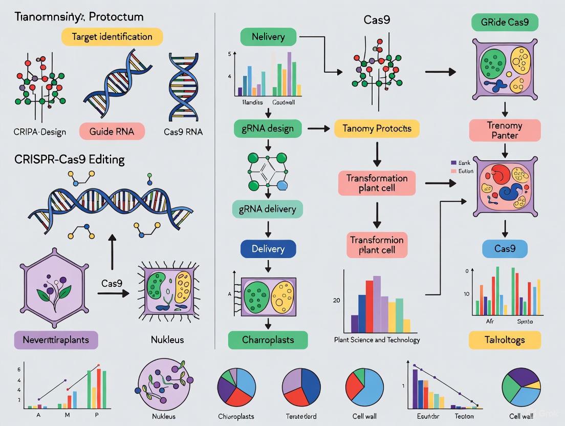
This article provides a comprehensive guide to CRISPR-Cas9 genome editing protocols for plant transformation, tailored for researchers and scientists.
Advanced CRISPR-Cas9 Protocols for Efficient Plant Transformation: A Comprehensive Guide from Design to Validation
Abstract
This article provides a comprehensive guide to CRISPR-Cas9 genome editing protocols for plant transformation, tailored for researchers and scientists. It covers foundational principles of CRISPR-Cas9 mechanisms and prerequisite genomic tools, detailed methodologies for stable and transient transformation across diverse plant species, advanced strategies for optimizing editing efficiency and troubleshooting common challenges, and robust techniques for validating edits and analyzing outcomes. By integrating the latest research and practical protocols, this resource aims to empower plant biotechnologists in developing improved crops with enhanced traits such as yield, disease resistance, and stress tolerance.
Core Principles and Prerequisites for Plant Genome Editing
The CRISPR-Cas9 system, derived from a bacterial adaptive immune mechanism, has revolutionized genetic engineering by providing an efficient, precise, and relatively easy genome editing tool [1]. This technology has initiated a new chapter in genetic engineering, enabling researchers to introduce targeted modifications in living cells across diverse organisms, including plants [2] [1]. For plant transformation research, CRISPR-Cas9 offers unprecedented opportunities to accelerate functional genomics studies and crop improvement programs by facilitating the development of plants with enhanced traits such as disease resistance, improved nutritional profiles, and better adaptability to environmental stresses [3] [4].
The fundamental CRISPR-Cas9 system consists of two key components: a DNA-binding domain made of a single guide RNA (sgRNA) and a DNA-cleaving domain comprising the Cas9 endonuclease protein [2]. These components work in concert to identify specific DNA sequences and introduce double-stranded breaks (DSBs) at predetermined genomic locations [1]. The cellular repair mechanisms that address these breaks then enable the introduction of desired genetic modifications [1]. This application note provides a comprehensive overview of the CRISPR-Cas9 mechanism, with detailed protocols and resources tailored for plant researchers engaged in transformation studies.
The Core Components of the CRISPR-Cas9 System
Cas9 Nuclease: The Molecular Scissors
The Cas9 protein, most commonly derived from Streptococcus pyogenes (SpCas9), is a large multi-domain DNA endonuclease (1368 amino acids) that functions as the catalytic engine of the system [1]. Structurally, Cas9 consists of two primary lobes: the recognition (REC) lobe and the nuclease (NUC) lobe [1]. The REC lobe, containing REC1 and REC2 domains, is responsible for binding the guide RNA [1]. The NUC lobe contains three critical domains: RuvC and HNH, which each cleave one DNA strand, and the PAM-interacting domain, which confers specificity for the Protospacer Adjacent Motif (PAM) sequence essential for target recognition [1].
The PAM sequence, a short conserved DNA sequence downstream of the cut site, is a critical component of target recognition [1]. For SpCas9, the PAM sequence is 5'-NGG-3', where N can be any nucleotide base [1]. The Cas9 nuclease becomes activated upon binding to both a valid PAM sequence and a complementary target DNA sequence specified by the guide RNA [1].
Table 1: Key Cas Protein Variants and Their Characteristics
| Cas Protein | Source Organism | PAM Sequence | DSB Pattern | Key Features |
|---|---|---|---|---|
| SpCas9 | Streptococcus pyogenes | 5'-NGG-3' | Blunt ends | Most widely used; requires G-rich PAM |
| Cas9 D10A | Engineered mutant | 5'-NGG-3' | Single-strand nick | Nickase; reduced off-target effects when used in pairs |
| Cas9 H840A | Engineered mutant | 5'-NGG-3' | Single-strand nick | Nickase; cleaves non-target strand |
| A.s. Cas12a (Cpfl) | Acidaminococcus sp. | 5'-TTTV-3' | Staggered ends with 5' overhangs | Shorter gRNA; useful for AT-rich regions |
Guide RNA: The Targeting System
The guide RNA is a synthetic hybrid molecule that combines two natural RNA components: the CRISPR RNA (crRNA) and the trans-activating crRNA (tracrRNA) [2] [1]. The crRNA contains a 20-nucleotide guide sequence that is complementary to the target DNA sequence, providing the targeting specificity, while the tracrRNA serves as a binding scaffold for the Cas9 nuclease [1]. For experimental use, these are typically combined into a single guide RNA (sgRNA) molecule through a synthetic hairpin-like loop (linker-loop) [1].
The guide RNA can be delivered in two primary formats, each with optimized lengths determined through empirical studies:
- Two-part gRNA system: Consists of separate crRNA (36 nucleotides optimal length) and tracrRNA (67 nucleotides optimal length) components [5]
- Single guide RNA (sgRNA): Combined molecule with an optimal length of 100 nucleotides [5]
The design of the target-specific spacer sequence is arguably the most critical factor determining CRISPR-Cas9 efficiency and specificity [2]. For plant genomes, which often exhibit high complexity, polyploidy, and repetitive sequences, careful gRNA design is particularly essential to maximize on-target activity while minimizing off-target effects [2].
Comprehensive gRNA Design Strategy for Plant Systems
Principles of Efficient gRNA Design
Designing highly specific gRNA with minimal off-target activity is a prerequisite for successful gene editing in plants [2]. The goal is to achieve the highest possible on-target activity while minimizing off-target effects, which can cause unwanted phenotypes including cell death [5]. The target sequence must be adjacent to a PAM sequence (NGG for SpCas9), but the PAM itself should not be included in the gRNA design [5].
Several factors must be considered during gRNA design, particularly for complex plant genomes like wheat, which has a hexaploid structure with three sub-genomes and a high proportion of repetitive DNA sequences (more than 80%) [2]. These complexities increase the possibility of off-target mutations and decrease editing specificity [2]. Key considerations include GC content (optimal 40-80%), avoidance of repetitive sequences, and ensuring uniqueness of the target across all sub-genomes in polyploid species [2].
Table 2: gRNA Design Parameters for Optimal Editing Efficiency
| Parameter | Optimal Range | Impact on Efficiency | Tool/Resource |
|---|---|---|---|
| GC Content | 40-80% | GC content outside this range decreases efficiency | IDT gRNA design tool [5] |
| gRNA Length | 20 nucleotides | Shorter sequences negatively impact on-target activity | IDT guidelines [5] |
| PAM Position | Immediate 5' of NGG | Essential for Cas9 recognition and cleavage | Cas9 specificity [1] |
| Off-target Potential | Minimal similarity | Reduces unintended editing events | BLAST analysis [2] |
| Secondary Structure | Minimal self-complementarity | Ensures gRNA availability for target binding | RNA folding tools [2] |
A Stepwise Protocol for gRNA Design in Plants
The process of designing gRNA for CRISPR-Cas9-SDN1 genome editing in plants can be divided into three phases: gene verification, gRNA designing, and gRNA analysis [2].
Phase 1: Gene Identification and Verification
- Gene Selection: Identify promising negative regulator genes through literature review of genome editing, RNAi, or TILLING studies [2]. The target gene should ideally have tissue-specific expression rather than pleiotropic effects [2].
- Sequence Retrieval: Obtain the gene sequence and chromosomal location using databases such as Ensembl Plants and KnetMiner [2].
- Homology Analysis: Use BLAST analysis to determine editing ability across various sub-genomes and identify potential off-target sites [2]. Assess similarity between the identified gene and genes in other plant species and sub-genomes using Clustal Omega software [2].
- Cultivar-Specific Considerations: For species with pan-genome resources (e.g., wheat), consult databases that incorporate presence-absence variations, structural variants, and diverse allelic forms across cultivars to enable precise cultivar-specific gRNA design [2].
Phase 2: gRNA Design and Selection
- Target Site Identification: Scan the target gene for sequences matching 5'-N(19-21)-NGG-3' [2].
- Specificity Validation: Verify target uniqueness across the entire genome using BLAST analysis against genomic and cDNA databases [2].
- Efficiency Prediction: Utilize bioinformatic tools to rank potential gRNAs based on predicted efficiency scores [2].
- Multi-gRNA Strategy: For polyploid species, design gRNAs that target all homoeologs simultaneously, or design specific gRNAs for each homoeolog if differential editing is desired [2].
Phase 3: gRNA Validation and Optimization
- Secondary Structure Analysis: Validate gRNA by testing its potential secondary structure, Gibbs free energy, and propensity to base pair within itself [2].
- Vector Compatibility: Check sequence similarity to the cloning binary vector to be used in the study [2].
- Experimental Validation: For critical applications, design multiple gRNAs (typically 3-5) for empirical testing to identify the most effective variant [2].
Figure 1: gRNA Design Workflow for Plant CRISPR Systems. This diagram illustrates the comprehensive process for designing effective guide RNAs for plant genome editing, from initial gene selection through experimental validation.
DNA Repair Mechanisms in CRISPR-Cas9 Genome Editing
Double-Strand Break Repair Pathways
After the CRISPR-Cas9 complex introduces a double-stranded break at the target site, cellular repair mechanisms are activated to repair the damage [1]. The Cas9 nuclease creates DSBs 3 base pairs upstream of the PAM sequence, generating predominantly blunt-ended breaks [1]. Two primary cellular repair pathways address these breaks: Non-Homologous End Joining (NHEJ) and Homology-Directed Repair (HDR) [1].
Non-Homologous End Joining (NHEJ) is an error-prone repair mechanism that functions throughout the cell cycle by directly ligating the broken DNA ends without requiring a template [1]. This process often results in small insertions or deletions (indels) at the cleavage site, which can disrupt gene function through frameshift mutations or premature stop codons, effectively creating gene knockouts [1]. In plants, NHEJ is the predominant DSB repair pathway and is highly efficient for generating gene knockouts [6].
Homology-Directed Repair (HDR) is a precise repair mechanism that requires a donor DNA template with homology to the sequences flanking the DSB [1]. HDR is most active during the late S and G2 phases of the cell cycle and can execute precise gene insertions or replacements by using donor DNA templates containing the desired sequence modifications flanked by homology arms [1]. While HDR offers precision, its efficiency in plants is typically much lower than NHEJ due to competition between the pathways and the infrequency of HDR in somatic plant cells [6].
Table 3: Comparison of DNA Repair Pathways in CRISPR-Cas9 Editing
| Parameter | Non-Homologous End Joining (NHEJ) | Homology-Directed Repair (HDR) |
|---|---|---|
| Template Requirement | No template required | Requires homologous donor template |
| Repair Precision | Error-prone (indels) | Precise (specific sequence changes) |
| Efficiency in Plants | High (predominant pathway) | Low (1.12% or less in potato protoplasts) [6] |
| Primary Application | Gene knockouts | Precise gene insertion or replacement |
| Cell Cycle Phase | All phases | Late S and G2 phases |
| Outcome | Random insertions/deletions | Precise, predictable edits |
Optimizing HDR in Plant Systems
HDR remains challenging in plant systems due to its inherently low efficiency in gene editing applications [6]. However, several strategies can improve HDR outcomes:
Donor Template Design: Use single-stranded DNA (ssDNA) donors with 30-97 nucleotide homology arms, which have shown success in potato protoplasts [6]. The target orientation (complementary to the gRNA) generally outperforms the non-target orientation [6].
RNP Delivery: Ribonucleoprotein (RNP) complex delivery of Cas9 and gRNA enables faster editing onset, reduces off-target effects, and eliminates the risk of random plasmid integration [7].
HDR Enhancement Strategies: Although chemical inhibitors of NHEJ pathways have shown success in animal systems, their efficacy in plants remains limited [6]. Instead, focus on optimizing donor template structure and delivery methods.
Protocol for HDR Donor Design [7]:
- Homology Arm Length: Design ssDNA donors with 30-40 nucleotide homology arms on each side for optimal efficiency in plant systems [6].
- Blocking Mutations: Incorporate silent mutations in the PAM sequence or seed region of the target site to prevent re-cleavage of successfully edited alleles [7].
- Strand Selection: When using ssDNA donors, the target strand (complementary to the gRNA) generally shows higher HDR efficiency than the non-target strand [7].
- Chemical Modifications: Consider adding phosphorothioate (PS) linkages to the ends of ssDNA donors to improve stability and HDR frequency [7].
Figure 2: DNA Repair Pathways After CRISPR-Cas9 Cleavage. This diagram illustrates the two primary cellular repair mechanisms that address double-strand breaks introduced by CRISPR-Cas9, leading to different editing outcomes.
Experimental Protocol for Plant Transformation
CRISPR-Cas9 Construct Assembly and Plant Transformation
The following protocol adapts established methods for tomato and grapevine transformation to provide a generalizable approach for dicot plants [8] [4].
Part 1: Vector Construction using Golden Gate Cloning [8]
- Select appropriate CRISPR backbone vector based on plant species and selection requirements (e.g., pGreen or pCAMBIA backbones) [9].
- Clone gRNA expression cassette into the vector using BsaI restriction sites for modular assembly [9]. For multiplex editing, assemble multiple gRNAs with tRNA spacers [4].
- Choose promoter for Cas9 expression: For dicot plants, the 35S promoter generally shows strong expression, though the RPS5a promoter may offer improved efficiency in some species [4].
- Select optimized Cas9 variant: Use plant-codon optimized Cas9 with intronic sequences (e.g., zCas9i) and two nuclear localization signals (NLS) for enhanced editing efficiency [4].
- Include visual selection marker: Incorporate DsRed2 or similar fluorescent protein under appropriate promoter for efficient screening of transformed tissues [4].
Part 2: Agrobacterium-mediated Plant Transformation [8]
- Prepare explant material: Surface-sterilize cotyledons or other explant tissues from sterile seedlings.
- Agrobacterium co-cultivation:
- Grow Agrobacterium strain EHA105 or GV3101 harboring the CRISPR construct overnight at 28°C
- Resuspend bacteria to OD₆₀₀ = 0.5-1.0 in liquid co-cultivation medium
- Immerse explants in bacterial suspension for 10-30 minutes
- Transfer to co-cultivation medium and incubate at 26°C for 2-3 days in darkness
- Selection and regeneration:
- Transfer explants to selection medium containing appropriate antibiotics (e.g., kanamycin) and bacteriostatic agents (e.g., timentin or cefotaxime)
- Subculture every 2-3 weeks onto fresh selection medium
- Monitor for fluorescent marker expression to identify successfully transformed tissues
- Plant regeneration:
- Transfer putative transgenic calli to shoot induction medium
- Develop shoots for 4-8 weeks, then transfer to root induction medium
- Acclimate regenerated plantlets to greenhouse conditions
Part 3: Molecular Analysis of Transformed Plants
- Genomic DNA extraction from putative transgenic plant leaves
- PCR amplification of target region and selection marker genes
- Sequence verification of edited loci through Sanger sequencing or next-generation sequencing
- Off-target analysis by examining potential off-target sites identified during gRNA design
Table 4: Essential Research Reagents for Plant CRISPR-Cas9 Experiments
| Reagent/Category | Specific Examples | Function/Purpose | Considerations for Plant Systems |
|---|---|---|---|
| Cas9 Variants | SpCas9, zCas9i, hCas9 | DNA cleavage enzyme | Plant-codon optimized versions (zCas9i) show higher efficiency [4] |
| gRNA Format | sgRNA, 2-part crRNA:tracrRNA | Targets Cas9 to specific genomic loci | sgRNA (100 nt) most common; 2-part system offers chemical modification options [5] |
| Delivery Method | Agrobacterium, RNP complexes | Introduces editing components to cells | RNP delivery reduces off-target effects; Agrobacterium enables stable transformation [7] |
| Donor Templates | ssODN, dsDNA with homology arms | Provides template for HDR repair | ssDNA with 30-40 nt homology arms effective in plants [6] |
| Selection Markers | DsRed2, NPTII, HPT | Identifies successfully transformed tissues | Fluorescent markers enable early visual screening [4] |
| Bioinformatics Tools | IDT HDR Design Tool, Ensembl Plants, BLAST | gRNA design and specificity analysis | Essential for addressing complex plant genomes with high repetition [2] [7] |
Advanced Applications in Plant Research
CRISPR Activation (CRISPRa) for Gain-of-Function Studies
Beyond gene knockouts, CRISPR technology has been adapted for transcriptional activation through CRISPR activation (CRISPRa) systems [3]. Unlike conventional CRISPR editing that introduces double-stranded breaks, CRISPRa employs a deactivated Cas9 (dCas9) fused to transcriptional activators to upregulate target gene expression without altering the DNA sequence [3]. This approach offers unique opportunities for gain-of-function studies, particularly when studying gene families with functional redundancy where knockouts may fail to reveal phenotypic changes [3].
CRISPRa has been successfully applied in plants to enhance disease resistance by upregulating defense-related genes [3]. For example, in tomato, CRISPRa was used to upregulate the PATHOGENESIS-RELATED GENE 1 (SlPR-1), enhancing plant defense against Clavibacter michiganensis infection [3]. Similarly, upregulation of the SlPAL2 gene through targeted epigenetic modifications led to enhanced lignin accumulation and increased defense [3].
Multiplex Genome Editing for Complex Traits
For polygenic traits controlled by multiple genes, multiplex genome editing enables simultaneous modification of several target loci [9]. Advanced CRISPR toolkits facilitate the assembly of one or more gRNA expression cassettes with high efficiency using modular cloning systems [9]. This approach is particularly valuable for addressing genetic redundancy in polyploid crops or for engineering complex metabolic pathways [9].
The CRISPR-Cas9 system provides plant researchers with a powerful and precise tool for genetic engineering, with applications ranging from basic functional genomics to applied crop improvement. Successful implementation requires careful attention to gRNA design, appropriate selection of CRISPR components, and optimization of transformation protocols for specific plant species. By following the comprehensive guidelines and protocols outlined in this application note, researchers can leverage CRISPR-Cas9 technology to address fundamental questions in plant biology and develop improved crop varieties with enhanced agricultural traits.
In plant biotechnology, the CRISPR-Cas9 system has emerged as a revolutionary tool for functional genomics and crop improvement, enabling researchers to develop climate-resilient varieties of major staple crops such as wheat, rice, and maize [10]. This RNA-guided endonuclease technology facilitates precise genomic modifications through targeted double-strand breaks (DSBs), which are subsequently repaired via endogenous cellular mechanisms [11]. The system's core components include the Cas9 nuclease and a single-guide RNA (sgRNA), with the latter conferring sequence specificity through complementary base pairing to the target DNA site, which must be adjacent to a protospacer adjacent motif (PAM) with the sequence NGG [11] [12].
The efficacy and precision of CRISPR-Cas9-mediated genome editing are fundamentally dependent on prior access to high-quality genome sequences and comprehensive structural annotations [13]. These genomic resources enable accurate sgRNA design by providing precise coordinates of functional elements, thereby minimizing off-target effects while maximizing editing efficiency. For plant species, this requirement presents unique challenges due to complex genome architectures, including high ploidy levels, extensive repetitive content, and substantial intron-exon structures [14]. This application note delineates the essential prerequisites of genome sequencing and annotation within the context of CRISPR-Cas9 protocols for plant transformation research, providing detailed methodologies and resources to support successful genome editing outcomes.
Genome Annotation Methodologies for Plant Research
Genome annotation encompasses two distinct bioinformatics processes: structural annotation, which identifies the physical locations and structures of functional elements (genes, transcripts, exons, coding sequences, and untranslated regions), and functional annotation, which assigns putative functions, gene symbols, and Gene Ontology terms to these elements [13]. For CRISPR-Cas9 applications, structural annotation is particularly critical as it directly informs target site selection.
Major Annotation Approaches
Current state-of-the-art genome annotation strategies fall into three primary categories, each with distinct strengths, limitations, and input requirements [13]:
Table 1: Comparison of Genome Annotation Approaches
| Method | Underlying Approach | Primary Output | Key Input Requirements | Best Suited For |
|---|---|---|---|---|
| Model-Based (BRAKER) | Hidden Markov Models (HMMs) | Protein-coding genes | Protein sequences from related species (e.g., OrthoDB) and/or RNA-seq data | Annotating proteomes without closely related reference genomes |
| RNA-seq Assembly (Stringtie-TransDecoder) | Transcriptome assembly from splice graphs | Complete transcripts (including UTRs) | Paired-end RNA-seq reads and protein BLAST database | Comprehensive transcriptome annotation when RNA-seq is available |
| Annotation Transfer (TOGA, Liftoff) | Liftover of annotations via whole-genome alignment | Homologous features from reference genome | High-quality annotated genome from closely related species | Rapid annotation when high-quality reference exists |
Decision Framework for Annotation Method Selection
The choice of an appropriate annotation strategy should be guided by research objectives, data availability, and evolutionary considerations. The following workflow provides a systematic approach for selecting the optimal annotation method:
Figure 1: Decision workflow for selecting appropriate genome annotation methods based on available data and research objectives [13].
Special Considerations for Plant Genomes
Plant genomes present unique challenges for annotation and subsequent CRISPR applications due to their distinctive characteristics [13]:
- High Repetitive Content: Many plant genomes contain substantial repetitive elements that can complicate assembly and annotation. For example, the Garra rufa genome has 51.95% of its sequence masked by WindowMasker [15], illustrating the repetitive nature of some genomes.
- Complex Genome Organization: Polyploidy and extensive gene families are common in plants, requiring specialized annotation approaches.
- Annotation Transfer Limitations: Whole-genome alignment between plant species can be challenging due to evolutionary divergence, making annotation transfer methods less reliable than for animal genomes.
For these reasons, empirical validation of genome annotations through RNA-seq data is strongly recommended for plant CRISPR projects, even when using annotation transfer approaches [13].
Experimental Protocols for Genome Annotation
Structural Annotation Using BRAKER2 Pipeline
The following protocol outlines the steps for generating structural annotations using the BRAKER2 pipeline, which employs a combination of GeneMark-ET and AUGUSTUS to predict protein-coding genes [13].
Materials and Reagents
Table 2: Key Research Reagent Solutions for Genome Annotation
| Reagent/Resource | Function/Purpose | Example Sources |
|---|---|---|
| Genome Assembly (FASTA) | Target for annotation | Institutional sequencing core or public repositories |
| OrthoDB Protein Set | Evolutionary-informed protein sequences for homology hints | OrthoDB database |
| RNA-seq Reads (FASTQ) | Transcriptional evidence for splice site prediction | NCBI SRA or in-house sequencing |
| BRAKER2 Software | Automated annotation pipeline | GitHub repository |
| BUSCO Dataset | Assessment of annotation completeness | BUSCO website |
Step-by-Step Procedure
Data Preparation
- Obtain genome assembly in FASTA format (e.g.,
assembly.fasta) - Download taxonomically appropriate protein sequences from OrthoDB
- Format the protein database using BLAST+
- Obtain genome assembly in FASTA format (e.g.,
Protein Alignment
- Align protein sequences to the genome using ProtHint
- Convert alignments to hints for AUGUSTUS
Gene Prediction
- Execute BRAKER2 with protein hints
- Run command:
braker.pl --genome=assembly.fasta --prot_seq=proteins.faa --cores=8 --species=YourSpecies - Generate output in GFF3 format
Quality Assessment
- Assess annotation completeness using BUSCO
- Run command:
busco -i annotation.gff -l actinopterygii_odb10 -o busco_results -m genome
Annotation Transfer Using Liftoff
For researchers with access to a high-quality annotated reference genome from a closely related species, annotation transfer offers a rapid alternative [13]:
Generate Whole-Genome Alignment
- Align reference and target genomes using minimap2 or LASTZ
Execute Annotation Transfer
- Run Liftoff to transfer annotations:
liftoff -g reference.gff target_assembly.fasta reference_assembly.fasta -o transferred_annotations.gff
- Run Liftoff to transfer annotations:
Validate Transferred Annotations
- Check for complete BUSCO scores compared to reference
- Manually inspect key gene families of interest
Integration with CRISPR-Cas9 Experimental Design
High-quality genome annotations directly inform multiple aspects of CRISPR-Cas9 experimental design in plants, significantly enhancing the probability of successful editing outcomes.
sgRNA Design and Target Selection
Comprehensive genome annotations enable strategic sgRNA design through the identification of:
- Exon-Intron Boundaries: sgRNAs should preferentially target exonic regions, particularly first exons downstream of start codons, to maximize probability of functional knockouts [16]
- Functional Domains: Annotations facilitate targeting of critical protein domains for complete loss-of-function mutations
- Sequence Uniqueness: Annotations enable BLAST searches to ensure sgRNA specificity and minimize off-target effects
For example, in tomato genome editing protocols, sgRNAs are designed within the first exon closer to the start codon to ensure disruption of the functional protein [16].
Assessment of Editing Outcomes
Following CRISPR-Cas9-mediated transformation, genome annotations facilitate molecular characterization of edited plants through:
- High-Resolution Melt (HRM) Analysis: Annotations provide coordinates for designing PCR primers flanking target sites
- Sanger Sequencing: Annotated reference sequences enable alignment and identification of induced mutations
- Variant Effect Prediction: Structural annotations allow prediction of functional consequences of edits (e.g., frameshifts, premature stop codons)
In potato editing protocols, HRM analysis coupled with annotations enables efficient screening of tetraploid mutants without the need for lengthy segregation [14].
Case Study: Fusarium oxysporum Genome Annotation for Virulence Gene Editing
A recent application of annotation-driven CRISPR editing targeted the SIX9 effector gene in Fusarium oxysporum f.sp. cubense race 1 (Foc1), a pathogen causing Fusarium wilt in bananas [17]. The experimental workflow demonstrates the critical role of prior genome annotation:
Figure 2: Workflow for CRISPR-Cas9-mediated editing of Fusarium oxysporum SIX9 effector gene [17].
This study relied on previously annotated Fusarium oxysporum genomes to identify the SIX9 gene as a candidate virulence factor. Researchers then designed two sgRNAs targeting this annotated locus and developed an optimized in vitro protocol to produce highly active Cas9 protein, demonstrating enzymatic activity comparable to commercial standards [17]. The success of this pathogen-focused editing approach underscores the value of comprehensive genome annotation for both plant and pathogen genomics in developing transformative crop protection strategies.
High-quality genome sequences and structural annotations represent foundational prerequisites for effective CRISPR-Cas9 genome editing in plants. By enabling precise sgRNA design, informing target selection strategies, and facilitating molecular characterization of edited lines, comprehensive annotations significantly enhance editing efficiency and functional outcomes. As CRISPR technologies continue to evolve toward more sophisticated applications—including base editing, prime editing, and multiplexed interventions—the importance of accurate genomic references will only intensify. Plant researchers should prioritize investment in robust annotation pipelines tailored to their species of interest, as these resources ultimately determine the success and reproducibility of genome editing initiatives aimed at crop improvement and climate resilience.
Within the framework of CRISPR-Cas9 genome editing protocols for plant transformation, tissue culture represents the fundamental bridge between genetic manipulation and the recovery of viable, genetically stable plants. While CRISPR-Cas9 systems provide the tools for precise genomic modifications, the success of entire editing initiatives hinges upon the ability to regenerate whole plants from single, transformed cells. This protocol details the establishment of robust regeneration pathways, specifically tailored for use with CRISPR-edited plants, to ensure the efficient recovery of non-transgenic, edited lines. The methodologies outlined herein are critical for converting edited cells into homozygous, transgene-free plants, thereby solidifying the functional genomics and trait improvement pipeline [10] [18].
The Critical Role of Regeneration in CRISPR-Cas9 Workflows
The regeneration phase is the most critical determinant of success and timeline in plant genome editing projects. A typical workflow to obtain an edited, transgene-free plant requires 6–12 months, with the majority of this time dedicated to the tissue culture and regeneration steps [18]. The regeneration protocol must be finely synchronized with the transformation and editing event. Following Agrobacterium-mediated transformation of plant explants with CRISPR-Cas9 constructs, the application of precisely formulated plant growth regulators in the culture media directs cell division and fate. Genetically edited, single cells must undergo dedifferentiation to form a callus, followed by redifferentiation into shoots and roots. The efficiency of this process directly impacts the number of independent edited events recovered, thereby influencing the statistical power of subsequent phenotypic analyses [18] [19]. Furthermore, the selection of regeneration strategy is pivotal for achieving transgene excision. By leveraging the sexual reproduction of regenerated T0 plants, transgene-free T1 progeny carrying the stable knockout mutation can be isolated through molecular screening, fulfilling the promise of CRISPR-Cas9 for non-transgenic plant improvement [18].
Table 1: Key Stages and Durations in a CRISPR-Cas9 Regeneration Pipeline for Tomato
| Stage | Process Description | Key Media/Treatments | Typical Duration |
|---|---|---|---|
| 1. Explant Preparation & Transformation | Sterilization and co-cultivation with Agrobacterium carrying CRISPR-Cas9 | Acetosyringone induction medium (CIM II) | 2-3 days |
| 2. Callus Induction & Selection | Dedifferentiation of explant tissue into callus; selection of transformed cells | Callus Induction Medium (CIM I, CIM II) with antibiotics | 2-4 weeks |
| 3. Shoot Regeneration | Redifferentiation of callus into shoot primordia | Shoot Induction Medium (SIM I, SIM II) with cytokinins | 4-8 weeks |
| 4. Root Regeneration | Development of roots from shoots | Root Induction Medium (RIM) with auxins | 2-4 weeks |
| 5. Acclimatization & Seed Set | Transfer to soil and growth to maturity in greenhouse | N/A | 8-12 weeks |
| 6. Molecular Screening | Identification of transgene-free edited progeny in T1 generation | PCR, ddPCR, sequencing | 4-8 weeks |
Experimental Protocol: Regeneration of CRISPR-Edited Tomato Plants
The following step-by-step protocol, adapted from a established method for generating knockout lines in tomato, provides a detailed methodology for the regeneration of edited plants, from explant to transgene-free progeny [18].
Materials and Reagents
Biological Materials
- Solanum lycopersicum cv. MoneyMaker (or other suitable cultivar)
- Agrobacterium tumefaciens strain GV3101 harboring the CRISPR-Cas9 binary vector (e.g., pZG23C04 or similar)
Media and Solutions Prepare all media according to the recipes listed in Section 3.2. Adjust pH to 5.8 before autoclaving. Add filter-sterilized hormones and antibiotics after the medium has cooled to approximately 50°C.
Media Formulations
Table 2: Detailed Composition of Tissue Culture Media for Tomato Regeneration
| Medium Name | Basal Salt/Vitamin Base | Carbon Source | Solidifying Agent | Growth Regulators | Other Additives (post-sterilization) |
|---|---|---|---|---|---|
| ½ MS (Pre-culture) | 2.15 g/L MS + Gamborg B5 vitamins | 10 g/L Sucrose | 8 g/L Agar | - | - |
| CIM I | 4.3 g/L MS + Gamborg B5 vitamins | 30 g/L Sucrose | 5.2 g/L Phytoagar | 1 mg/L 2,4-D, 0.2 mg/L Kinetin | 1 mg/L Thiamine HCl |
| CIM II | 4.3 g/L MS + Gamborg B5 vitamins | 30 g/L Sucrose | 5.2 g/L Phytoagar | 1 mg/L 2,4-D, 0.2 mg/L Kinetin | 1 mg/L Thiamine HCl, 200 μM Acetosyringone |
| SIM I | 4.3 g/L MS + Gamborg B5 vitamins | 30 g/L Sucrose | 5.2 g/L Phytoagar | 2 mg/L trans-Zeatin | 1 mg/L Thiamine HCl, 100 mg/L Kanamycin, 250 mg/L Timentin |
| SIM II | 4.3 g/L MS + Gamborg B5 vitamins | 30 g/L Sucrose | 5.2 g/L Phytoagar | 1 mg/L trans-Zeatin | 1 mg/L Thiamine HCl, 0.1 mg/L IAA, 100 mg/L Kanamycin, 250 mg/L Timentin |
| RIM | 4.3 g/L MS + Gamborg B5 vitamins | 30 g/L Sucrose | 5.2 g/L Phytoagar | 1 mg/L IAA | 50 mg/L Kanamycin, 250 mg/L Timentin |
Step-by-Step Methodology
Step 1: Explant Preparation and Transformation
- Surface Sterilization: Sterilize tomato seeds in 70% ethanol for 5 minutes, followed by immersion in a 50% sodium hypochlorite solution for 30 minutes. Rinse thoroughly with sterile water [18].
- Germination: Aseptically place seeds on half-strength MS medium and culture in a growth chamber for 14 days.
- Co-cultivation: Excise cotyledons or other explants from seedlings and immerse in an Agrobacterium suspension (OD~600 = 0.5-1.0) prepared in CIM II medium containing acetosyringone. Co-cultivate for 2-3 days in the dark.
Step 2: Callus Induction and Selection
- Transfer explants to CIM I medium containing timentin to suppress Agrobacterium growth. Culture for one week.
- Subsequently, transfer explants to fresh CIM I medium supplemented with both kanamycin (for selection of transformed cells) and timentin. Subculture to fresh medium every two weeks for a total of 2-4 weeks until callus formation is observed.
Step 3: Shoot Regeneration
- Move developed calli to SIM I medium to initiate shoot organogenesis. Maintain cultures under a 16/8 hour light/dark photoperiod.
- After two weeks, transfer developing shoot primordia to SIM II medium to promote further shoot elongation. Subculture every two weeks until shoots are 2-3 cm tall.
Step 4: Root Regeneration and Acclimatization
- Excise elongated shoots and transfer to RIM medium to induce root formation.
- Once a robust root system has developed, carefully remove plantlets from the culture vessels, gently wash off agar, and transfer to sterile soil in small pots.
- Maintain high humidity for the first week by covering pots with transparent domes, gradually reducing humidity to acclimate plants to greenhouse conditions.
Step 5: Molecular Screening for Edited, Transgene-Free Plants
- Genomic DNA Extraction: Extract genomic DNA from leaf tissue of regenerated T0 plants and subsequent T1 progeny.
- Mutation Analysis: Use PCR to amplify the target genomic region, followed by Sanger sequencing or next-generation sequencing to identify indel mutations [18] [19].
- Transgene Segregation: Screen T1 progeny for the absence of the Cas9/sgRNA transgene cassette using PCR with primers specific to the HPT (hygromycin) or other selectable marker gene. Plants lacking the transgene but harboring the desired mutation are the final product [18].
The Scientist's Toolkit: Research Reagent Solutions
Table 3: Essential Reagents for CRISPR-Cas9 Plant Regeneration Protocols
| Reagent/Category | Specific Examples | Function in the Protocol |
|---|---|---|
| CRISPR Vector System | pZG23C04, pZNH2GTRU6, pZD202-Cas3 [18] [19] | Provides the genetic machinery for genome editing; contains Cas9/Cas3 nuclease, sgRNA expression cassette, and selectable marker. |
| Plant Growth Regulators | 2,4-Dichlorophenoxyacetic acid (2,4-D), Kinetin, trans-Zeatin, IAA [18] | Directs cell fate: auxins like 2,4-D promote callus formation, while cytokinins like zeatin stimulate shoot initiation. |
| Selection Agents | Kanamycin, Hygromycin [18] [19] | Selects for plant cells that have integrated the T-DNA from the binary vector by conferring antibiotic resistance. |
| Antibiotics for Microbiology | Ampicillin, Rifampicin, Gentamicin, Timentin [18] | Used for bacterial culture selection (Amp, Rif, Gen) and plant culture decontamination (Timentin eliminates Agrobacterium post-co-cultivation). |
| Enzymes for Molecular Cloning | BsaI, BbsI, T4 DNA Ligase [18] [9] | Restriction enzymes and ligases for Golden Gate assembly of sgRNA sequences into the CRISPR binary vector. |
| Molecular Validation Kits | PCR Purification Kit, Plasmid DNA Purification Kit, DNeasy Plant Mini Kit [18] | Essential for molecular biology workflows, including vector construction and genomic DNA extraction for genotyping. |
Workflow Visualization: From Explant to Edited Plant
The following diagram illustrates the complete experimental workflow, integrating both the molecular and tissue culture stages.
The selection of an appropriate editing tool is a critical first step in designing successful plant genome engineering experiments. CRISPR-based systems have evolved from simple nucleases that create double-strand breaks (DSBs) to more sophisticated base editors that enable precise nucleotide conversions without DSBs [20] [21]. This Application Note provides a structured comparison between Cas nucleases and base editors, offering detailed protocols for their application in plant transformation research. The guidance is tailored for researchers and scientists engaged in plant functional genomics and crop improvement programs, with a focus on practical implementation considerations.
Core Technology Comparison: Mechanisms and Outcomes
Fundamental Editing Mechanisms
Cas Nucleases generate double-stranded breaks (DSBs) at targeted genomic locations [20] [11]. These breaks are primarily repaired through either the error-prone non-homologous end joining (NHEJ) pathway, which often results in insertions or deletions (indels) that disrupt gene function, or the homology-directed repair (HDR) pathway, which requires a donor template for precise edits [20] [21]. The classic example is Streptococcus pyogenes Cas9 (SpCas9), which recognizes a 5'-NGG-3' protospacer adjacent motif (PAM) and creates blunt-ended DSBs [20] [11].
Base Editors achieve precise nucleotide conversions without creating DSBs by fusing a catalytically impaired Cas nuclease (nickase or dead Cas) to a deaminase enzyme [20] [21]. Cytosine Base Editors (CBEs) mediate C•G to T•A transitions, while Adenine Base Editors (ABEs) mediate A•T to G•C transitions [20] [22]. This approach minimizes unintended indels and is particularly valuable for introducing specific single nucleotide polymorphisms (SNPs) or creating premature stop codons [21].
Comparative Analysis of Editing Tools
Table 1: Comparative Characteristics of Cas Nucleases and Base Editors
| Feature | Cas Nucleases | Base Editors |
|---|---|---|
| Primary Mechanism | Creates DSBs | Chemical conversion of bases without DSBs [21] |
| DNA Repair Pathway | NHEJ, HDR [20] | Base excision repair [21] |
| Typical Editing Outcomes | Indels (insertions/deletions), gene knockouts, large deletions [20] [11] | C→T or A→G transitions (point mutations) [20] [22] |
| Product Purity | Mixed outcomes (indels) [20] | High (typically >90% desired base conversion without indels) [21] |
| PAM Requirement | Yes (e.g., NGG for SpCas9) [20] [11] | Yes (determined by the Cas moiety) [21] |
| Multiplexing Capability | High (via multiple gRNAs) [21] [22] | Moderate |
| Optimal Editing Window | Precise cut site | ~3-5 nucleotide window within the protospacer [20] |
| Delivery Size | ~4.2 kb for SpCas9 | Larger (~5-6 kb) due to added deaminase domains |
| Common Applications | Gene knockouts, gene insertions (with donor), large deletions | SNP introduction, corrective point mutations, creating stop codons [21] |
Decision Framework for Tool Selection
The following diagram illustrates the decision-making workflow for selecting between Cas nucleases and base editors based on research objectives and sequence context:
Experimental Protocols
Protocol 1: Multiplexed Gene Knockout Using Cas9 Nuclease
Objective: Simultaneously disrupt multiple genes in Nicotiana benthamiana using SpCas9 and tRNA-sgRNA polycistronic vectors [22].
Materials:
- Golden Gate modular cloning toolkit [22]
- pIZZA-BYR-SpCas9 and pBYR2eFa-U6-sgRNA binary vectors [23]
- Agrobacterium tumefaciens GV3101
- N. benthamiana plants (4-6 weeks old)
Procedure:
sgRNA Design and Vector Assembly:
Plant Transformation:
- Introduce the final construct into Agrobacterium.
- Infiltrate N. benthamiana leaves using standard procedures.
- Incubate plants for 7 days post-infiltration.
Editing Efficiency Analysis:
- Extract genomic DNA from infiltrated tissue.
- Amplify target regions and quantify editing efficiency using T7E1 assay or amplicon sequencing [23].
Expected Results: Editing efficiencies typically range from 0.1% to >30% across different sgRNA targets [23]. Multiplexing enables simultaneous knockout of up to six genes in a single transformation.
Protocol 2: Precision Base Editing in Rice Protoplasts
Objective: Introduce a specific C-to-T point mutation in the OsEPSPS gene using a cytidine base editor.
Materials:
- Plant-optimized cytidine base editor (e.g., pTarget-AID) [21]
- Rice protoplasts isolated from etiolated seedlings
- PEG transformation solution
Procedure:
Base Editor Design and Delivery:
- Design sgRNA with target cytidine within positions 3-8 of the protospacer.
- Co-deliver base editor and sgRNA constructs to rice protoplasts via PEG-mediated transformation.
Editing Analysis:
- Harvest protoplasts 48 hours post-transformation.
- Extract genomic DNA and amplify target region.
- Detect base conversions using restriction fragment length polymorphism (RFLP) or Sanger sequencing.
Off-Target Assessment:
Expected Results: Typical base editing efficiencies of 1-20% in rice protoplasts with minimal indels (<1%). Product purity can exceed 90% [21].
Table 2: Key Research Reagent Solutions for Plant Genome Editing
| Reagent Type | Specific Examples | Function & Application Notes |
|---|---|---|
| Cas Nucleases | SpCas9, SaCas9, StCas9, ScCas9, FnCas12a, LbCas12a [22] | SpCas9 (NGG PAM) most common; SaCas9 (NNGRRT PAM) smaller size; Cas12a (TTTV PAM) creates staggered ends |
| Base Editors | Target-AID (CBE), ABE7.10 [22] | Target-AID for C-to-T conversions; ABE for A-to-G conversions |
| Promoters (Monocot) | OsU3p, OsU6-2p, TaU3p [22] | Drive gRNA expression in monocots |
| Promoters (Dicot) | AtU6-26p [22] | Drives gRNA expression in dicots |
| Delivery Vectors | pIZZA-BYR-SpCas9, pBYR2eFa-U6-sgRNA [23] | Binary vectors for Agrobacterium-mediated transformation |
| Cloning Systems | Golden Gate Modular Toolkit [22] | Enables rapid assembly of multigene constructs |
| Detection Reagents | T7E1, RFLP enzymes, AmpSeq kits [23] | For quantifying editing efficiency |
| Bioinformatics Tools | CRISPOR, CRISPR-P 2.0 [23] [25] | sgRNA design and specificity checking |
Advanced Applications and Emerging Technologies
Expanding Targeting Scope with Engineered Cas Variants
The PAM requirement represents a significant limitation for targeting specific genomic regions. Engineered Cas variants with altered PAM specificities have substantially expanded the targeting scope [20] [22]. SpCas9-NG recognizes NG PAMs instead of NGG, while xCas9 recognizes NG, GAA, and GAT PAMs [20] [22]. ScCas9 from Streptococcus canis recognizes NNG PAMs, further expanding potential target sites [22]. When planning experiments requiring targeting of specific sequences with restricted PAM availability, these variants provide valuable alternatives to wild-type SpCas9.
Editing Efficiency Optimization Strategies
Editing efficiency varies significantly based on genomic context, chromatin accessibility, and sgRNA design. Several strategies can enhance editing efficiency:
- gRNA Design: Select guides with high predicted efficiency scores (e.g., Doench'16 score) [23]
- Regulatory Elements: Use appropriate Pol III promoters (U3/U6) matched to your plant species [22]
- Expression Optimization: Implement egg cell-specific promoters (e.g., DD45) for heritable edits in Arabidopsis [21]
- Delivery Method Selection: Choose between Agrobacterium, biolistics, or nanoparticle-based delivery based on plant species and application [26] [27]
Recent advances in nanoparticle-mediated delivery and viral vectors have shown promising results for improving editing efficiency, particularly in difficult-to-transform species [27].
The selection between Cas nucleases and base editors represents a fundamental decision point in plant genome engineering experimental design. Cas nucleases remain the tool of choice for gene knockouts and large-scale modifications, while base editors offer superior precision for single-nucleotide changes. The continued development of engineered Cas variants with expanded PAM compatibilities, improved specificity, and novel functionalities promises to further enhance our capability to precisely modify plant genomes. By following the structured decision framework and optimized protocols outlined in this Application Note, researchers can systematically select the most appropriate editing tools for their specific plant transformation research objectives.
The classification of genome editing applications into SDN-1, SDN-2, and SDN-3 provides a critical framework for researchers navigating the regulatory landscape of plant biotechnology. These categories, defined by Friedrichs et al. (2019), differentiate genome editing techniques based on their molecular mechanisms and outcomes, with direct bearing on regulatory considerations [28]. This classification system helps distinguish between edits that result in small, targeted mutations versus those that incorporate larger DNA sequences, which has implications for risk assessment and regulatory oversight. Within the context of CRISPR-Cas9 genome editing protocols for plant transformation, understanding these categories is essential for designing experiments that align with both research objectives and regulatory requirements.
The CRISPR-Cas9 system has revolutionized plant molecular biology by providing powerful tools for precise gene manipulation [16]. This technology utilizes guide RNAs (gRNAs) that direct the Cas9 endonuclease to generate double-stranded breaks (DSBs) at targeted genomic locations [29]. The cellular repair of these breaks then leads to specific mutations. The SDN classification system specifically addresses how these breaks are repaired and whether external DNA templates are used, creating a spectrum of technical approaches with differing regulatory implications.
SDN Classification System: Mechanisms and Applications
Molecular Mechanisms and Technical Distinctions
The SDN categorization is fundamentally based on the DNA repair pathways employed following the creation of a targeted double-strand break. Each category represents a distinct approach to genome editing with specific technical considerations and outcomes.
Table 1: Comparative Analysis of SDN Classification Categories
| Classification | Repair Mechanism | Template Required | Typical Outcome | Primary Applications in Plants |
|---|---|---|---|---|
| SDN-1 | Non-Homologous End Joining (NHEJ) | No | Small insertions or deletions (indels), gene knockout | Gene silencing, loss-of-function mutations, functional genomics [28] |
| SDN-2 | Homology-Directed Repair (HDR) | Short single-stranded DNA oligonucleotide | Introduction of small, specific point mutations | Precise amino acid changes, fine-tuning gene function [28] |
| SDN-3 | Homology-Directed Repair (HDR) | Large double-stranded DNA vector | Insertion of large DNA sequences (e.g., genes) | Gene insertion, trait stacking, metabolic engineering [28] |
SDN-1 involves the unguided repair of a specific DSB by the Non-Homologous End Joining (NHEJ) pathway [28]. This error-prone repair process often results in small insertions or deletions (indels) at the target site. These mutations can modify a gene's activity, cause gene silencing, or create a knockout by disrupting the reading frame. SDN-1 is considered an efficient method with many applications already demonstrated in various crops [28]. It is particularly valuable for creating loss-of-function mutations to study gene function or to deactivate undesirable genes.
SDN-2 utilizes a short nucleic acid sequence donor, typically a single-stranded DNA oligonucleotide, to direct the repair of a specific DSB through Homology-Directed Repair (HDR) [28]. The donor template is designed with one or more desired mutations flanked by homology sequences that match the regions on either side of the DSB. This allows for precise, predefined changes to be introduced at the target locus. SDN-2 is more complex than SDN-1 due to the lower efficiency of HDR in plants, but it enables more subtle edits than complete gene knockouts.
SDN-3 employs a larger sequence donor, usually a double-stranded DNA molecule carrying a gene or extended genetic element, to direct the repair of a targeted DSB via HDR [28]. The donor typically features long homology arms (often exceeding 800 base pairs each) that flank the insert, facilitating its integration at the target site. SDN-3 is technically the most challenging approach but allows for the introduction of entirely new functions, such as inserting a gene for disease resistance or enhancing nutritional content.
Visualizing the SDN Classification Workflow
The following diagram illustrates the conceptual workflow and decision-making process for selecting and implementing the different SDN categories in a plant genome editing project.
Diagram 1: SDN Category Selection Workflow. This diagram outlines the decision-making process for selecting the appropriate SDN classification based on the desired editing outcome in plant genome editing projects.
Experimental Protocols for SDN-Based Plant Genome Editing
Core Protocol: CRISPR-Cas9 System Assembly and Plant Transformation
The following protocol provides a detailed methodology for implementing SDN-1 type editing (gene knockout) in tomato plants, which can be adapted with modifications for SDN-2 and SDN-3 approaches.
Background: Tomato (Solanum lycopersicum) serves as an important model organism for crop improvement studies [16]. CRISPR-Cas9 provides an effective tool for uncovering the complex functions of tomato genes. The primary objective of this protocol is to establish a robust strategy for producing knockout lines (SDN-1) in tomato plants, which could be adapted for SDN-2 and SDN-3 approaches with the inclusion of appropriate repair templates.
Key Features [16]:
- Two sgRNAs employed for increased efficiency
- Takes 6–12 months to generate edited transgene-free plants
- Specifically optimized for tomato cv. MoneyMaker
Materials and Reagents:
Table 2: Essential Research Reagent Solutions for CRISPR Plant Transformation
| Reagent/Category | Specific Examples | Function/Purpose | Reference |
|---|---|---|---|
| CRISPR Vector System | pZG23C04, pICH47742::2x35S-5'UTR-hCas9(STOP)-NOST | Carries Cas9 and sgRNA expression cassettes | [16] |
| Cloning Enzymes | BpiI (BbsI), BsaI HF, T4 DNA Ligase | Golden Gate assembly of sgRNAs into vectors | [16] |
| Plant Transformation | Agrobacterium tumefaciens GV3101 | Delivery of CRISPR constructs to plant cells | [16] [30] |
| Selection Agents | Kanamycin, Timentin | Selection of transformed plant tissue | [16] |
| Plant Growth Regulators | trans-Zeatin, 2,4-D, IAA, Kinetin | Direct shoot and root regeneration | [16] |
| Culture Media | CIM I, CIM II, SIM I, SIM II, RIM | Support different stages of plant tissue development | [16] |
Procedure:
sgRNA Design and Cloning:
- Design two sgRNAs targeting the first exon downstream and closer to the start codon of the gene of interest [16].
- Use online tools (e.g., CRISPR-Plant, CRISPOR) to minimize off-target effects [26].
- Assemble sgRNA expression cassettes using Golden Gate cloning with BpiI (BbsI) or BsaI enzymes into the final CRISPR-Cas9 binary vector [16] [30].
Plant Transformation:
- Introduce the assembled plasmid into Agrobacterium tumefaciens strain GV3101 [16].
- Use 7-8 day old tomato cotyledons (S. lycopersicum cv. MoneyMaker) as explants [30].
- Perform co-cultivation with Agrobacterium for 2 days [30].
- Transfer explants to selection media (SIM I) containing kanamycin (100 mg/L) and timentin (250 mg/L) to inhibit Agrobacterium growth [16].
Plant Regeneration [16]:
- Culture explants on Shoot Induction Medium (SIM I and SIM II) with appropriate plant growth regulators (trans-zeatin) to promote shoot formation.
- Transfer developed shoots to Root Induction Medium (RIM) containing auxins to encourage root development.
- Maintain cultures at 25°C with a 16/8 hour light/dark cycle.
Screening and Molecular Characterization:
- Extract genomic DNA from regenerated plantlets.
- Use PCR amplification of the target region followed by restriction enzyme digestion or high-resolution melt (HRM) analysis to detect mutations [14].
- Sequence PCR products to confirm the exact nature of induced mutations [14].
- Screen for transgene-free plants by analyzing segregation in the next generation [16].
Adaptation for SDN-2 and SDN-3 Approaches
For SDN-2 applications, include a single-stranded oligodeoxynucleotide (ssODN) donor template in the transformation procedure. This template should contain the desired point mutation(s) flanked by homology arms (approximately 40-80 bp) matching the sequence on either side of the cleavage site [28].
For SDN-3 approaches, a larger double-stranded DNA donor must be provided. This is typically a vector containing the gene or genetic element to be inserted, flanked by long homology arms (often >800 bp each) corresponding to the sequences surrounding the target site [28]. The delivery of this large donor template can be challenging and may require optimization of concentration and delivery method.
Regulatory Considerations and Concluding Remarks
The SDN classification framework provides a structured approach to categorizing genome editing outcomes that has important implications for regulatory science. Generally, SDN-1 and some SDN-2 applications may face simpler regulatory pathways in many jurisdictions, as the resulting plants may contain only small mutations indistinguishable from those obtained through conventional breeding or chemical mutagenesis, and often contain no foreign DNA [31]. In contrast, SDN-3 approaches typically fall under stricter regulatory oversight similar to traditional transgenic crops, as they involve the insertion of larger DNA sequences, potentially including genes from unrelated species.
The experimental protocols detailed herein for tomato can be adapted to other crop species with modifications to the transformation and regeneration methods. The continuous refinement of CRISPR-Cas9 technology, including the development of base editors and prime editors that can create precise changes without double-strand breaks, further expands the toolbox available to plant scientists [26] [31]. When planning genome editing projects, researchers should consider both the technical feasibility of different SDN approaches and the regulatory implications of their chosen strategy, keeping abreast of evolving policies in their target countries.
Practical Transformation Methods and Trait Engineering Applications
Agrobacterium-mediated stable transformation remains a cornerstone technique in plant biotechnology, enabling the precise integration of foreign DNA into plant genomes. Within modern functional genomics and breeding programs, this method has become indispensable for delivering CRISPR-Cas9 components, facilitating advanced genome editing in a wide range of plant species [10] [32] [33]. The natural ability of Agrobacterium tumefaciens to transfer T-DNA from its Ti plasmid to plant cells provides a highly efficient system for generating transgenic plants with stable, single-copy insertion events, which are crucial for consistent transgene expression and regulatory compliance [34] [33]. This application note details current vector systems and optimized protocols that leverage Agrobacterium-mediated transformation for CRISPR-Cas9 genome editing, supported by quantitative efficiency data and standardized methodologies for reproducible results across diverse plant species.
Vector Systems for Agrobacterium-mediated Transformation
Conventional and Gateway Binary Vectors
Traditional binary vectors for Agrobacterium-mediated transformation contain the necessary components for T-DNA transfer: left and right border sequences, multiple cloning sites for gene insertion, selectable marker genes for plants, and bacterial resistance markers. These vectors replicate in both E. coli and Agrobacterium, facilitating molecular cloning and plant transformation workflows [34] [35].
The Gateway Technology has significantly streamlined vector construction through site-specific recombination, eliminating dependence on restriction enzymes. This system uses BP and LR Clonase enzyme mixes to efficiently shuttle genes of interest from Entry clones into various Destination vectors [35]. A key advantage is the ccdB negative selection system, which prevents growth of non-recombinant colonies after the LR reaction, ensuring high cloning efficiency. When using vectors with identical antibiotic resistance markers, the differential replication origins (e.g., ColE1 for E. coli and pVS1 for Agrobacterium) enable successful selection. The pENTR vector cannot replicate in Agrobacterium, allowing for direct transformation of LR reaction mixtures and selective recovery of the desired binary vector in this host [35].
CRISPR-Cas9 Expression Vectors
Specialized binary vectors have been developed to express the CRISPR-Cas9 system in plants. These typically feature:
- A plant codon-optimized Cas9 gene driven by constitutive promoters such as CaMV 35S or maize Ubiquitin1 [32] [33]
- RNA polymerase III promoters (e.g., AtU6, TaU3, TaU6) to drive single-guide RNA (sgRNA) expression [33]
- Multiple cloning sites for inserting sgRNA expression cassettes to enable multiplexed genome editing
- Optional donor DNA templates for homology-directed repair (HDR)
The pYLCRISPR/Cas9 system has been successfully deployed in wheat, rice, and tomato, demonstrating the versatility of these vector platforms across diverse crops [32] [33].
Quantitative Transformation Efficiencies Across Plant Systems
Transformation efficiency varies significantly across plant species, cultivars, and experimental conditions. The table below summarizes reported efficiencies for different plant systems using Agrobacterium-mediated transformation.
Table 1: Transformation Efficiencies in Various Plant Systems
| Plant Species | Genotype/Cultivar | Target Gene | Transformation Efficiency | Key Factors | Citation |
|---|---|---|---|---|---|
| Aspergillus carbonarius (Fungus) | - | ayg1 (conidial pigment) | High (Method-dependent) | Agrobacterium strain selection critical | [36] |
| Common Wheat | Fielder | DA1 | 54.17% (T0 mutation rate) | Agrobacterium strain EHA105; immature embryos | [33] |
| Tomato | M82 | ALC | 72.73% (T0 mutation rate) | Hypocotyl explants; 35S promoter for Cas9 | [32] |
| Carrot | - | - | >85% | Somatic embryogenesis; 2,4-D hormone | [37] |
| Japonica Rice | Taichung 65 | - | High (Protocol-optimized) | Meropenem for bacterial control; mature embryos | [34] |
Detailed Experimental Protocols
Streamlined Cloning Protocol Using Gateway Technology
This protocol enables efficient cloning of genes into binary vectors for Agrobacterium transformation, specifically addressing challenges when vectors share identical antibiotic resistance markers [35].
Materials
- pENTR/D-TOPO cloning kit
- Gateway-compatible binary vector (e.g., pMDC series)
- Chemically competent E. coli (TOP10, DH5α)
- Chemically competent Agrobacterium (EHA105, EHA101)
- LB and YEP media with appropriate antibiotics
- Gateway LR Clonase enzyme mix
Procedure
Entry Clone Construction
- Design gene-specific primers with CACC overhang at the 5' end of the forward primer
- Amplify the gene of interest using high-fidelity DNA polymerase
- Purify the PCR product and set up TOPO cloning reaction
- Transform into competent E. coli and select on kanamycin plates
- Verify positive clones by colony PCR and sequencing
LR Reaction for Binary Vector Construction
- Set up LR recombination reaction mixing Entry clone with Destination vector
- Transform the entire LR reaction mixture into competent Agrobacterium cells
- Plate on YEP medium with appropriate antibiotic (e.g., kanamycin)
- Isolate single colonies and verify recombinant binary vectors by PCR
Critical Note: The pENTR vector cannot replicate in Agrobacterium, so only cells containing the recombined binary vector will grow, providing effective selection even when antibiotic resistance markers are identical [35].
Agrobacterium-mediated Transformation of Japonica Rice
This optimized protocol for japonica rice cv. Taichung 65 enables production of transgenic plants within approximately 90 days using mature embryos [34].
Materials
- Mature seeds of japonica rice cv. Taichung 65
- Agrobacterium tumefaciens strain EHA101 or EHA105
- Binary vector with gene of interest and selection marker (e.g., hygromycin resistance)
- Sterilization solution: 70% ethanol, 1% NaClO with Tween 20
- Co-cultivation medium: N6-based with sucrose, glucose, casamino acid, proline
- Selection medium: Hygromycin B with meropenem
- Regeneration medium: N6-based with hormones
Procedure
Callus Induction from Mature Seeds
- Sterilize mature seeds in 70% ethanol (30 sec) followed by 1% NaClO (15 min)
- Rinse 5 times with sterile water
- Culture seeds on callus induction medium in dark at 25°C for 3-4 weeks
- Select friable, yellowish-white embryogenic calli for transformation
Agrobacterium Preparation and Infection
- Grow Agrobacterium harboring binary vector in LB medium with appropriate antibiotics
- Resuspend bacteria in co-cultivation medium to OD~600~ = 0.1
- Immerse selected calli in bacterial suspension for 30 minutes
- Blot dry on sterile filter paper and transfer to co-cultivation medium
- Incubate in dark at 25°C for 2-3 days
Selection and Regeneration of Transgenic Plants
- Transfer co-cultivated calli to selection medium containing hygromycin and meropenem
- Subculture every 2 weeks onto fresh selection medium
- Transfer developing calli to regeneration medium under light (5000 lux)
- Transfer regenerated shoots to rooting medium
- Acclimate plantlets in greenhouse conditions
Key Optimization: Using meropenem instead of carbenicillin or cefotaxime for Agrobacterium elimination significantly improves shoot regeneration rates in rice [34].
CRISPR-Cas9 Mutagenesis in Common Wheat
This protocol demonstrates successful Agrobacterium-mediated delivery of CRISPR-Cas9 to common wheat, achieving high mutation rates in the T~0~ generation [33].
Materials
- Common wheat cultivar Fielder
- Binary vector pYLCRISPR/Cas9Pubi-B with sgRNA expression cassette
- Agrobacterium tumefaciens strain EHA105
- Immature wheat embryos (14-16 days post-anthesis)
Procedure
Vector Design and Construction
- Design sgRNAs targeting genes of interest (e.g., Pinb, waxy, DA1)
- Clone sgRNA expression cassettes under TaU6 or TaU3 promoters
- Assemble final CRISPR/Cas9 construct using binary vector system
Wheat Transformation
- Collect immature wheat spikes at 14-16 DPA
- Surface sterilize with 75% ethanol (30 sec) and 1% NaClO (15 min)
- Isolate immature embryos and incubate with Agrobacterium suspension for 5 min
- Co-cultivate on medium for 2 days in darkness at 25°C
- Remove embryonic axes and transfer scutella to callus induction medium
- Implement progressive selection and regeneration as described [33]
Mutation Analysis
- Extract genomic DNA from T~0~ transgenic plants
- PCR-amplify target regions and sequence products
- Analyze sequencing chromatograms for indel mutations
- Screen for off-target effects in highly homologous genomic regions
Efficiency Note: This protocol achieved 54.17% mutation frequency in T~0~ wheat plants with no detected off-target mutations, demonstrating the precision of Agrobacterium-mediated CRISPR-Cas9 delivery [33].
Visualization of Agrobacterium-mediated Transformation Workflow
Figure 1: Agrobacterium-mediated Plant Transformation Workflow. This diagram outlines the key stages from vector preparation to molecular confirmation of transgenic plants.
The Scientist's Toolkit: Essential Research Reagents
Table 2: Key Research Reagents for Agrobacterium-mediated Transformation
| Reagent/Equipment | Function/Application | Examples/Specifications |
|---|---|---|
| Agrobacterium Strains | T-DNA delivery to plant cells | EHA101, EHA105, AGL1, LBA4404 [36] [34] [33] |
| Binary Vectors | Carrying gene of interest between T-DNA borders | pMDC series, pYLCRISPR/Cas9Pubi-B [33] [35] |
| Selection Antibiotics | Selection of transformed plant tissues | Kanamycin, Hygromycin B [34] [35] |
| Agrobacterium Suppressors | Eliminating bacterial overgrowth after co-cultivation | Meropenem, Carbenicillin, Cefotaxime [34] |
| Plant Growth Regulators | Inducing callus formation and regeneration | 2,4-D (somatic embryogenesis), Cytokinins, Auxins [37] |
| Gateway Cloning System | Efficient vector construction without restriction enzymes | pENTR/D-TOPO, LR Clonase enzyme mix [35] |
Agrobacterium-mediated stable transformation continues to evolve as an essential platform for plant genome engineering, particularly with the integration of CRISPR-Cas9 technologies for precise genome editing. The protocols and vector systems detailed in this application note provide researchers with standardized methodologies that have demonstrated high efficiency across diverse plant species, from model plants to agriculturally important crops. As plant biotechnology advances toward more sophisticated applications, these foundational transformation techniques will remain crucial for functional genomics, trait development, and the creation of climate-resilient crops to address global agricultural challenges.
Within the broader scope of CRISPR-Cas9 genome editing protocols for plant transformation research, transient transformation systems are indispensable tools for rapid functional genomics analysis. Unlike stable transformation, which integrates transgenes into the plant genome, transient transformation involves temporary gene expression, enabling quick assessment of gene editing efficiency and function before committing to lengthy stable transformation and regeneration processes. Two predominant systems—protoplast isolation and hairy root assays—provide versatile platforms for validating CRISPR constructs, studying gene function, and characterizing cellular processes. This document details the application, optimization, and methodology of these systems, providing structured protocols and quantitative data to support researchers in plant biotechnology and drug development who seek to implement these approaches for accelerated genome editing workflows.
Protoplast-Based Transient Transformation Systems
Protoplasts are plant cells that have had their cell walls removed enzymatically, creating a versatile platform for transient expression assays. The polyethylene glycol (PEG)-mediated transformation of protoplasts enables efficient delivery of CRISPR/Cas9 components, including plasmid DNA, in vitro transcripts, and pre-assembled ribonucleoprotein (RNP) complexes [38]. This system is particularly valuable for rapid validation of guide RNA (gRNA) efficiency and nuclease activity before embarking on stable transformation. Applications extend to subcellular localization, protein interaction studies, transcriptional regulation analysis via dual-luciferase assays, and multi-omics research [39]. A significant advantage of RNP delivery is the generation of transgene-free edited plants, addressing regulatory concerns associated with genetically modified organisms [38].
Quantitative Data from Optimization Studies
Recent optimization studies across diverse plant species have yielded critical quantitative data for protocol establishment. The tables below summarize key parameters for protoplast isolation and transformation.
Table 1: Optimized Protoplast Isolation Parameters Across Plant Species
| Plant Species | Optimal Enzyme Composition | Optimal Osmoticum (Mannitol) | Incubation Conditions | Yield (Protoplasts/g FW) | Viability | Citation |
|---|---|---|---|---|---|---|
| Uncaria rhynchophylla | 1.25% Cellulase R-10 + 0.6% Macerozyme R-10 | 0.8 M | 5 h, 26°C, 40 rpm | 1.5 × 10⁷ | >90% | [39] |
| Banana (Cavendish) | 1.25% Cellulase R-10 + 0.6% Macerozyme R-10 | 0.8 M | 5 h, 26°C, dark | Not specified | >90% | [38] |
| Wheat (cv. Roblin) | Not specified | Not specified | Not specified | Not specified | ~60% Transfection Efficiency | [40] |
Table 2: Optimized PEG-Mediated Protoplast Transformation Parameters
| Parameter | Uncaria rhynchophylla [39] | Banana [38] | Wheat [40] |
|---|---|---|---|
| PEG Concentration | 40% | 50% | Not specified |
| Plasmid DNA Amount | 40 µg | Not specified | Not specified |
| Transformation Duration | 40 min | 30 min | Not specified |
| Incubation Temperature | 24°C (overnight) | Not specified | Not specified |
| Transformation Efficiency | 71% | 5.6% | ~60% |
Detailed Experimental Protocol: PEG-Mediated Transformation of Protoplasts with CRISPR/Cas9 RNPs
Principle: This protocol describes the isolation of mesophyll protoplasts from leaf tissue and their subsequent transfection with pre-assembled CRISPR/Cas9 ribonucleoprotein (RNP) complexes via PEG-mediated transformation. The method is adapted from established procedures in banana [38] and Uncaria rhynchophylla [39].
Materials:
- Plant Material: Young, fully expanded leaves from in vitro plantlets or healthy greenhouse-grown plants.
- Enzyme Solution: Comprising Cellulase R-10, Macerozyme R-10, and osmoticum (e.g., 0.6 M mannitol or 0.8 M D-mannitol) in an appropriate salt solution (e.g., MMG solution: 0.6 M mannitol, 15 mM MgCl₂, 4 mM MES, pH 5.7).
- Washing Solution (WS): 0.6 M mannitol, 20 mM KCl, 4 mM MES, pH 5.7.
- PEG Solution: 40% (w/v) PEG 4000 in MMG solution or 0.6 M mannitol and 0.1 M CaCl₂.
- CRISPR Reagents: Purified Cas9 protein and synthesized target-specific sgRNA.
Procedure:
- Protoplast Isolation: a. Slice leaves into thin strips (0.5–1.0 mm) using a sharp razor blade. b. Submerge tissue in the pre-warmed enzyme solution. c. Incubate in the dark for 4-6 hours at 26°C with gentle shaking (e.g., 40 rpm). d. Gently release protoplasts by swirling the flask. Filter the mixture through a 70-100 μm nylon mesh to remove undigested debris. e. Centrifuge the filtrate at 100 × g for 5 minutes to pellet protoplasts. Carefully remove the supernatant. f. Resuspend the pellet in WS and centrifuge again. Repeat this wash step. g. Resuspend the final protoplast pellet in an appropriate volume of MMG solution. Count protoplasts using a hemocytometer and adjust the density to 2 × 10⁵ cells/mL.
RNP Complex Assembly: a. Pre-assemble the RNP complex by mixing purified Cas9 protein with a molar excess of sgRNA in a suitable buffer. b. Incubate the mixture at 25°C for 15-30 minutes to allow complex formation.
PEG-Mediated Transformation: a. Aliquot 2 × 10⁵ protoplasts (in 100 μL MMG) into a round-bottom tube. b. Add the pre-assembled RNP complex (e.g., 10-20 μg Cas9 protein with corresponding sgRNA). c. Add an equal volume of 40% PEG solution (e.g., 100 μL) dropwise, gently mixing after each addition. d. Incubate the transformation mixture at room temperature for 30-40 minutes. e. Carefully stop the reaction by gradually adding 4-5 volumes of WS with gentle mixing. f. Centrifuge at 100 × g for 5 minutes to pellet the transfected protoplasts. Remove the supernatant. g. Resuspend the protoplasts in an appropriate culture medium and incubate in the dark at 24-26°C for 48-72 hours to allow for genome editing to occur before analysis.
Mutation Analysis: a. Extract genomic DNA from transfected protoplasts after the incubation period. b. Amplify the target genomic region by PCR. c. Analyze editing efficiency using methods such as: - PCR-Restriction Enzyme (PCR-RE) assay if the edit disrupts a restriction site [38] [40]. - T7 Endonuclease I or Surveyor nuclease assay. - Sanger sequencing of cloned PCR amplicons or deep amplicon sequencing for a quantitative assessment [38].
Figure 1: Workflow for Protoplast Isolation and RNP Transformation. This diagram outlines the key steps for establishing a transient CRISPR/Cas9 system in plant protoplasts, highlighting critical optimized parameters from recent studies [38] [39].
Hairy Root-Based Transient Transformation Systems
Hairy root transformation utilizes the natural DNA transfer capability of Agrobacterium rhizogenes (Rhizobium rhizogenes). This soil-borne bacterium infects wounded plant sites and transfers T-DNA from its Root-Inducing (Ri) plasmid into the plant genome, leading to the development of genetically transformed "hairy roots" [41]. This system provides a rapid and convenient means to obtain transgenic roots within a few weeks, making it particularly valuable for studying root biology, root-microbe interactions, and the production of root-derived secondary metabolites. When combined with CRISPR/Cas9, it serves as a powerful platform for functional gene validation in roots, especially in species where stable plant regeneration is difficult or time-consuming. The system has been successfully applied in 26 different plant species, including legumes like soybean and peanut, for CRISPR/Cas-mediated genome editing [41].
Key Agrobacterium rhizogenes Strains and Vector Components
The choice of A. rhizogenes strain and CRISPR vector design are critical for successful genome editing in hairy roots.
Table 3: Widely Used Agrobacterium rhizogenes Strains for Hairy Root Transformation [41]
| Strain | Also Known As | Origin/Source | Key Features |
|---|---|---|---|
| ATCC15834 | LBA9340, 15834, AR15834 | Isolated from rose | One of the first wild-type strains widely used; contains pRi15834 plasmid. |
| A4 | ATCC43057 | Isolated from rose | Wild-type strain; contains pRiA4 plasmid; gave rise to derivative A4RS. |
| A4RS | - | Derivative of A4 | Resistant to rifampicin and spectinomycin; lacks pArA4a plasmid; frequently used. |
| K599 | NCPPB2659 | Isolated from cucumber | Widely used for hairy root transformation in legumes (soybean, peanut). |
| NCPPB1855 | LBA9400 | Isolated from Rosa sp. | Wild-type strain; rifampicin-resistant derivative LBA9402 is available. |
CRISPR/Cas vectors for hairy root transformation typically consist of:
- Cas9 Nuclease: Driven by constitutive promoters like the Cauliflower Mosaic Virus 35S (CaMV 35S) promoter, its enhanced variants (2x35S), or other strong promoters such as maize Ubiquitin (pUbi) [41] [12].
- Guide RNA (gRNA): Expressed under RNA polymerase III-dependent promoters, such as Arabidopsis U6 (AtU6-26) or rice U3 (OsU3) promoters [12].
- Selectable/Screenable Markers: To identify transformed roots, selectable markers (e.g., antibiotic resistance) or screenable markers (e.g., fluorescent proteins like DsRed1 or GFP) are used. Fluorescent markers allow for visual screening and isolation of transformed roots without antibiotic selection [41].
Detailed Experimental Protocol: Hairy Root Transformation for CRISPR/Cas9 Gene Editing
Principle: This protocol involves the co-cultivation of explants (e.g., leaf discs, stem segments, or cotyledons) with Agrobacterium rhizogenes harboring a CRISPR/Cas9 construct. The T-DNA is transferred to plant cells, leading to the development of transgenic hairy roots at the infection sites. These roots can be screened and used for functional analysis of gene edits.
Materials:
- Plant Material: Sterile seedlings or surface-sterilized explants.
- Agrobacterium rhizogenes Strain: e.g., A4RS or K599, carrying the binary CRISPR/Cas9 vector.
- Induction Medium: Solid culture medium without antibiotics for co-cultivation.
- Selection Medium: Solid culture medium containing antibiotics to suppress Agrobacterium growth (e.g., cefotaxime) and, if applicable, to select for transformed roots (e.g., kanamycin).
- CRISPR Vector: Binary vector with Cas9 and species-specific gRNA expression cassettes.
Procedure:
- Vector Construction and Agrobacterium Preparation: a. Clone the species-specific gRNA(s) targeting your gene of interest into a binary CRISPR/Cas9 vector. b. Transform the construct into an appropriate A. rhizogenes strain. c. Inoculate a single bacterial colony into liquid medium with appropriate antibiotics and grow overnight at 28°C with shaking until the culture reaches an OD₆₀₀ of 0.5-1.0. d. Pellet the bacteria by centrifugation and resuspend in a liquid induction medium (e.g., with acetosyringone) to the same OD.
Plant Inoculation and Co-cultivation: a. Prepare explants (e.g., wound leaf discs or cut stem segments) from sterile plants. b. Immerse the explants in the prepared Agrobacterium suspension for 10-30 minutes. c. Blot the explants dry on sterile filter paper and transfer them onto solid induction medium. d. Co-cultivate the explants with Agrobacterium in the dark at 22-25°C for 2-3 days.
Hairy Root Induction and Selection: a. After co-cultivation, transfer the explants to selection medium containing antibiotics to kill the Agrobacterium. b. Maintain the cultures under a light/dark cycle at 25°C. Hairy roots typically emerge from the wound sites within 1-3 weeks. c. Excise emerging roots and transfer them to fresh selection medium. If a fluorescent marker is used (e.g., DsRed1), visually screen for positive roots under a fluorescence microscope [41].
Genotyping and Phenotypic Analysis: a. Isolate genomic DNA from the hairy root tips. b. Amplify the target region by PCR and analyze for mutations using methods like restriction enzyme digestion (if the edit disrupts a site), T7E1 assay, or sequencing. c. For phenotypic analysis, the transgenic hairy roots can be propagated in vitro or used in subsequent bioassays depending on the target gene's function.
Figure 2: Workflow for Hairy Root Transformation and CRISPR Analysis. This diagram illustrates the process of generating CRISPR/Cas9-edited hairy roots, highlighting the rapid timeline and key decision points for successful transformation [41].
The Scientist's Toolkit: Essential Research Reagent Solutions
The table below catalogues key reagents and their functions essential for establishing robust transient transformation and genome editing systems.
Table 4: Essential Research Reagents for Transient CRISPR/Cas9 Systems
| Reagent / Material | Function / Application | Examples & Notes |
|---|---|---|
| Cellulase R-10 | Enzyme for cell wall degradation in protoplast isolation. Hydrolyzes cellulose. | Used in combination with Macerozyme R-10. Concentration typically 1.25% [38] [39]. |
| Macerozyme R-10 | Enzyme for cell wall degradation in protoplast isolation. Degrades pectin. | Used in combination with Cellulase R-10. Concentration typically 0.6% [38] [39]. |
| D-Mannitol | Osmoticum. Maintains osmotic pressure to prevent protoplast bursting. | Critical concentration; typically 0.6-0.8 M in enzyme and washing solutions [39]. |
| PEG 4000 | Polymer that induces membrane perturbation and facilitates delivery of macromolecules into protoplasts. | Concentration is critical for efficiency; optimal range 40-50% [38] [39]. |
| Purified Cas9 Protein | Core nuclease component for DNA cleavage in RNP-based editing. | For DNA-free editing; used in pre-assembled RNP complexes with sgRNA [38]. |
| Agrobacterium rhizogenes Strains | Natural vector for DNA transfer to plant cells to induce hairy roots. | Strains like A4RS, K599; choice depends on plant species [41]. |
| sgRNA Scaffold & Promoters | Guides Cas9 to specific genomic target; expressed from Pol III promoters. | Enhanced scaffolds improve efficiency. Promoters: AtU6, OsU3, TaU3; species-specific choice is key [41] [12]. |
| Fluorescent Protein Markers (e.g., GFP, DsRed1) | Screenable markers for rapid, non-destructive identification of transformed tissues. | Allows visual selection of transfected protoplasts or transgenic hairy roots without antibiotics [41] [38]. |
The advent of Clustered Regularly Interspaced Short Palindromic Repeats (CRISPR)-Cas9 technology has revolutionized plant genome engineering, offering unprecedented precision for crop improvement [42] [43]. Among the various delivery formats, Ribonucleoprotein (RNP) complexes—pre-assembled complexes of Cas9 nuclease and guide RNA (gRNA)—represent a cutting-edge approach for transgene-free genome editing [44] [38]. This Application Note details the establishment of RNP-mediated transformation protocols across diverse plant species, enabling researchers to bypass the regulatory and technical hurdles associated with foreign DNA integration.
RNP delivery offers significant advantages over DNA-based methods, including reduced off-target effects due to transient activity, elimination of DNA vector design and codon optimization, and immediate nuclease function upon delivery [45] [46]. Most importantly, RNP-based editing produces edited plants without integrated transgenes, which can simplify regulatory approval and public acceptance [43] [47]. This document provides a comprehensive technical overview of RNP complex delivery, featuring optimized protocols, efficiency data, and practical resources for implementation in plant transformation pipelines.
Technical Specifications and Comparative Efficiency
The application of RNP complexes has been successfully demonstrated in a growing number of plant species, with editing efficiencies quantified through advanced sequencing methods. The table below summarizes key performance metrics from recent studies.
Table 1: Editing Efficiencies of RNP-Mediated Transformation in Various Plant Species
| Plant Species | Target Gene | Delivery Method | Editing Efficiency | Confirmation Method | Reference |
|---|---|---|---|---|---|
| Raspberry (Rubus idaeus) | Phytoene desaturase (PDS) | PEG-mediated protoplast transfection | 19% | Amplicon sequencing | [42] |
| Banana (Cavendish) | Phytoene desaturase (PDS) | PEG-mediated protoplast transfection | Up to 0.19% (RNP) | Deep amplicon sequencing | [38] |
| Wheat (Triticum aestivum) | GW2-B, PinB-D, ASN2-A | PEG-mediated protoplast transfection | Comparable to plasmid methods | T7EI assay & Sanger sequencing | [43] |
| Phytophthora cactorum (oomycete) | ORP1 | PEG-mediated protoplast transfection (plasmid-RNP co-transformation) | Mutants obtained | Phenotypic screening | [48] |
| Tomato & Potato | Various | PEG-mediated protoplast transfection | High efficiency reported | Not specified | [45] |
Established Experimental Protocols
PEG-Mediated Protoplast Transformation in Raspberry
This protocol, a landmark for the species, enables DNA-free mutagenesis in raspberry, preserving the genetic background of elite cultivars [42].
- Protoplast Isolation: Stem cultures from the raspberry cultivar BWP102 serve as the tissue source. Tissues are digested in an enzymatic solution containing cellulase, macerozyme, mannitol, and calcium chloride to liberate protoplasts. The optimal PEG concentration for transfection is 50% with a 30-minute induction time, yielding a transformation efficiency of approximately 5.6% as confirmed by flow cytometry for GFP-positive cells [38].
- RNP Transfection: Pre-assembled RNP complexes, comprising commercially sourced Cas9 protein and synthetic gRNAs targeting two loci within the PDS gene, are delivered to the protoplasts via PEG-mediated transformation.
- Analysis of Editing: Mutagenesis is confirmed and quantified using amplicon sequencing, which is sensitive enough to detect editing events in low-efficiency samples. A restriction enzyme (Eco47I) digestion (PCR-RE) assay can also be used for initial screening, where successful editing disrupts the enzyme's recognition site [38].
Biolistic RNP Delivery in Wheat
For species where protoplast regeneration is challenging, biolistic delivery offers an alternative pathway.
- RNP Preparation: The protocol involves the in vitro transcription of single-guide RNA (sgRNA) and purification of Cas9 protein. The pre-assembled RNP complexes are then purified [47].
- Bombardment: Mature seed-derived embryos, pre-cultured on callus induction medium for 7 days, are bombarded using a PDS-1000/He system. To facilitate selection, a plasmid carrying a selectable marker (e.g., hygromycin resistance) is often co-delivered with the RNPs [49].
- Regeneration and Screening: Following bombardment, tissues are selected on hygromycin-containing media. Putative edited events are first screened via PCR, and the editing efficiency and specificity are determined by T7 endonuclease I (T7EI) assay and Sanger sequencing of the target region [49] [43].
The Scientist's Toolkit: Essential Research Reagents
Successful implementation of RNP-based editing requires a suite of specialized reagents. The following table catalogues the core components and their functions.
Table 2: Key Reagent Solutions for RNP-Mediated Genome Editing
| Reagent / Kit | Function | Example Use-Case | Reference |
|---|---|---|---|
| EnGen Spy Cas9 NLS (NEB) | High-purity Cas9 nuclease for RNP assembly. | Used in wheat and raspberry protoplast editing. | [43] [50] |
| HiScribe T7 High Yield RNA Synthesis Kit (NEB) | In vitro transcription of sgRNA. | Synthesis of target-specific sgRNAs for RNP complexes. | [50] |
| Cellulase "Onozuka" R-10 & Macerozyme R-10 | Enzymatic digestion of plant cell walls for protoplast isolation. | Isolation of protoplasts from raspberry stem cultures and banana leaves. | [42] [45] |
| Polyethylene Glycol (PEG) 3350/4000 | Mediates the delivery of RNP complexes into protoplasts. | PEG-mediated transfection in banana, raspberry, and wheat protoplasts. | [45] [38] |
| Electroporation Enhancer (IDT) | Single-stranded DNA carrier to improve RNP delivery during electroporation. | Enhances editing efficiency in hard-to-transfect cell types. | [46] |
Workflow and Strategic Decision Pathways
The journey from experimental design to a regenerated, edited plant involves critical decision points. The workflow below outlines the primary pathway for RNP delivery in plants, highlighting key methodological choices.
Diagram 1: RNP delivery workflow for transgene-free plants.
Critical Considerations and Troubleshooting
A successful RNP editing project requires attention to several technical nuances.
- Protoplast Viability and Regeneration: The single greatest bottleneck is often the regeneration of fertile plants from transfected protoplasts. Success is highly species-dependent and relies on optimized tissue culture media and conditions [45]. Using young, actively growing tissue as a source for protoplasts is crucial for high yield and viability [45].
- Co-delivery with Marker Plasmids: A common strategy to enrich for transfected cells is to co-deliver the RNP with a plasmid carrying a selectable marker gene. However, a critical study in rice revealed that this can lead to a high frequency (over 14%) of random plasmid or chromosomal DNA fragment insertion at the CRISPR target site [49]. This unintended consequence is not observed with Agrobacterium-mediated delivery and must be accounted for in molecular screening [49].
- gRNA Design and Validation: Prior validation of sgRNA cleavage efficiency in vitro before proceeding to protoplast transformation is highly recommended to save time and resources [43] [38]. Accessibility of the target genomic region, which can be hindered by chromatin state, can be tested using a streamlined protoplast transfection assay [43].
Multiplex genome-editing (MGE) represents a transformative advancement in molecular biology, enabling researchers to modify two or more specific DNA loci within a single genome in a single experimental round [51]. This capability is particularly valuable in plant science, where it facilitates the functional analysis of gene families with redundant functions, allows for the dissection of complex epistatic relationships in genetic pathways, and provides an unprecedented platform for sophisticated metabolic pathway engineering [12] [51]. The emergence of CRISPR/Cas systems has dramatically simplified MGE by leveraging the power of RNA-guided DNA recognition, eliminating the need for engineering custom proteins for each new target site [51]. This technical overview details the core strategies, protocols, and reagent toolkits that empower researchers to implement multiplexed CRISPR/Cas systems effectively in plant systems, thereby accelerating both fundamental research and crop improvement programs.
Core Strategies for Multiplexed Editing
Multiplexing via Synthetic gRNA Arrays
A predominant strategy for MGE involves the assembly of multiple single-guide RNA (sgRNA) expression cassettes into a single transfer DNA (T-DNA) [52]. This approach often employs plant RNA polymerase III-dependent promoters (e.g., U6 or U3 promoters) to drive the expression of each sgRNA. A highly efficient method for building these arrays utilizes Golden Gate cloning with type IIS restriction enzymes (e.g., BsaI or BpiI), which create unique, non-palindromic overhangs, allowing for the scarless, sequential, and directional assembly of multiple transcriptional units [12] [52]. The MoClo (Modular Cloning) system is a refined implementation of this principle, enabling the hierarchical assembly of genetic parts into increasingly complex constructs [52]. This system allows researchers to first create individual Level 1 sgRNA genes and then combine them into a single Level 2 multiplex array, with no theoretical limit to the number of guides that can be assembled.
tRNA-Based Polycistronic Systems
An alternative and powerful strategy exploits the endogenous tRNA processing system. In this approach, multiple sgRNA units are linked in a single transcript, with each sgRNA flanked by tRNA sequences. The cellular machinery precisely cleaves the tRNA-sgRNA polycistronic transcript, releasing multiple mature, functional sgRNAs from a single promoter [53]. This tRNA–gRNA system has been successfully implemented in plants to enable robust simultaneous editing of up to seven genes, as demonstrated in rice with the CRISPR–Act3.0 system [53]. This method reduces the need for multiple strong promoters and simplifies vector construction.
Orthogonal Editing with CRISPR-Combo
A recent innovation, CRISPR-Combo, allows for orthogonal genome editing and transcriptional activation simultaneously. This platform engineers the sgRNA structure to function in both roles. For instance, one sgRNA can be designed to target a gene for knockout via the Cas9 nuclease, while another sgRNA can be modified to recruit transcriptional activators to upregulate a different gene, all within the same plant cell. This has been applied to speed breed transgene-free Arabidopsis plants and to enhance the regeneration efficiency of edited rice cells in a hormone-free manner [54].
Table 1: Comparison of Major Multiplex Editing Strategies
| Strategy | Key Principle | Typical Number of Targets | Key Advantages | Documented Applications |
|---|---|---|---|---|
| Synthetic gRNA Arrays | Multiple individual sgRNA cassettes assembled in a vector [12]. | 2-6+ targets [55] [52] | Flexible promoter use; well-established cloning methods. | Targeting 6 PYL genes in Arabidopsis [55]. |
| tRNA-Polycistronic Systems | Single transcript processed into multiple sgRNAs via endogenous tRNA machinery [53]. | Up to 7 targets demonstrated [53]. | Efficient processing; compact vector design; simplified assembly. | Multiplexed gene activation in rice [53]. |
| CRISPR-Combo Systems | Engineered sgRNAs enable simultaneous editing and gene activation [54]. | Varies by design for orthogonal functions. | Multi-functional (editing + activation); enhances regeneration. | Speed breeding in Arabidopsis; hormone-free rice regeneration [54]. |
Detailed Experimental Protocols
Protocol 1: Golden Gate Assembly of a Multiplex gRNA Array for Maize
This protocol outlines the construction of a custom guide array using the MoClo system [52].
Materials & Reagents
- MoClo Toolkit (e.g., Addgene Kit #1000000044)
- Type IIS Restriction Enzymes: BsaI-HFv2, BpiI (or BbsI-HF)
- T4 DNA Ligase
- High-fidelity DNA Polymerase (e.g., Phusion)
- Chemically competent E. coli cells
- Vectors: pENTR/D-TOPO, pMCG1005 (or similar Cas9 binary vector)
- Gateway LR Clonase II Enzyme mix
- Maize B73 genomic DNA
Procedure
- Design and Generate Level 0 Parts: For each target site, amplify a ~400bp fragment containing the maize U6 promoter. For the guide spacer, design and order oligonucleotides that, when annealed, produce a duplex with the correct BsaI overhangs. The sgRNA scaffold is available as a standard Level 0 part in the MoClo toolkit.
- Assemble Level 1 Units: For each target gene, perform a Golden Gate reaction to combine the three Level 0 parts (promoter, guide spacer, and sgRNA scaffold) into a Level 1 acceptor vector. The reaction mixture includes the parts, BsaI, T4 DNA Ligase, and the appropriate buffer. The thermocycler program is: (37°C for 5 minutes; 20°C for 5 minutes) × 25 cycles; 50°C for 5 minutes; 80°C for 5 minutes.
- Assemble Level M (Multiplex) Array: Combine multiple confirmed Level 1 plasmids (each with a unique gRNA) in a single Golden Gate reaction using BpiI (or BbsI) and T4 DNA Ligase. This step assembles the individual gRNA units into a single, tandem array in a Level 2 acceptor vector.
- Final Assembly into Binary Vector: Use Gateway LR recombination to transfer the completed Level M gRNA array from the entry vector into a destination binary vector (e.g., pMCG1005) that already contains a maize-codon optimized Cas9 gene. Transform the final construct into Agrobacterium tumefaciens strain EHA101 for maize transformation.
Protocol 2: Eliminating Selection Markers via Multiplex CRISPR
This protocol describes using MGE to excise a selectable marker gene (SMG) from established transgenic tobacco lines, a key step in producing clean, market-ready engineered crops [56].
Materials & Reagents
- Transgenic tobacco line harboring the SMG (e.g., DsRED) and gene of interest (GOI).
- CRISPR binary vector with four gRNAs targeting the flanking regions of the SMG cassette.
- Agrobacterium tumefaciens strain LBA4404.
- Shoot regeneration medium (3% MS media + 2 mg/L Kinetin + 1 mg/L IAA).
Procedure
- Vector Design: Design four gRNAs such that two target the 5' and two target the 3' flanking sequences of the SMG cassette. This design induces two double-strand breaks on either side of the SMG, promoting its deletion via the error-prone non-homologous end joining (NHEJ) pathway.
- Plant Re-transformation: Re-transform leaf discs from the established transgenic tobacco line with the Agrobacterium strain carrying the quadruple gRNA CRISPR vector.
- Regeneration and Screening: Regenerate shoots on medium without the original selection agent. Screen approximately 20% of the regenerated shoots for the loss of the SMG phenotype (e.g., loss of red fluorescence). Confirm the excision of the SMG cassette via PCR and sequencing, expecting a deletion event and small indels at the gRNA target sites. This method has achieved an SMG excision efficiency of around 10% [56].
- Segregation to Obtain Clean Plants: Grow the confirmed SMG-free T0 plants to maturity and collect seeds (T1 generation). Screen the T1 progeny for plants that have segregated away from the CRISPR/Cas9 transgene, resulting in marker-free and Cas9-free transgenic plants possessing only the GOI [56].
Figure 1: Workflow for Selectable Marker Gene (SMG) Excision.
The Scientist's Toolkit: Essential Research Reagents
A successful multiplex editing experiment relies on a carefully selected set of molecular tools and reagents. The table below catalogs the key components required for constructing and delivering multiplex CRISPR systems in plants.
Table 2: Essential Reagent Toolkit for Plant Multiplex Editing
| Reagent / Tool Category | Specific Examples | Function & Importance |
|---|---|---|
| Cloning Systems | Golden Gate (MoClo) Toolkit [52], Gateway | Enables modular, scarless assembly of multiple gRNA expression cassettes into binary vectors. Essential for building complex arrays. |
| CRISPR Vectors | pGreen-based, pCAMBIA-based vectors [12]; CRISPR-Act3.0 vectors [53] | Binary backbones for plant transformation. They harbor codon-optimized Cas9 and sites for gRNA array integration. Specialized vectors enable activation (CRISPRa). |
| Plant Promoters | AtU6-26p, OsU3p, TaU3p [12]; ZmUbi [53] | Drive expression of gRNAs (Pol III promoters) or Cas9 (Pol II promoters like Ubiquitin). Promoter choice significantly impacts efficiency [12]. |
| Restriction Enzymes | BsaI-HFv2, BpiI, BbsI-HF [52] | Type IIS enzymes critical for Golden Gate assembly. They cut outside their recognition sequence, enabling predictable fusion of DNA parts. |
| Agrobacterium Strains | EHA101 [52], LBA4404 [56] | Used for stable transformation of dicot and monocot plants. The strain must be compatible with the binary vector's replication origin. |
| Selection Agents | Hygromycin, Kanamycin, Basta [12] | Allow for the selection of transformed plant tissues. The choice of agent depends on the selectable marker gene present in the binary vector. |
Multiplex genome editing has fundamentally expanded the scope of what is possible in plant genetic engineering. The strategies outlined here—from gRNA arrays and tRNA-processing systems to multi-functional CRISPR-Combo platforms—provide researchers with a powerful and adaptable arsenal. The detailed protocols for array assembly and marker excision offer practical roadmaps for implementation, supported by a defined toolkit of essential reagents. As these technologies continue to evolve, they will undoubtedly unlock deeper insights into plant biology and pave the way for the next generation of precision-bred crops.
The CRISPR-Cas9 system has revolutionized plant genome editing, providing researchers with a precise and efficient tool for functional genomics and crop improvement. This technology enables the development of novel plant varieties with enhanced traits, such as disease resistance, abiotic stress tolerance, and improved nutritional quality, which are crucial for addressing the challenges of global food security and climate change [10]. The application of CRISPR-Cas9 in plant biotechnology has expanded rapidly due to its simplicity, high specificity, and versatility compared to previous genome editing techniques like ZFNs and TALENs [11]. This protocol article provides detailed methodologies for implementing trait-specific CRISPR-Cas9 editing in various plant species, supporting researchers in developing improved crop varieties with precision and efficiency.
Disease Resistance Engineering
Protocol: Engineering Phytophthora Resistance in Cacao via TcNPR3 Gene Editing
Background: Cacao production faces significant threats from Phytophthora species, which cause black pod disease and can lead to substantial yield losses. This protocol details the generation of cacao plants with enhanced disease resistance through CRISPR-Cas9-mediated editing of the TcNPR3 gene, a suppressor of plant defense mechanisms [57].
Materials:
- Plant Material: Cacao genotypes (e.g., Kerl-L, Scavina-6)
- Vector: Binary vector pGSh16.1010 containing Cas9 and gene-specific guide RNAs targeting TcNPR3
- Agrobacterium Strain: For transformation
- Selection Antibiotics: Appropriate for the vector system
- PCR Reagents: For genotyping and mutation detection
- Plant Growth Regulators: For regeneration medium
Methodology:
- Vector Design and Construction: Design guide RNAs targeting specific exons of the TcNPR3 gene. Clone these into the binary vector pGSh16.1010 containing the Cas9 nuclease expression cassette.
- Plant Transformation: Transform cacao somatic embryos using Agrobacterium-mediated transformation with the constructed vector.
- Selection and Regeneration: Select transformed tissues using appropriate antibiotics and regenerate plants through somatic embryogenesis.
- Crossing to Eliminate Transgenes: Cross transgenic T0 plants with wild-type cacao genotypes to obtain non-transgenic progeny.
- Genotyping: Screen F1 progeny for desired mutations using PCR amplification and amplicon sequencing. Confirm absence of T-DNA through whole-genome sequencing.
- Phenotypic Validation: Conduct foliar bioassays by inoculating leaves with Phytophthora palmivora and measure lesion size after 48 hours. Compare edited plants to wild-type controls.
Results and Validation: In a recent study, this approach successfully generated non-transgenic cacao progeny with enhanced resistance to Phytophthora [57]. Mutant plants exhibited a 42% reduction in lesion size (0.92 cm² in mutants vs. 1.5 cm² in controls) following pathogen inoculation. Transcriptome analysis revealed 119 differentially expressed genes in npr3 mutants, including upregulated pathogenesis-related genes. The edited plants showed normal growth and development, indicating no pleiotropic effects from the mutation.
Research Reagent Solutions for Disease Resistance
Table 1: Essential Reagents for Engineering Disease Resistance in Plants
| Reagent/Category | Specific Examples | Function/Application |
|---|---|---|
| CRISPR Vector System | pGSh16.1010 binary vector; GoldenGate modular cloning system [16] [30] | Delivery of Cas9 and guide RNA expression cassettes |
| Plant Transformation | Agrobacterium strain GV3101; acetosyringone [16] [30] | Facilitates DNA transfer into plant cells |
| Selection Agents | Kanamycin; timentin [16] | Selection of transformed tissues and elimination of Agrobacterium |
| Plant Growth Regulators | 2,4-D; kinetin; zeatin; IAA [58] [16] | Promote callus formation, organogenesis, and plant regeneration |
| Genotyping Reagents | PCR primers; restriction enzymes (T7E1, CELI) [11] | Detection of targeted mutations and editing efficiency |
Abiotic Stress Tolerance
Advancements in Engineering Drought, Salinity, and Extreme Temperature Tolerance
CRISPR-Cas9 technology has emerged as a powerful approach for developing crops with enhanced tolerance to abiotic stresses, including drought, salinity, and extreme temperatures [10] [59]. Recent advances have identified key genes and pathways that can be targeted to improve stress resilience in important crop species.
Key Target Genes and Pathways:
- Drought Tolerance: Genes involved in root architecture (e.g., DRO1), stomatal regulation, osmotic adjustment, and ABA signaling pathways [59] [60].
- Salinity Tolerance: Genes encoding ion transporters (e.g., NHX, HKT), ROS scavenging enzymes, and components of the SOS pathway [59].
- Temperature Stress: Heat shock proteins, transcription factors (e.g., HSFs), and genes involved in membrane fluidity and antioxidant defense [59].
Multiplex Editing for Stress Tolerance: Multiplex genome editing approaches allow simultaneous modification of multiple genes involved in stress response pathways. For example, editing entire gene families (e.g., AITR genes in Arabidopsis) has resulted in enhanced drought and salinity tolerance without fitness costs [59]. In wheat, multiplex editing of drought-responsive genes has shown promising results in improving water use efficiency and yield under water-limited conditions [59].
Protocol Overview for Abiotic Stress Tolerance:
- Target Identification: Select stress-responsive genes based on transcriptomic studies and prior knowledge.
- Guide RNA Design: Design multiple guide RNAs for each target gene to improve editing efficiency.
- Vector Construction: Use multiplex CRISPR systems capable of expressing multiple guide RNAs.
- Plant Transformation and Screening: Transform appropriate explants and screen for edited lines using molecular methods.
- Phenotypic Evaluation: Evaluate stress tolerance under controlled environmental conditions and field trials.
Table 2: Quantified Improvements in Abiotic Stress Tolerance via CRISPR Editing
| Crop Species | Target Gene | Stress Target | Editing Efficiency | Documented Improvement |
|---|---|---|---|---|
| Arabidopsis | AITR gene family | Drought & Salinity | High | Enhanced tolerance without fitness costs [59] |
| Wheat | Multiple drought-responsive genes | Drought | Variable | Improved water use efficiency [59] |
| Maize | ZmDnaJ-ZmNCED6 module | Drought | Not specified | Enhanced stomatal regulation [59] |
| Rice | OsVPE2 | Chilling | Not specified | Decreased chilling tolerance [59] |
| Tomato | Multiple genes | Cold | Not specified | Enhanced starch degradation via β-amylase [59] |
Diagram: Experimental Workflow for Trait-Specific Genome Editing
Quality Trait Improvement
Protocol: Efficient Protoplast Regeneration and Transfection for Brassica carinata
Background: This protocol describes a highly efficient protoplast regeneration and transfection system for Brassica carinata, enabling DNA-free genome editing to improve quality traits such as oil composition [58].
Materials:
- Plant Material: Brassica carinata genotypes (e.g., S-67 x Holetta-1, Tesfa, Derash)
- Enzyme Solution: 1.5% (w/v) cellulase Onozuka R10, 0.6% (w/v) Macerozyme R10, 0.4 M mannitol, 10 mM MES, 0.1% BSA, 1 mM CaCl₂, 1 mM β-mercaptoethanol
- Solutions: W5 solution (154 mM NaCl, 125 mM CaCl₂, 5 mM KCl, 5 mM glucose), plasmolysis solution (0.4 M mannitol)
- Media: Five-stage media protocol (MI-MV) with specific plant growth regulator combinations
- Transfection: PEG-mediated transfection
Methodology:
- Protoplast Isolation:
- Harvest fully expanded leaves from 3-4 week-old seedlings.
- Slice leaves finely and incubate in plasmolysis solution for 30 minutes in dark.
- Digest with enzyme solution for 14-16 hours with gentle shaking.
- Filter through 40 μm nylon mesh and centrifuge at 100 × g for 10 minutes.
- Wash protoplasts twice with W5 solution.
Protoplast Culture and Regeneration:
- Culture protoplasts in a five-stage media system:
- MI: High auxin (NAA, 2,4-D) for cell wall formation
- MII: Lower auxin relative to cytokinin for cell division
- MIII: High cytokinin-to-auxin ratio for callus growth and shoot induction
- MIV: Very high cytokinin-to-auxin ratio for shoot regeneration
- MV: Low BAP and GA₃ for shoot elongation
- Maintain appropriate osmotic pressure at early stages.
- Adjust culture duration at each stage based on protoplast development.
- Culture protoplasts in a five-stage media system:
Transfection:
- Transfert protoplasts using PEG-mediated transformation with CRISPR-Cas9 ribonucleoproteins.
- Use GFP marker gene to assess transfection efficiency.
Results and Validation: This optimized protocol achieved an average regeneration frequency of up to 64% and transfection efficiency of 40% using the GFP marker gene [58]. The systematic optimization of media composition and culture duration addressed previous challenges in Brassica carinata protoplast regeneration, enabling efficient application of CRISPR systems for quality trait improvement.
Nutritional Quality Enhancement Approaches
CRISPR-Cas9 has been successfully applied to improve nutritional quality in staple crops through biofortification and modification of metabolic pathways:
Biofortification: Enhancement of micronutrient content in cereals, such as increasing Vitamin A, iron, and zinc in rice and maize [60]. This addresses "hidden hunger" and micronutrient deficiencies in populations relying heavily on cereal-based diets.
Oil Quality Modification: Targeting genes involved in fatty acid biosynthesis to improve oil composition. In Brassica carinata, editing genes associated with erucic acid content can enhance nutritional quality [58].
Reduction of Anti-nutritional Factors: Editing genes encoding compounds that interfere with nutrient absorption or have adverse health effects.
Table 3: Media Formulation for Brassica carinata Protoplast Regeneration
| Media Stage | Key Components | Plant Growth Regulators | Function | Culture Duration |
|---|---|---|---|---|
| MI | MS salts, sucrose, agar | High NAA and 2,4-D | Cell wall formation | 7-10 days |
| MII | MS salts, sucrose, agar | Lower auxin:cytokinin ratio | Active cell division | 14-21 days |
| MIII | MS salts, sucrose, agar | High cytokinin:auxin ratio | Callus growth and shoot induction | 21-28 days |
| MIV | MS salts, sucrose, agar | Very high cytokinin:auxin ratio | Shoot regeneration | 21-28 days |
| MV | MS salts, sucrose, agar | Low BAP and GA₃ | Shoot elongation | 14-21 days |
Emerging Technologies and Future Directions
Advanced CRISPR Systems and Delivery Methods
The field of plant genome editing continues to evolve with the development of more precise editing tools and improved delivery methods:
Advanced Editing Systems:
- Base Editing: Enables precise nucleotide changes without creating double-strand breaks, allowing for more controlled modifications [59].
- Prime Editing: Offers even greater precision with the ability to make all possible base substitutions, as well as small insertions and deletions, without donor DNA templates [59].
- Multiplex Genome Editing: Allows simultaneous editing of multiple genes, enabling engineering of complex traits controlled by multiple genetic factors [59].
Delivery Method Innovations:
- DNA-Free Editing: Using preassembled CRISPR-Cas9 ribonucleoproteins (RNPs) to eliminate integration of foreign DNA, simplifying regulatory approval [58] [61].
- Protoplast-Based Transformation: Provides a efficient system for DNA-free editing, though regeneration efficiency remains a challenge in some species [58].
- Novel Delivery Systems: Companies like BetterSeeds are developing advanced CRISPR delivery solutions to overcome the current limitation of application to approximately 10 crops, aiming to make the technology applicable to a much wider range of crops [61].
Regulatory Considerations and Commercialization
The successful implementation of CRISPR-edited crops requires navigating regulatory frameworks and addressing public acceptance:
- Regulatory Distinctions: Many countries have established clearer, faster pathways for CRISPR-edited plants that do not contain foreign DNA, distinguishing them from traditional GMOs [60].
- Transgene Elimination: Strategies such as outcrossing transgenic T0 plants with wild-type genotypes to obtain non-transgenic progeny, as demonstrated in cacao [57], can facilitate regulatory approval.
- Commercial Adoption: As regulatory clarity improves, commercial adoption of CRISPR-edited crops is accelerating, with several products reaching field trial and commercialization stages [60].
The protocols and methodologies presented in this article provide researchers with comprehensive guidelines for implementing trait-specific CRISPR-Cas9 genome editing in plants. The case studies and experimental workflows demonstrate the potential of this technology for addressing critical challenges in agriculture, including disease management, abiotic stress tolerance, and nutritional quality improvement. As the field continues to advance with the development of more precise editing tools and efficient delivery methods, CRISPR-Cas9 is poised to play an increasingly important role in global efforts to enhance food security and develop sustainable agricultural systems.
Enhancing Efficiency and Overcoming Technical Challenges
The success of CRISPR-Cas9 genome editing in plants hinges critically on the selection of highly efficient and specific guide RNAs (gRNAs). These gRNAs are short RNA sequences that direct the Cas9 nuclease to precise genomic locations, where it induces double-strand breaks (DSBs). The cellular repair of these breaks through error-prone non-homologous end joining (NHEJ) often results in insertions or deletions (indels) that can knockout gene function. The design process involves selecting a 20-nucleotide sequence that is complementary to the target DNA and located immediately upstream of a Protospacer Adjacent Motif (PAM), which for the most commonly used Streptococcus pyogenes Cas9 is 5'-NGG-3' [62]. However, not all gRNAs perform equally well; their efficiency and specificity vary substantially based on multiple sequence and structural features [63].
The challenge of gRNA design is particularly pronounced in plant species, especially polyploid crops like wheat, where genetic redundancy can obscure editing outcomes and where algorithms developed using animal data may not perform optimally [64] [65]. In fact, a 2020 study examining eight different online gRNA design tools found "little consensus among the rankings by the different algorithms, nor a statistically significant correlation between rankings and in vivo effectiveness" in plant systems [64]. This underscores the importance of a multi-faceted approach to gRNA design that combines computational prediction with experimental validation. This protocol provides a comprehensive framework for optimizing gRNA design for plant transformation research, integrating both computational and empirical methods to maximize editing efficiency while minimizing off-target effects.
Computational Tools and Scoring Algorithms
Numerous computational tools have been developed to predict gRNA efficacy and specificity, employing diverse algorithms ranging from simple alignment-based methods to sophisticated machine learning approaches. These tools evaluate gRNAs based on factors including sequence composition, GC content, positional nucleotide preferences, chromatin accessibility, and potential off-target sites across the genome [63]. For plant researchers, several specialized platforms have been developed, including CRISPR-P, which supports gRNA design for approximately 50 plant species and provides scoring for both on-target efficiency and off-target effects, and CRISPR-PLANT, which calculates gRNA specificity based on mismatch number and position [63].
The predictive algorithms underlying these tools can be broadly categorized into three groups: (1) Alignment-based methods that identify gRNA candidates purely by locating PAM sequences in the target genome; (2) Hypothesis-driven approaches that score gRNAs empirically by compiling information about factors known to impact editing efficiency; and (3) Machine and Deep learning-based methods that predict gRNA efficiency from training models incorporating multiple features [63]. Recent evidence suggests that hypothesis-driven and learning-based strategies generally outperform simple alignment-based methods, with deep learning approaches emerging as particularly promising due to their ability to recognize complex patterns in large datasets [63].
Key Parameters for gRNA Efficiency
Computational tools evaluate numerous parameters to predict gRNA efficiency. The seed sequence (10 nucleotides closest to the PAM site) is particularly critical, as it requires perfect complementarity for efficient cleavage [63]. GC content between 40-60% is generally associated with higher efficiency, while extreme GC values can diminish performance [63]. Secondary structure formation in the gRNA itself can impede its interaction with Cas9 or the target DNA, with self-folding free energy strongly influencing cleavage efficiency [66]. Nucleotide preferences at specific positions also impact efficiency; for instance, a guanine at position 20 and a cytosine at position 19 are often associated with higher activity [63]. Chromatin accessibility and epigenetic features such as DNA methylation and histone modifications can further influence target site accessibility, though these factors are less consistently incorporated in current plant-specific prediction algorithms [63].
Table 1: Key Parameters for Predicting gRNA On-Target Efficiency
| Parameter | Optimal Characteristic | Impact on Efficiency |
|---|---|---|
| Seed Sequence | Perfect complementarity in PAM-proximal 8-12 nt | Critical for DNA recognition and cleavage |
| GC Content | 40-60% | Balanced stability; extremes reduce efficiency |
| Positional Nucleotides | G at position 20, C at position 19 | Higher predicted activity |
| Secondary Structure | Low self-folding free energy | Prevents gRNA obstruction |
| Chromatin Accessibility | Open chromatin regions | Enhances Cas9 access to target site |
Specificity and Off-Target Prediction
Minimizing off-target effects is equally crucial in gRNA design, particularly for potential agricultural applications where regulatory approval requires comprehensive molecular characterization. Off-target activity occurs when gRNAs bind to and cleave genomic loci with high sequence similarity to the intended target, especially when mismatches occur outside the seed region [65]. Several algorithms have been developed to predict off-target effects, including Cutting Frequency Determination (CFD), Mismatch Count, and MIT specificity scores [66]. Recent studies have indicated that "the CFD score compared to the MIT score and Mismatch Count method in predicting off-target effects during gRNA design for CRISPR-Cas9 applications in plants" shows superior reliability and accuracy [66]. These algorithms employ scoring systems based on the number, position, and type of mismatches to anticipate potential off-target activity.
When designing gRNAs for polyploid plants like wheat, which contain multiple homoeologous genomes, specificity takes on additional complexity. In such species, researchers may need to design either genome-specific gRNAs that target individual subgenomes or broad-spectrum gRNAs that simultaneously edit all homoeologs [65]. For the latter approach, careful verification of sequence identity across all target sites is essential. A study in hexaploid wheat demonstrated that the presence of even a single mismatch within the seed region "greatly reduced but did not abolish gRNA activity, whereas the presence of an additional mismatch, or the absence of a PAM, all but abolished gRNA activity" [65].
Experimental Validation of gRNA Efficiency
Transient Expression in Protoplasts
While computational predictions provide valuable initial screening, empirical validation of gRNA efficiency remains essential, particularly for important projects requiring high efficiency. Transient expression in protoplasts offers a rapid validation system that can save considerable time and resources before embarking on stable plant transformation. This approach is particularly valuable for polyploid species like wheat, where genetic redundancy complicates editing outcomes [65]. The protoplast validation method enables quantitative assessment of editing efficiency through TIDE (Tracking of Indels by DEcomposition) analysis of Sanger sequencing traces or CRISPResso analysis of amplicon sequencing data [65].
A well-optimized protoplast transformation protocol for wheat achieves transformation efficiencies of 64-72% as measured by YFP expression [65]. Key to this high efficiency is diluting protoplasts to an optimal concentration of 3.0 × 10⁵ cells/mL (rather than the 2.5 × 10⁶ cells/mL described in some protocols) and minimizing the incubation time of DNA with protoplasts before adding PEG [65]. Following transformation, target regions are amplified by PCR and subjected to sequencing analysis. The TIDE method is particularly valuable as it can detect indels at frequencies as low as approximately 1% and provides a quantitative assessment of editing efficiency without requiring cloning [65]. This sensitive detection is crucial for evaluating gRNAs with modest activity that might still be useful for specific applications.
Table 2: gRNA Validation Methods in Plant Systems
| Validation Method | Key Features | Detection Sensitivity | Typical Timeframe |
|---|---|---|---|
| Protoplast Transient Assay | Rapid screening, quantitative | ~1% (with TIDE analysis) | 1-2 weeks |
| Agrobacterium-Mediated Transient Expression | Tissue-specific expression possible | Variable | 2-3 weeks |
| Stable Transformation | Gold standard, reveals heritability | N/A | 3-6 months (species-dependent) |
| In Vitro Cleavage Assay | Cell-free system, rapid | Qualitative | 1-2 days |
Analysis of Validation Data
Interpreting validation data requires understanding both the quantitative and qualitative aspects of editing outcomes. Editing efficiency is typically reported as the percentage of indel-containing reads in the total sequenced amplicons, with effective gRNAs in wheat protoplasts achieving mean indel frequencies from 0% to approximately 20% in validated cases [65]. Beyond the overall efficiency, the spectrum of induced mutations provides valuable information; a diverse array of indels suggests robust activity, while a limited repertoire might indicate constrained accessibility or other issues. Perhaps surprisingly, research in wheat has revealed that "large insertions (≥20 bp) of DNA vector-derived sequence were detected at frequencies up to 8.5% of total indels" [65], highlighting the importance of examining the nature—not just the frequency—of editing outcomes.
When validation results contradict computational predictions, researchers should carefully examine the specific gRNA characteristics to improve future designs. The absence of a clear correlation between predicted and observed efficiencies in plant studies [64] [65] suggests that important plant-specific factors affecting gRNA performance and/or target site accessibility remain to be elucidated and incorporated into prediction algorithms. This disparity underscores why experimental validation remains indispensable in plant CRISPR workflows, particularly for non-model species or polyploid crops where existing algorithms have limited training data.
Implementation in Plant Transformation
Vector Design and Toolkit Selection
Effective implementation of optimized gRNAs requires careful selection of appropriate transformation vectors and functional components. Modular CRISPR/Cas9 toolkit systems based on pGreen or pCAMBIA backbones have been developed specifically for plant applications, enabling efficient assembly of one or more gRNA expression cassettes using Golden Gate or Gibson Assembly methods [12]. These toolkits typically include plant codon-optimized Cas9 variants and options for various selectable markers (hygromycin, kanamycin, or Basta resistance) to accommodate different plant species and transformation systems [12]. The vector design must include appropriate promoters for driving Cas9 and gRNA expression; strong constitutive promoters like 2X35S are commonly used for Cas9, while U3 or U6 snRNA promoters are preferred for gRNA expression [12].
Comparative studies have revealed that promoter choice significantly impacts editing efficiency. In maize protoplasts, maize codon-optimized Cas9 performed considerably better than human codon-optimized versions, and among Pol III promoters for gRNA expression, the TaU3 promoter outperformed OsU3, which in turn performed much better than the AtU6-26 promoter [12]. For multiplex editing, strategies such as polycistronic tRNA-gRNA systems have been shown to enhance editing efficiency by enabling coordinated expression of multiple gRNAs from a single transcript [67]. The development of specialized databases like SiMul-db for specific crops (e.g., hazelnut) provides valuable resources for identifying single and multi-target gRNAs, particularly for species without established design tools [66].
The Scientist's Toolkit: Essential Reagents
Table 3: Research Reagent Solutions for Plant CRISPR/Cas9 Experiments
| Reagent Category | Specific Examples | Function in Experiment |
|---|---|---|
| CRISPR Vectors | pGreen- and pCAMBIA-based backbones [12] | Delivery of Cas9 and gRNA to plant cells |
| Cas9 Variants | SpCas9, xCas9, SpCas9-NG, SpRY [67] | DNA cleavage with varying PAM specificities |
| Selectable Markers | Hygromycin, Kanamycin, Basta resistance genes [12] | Selection of successfully transformed plant tissue |
| Promoters | 2X35S (Cas9), U3/U6 snRNA (gRNA) [12] | Drive expression of CRISPR components |
| gRNA Design Tools | CRISPR-P, CRISPR-PLANT, CRISPOR [63] [68] | Computational prediction of gRNA efficiency |
| Validation Tools | TIDE, CRISPResso [65] | Analysis of editing efficiency and specificity |
Optimizing gRNA design for plant CRISPR-Cas9 experiments requires a integrated approach that combines computational prediction with empirical validation. While numerous sophisticated algorithms exist for predicting gRNA efficiency and specificity, their performance in plant systems remains variable, necessitating experimental confirmation, particularly for challenging species or critical applications. The protocol outlined here—from computational screening through protoplast validation to stable transformation—provides a robust framework for identifying highly active gRNAs while minimizing off-target effects in plant genomes.
Future directions in gRNA optimization will likely incorporate more plant-specific training data into machine learning algorithms, develop improved Cas variants with expanded PAM recognition and enhanced specificity, and create integrated platforms that combine gRNA design with downstream validation workflows. As these tools evolve, the efficiency and reliability of plant genome editing will continue to improve, accelerating both basic research and crop improvement programs. By adhering to the comprehensive optimization strategy detailed in this protocol, researchers can significantly enhance the success rate of their plant genome editing projects while minimizing costly and time-consuming empirical testing of suboptimal gRNAs.
In plant genome editing, the successful application of the CRISPR-Cas9 system hinges on achieving high editing efficiency, which is critically dependent on two fundamental aspects: the selection of appropriate promoters to drive component expression and the strategic engineering of the Cas9 protein itself. The CRISPR-Cas9 system, derived from bacterial immune systems, enables precise genetic modifications by using a guide RNA (gRNA) to direct the Cas9 nuclease to specific genomic loci [69]. While the technology has revolutionized functional genomics and crop improvement, its adoption in plant systems faces challenges including variable mutation rates, off-target effects, and the recalcitrance of many plant species to genetic transformation [8] [70].
This application note examines the interconnected roles of promoter selection and protein engineering in optimizing CRISPR-Cas9 editing efficiency for plant transformation research. We provide a structured analysis of quantitative performance data, detailed protocols for implementing these strategies, and visual workflows to guide researchers in selecting and applying these technologies effectively. By addressing both the regulatory elements that control editor expression and the protein engineering approaches that enhance Cas9 functionality, plant biotechnologists can significantly improve the success rates of their genome editing projects across diverse crop species.
Promoter Selection for Enhanced Expression
The choice of promoters directly influences the concentration and timing of Cas9 and gRNA expression, which are critical determinants of editing efficiency. Strong constitutive promoters often serve as the default choice, but growing evidence suggests that strategic promoter selection can dramatically improve outcomes in plant systems.
Quantitative Analysis of Promoter Performance
Table 1: Promoter Types and Their Documented Efficiencies in Plant Systems
| Promoter Type | Specific Example | Target Plant | Editing Efficiency | Key Advantages |
|---|---|---|---|---|
| Constitutive | CaMV 35S | Tomato | ~10% independent mutant lines per explant [8] | High expression across tissues |
| Constitutive | CaMV 35S | Arabidopsis | 46% mutagenesis frequency (wild-type background) [71] | Reliable, well-characterized |
| Constitutive | Ubiqutin | Apple, Grapevine | Highly efficient mutations in protoplasts [72] | Broad applicability across species |
| Tissue-specific | Various | Multiple | Variable; enables targeted editing | Reduces pleiotropic effects |
| Inducible | Chemical-induced | Experimental systems | Temporal control demonstrated | Precise temporal control |
Overcoming Plant RNA Silencing Pathways
A significant challenge in achieving high editing efficiency in plants is the inherent RNA silencing machinery that targets transgene transcripts. Research has demonstrated that co-expression of viral suppressors of RNA silencing (VSRs), such as the p19 protein from tomato bushy stunt virus, can dramatically increase both Cas9 and sgRNA transcript levels, resulting in significantly higher mutagenesis frequencies [71]. In Arabidopsis mutants defective in post-transcriptional gene silencing pathways (ago1-27, dcl1-3, and dcl2-1/dcl3-1/dcl4-2), CRISPR-Cas9 editing efficiency increased to 71%, 62%, and 73% respectively, compared to 46% in wild-type controls [71].
The following diagram illustrates how manipulating RNA silencing pathways enhances CRISPR-Cas9 editing efficiency in plants:
Diagram 1: Enhancing CRISPR efficiency by manipulating plant RNA silencing. The pathway shows how viral suppressors of RNA silencing (VSRs) or AGO1 silencing counteracts plant defense mechanisms, leading to higher Cas9/sgRNA levels and improved editing efficiency.
Protocol: Evaluating Promoter Efficiency in Plant Systems
Materials: Binary vectors with candidate promoters driving Cas9, gRNA constructs, Agrobacterium strains, plant explants, tissue culture media, PCR reagents, sequencing primers.
Vector Construction (5-7 days)
- Clone candidate promoters (e.g., CaMV 35S, Ubiqutin, tissue-specific) upstream of Cas9 in binary vectors
- Incorporate identical gRNA expression cassettes targeting a visible marker gene (e.g., TT4 in Arabidopsis)
- Include selectable markers (e.g., kanamycin resistance) for plant transformation
Plant Transformation (4-8 weeks)
- Transform tomato cotyledon explants via Agrobacterium-mediated method [8]
- Culture on selective regeneration medium with appropriate hormones
- Regenerate shoots and root on appropriate media
Efficiency Assessment (2-3 weeks)
- Genotype T0 plants by PCR amplification of target region
- Sequence amplicons to detect induced mutations
- Calculate editing efficiency as percentage of transformed plants with mutations
- For quantitative comparison: Use GUUS reporter system to measure Gene Relative Integrity (RI) [71]
Data Analysis
- Compare mutation rates across promoter constructs
- Assess mutation patterns (homozygous, heterozygous, chimeric)
- Evaluate potential correlation between transgene expression levels and editing efficiency
Protein Engineering Strategies
Protein engineering addresses inherent limitations of wild-type Cas9, including off-target effects, PAM restrictions, and delivery constraints. These approaches expand targeting scope and enhance precision in plant genome editing.
Engineered Cas9 Variants and Their Applications
Table 2: Protein Engineering Approaches for Enhanced Cas9 Functionality
| Engineering Approach | Representative Variants | Primary Application | Key Improvements |
|---|---|---|---|
| PAM Specificity Alteration | xCas9, SpCas9-NG | Expanded targeting range | Relaxed PAM requirements (NG, GAA) |
| Off-Target Reduction | eSpCas9(1.1), SpCas9-HF1 | High-fidelity editing | Reduced off-target activity through engineered contacts |
| Domain Fusion | dCas9-effector domains | Gene regulation, base editing | Transcriptional activation/repression, precise base changes |
| Directed Evolution | Enhanced S. pyogenes Cas9 | Improved functionality | Optimized for eukaryotic systems |
| RNP Delivery Optimization | Cas9 protein purification | DNA-free editing | Direct delivery of ribonucleoproteins [72] |
DNA-Free Editing Using Ribonucleoproteins
The direct delivery of preassembled CRISPR-Cas9 ribonucleoproteins (RNPs) offers a powerful DNA-free editing approach that eliminates transgene integration and reduces off-target effects. In apple and grapevine, RNP delivery to protoplasts achieved highly efficient targeted mutagenesis in as little as 2-3 weeks, compared to >3 months for plasmid-mediated procedures [72]. This strategy is particularly valuable for commercial crop development where regulatory concerns about transgenic plants persist.
Protocol: Ribonucleoprotein Delivery to Plant Protoplasts
Materials: Cas9 protein (commercial or purified [17]), in vitro transcription kit for gRNA, protoplast isolation enzymes, PEG transformation solution, protoplast culture media.
RNP Complex Assembly (1 day)
- Purify Cas9 protein using affinity chromatography or obtain commercially
- Synthesize target-specific gRNA using in vitro transcription
- Assemble RNP complexes by incubating 10μg Cas9 with 5μg gRNA in nuclease-free buffer at 25°C for 15 minutes
Protoplast Isolation (1 day)
- Harvest young leaves from in vitro plantlets (apple, grapevine)
- Digest with enzyme solution (cellulase, macerozyme) for 12-16 hours in dark
- Filter through 100μm mesh, wash with W5 solution
- Purify protoplasts by sucrose gradient centrifugation
PEG-Mediated Transformation (1 day)
- Resuspend protoplasts at 1×10^6 cells/mL in MaMg solution
- Mix 100μL protoplasts with 20μL RNP complexes
- Add equal volume of 40% PEG solution, incubate 15-30 minutes
- Dilute gradually with W5 solution, pellet protoplasts
Culture and Regeneration (4-12 weeks)
- Culture transfected protoplasts in agarose-solidified medium
- Monitor cell division and microcallus formation
- Transfer developing calli to regeneration media
- Regenerate whole plants through somatic embryogenesis [72]
Mutation Analysis
- Extract genomic DNA from regenerated calli or plants
- Amplify target region by PCR
- Detect mutations using sequencing or mismatch detection assays
The following workflow summarizes the complete experimental process from planning to validation:
Diagram 2: Complete workflow for optimizing CRISPR editing efficiency. The process integrates promoter selection, protein engineering, careful gRNA design, appropriate delivery methods, and comprehensive analysis to maximize editing success in plants.
The Scientist's Toolkit: Research Reagent Solutions
Table 3: Essential Reagents for CRISPR-Cas9 Plant Genome Editing
| Reagent Category | Specific Examples | Function | Application Notes |
|---|---|---|---|
| Cas9 Nuclease | Wild-type SpCas9, High-fidelity variants | DNA cleavage at target sites | Choose based on PAM requirements and fidelity needs |
| Expression Vectors | pRGEB31, pBUN411 | Delivery of CRISPR components | Binary vectors for Agrobacterium-mediated transformation |
| gRNA Design Tools | CasOT, CRISPR-P 2.0 | Target selection and off-target prediction | Essential for designing specific gRNAs with minimal off-targets |
| Delivery Tools | Agrobacterium strains, PEG for protoplasts | Introduction of editors into plant cells | Selection depends on plant species and experimental goals |
| Detection Assays | Genomic Cleavage Detection (GCD), Next-generation sequencing | Mutation verification and efficiency assessment | NGS enables high-throughput screening of mutants [73] |
| Plant Materials | Tomato cotyledons, Arabidopsis mutants (ago1-27, dcl mutants) | Transformation and efficiency testing | Silencing mutants enhance editing efficiency [71] |
The strategic integration of optimized promoter systems and engineered Cas9 variants provides a powerful approach for enhancing editing efficiency in plant transformation research. Key findings demonstrate that manipulating plant RNA silencing pathways through viral suppressors or in silencing-deficient mutant backgrounds can increase editing efficiency by up to 25% compared to wild-type systems [71]. Simultaneously, DNA-free editing using ribonucleoproteins enables rapid mutation generation in as little as 2-3 weeks while avoiding transgene integration [72].
For researchers designing CRISPR experiments in plants, we recommend: (1) employing strong constitutive promoters coupled with silencing suppression strategies for maximum expression, (2) selecting high-fidelity Cas9 variants with appropriate PAM specificities for the target sequence, and (3) considering RNP delivery for rapid editing without DNA integration. These approaches, combined with careful gRNA design and efficient transformation protocols, provide a comprehensive strategy for optimizing editing efficiency across diverse plant species.
As CRISPR technology continues to evolve, further improvements in editing efficiency will emerge through continued promoter development, novel Cas9 engineering, and optimized delivery methods. By systematically applying the principles and protocols outlined in this application note, plant biotechnologists can accelerate their research in functional genomics and crop improvement.
In CRISPR-Cas9-mediated plant transformation, off-target effects represent a significant challenge that can compromise experimental results and confound phenotypic analysis. These unintended edits occur when the Cas9 nuclease cleaves genomic sites with sequence similarity to the intended target, potentially disrupting non-target genes and regulatory elements [74] [75]. The specificity of CRISPR systems is influenced by multiple factors, including guide RNA (gRNA) design, Cas9 variant selection, delivery method, and the cellular context of the target plant species [76] [75]. As CRISPR technologies advance toward commercial agricultural applications and regulatory approval, establishing robust protocols for predicting and minimizing off-target activity becomes paramount for developing precisely edited plant varieties with predictable traits [74].
The fundamental mechanisms driving off-target effects stem from the natural biochemistry of Cas9-DNA interactions. While Cas9 requires complementarity between the gRNA spacer sequence and target DNA, it can tolerate mismatches, particularly outside the seed region adjacent to the PAM site [75]. This promiscuity means that genomic sites with high sequence similarity to the intended target, especially those with the correct PAM sequence (5'-NGG-3' for standard SpCas9), are vulnerable to off-target cleavage [77] [75]. In plants, where complex genomes often contain duplicated regions and gene families, the risk of off-target editing is particularly acute, necessitating specialized prediction tools and experimental strategies tailored to plant systems [77] [45].
Computational Prediction of Off-Target Effects
gRNA Design Principles and Tools
Effective gRNA design constitutes the first and most crucial defense against off-target effects in plant genome editing. Computational prediction tools leverage algorithms to identify target sequences with maximal on-target activity and minimal potential for off-target binding [74]. Specificity and efficiency represent the dual objectives in gRNA selection, requiring careful balance to ensure successful editing while minimizing unintended consequences [78].
Plant researchers benefit from both general CRISPR design tools and platforms specifically developed for plant genomes. The CRISPR-PLANT database enables researchers to design gRNA spacers for eight model plant species, ranking them by specificity (with classes 0.0 and 1.0 recommended) to avoid off-target editing [77]. When designing gRNAs, the following sequence characteristics should be prioritized:
- Specificity ranking: Select gRNAs ranked with high specificity (class 0.0 or 1.0 in CRISPR-PLANT) [77]
- GC content: Maintain 40-60% GC content for optimal stability and specificity [75]
- Seed region: Ensure perfect complementarity in the PAM-proximal 8-12 nucleotides [75]
- Off-target sites: Avoid targets with closely related sequences elsewhere in the genome, especially with fewer than three mismatches [77]
Table 1: Computational Tools for gRNA Design and Off-Target Prediction in Plants
| Tool Name | Primary Application | Key Features | Plant-Specific Optimization |
|---|---|---|---|
| CRISPR-PLANT [77] | gRNA design | Specificity ranking for 8 plant species | Yes |
| CRISPOR [74] | gRNA design & evaluation | Integrates multiple scoring algorithms | Limited |
| CHOPCHOP [79] | gRNA design | Supports Cas9, Cpf1, Cas13, TALENs | Limited |
| GuideScan [75] | gRNA design | Considers chromatin accessibility | No |
| CRISPR-DO [78] | gRNA design | Targets coding and non-coding regions | No |
Advanced Prediction Models
Recent advances in machine learning have significantly enhanced off-target prediction capabilities. Deep learning models now outperform traditional scoring methods by discovering complex relationships between sequence features and editing outcomes [79]. Transfer learning approaches have emerged as particularly valuable for plant research where large-scale training data may be limited. These methods leverage knowledge from large source datasets to improve predictions for smaller target datasets, with cosine distance proving an effective metric for identifying optimal source-target pairs [79].
The CRISPR-FMC framework represents a cutting-edge approach that integrates One-hot encoding with contextual embeddings from a pre-trained RNA-FM model [80]. This dual-branch hybrid network employs multi-scale convolution, BiGRU, and Transformer blocks to extract hierarchical sequence features, demonstrating strong performance across multiple CRISPR-Cas9 datasets, especially under low-resource conditions common in plant research [80]. Such models are particularly adept at capturing the importance of the PAM-proximal region, aligning with biological evidence that this area is critical for target recognition and cleavage specificity [80].
Experimental Strategies to Minimize Off-Target Effects
CRISPR System Selection and Engineering
The choice of CRISPR system profoundly influences off-target profiles in plant transformations. Beyond wild-type SpCas9, several engineered alternatives offer enhanced specificity:
High-fidelity Cas9 variants, such as eSpCas9(1.1) and SpCas9-HF1, contain mutations that reduce non-specific interactions with DNA while maintaining on-target activity [75] [80]. These variants demonstrate significantly reduced off-target editing across diverse plant systems. Additionally, Cas12a (Cpf1) systems provide an alternative with different PAM requirements and minimal off-target activity, expanding the targeting range while maintaining specificity [74].
For applications requiring precise editing without double-strand breaks, base editing and prime editing systems offer compelling alternatives. These technologies use catalytically impaired Cas9 variants (dCas9 or nCas9) fused to effector domains that mediate precise nucleotide changes without generating double-strand breaks, dramatically reducing off-target effects [76] [74]. CRISPR activation (CRISPRa) systems also employ dCas9 fused to transcriptional activators, enabling gene upregulation without DNA cleavage, thus eliminating off-target mutagenesis concerns associated with nuclease activity [3].
Table 2: CRISPR Systems and Their Off-Target Profiles
| System Type | Key Characteristics | Off-Target Risk | Best Applications in Plants |
|---|---|---|---|
| Wild-type SpCas9 | NGG PAM, high activity | Moderate to high | Preliminary proof-of-concept studies |
| High-fidelity Cas9 variants | Engineered for specificity | Low | Production of edited lines for phenotypic analysis |
| Cas12a (Cpf1) | T-rich PAM, staggered cuts | Low | AT-rich genomic regions |
| Base editors | No DSBs, C•G to T•A or A•T to G•C conversions | Very low | Precision breeding for trait improvement |
| Prime editors | No DSBs, all possible transitions/transversions | Very low | Correction of specific pathogenic variants |
| CRISPRa (dCas9) | No DNA cleavage, transcriptional activation | None (binding only) | Functional genomics, trait enhancement |
Delivery Method Optimization
The method used to deliver CRISPR components into plant cells significantly influences off-target rates by controlling the duration and concentration of editing components. Agrobacterium-mediated transformation, while established for many plant species, results in prolonged Cas9 expression that increases the window for off-target activity [45] [81].
Ribonucleoprotein (RNP) delivery to protoplasts represents a superior approach for minimizing off-target effects. This DNA-free method involves direct delivery of pre-assembled Cas9-gRNA complexes, leading to rapid degradation of editing components after the initial editing window [45]. The transient nature of RNP activity substantially reduces off-target potential while eliminating transgenic integration concerns. The protocol involves:
- Protoplast isolation: Using enzymatic digestion with cellulase (1.5-2%), macerozyme, and pectinase to remove cell walls from young leaf tissue or hypocotyls [45]
- RNP transfection: Introducing pre-complexed Cas9 protein and sgRNA into protoplasts via polyethylene glycol (PEG)-mediated transformation [45]
- Plant regeneration: Inducing cell wall regeneration, callus formation, and shoot organogenesis from successfully edited protoplasts [45]
For species where protoplast regeneration remains challenging, transient transformation systems using plasmid vectors without integration or virus-based delivery systems can limit Cas9 exposure duration compared to stable Agrobacterium-mediated transformation [45].
gRNA Engineering and Modification
Strategic gRNA modifications can further enhance specificity without compromising on-target efficiency:
Truncated gRNAs (tru-gRNAs) shorten the spacer sequence by 2-3 nucleotides, increasing specificity by reducing mismatch tolerance while potentially maintaining robust on-target activity [76] [75]. Chemical modifications such as 2'-O-methyl analogs (2'-O-Me) and 3' phosphorothioate bonds (PS) increase gRNA stability and can reduce off-target editing while potentially enhancing on-target efficiency [74]. Additionally, dual nickase systems that employ two offset gRNAs with Cas9 nickase dramatically improve specificity by requiring simultaneous binding at adjacent sites to generate double-strand breaks [76].
Experimental Protocols for Off-Target Assessment
Off-Target Detection Methods
Comprehensive off-target assessment is essential for characterizing editing specificity in plant transformants. Multiple experimental approaches are available with varying sensitivity, scalability, and technical requirements:
Candidate site sequencing represents the most accessible method, involving PCR amplification and sequencing of in silico predicted off-target sites [74]. While cost-effective, this approach may miss unpredicted off-target events.
Genome-wide methods provide more comprehensive off-target profiling:
- GUIDE-seq uses double-stranded oligodeoxynucleotides to tag double-strand breaks for genome-wide identification [74] [75]
- CIRCLE-seq employs in vitro circularization and sequencing to sensitively detect potential cleavage sites [74]
- Whole genome sequencing (WGS) represents the most comprehensive approach but requires substantial bioinformatic analysis and higher sequencing coverage [74]
For plant species with established transformation protocols, we recommend a tiered approach: begin with computational prediction and candidate site sequencing for initial characterization, progressing to more comprehensive methods like GUIDE-seq for lines intended for regulatory approval or commercial development.
Protocol: Off-Target Assessment via Candidate Site Sequencing
This protocol describes a targeted approach for identifying potential off-target mutations in CRISPR-edited plants using computationally predicted sites.
Materials and Reagents
- CTAB buffer (for DNA extraction) [77]
- Plant genomic DNA extraction kit [81]
- GoTaq DNA polymerase or similar PCR reagents [77]
- T7 endonuclease I (for mismatch detection) [77]
- Sanger sequencing reagents
Procedure
Genomic DNA Extraction
- Harvest 50-100 mg of leaf tissue from transgenic and wild-type control plants
- Grind tissue in liquid nitrogen using a mortar and pestle
- Add 200 μL of heated (60°C) CTAB buffer and incubate at 60°C for 10 min with occasional mixing [77]
- Add 80 μL of chloroform, vortex, and incubate at room temperature for 20 min
- Centrifuge at 14,000 × g for 5 min and transfer aqueous phase to a new tube
- Add 1/10 volume sodium acetate (3 M, pH 5.3) and 2.5 volumes 100% ethanol
- Incubate on ice for 10 min, centrifuge at 14,000 × g for 5 min
- Wash pellet with 70% ethanol, air dry, and resuspend in 50-100 μL H₂O [77]
PCR Amplification of Predicted Off-Target Loci
- Design primers flanking each computationally predicted off-target site
- Set up 50 μL PCR reactions:
- Genomic DNA: 100 ng
- Forward primer (10 μM): 1 μL
- Reverse primer (10 μM): 1 μL
- dNTPs (10 mM): 1 μL
- 5× Green GoTaq Reaction Buffer: 10 μL
- Taq polymerase: 1 μL
- H₂O to 50 μL [77]
- Amplify using appropriate cycling conditions (annealing temperature and extension time depend on primer characteristics) [77]
Mutation Detection
- Option A: T7 Endonuclease I Assay
- Hybridize PCR products from transgenic and wild-type plants
- Digest with T7EI at 37°C for 15-60 min
- Analyze fragments by agarose gel electrophoresis [77]
- Option B: Direct Sequencing
- Purify PCR products and submit for Sanger sequencing
- Analyze sequences for indels using alignment tools (e.g., ICE analysis tool) [74]
- Option A: T7 Endonuclease I Assay
Off-Target Assessment Workflow for Plant Genome Editing
Table 3: Research Reagent Solutions for Off-Target Assessment and Minimization
| Reagent/Resource | Function | Application Notes |
|---|---|---|
| High-fidelity Cas9 variants | Engineered nucleases with reduced off-target activity | Use SpCas9-HF1 or eSpCas9(1.1) for critical applications requiring high specificity [75] [80] |
| Cas9 ribonucleoprotein (RNP) complexes | Pre-assembled Cas9-gRNA complexes for transient expression | Reduces off-target effects through rapid degradation; ideal for protoplast transfection [45] |
| T7 Endonuclease I | Detection of mismatches in heteroduplex DNA | Cost-effective method for initial screening of editing efficiency and potential off-target events [77] |
| CTAB buffer | Plant genomic DNA extraction | Effective for difficult plant tissues containing polysaccharides and polyphenols [77] |
| CELLULASE "ONOZUKA" RS | Enzymatic cell wall digestion for protoplast isolation | Use at 1.5-2% concentration in combination with macerozyme for efficient protoplast isolation [45] |
| PEG solution (Polyethylene glycol) | Mediates delivery of RNPs into plant protoplasts | Critical for RNP transfection efficiency; concentration typically 20-40% [45] |
| Guide RNA modification reagents | 2'-O-methyl and phosphorothioate modifications | Enhance gRNA stability and reduce off-target effects [74] |
A multi-layered strategy integrating computational prediction, CRISPR system selection, delivery method optimization, and comprehensive off-target assessment provides the most effective approach to addressing off-target effects in plant genome editing. By implementing these protocols and principles, plant researchers can significantly enhance the specificity of their CRISPR interventions, generating more reliable data and developing precisely edited plant varieties with minimal unintended mutations. As CRISPR technologies continue evolving toward more precise editing systems and more sophisticated prediction algorithms, the research community moves closer to achieving the precision necessary for both fundamental plant science and commercial crop development.
Overcoming Genotype-Dependent Regeneration Barriers
A major bottleneck in plant biotechnology and CRISPR-Cas9 genome editing is genotype-dependent regeneration, where the genetic background of a plant significantly influences its ability to regenerate whole plants from transformed cells [26]. This limitation restricts CRISPR applications to only a few laboratory-adapted model genotypes, creating a signifcant barrier to editing recalcitrant but agronomically important species and varieties. Even within well-studied species, efficient transformation and regeneration protocols established for one cultivar often fail in others [82]. The development of robust, genotype-independent regeneration systems is therefore fundamental to advancing plant transformation research and expanding the scope of CRISPR-Cas9 genome editing across diverse germplasm.
This application note details integrated strategies to overcome these barriers, providing a systematic framework for establishing efficient regeneration and transformation systems for previously recalcitrant species. We present quantitative data from successful case studies, detailed methodologies for key experiments, and essential reagent solutions to support researchers in adapting these protocols to their specific plant systems.
Quantitative Analysis of Regeneration Success Across Species
Data aggregated from recent studies demonstrate that optimizing medium composition and transformation methods can achieve high regeneration and mutation efficiencies even in challenging species.
Table 1: Regeneration and Editing Efficiencies in Recalcitrant Species
| Plant Species | Baseline Regeneration Efficiency | Optimized Regeneration Efficiency | Key Optimization Factors | CRISPR Mutation Efficiency | Citation |
|---|---|---|---|---|---|
| Lycium ruthenicum (Black Wolfberry) | Not established | ~100% callus induction; High differentiation rate | Hormonal combination (6-BA/NAA); Agrobacterium concentration; Co-cultivation duration | 95.45% (T0 transgenic lines) | [82] |
| Tomato (S. lycopersicum cv. MoneyMaker) | Protocol-dependent | Transgene-free edited plants in 6-12 months | Two sgRNA design; Specific tissue culture media | High (exact % not specified) | [16] |
| Arabidopsis thaliana | Model system | 78.6% increase in T1 mutation rate vs. GFP/Cas9 | RNA aptamer (3WJ-4×Bro) reporter system | Homozygous mutation rate: 1.78% (T1) | [83] |
| Torenia (T. fournieri) | -- | ~80% of regenerated lines with edited flower color | Agrobacterium-mediated transformation; Target gene (F3H) selection | >60% of lines with biallelic mutations | [84] |
Table 2: Impact of Hormonal Combinations on Callus Differentiation in Lycium ruthenicum [82]
| Medium Code | 6-BA (mg/L) | NAA (mg/L) | Differentiation Rate | Multiplication Coefficient | Callus Status |
|---|---|---|---|---|---|
| B1-B4 | > 0.5 | 0.05 | Low | Low | Not specified |
| B5 | 0 | 0.05 | Not differentiated/low | Low | Not specified |
| B7 | 0.2 | 0.05 | Highest | Highest | Low browning/vitrification |
| B6, B8-B10 | 0.1 - 0.5 | 0.02 - 0.1 | Significantly increased | Significantly increased | Low browning/vitrification |
Core Experimental Protocol: An Integrated Workflow
This section provides a detailed, sequential protocol for establishing a regeneration and CRISPR-Cas9 transformation system for a recalcitrant species, based on the successful example in Lycium ruthenicum [82].
Stage 1: Establishment of a High-Efficiency Regeneration System
Principle: Identify the optimal hormonal balance to induce callus formation and subsequent shoot differentiation from explant tissues, which is the most genotype-dependent step.
Materials:
- Plant Material: Surface-sterilized seeds or sterile juvenile leaf segments.
- Basal Media: MS (Murashige and Skoog) medium with Gamborg B5 vitamins.
- Hormones: 6-Benzylaminopurine (6-BA), 1-Naphthaleneacetic acid (NAA), Indole-3-acetic acid (IAA).
- Other Additives: Sucrose, Phytoagar, Thiamine HCl.
Procedure:
- Callus Induction (15 days):
- Inoculate leaf explants onto callus induction media (CIM) with varying hormonal combinations.
- Optimal Medium (A3) for L. ruthenicum: MS + 0.5 mg/L 6-BA + 0.5 mg/L NAA + 1 mg/L Thiamine HCl + 30 g/L sucrose [82].
- Maintain cultures at 25 ± 2°C with a 16/8 h light/dark cycle.
- Assess callus induction rate, growth status, and morphology (color, structure, browning).
Shoot Differentiation (30 days):
- Transfer high-quality, green, loose-structure callus to shoot induction media (SIM).
- Optimal Medium (B7) for L. ruthenicum: MS + 0.2 mg/L 6-BA + 0.05 mg/L NAA + 1 mg/L Thiamine HCl + 30 g/L sucrose [82].
- Sub-culture every 30 days and monitor differentiation rate and multiplication coefficient.
Rooting (15 days):
- Separate differentiated shoots (≥ 2 cm) and transfer to half-strength MS medium without hormones or with low auxin (e.g., 0.1 mg/L IAA) to induce rooting.
Stage 2: CRISPR Vector Construction and Transformation
Principle: Design and clone highly specific sgRNAs into a CRISPR-Cas9 vector system and introduce it into the plant cells using optimized Agrobacterium-mediated transformation.
Materials:
- Vector System: A modular cloning system (e.g., GoldenGate compatible plasmids like pICH47742::2x35S-5'UTR-hCas9(STOP)-NOST for Cas9 and pICSL01009::AtU6p for sgRNA expression) [16].
- Enzymes: Restriction enzymes (e.g., BsaI, BbsI), T4 DNA Ligase.
- Bacterial Strains: Agrobacterium tumefaciens strain GV3101.
- Antibiotics: Kanamycin, Hygromycin, Timentin, Carbenicillin.
- Chemical Inducers: Acetosyringone.
Procedure:
- sgRNA Design and Cloning:
- Select target sites within the first exons of the gene of interest, close to the start codon [16].
- Use two sgRNAs per gene to increase knockout efficiency [16].
- Design sgRNAs with 20-nt binding sequences, NGG PAM, 40-70% GC content, and avoid sequences with >4 consecutive T bases or off-targets [82].
- Clone the sgRNA expression cassettes into the destination vector containing Cas9 using GoldenGate assembly [16].
- Agrobacterium-Mediated Transformation:
- Transformation of A. tumefaciens: Introduce the final CRISPR plasmid into GV3101 via electroporation or freeze-thaw method.
- Pre-conditioning: Grow a single colony of transformed Agrobacterium in liquid LB with appropriate antibiotics until OD₆₀₀ ≈ 0.8.
- Inoculation: Harvest bacteria and re-suspend in liquid CIM II medium supplemented with 200 µM acetosyringone.
- Infection: Immerse high-quality calli or leaf explants in the Agrobacterium suspension (OD₆₀₀ = 0.2) for 10 minutes with gentle agitation [82].
- Co-cultivation (Critical Step): Blot-dry explants and co-cultivate on solid CIM II medium with acetosyringone for 2-3 days in the dark at 25°C.
Stage 3: Selection, Regeneration, and Molecular Analysis
Principle: Select successfully transformed cells, regenerate whole plants, and identify those with desired mutations, while efficiently segregating out the transgenes.
Procedure:
- Selection and Regeneration of Transformed Tissues:
- After co-cultivation, transfer explants to SIM I medium (MS + 0.2 mg/L 6-BA + 0.05 mg/L NAA + 1 mg/L Thiamine HCl) containing selection agents (e.g., 100 mg/L Kanamycin) and bacteriostats (e.g., 250 mg/L Timentin) to inhibit Agrobacterium growth [82].
- Sub-culture developing resistant shoots onto SIM II medium (MS + 1 mg/L trans-Zeatin + 0.1 mg/L IAA + antibiotics) to promote further growth.
- Transfer shoots to rooting induction medium (RIM) containing selection agents to encourage root development from transformed shoots.
Molecular Identification of Mutants:
- Extract genomic DNA from regenerated T0 plant leaves.
- PCR-amplify the target genomic region and analyze mutations via Sanger sequencing or next-generation amplicon sequencing (e.g., Illumina Mi-seq) for higher resolution [84].
- Identify homozygous/biallelic mutants in the T0 generation.
Generation of Transgene-Free Plants:
- Allow T0 plants to self-pollinate and collect T1 seeds.
- Screen T1 populations for the absence of Cas9/sgRNA transgenes by PCR and for the presence of the desired genomic edit.
- Advanced Method: Implement an RNA aptamer-assisted system (e.g., 3WJ-4×Bro) for fluorescence-based visual screening of Cas9-free T2 mutants, which can improve sorting efficiency by over 30% compared to GFP-based methods [83].
Diagram Title: Workflow for Overcoming Regeneration Barriers
The Scientist's Toolkit: Essential Research Reagent Solutions
Successful implementation of the protocol relies on key reagents and materials. The following table catalogs essential solutions.
Table 3: Essential Research Reagent Solutions
| Reagent / Tool | Function / Application | Specific Examples / Notes | Citation |
|---|---|---|---|
| Modular Cloning System | Enables flexible assembly of multiple sgRNA and Cas9 expression cassettes. | Plasmids: pICH47742 (Cas9), pICSL01009 (sgRNA), pICH47751, pICH47761. GoldenGate assembly. | [16] |
| CRISPR Variants & Effectors | Increases specificity, reduces off-target effects, or enables different editing modes. | High-fidelity Cas9 (e.g., eSpCas9, SpCas9-HF1); deactivated Cas9 (dCas9) for CRISPRa; Cas9 nickase (Cas9n). | [10] [85] [86] |
| RNA Aptamer Reporter | Visual selection of transformed cells and identification of transgene-free progeny without fluorescent proteins. | 3WJ-4×Bro aptamer binds DFHBI-1T dye, producing fluorescence. Increases T1 mutation rate by 78.6%. | [83] |
| Agrobacterium Strain | Delivery of T-DNA containing CRISPR machinery into plant cells. | A. tumefaciens GV3101. Optimization of OD600 (0.2) and infection time (10 min) is critical. | [16] [82] |
| Hormone Stock Solutions | Directing cell fate in tissue culture (callogenesis, organogenesis). | 6-BA (cytokinin): promotes shoot formation. NAA, IAA (auxins): promote rooting and callus induction. | [82] |
| Chemical Inducers & Selective Agents | Enhance T-DNA transfer; select transformed tissues; eliminate Agrobacterium post-co-cultivation. | Acetosyringone (200 µM); Antibiotics: Kanamycin (50-100 mg/L), Hygromycin; Bacteriostats: Timentin (250 mg/L), Carbenicillin. | [16] [82] |
Overcoming genotype-dependent regeneration barriers is an achievable goal through systematic optimization of tissue culture conditions and transformation protocols. The integrated strategies outlined in this application note—combining hormonal optimization, efficient delivery methods, and advanced screening technologies—provide a robust framework for extending the benefits of CRISPR-Cas9 genome editing to a wider range of plant species and elite cultivars. This expansion is critical for developing improved crops with enhanced climate resilience [10], disease resistance [86], and nutritional quality [87] to meet global agricultural challenges.
Within the broader context of CRISPR-Cas9 genome editing protocols for plant transformation, the selection of successfully edited lines represents a critical bottleneck. Fluorescent marker-based screening systems provide researchers with powerful tools to identify and isolate transformed cells and tissues rapidly and non-destructively. While traditional fluorescent proteins like Green Fluorescent Protein (GFP) have served as valuable reporters in plant transformation [88], recent innovations in RNA aptamer-based systems now offer enhanced capabilities for generating Cas9-free edited plants [83]. This application note details both established and emerging fluorescent screening methodologies, providing comprehensive protocols and performance data to support researchers in implementing these advanced selection systems for plant genome editing applications.
Performance Comparison of Fluorescent Screening Systems
The table below summarizes key performance characteristics of major fluorescent screening systems used in plant CRISPR-Cas9 workflows, synthesized from recent research findings:
Table 1: Comparative Performance of Fluorescent Screening Systems in Plant CRISPR-Cas9 Applications
| System Type | Representative Marker | Mutation Efficiency | Selection Accuracy | Key Advantages | Reported Limitations |
|---|---|---|---|---|---|
| Protein-Based Fluorescent Reporter | GFP | Baseline | Moderate | Well-established protocols [88], Non-destructive visualization [88] | Potential interference with Cas9 activity, Lower sorting efficiency [83] |
| RNA Aptamer System | 3WJ-4×Bro | 78.6% increase over GFP/Cas9 [83] | 30.2% improvement over GFP-based method [83] | No exogenous protein expression, Higher homozygous mutation rate (1.78%) [83] | Requires DFHBI-1T dye for fluorescence [83] |
| Polymerized RNA Aptamer | 3WJ-8×Bro | High (in vitro) | Not reported | Enhanced fluorescence intensity, Superior photostability [83] | Requires empirical optimization for each plant system |
| Polymerized RNA Aptamer | 3WJ-12×Bro | Highest (in vitro) | Not reported | Maximum fluorescence signal [83] | Faster fluorescence decay under light [83] |
Table 2: Biochemical Properties of Engineered RNA Aptamers for Plant Screening
| Aptamer Variant | Relative Fluorescence Intensity | Photostability (Decay Rate) | Thermal Stability (Tₘ) | Ion Dependence |
|---|---|---|---|---|
| 3WJ-4×Bro | Baseline | Moderate | ≥58°C [83] | Saturation at 60 mM K⁺, 8 mM Mg²⁺ [83] |
| 3WJ-8×Bro | Significantly higher than 4×Bro [83] | Slowest decay among variants [83] | ≥58°C [83] | Saturation at 60 mM K⁺, 8 mM Mg²⁺ [83] |
| 3WJ-12×Bro | Highest among variants [83] | Fastest decay under light [83] | ≥58°C [83] | Saturation at 60 mM K⁺, 8 mM Mg²⁺ [83] |
Experimental Protocols
RNA Aptamer-Assisted CRISPR/Cas9 System Implementation
Principle: The 3WJ-4×Bro RNA aptamer functions as a transcriptional reporter when fused to Cas9 transcripts, enabling visual screening without protein-level interference [83]. The aptamer binds the small-molecule dye DFHBI-1T, generating fluorescence that facilitates selection of positive transformants and identification of Cas9-free mutants in subsequent generations.
Reagents Required:
- 3WJ-4×Bro/Cas9 binary vector system
- DFHBI-1T dye solution (10-100 µM in appropriate buffer)
- Plant transformation materials (Agrobacterium strains or biolistics equipment)
- Tissue culture media and selection antibiotics
- Fluorescence microscopy equipped with GFP filter sets
Procedure:
- Vector Construction: Assemble the CRISPR/Cas9 construct using Golden Gate or Gibson Assembly methods with the 3WJ-4×Bro aptamer incorporated into the Cas9 expression cassette [83] [12].
- Plant Transformation: Introduce the construct into plant cells via Agrobacterium-mediated transformation or other suitable methods [89].
- T1 Generation Screening:
- Treat freshly transformed tissues with DFHBI-1T dye
- Visualize using fluorescence microscopy
- Select fluorescence-positive transformants for further growth
- T2 Generation Cas9-Free Mutant Identification:
- Screen T2 progeny for fluorescence absence indicating Cas9 segregation
- Confirm Cas9-free status through molecular analysis
- Validate gene edits in selected lines
Technical Notes: The 3WJ-4×Bro system demonstrated a 78.6% increase in T1 mutation rate compared to conventional GFP/Cas9, with homozygous mutation rates reaching 1.78% in Arabidopsis [83]. For Cas9-free identification, this system improved sorting efficiency by 30.2% over GFP-based methods [83].
Conventional GFP-Based Screening Protocol
Principle: GFP serves as a visual marker for transformation success, with fluorescence indicating stable integration of transgenes without the need for exogenous substrates [88].
Reagents Required:
- GFP/Cas9 binary vector
- Selective agents (antibiotics or herbicides)
- Tissue culture media
- Fluorescence microscopy equipment
Procedure:
- Vector Delivery: Introduce GFP/Cas9 constructs via Agrobacterium-mediated transformation or direct DNA delivery [89] [88].
- Initial Selection: Apply appropriate selection pressure to identify transformed tissues.
- Fluorescence Screening:
- Visualize tissues under blue light excitation
- Identify GFP-positive regions
- Islect fluorescent sectors for regeneration
- Molecular Validation:
- Confirm edits through PCR and sequencing
- Screen subsequent generations for Cas9 segregation
Technical Notes: While widely used, GFP-based systems may show variation in fluorescence levels among different tissues and organs, and fluorescence may diminish in older tissues [88]. Newer soluble, highly fluorescent GFP variants can help address some of these limitations [88].
Workflow Visualization
The Scientist's Toolkit: Essential Research Reagents
Table 3: Essential Reagents for Implementing Fluorescent Marker-Based Screening Systems
| Reagent/Category | Specific Examples | Function in Screening Workflow | Implementation Notes |
|---|---|---|---|
| Fluorescent Reporters | GFP, RFP [88]; 3WJ-4×Bro, 3WJ-8×Bro, 3WJ-12×Bro aptamers [83] | Visual identification of transformed cells and tissues | RNA aptamers require cognate dyes (DFHBI-1T) for fluorescence [83] |
| CRISPR-Cas9 Components | Maize-codon optimized Cas9 [12]; gRNA expression cassettes [12] | Targeted genome editing | Codon-optimized Cas9 improves efficiency in plants [12] |
| Delivery Vectors | pGreen-based vectors [12]; pCAMBIA-derived binary vectors [12] | Delivery of editing components | Binary vectors compatible with Agrobacterium-mediated transformation [12] [89] |
| Detection Dyes/Chemicals | DFHBI-1T [83]; Selection antibiotics | Activation of aptamer fluorescence; selection pressure | DFHBI-1T concentration typically 10-100 µM for plant tissues [83] |
| Plant Transformation Materials | Agrobacterium strains; Protoplast isolation reagents; Tissue culture media [89] | Introduction and regeneration of edited plants | Regeneration capacity is fundamental to transformation success [89] |
Technical Considerations and Optimization
Factors Influencing System Performance
The effectiveness of fluorescent screening systems depends on several critical factors. For RNA aptamers, fluorescence intensity increases with multimerization (e.g., 3WJ-8×Bro, 3WJ-12×Bro) but must be balanced against photostability concerns, as 3WJ-12×Bro displays faster fluorescence decay under continuous light [83]. All polymerized RNA aptamers show equivalent resistance to enzymatic cleavage, an important consideration for in vivo applications [83]. For GFP-based systems, fluorescence can vary across tissues and diminish in older plant tissues, requiring careful timing of screening procedures [88].
Applications in Multiplex Genome Editing
Advanced fluorescent screening systems are particularly valuable in multiplex editing scenarios. The CRISPR/Cas9 toolkit enables assembly of multiple gRNA expression cassettes using Golden Gate or Gibson Assembly methods [12]. When combined with fluorescent markers, this allows efficient generation of homozygous double-target mutants. The 3WJ-4×Bro/Cas9 system has demonstrated particular effectiveness in creating such multiplex mutants compared to conventional GFP/Cas9 systems [83].
Fluorescent marker-based screening systems represent essential tools in modern plant genome editing workflows. While conventional GFP-based systems provide established methodology for transformation identification [88], emerging RNA aptamer technologies offer significant advantages in editing efficiency and Cas9-free mutant identification [83]. The 3WJ-4×Bro system demonstrates how engineered RNA aptamers can overcome limitations of protein-based reporters, enabling more efficient generation of non-transgenic edited plants. As CRISPR-Cas9 technologies continue evolving toward more sophisticated applications in plant research and crop improvement [87] [90], these advanced screening methods will play an increasingly vital role in accelerating the development of improved plant varieties.
Edit Confirmation, Phenotypic Analysis, and Technology Assessment
The successful application of the CRISPR-Cas9 system in plant transformation research necessitates rigorous molecular validation to confirm the introduction of intended genetic modifications and verify the absence of unintended edits. While the CRISPR-Cas9 system provides the tools for precise genome editing, confirmation of successful editing requires a suite of validation techniques that span from foundational methods to advanced high-throughput approaches [91] [92]. The selection of appropriate validation strategies is critical for accurately characterizing edited plant lines, as each method offers distinct advantages in terms of specificity, sensitivity, throughput, and cost [91]. Within the broader context of CRISPR-Cas9 genome editing protocols for plant transformation, this application note provides a comprehensive overview of current molecular validation techniques, their specific applications, and detailed protocols for implementation.
The fundamental principle underlying CRISPR validation involves detecting the DNA sequence alterations introduced by the cellular repair of CRISPR-Cas9-induced double-strand breaks [92]. These alterations primarily manifest as insertions or deletions (indels) resulting from the error-prone non-homologous end joining (NHEJ) repair pathway, or as precise edits introduced through homology-directed repair (HDR) [93]. This review systematically addresses the most widely employed validation methods, organized from targeted techniques to comprehensive sequencing approaches, providing researchers with a structured framework for confirming CRISPR editing outcomes in plant systems.
Enzyme Mismatch Cleavage Assays
Enzyme mismatch cleavage (EMC) techniques, particularly the T7 Endonuclease I (T7E1) assay, serve as accessible, cost-effective methods for initial screening of CRISPR-induced mutations [91] [92]. These methods leverage enzymes that recognize and cleave DNA heteroduplexes formed when wild-type and mutant DNA strands hybridize, creating mismatches at the site of indels.
The T7E1 assay begins with PCR amplification of the target region from genomic DNA of putative edited plants using high-fidelity DNA polymerase to prevent introduction of polymerase-generated errors that could lead to false positives [91]. The resulting PCR products are then denatured and reannealed through heating and cooling cycles, allowing formation of heteroduplexes between wild-type and mutant strands. These heteroduplexes contain mismatched sequences that T7E1 enzyme recognizes and cleaves, producing DNA fragments of predictable sizes based on the gRNA target location. The cleavage products are separated by agarose gel electrophoresis, and editing efficiency can be estimated by comparing the intensity ratio of cleaved versus uncleaved bands [91].
While EMC methods provide rapid, equipment-accessible validation, they cannot determine the specific sequence changes introduced and may yield false positives from naturally occurring polymorphisms [91]. They are therefore most appropriate as initial screening tools before proceeding to sequencing-based confirmation.
Sequencing-Based Validation Methods
Sanger Sequencing and TIDE Analysis
Sanger sequencing represents the gold standard for validation of CRISPR edits due to its reliability, sensitivity, and ability to precisely identify specific mutations [92]. Traditional Sanger sequencing requires establishment of clonal cell populations before sequencing, making the process time-consuming and labor-intensive [92].
The Tracking of Indels by Decomposition (TIDE) method enhances Sanger sequencing by enabling analysis of mixed cell populations [91] [92]. In this approach, the target region is amplified from pooled DNA of transfected cells and subjected to Sanger sequencing. The resulting chromatograms, which display overlapping sequences due to indels in the mixed population, are analyzed by specialized software that decomposes the sequence traces to identify indel mutations, determine their sequences, and estimate their frequency within the population [92]. While TIDE reduces costs by eliminating the need for cloning and provides quantitative information about editing efficiency, it cannot distinguish between alleles of the same length and has limited sensitivity for detecting rare alleles [92].
Next-Generation Sequencing
Next-generation sequencing (NGS) offers the highest sensitivity for detecting low-frequency mutations and enables comprehensive assessment of both on-target and off-target editing events [91] [92]. Unlike other methods, NGS can identify rare mutations in heterogeneous cell populations without requiring establishment of clonal lines, making it particularly valuable for early screening of editing events [92]. The massively parallel sequencing capability of NGS platforms allows for deep sequencing of target regions, providing quantitative data on mutation frequencies with high accuracy.
Despite advantages in sensitivity and throughput, NGS approaches historically incurred higher costs per run and exhibited error rates that complicated detection of very low-frequency edits, though continuous technological advancements are mitigating these limitations [92]. For applications requiring regulatory compliance or clinical translation, particularly in animal and human models, NGS validation is often mandatory [92].
Table 1: Comparison of Major CRISPR Validation Techniques
| Method | Detection Principle | Sensitivity | Throughput | Key Advantages | Main Limitations |
|---|---|---|---|---|---|
| T7E1 Assay | Enzyme cleavage of heteroduplex DNA | Moderate | Low | Rapid, inexpensive, simple equipment | Cannot identify specific mutations, false positives from polymorphisms |
| Sanger Sequencing | Chain-termination sequencing | High | Low | High precision, identifies specific mutations | Requires cloning, labor-intensive, low throughput |
| TIDE Analysis | Decomposition of Sanger chromatograms | Moderate | Medium | Quantitative, no cloning required, cost-effective | Cannot distinguish same-length alleles, low sensitivity for rare alleles |
| NGS | Massively parallel sequencing | Very High | High | Detects rare mutations, assesses off-target effects | Higher cost, complex data analysis, error rates |
Experimental Protocols
T7E1 Assay for Initial Mutation Screening
Materials and Reagents:
- High-fidelity DNA polymerase (e.g., AccuTaq LA DNA Polymerase)
- PCR purification kit
- T7 Endonuclease I enzyme
- Agarose gel electrophoresis equipment
- Target-specific PCR primers flanking the gRNA target site
Procedure:
- Genomic DNA Isolation: Harvest plant tissue from putative edited lines and wild-type controls. Extract genomic DNA using standard protocols, ensuring DNA quality and concentration are adequate for PCR amplification [91].
- PCR Amplification: Amplify the target region using high-fidelity DNA polymerase to minimize PCR-introduced errors. Primers should flank the gRNA target site at an appropriate distance to allow clear resolution of cleavage products [91].
- DNA Denaturation and Renaturation: Purify PCR products using a PCR purification kit. Denature and reanneal the DNA using a thermal cycler program: 95°C for 10 minutes, ramp down to 85°C at -2°C/second, then to 25°C at -0.1°C/second [91].
- T7E1 Digestion: Incubate reannealed DNA with T7 Endonuclease I enzyme according to manufacturer recommendations (typically 15-30 minutes at 37°C).
- Analysis: Separate digestion products by agarose gel electrophoresis. Identify cleaved fragments by comparing their sizes to DNA markers and calculate editing efficiency using the formula: % editing = (1 - √(1 - (a + b)/(a + b + c))) × 100, where a and b are intensities of cleavage products and c is the intensity of the uncleaved product [91].
TIDE Analysis for Quantitative Indel Assessment
Materials and Reagents:
- Sanger sequencing facilities
- TIDE analysis software (publicly available online)
- PCR purification kit
- Target-specific PCR primers
Procedure:
- Sample Preparation: Amplify the target region from both edited populations and wild-type controls as described for the T7E1 assay.
- Sanger Sequencing: Submit purified PCR products for Sanger sequencing using one of the PCR primers.
- Chromatogram Analysis: Upload sequencing chromatograms from both edited and control samples to the TIDE web tool. The software compares the traces to decompose the edited sample chromatogram into its constituent sequences.
- Interpretation: Review the TIDE output, which provides information on the specific indels present, their sequences, frequencies, and statistical significance. Indels with p-values <0.05 are typically considered significant [92].
Next-Generation Sequencing for Comprehensive Analysis
Materials and Reagents:
- NGS library preparation kit
- Target enrichment system (e.g., hybrid capture or amplicon-based)
- NGS platform (Illumina, Ion Torrent, etc.)
- Bioinformatics tools for sequence alignment and variant calling
Procedure:
- Library Preparation: Prepare sequencing libraries from genomic DNA of edited and control plants following manufacturer protocols. For targeted sequencing, include a step for enrichment of regions of interest.
- Sequencing: Run libraries on an appropriate NGS platform with sufficient sequencing depth (typically >1000x coverage for sensitive variant detection).
- Bioinformatic Analysis:
- Align sequences to the reference genome using tools like BWA or Bowtie2
- Call variants using specialized CRISPR analysis tools (e.g., CRISPResso2)
- Filter variants to distinguish true mutations from sequencing artifacts
- Quantify editing efficiency and characterize spectrum of induced mutations
- Off-target Analysis: Include potential off-target sites predicted by bioinformatic tools or identified through methods like GUIDE-seq for comprehensive risk assessment.
CRISPR Experimental Controls and Best Practices
Essential Experimental Controls
Proper controls are fundamental to rigorous CRISPR validation, providing the basis for sound analysis and interpretation [91] [92].
Negative Controls typically consist of a gRNA that does not target any known sequence in the experimental system, introduced into cells using the same reagents and methods as the experimental gRNA [91] [92]. This control ensures that observed phenotypes result from specific loss of function of the target gene rather than non-specific effects of the reagents or procedures.
Positive Controls include at least one pre-validated, high-efficiency gRNA under identical experimental conditions to the gRNAs being tested [91]. Housekeeping genes are commonly used for this purpose. Positive controls are particularly crucial when no editing is observed, as they distinguish between failed editing versus limitations in detection methods [91].
Validation of Protein Expression Changes
Successful introduction of indel mutations does not guarantee disruption of protein expression or function [92]. Therefore, validation should extend to confirming expected changes at the protein level. Western blotting with well-validated antibodies represents the most direct approach, preferably using antibodies recognizing epitopes toward the N-terminus of the protein to detect potential truncated forms [92]. Additional methods include functional assays specific to the target protein and phenotypic analyses correlated with gene disruption.
Research Reagent Solutions
Table 2: Essential Reagents for CRISPR Validation Experiments
| Reagent Category | Specific Examples | Function and Application |
|---|---|---|
| Nucleases for EMC Assays | T7 Endonuclease I | Recognizes and cleaves mismatched DNA in heteroduplexes for initial editing screening |
| High-Fidelity Polymerases | AccuTaq LA DNA Polymerase | Amplifies target regions with minimal errors to prevent false positives in validation assays |
| Sequencing Reagents | Chain-termination PCR reagents, NGS library prep kits | Enable determination of specific sequence modifications introduced by CRISPR editing |
| Validation Controls | Validated gRNAs for housekeeping genes, non-targeting gRNAs | Provide reference points for editing efficiency and specificity in experimental systems |
| Bioinformatic Tools | TIDE software, CRISPResso2, NGS analysis pipelines | Facilitate decomposition of complex editing outcomes and comprehensive mutation profiling |
Workflow Integration and Decision Framework
The following diagram illustrates the strategic workflow for selecting and implementing appropriate validation methods based on experimental requirements and resources:
Molecular validation represents an indispensable component of CRISPR-Cas9 genome editing protocols in plant transformation research. The progression from initial screening methods like T7E1 to sophisticated sequencing approaches mirrors the increasing rigor required to fully characterize edited plants. As CRISPR technologies continue evolving toward more sophisticated applications—including base editing, prime editing, and gene replacement—validation methodologies must similarly advance to address new challenges in detecting diverse editing outcomes [94].
The optimal validation strategy typically employs a tiered approach, beginning with rapid, cost-effective screening methods to identify successfully edited lines, followed by precise sequencing techniques to characterize specific mutations, and culminating with functional validation at the protein and phenotypic levels. This multifaceted approach ensures comprehensive assessment of both intended edits and potential unintended consequences, forming the foundation for robust, reproducible plant genome engineering outcomes. As the field progresses toward field applications of CRISPR-edited crops [95], rigorous validation will remain paramount for confirming trait improvements and ensuring regulatory compliance.
Detecting and Isolating Transgene-Free Edited Plants
The CRISPR-Cas9 system has revolutionized plant molecular biology, providing a powerful tool for precise gene function analysis and the development of new agricultural traits [16] [96]. A primary goal in both academic research and crop improvement is obtaining transgene-free edited plants—those that possess the desired genetic mutation but lack any integrated foreign DNA from the editing machinery itself. Achieving this status addresses regulatory concerns and facilitates public acceptance, as these plants may be indistinguishable from those developed through conventional breeding [97]. This protocol details established and emerging methods for the critical steps of detecting and isolating these transgene-free edited plants, a cornerstone of modern plant biotechnology.
Detection Methods for Transgene-Free Edited Plants
Confirming the absence of transgenes involves a multi-tiered analytical approach. The following table summarizes the key techniques employed.
Table 1: Methods for Detecting Transgene-Free Edited Plants
| Method | Target of Analysis | Key Principle | Indication of Transgene-Free Status |
|---|---|---|---|
| PCR Analysis [98] | Specific DNA sequences from the CRISPR vector (e.g., Cas9, sgRNA expression cassette) | Amplification of transgene-specific sequences using standard or digital PCR (dPCR) platforms [99]. | No amplification of transgene-specific fragments. |
| Southern Blot Analysis [98] | Integrated T-DNA/vector DNA | Hybridization of digested genomic DNA with a transgene-specific probe under non-stringent conditions. | Absence of hybridization bands corresponding to the transgene. |
| Segregation Analysis [83] | Progeny (T1 generation) of a primary (T0) transformed plant | Mendelian segregation of the transgene is tracked alongside the desired mutation. | Identification of progeny that carry the edit but not the transgene. |
| Fluorescence-Based Screening [83] | Visual reporter (e.g., GFP, RNA aptamer) co-expressed with Cas9 | Transgenic tissues/plants fluoresce under specific light, allowing visual isolation of non-fluorescent, Cas9-free individuals. | Absence of fluorescence in edited tissues or progeny. |
The most straightforward initial test is a PCR assay targeting multiple regions of the CRISPR/Cas9 construct (e.g., Cas9, promoter, or terminator sequences) [98]. While standard PCR is highly sensitive, digital PCR (dPCR) platforms offer an even more precise method for absolute quantification and can be crucial for detecting very low levels of persistent transgenes [99]. For conclusive evidence, Southern blot analysis remains the gold standard, as it can reveal the presence and copy number of any integrated T-DNA, with its absence confirming the plant is transgene-free [98].
As an alternative to destructive DNA-based methods, visual screening systems using fluorescent reporters provide a high-throughput way to identify Cas9-free plants. Conventional systems use Green Fluorescent Protein (GFP) fused to Cas9, but a novel RNA aptamer-assisted system (3WJ-4×Bro/Cas9) has been developed. This system uses a small, structured RNA that binds a dye to produce fluorescence, reporting Cas9 presence at the transcriptional level without the potential interference of a protein tag. This method has been shown to improve screening efficiency for Cas9-free mutants by over 30% compared to GFP-based systems [83].
Strategies for Isolating Transgene-Free Plants
The choice of initial delivery and regeneration strategy significantly influences the efficiency of obtaining transgene-free plants. The following workflow diagrams and table compare the two primary approaches.
Diagram 1: Transgene-Free Plant Isolation Workflows
Transient Expression and DNA-Free Methods
These methods avoid using integrating DNA vectors from the outset, dramatically increasing the proportion of transgene-free edited plants in the T0 generation.
- RNP Transfection into Protoplasts: This method involves delivering pre-assembled Cas9-gRNA ribonucleoprotein (RNP) complexes directly into plant protoplasts (cells without cell walls) [100]. As no foreign DNA is introduced, the system is inherently transgene-free. The RNP complex acts quickly and degrades rapidly, minimizing off-target effects and eliminating the possibility of integration [100]. The major challenge lies in the efficient regeneration of fertile plants from the transfected protoplasts, a process that can be technically demanding and species-dependent [100].
- Transient Expression of DNA or RNA: This strategy involves delivering CRISPR/Cas9 components as DNA plasmids or in vitro transcribed RNA into callus cells or immature embryos, typically via particle bombardment, but without relying on stable integration [98]. The components are expressed transiently, creating edits before being degraded. In wheat, the TECCDNA (Transient Expression of CRISPR/Cas9 DNA) method produced transgene-free mutants at frequencies of 44% to 87% in the T0 generation, while the TECCRNA (Transient Expression of CRISPR/Cas9 RNA) method completely eliminated DNA integration concerns [98].
Segregation of Stable Transformants
For plants regenerated from traditional Agrobacterium-mediated transformation, where T-DNA is stably integrated, transgene-free edited plants can be isolated in the next generation through genetic segregation [83]. The primary (T0) plant is typically a genetic mosaic. When it produces seeds (T1 generation), the integrated transgene and the edited gene(s) will segregate according to Mendelian principles. By genotyping the T1 population, researchers can identify individuals that have inherited the desired mutation but not the Cas9/sgRNA transgene [83].
Table 2: Comparison of Strategies for Isolating Transgene-Free Plants
| Strategy | Key Advantage | Primary Limitation | Reported Efficiency |
|---|---|---|---|
| Protoplast RNP Transfection [100] | Inherently transgene-free; low off-target effects; no codon optimization needed. | Technically challenging; low regeneration efficiency for many species. | Highly efficient editing reported; regeneration is the bottleneck. |
| Transient DNA/RNA in Callus [98] | Avoids protoplast regeneration; applicable to major crops like wheat. | Requires optimization of delivery (e.g., bombardment); some DNA methods may have low integration. | 44% - 87% transgene-free T0 plants (wheat). |
| Segregation in Progeny [83] | Leverages standard transformation protocols; reliable for many species. | Requires an additional generation (T1), extending timeline. | Standard Mendelian ratio (e.g., ~25% for a single locus). |
Experimental Protocols
PCR-Based Detection of Transgene Integration
This protocol is adapted from the validation steps used in wheat to confirm transgene-free plants [98].
- DNA Extraction: Extract high-quality genomic DNA from leaf tissue of putative edited plants (T0) or their progeny (T1) using a commercial plant DNA extraction kit.
- Primer Design: Design multiple PCR primer pairs targeting different regions of the CRISPR/Cas9 construct (e.g., Cas9 coding sequence, plant promoter, and terminator sequences) [98].
- PCR Amplification: Perform standard PCR reactions using the extracted DNA as a template. Always include appropriate controls: a positive control (plasmid DNA containing the CRISPR construct) and a negative control (wild-type plant DNA).
- Gel Electrophoresis: Analyze the PCR products by agarose gel electrophoresis.
- Interpretation: Plants showing no amplification for all transgene-specific primers, while showing a positive amplification with a control primer set for an endogenous plant gene (e.g., actin), are considered transgene-free candidates [98]. These should be advanced to Southern blot analysis for final confirmation.
RNA Aptamer-Assisted Visual Screening
This novel protocol leverages the 3WJ-4×Bro RNA aptamer system for high-throughput screening [83].
- Vector Construction: Assemble a binary vector where the Cas9 coding sequence is transcriptionally fused to the 3WJ-4×Bro RNA aptamer sequence.
- Plant Transformation & Regeneration: Transform plants via Agrobacterium and regenerate T0 plants using standard methods for your species.
- Screening T1 Progeny for Cas9-Free Mutants:
- Sow seeds from a self-pollinated, edited T0 plant to generate the T1 population.
- Treat T1 seedlings with the DFHBI-1T dye, which binds the aptamer to produce fluorescence.
- Using a fluorescence microscope or scanner, identify and isolate non-fluorescent seedlings. These plants have not inherited the Cas9-aptamer transgene.
- Genomically DNA from these non-fluorescent plants to confirm the presence of the desired mutation and the absence of the Cas9 transgene via PCR [83].
The Scientist's Toolkit
Table 3: Essential Research Reagents and Kits
| Reagent / Kit | Function | Example Use in Protocol |
|---|---|---|
| PrimeTime qPCR Probe Assays [101] | Highly efficient qPCR for gene expression or transgene detection. | Quantifying very low levels of transgene persistence in candidate plants using probe-based chemistry. |
| Amplicon-EZ Sequencing [99] | Deep, next-generation sequencing of PCR amplicons. | Validating CRISPR-induced mutations and analyzing editing efficiency in a pooled sample (e.g., transfected protoplasts). |
| Plasmid DNA Purification Kit [16] | High-purity plasmid isolation from E. coli. | Preparing the CRISPR/Cas9 expression vector for transformation or in vitro transcription. |
| Plant Genomic DNA Extraction Kit [81] | Isolation of PCR-quality genomic DNA from plant tissues. | Preparing template DNA for PCR-based genotyping and transgene detection. |
| pYLCRISPR/Cas9P35S-N Vector [81] | A modular, ready-to-use vector for assembling CRISPR/Cas9 constructs in plants. | Cloning sgRNAs for plant transformation. |
Workflow for Transgene-Free Plant Production
The following diagram provides a consolidated, decision-based workflow for generating transgene-free plants, integrating the strategies and methods detailed in this protocol.
Diagram 2: Decision Workflow for Transgene-Free Plant Production
In modern plant biology, phenotypic characterization is the critical bridge that connects genomic modifications to their observable biological outcomes. With the advent of precise genome editing technologies like CRISPR-Cas9, the ability to link specific genetic alterations to trait performance has become fundamental to advancing both basic research and applied crop improvement. This protocol establishes a framework for comprehensive phenotypic assessment following CRISPR-Cas9 mediated genome editing in plants, enabling researchers to quantitatively evaluate the functional consequences of targeted genetic modifications across multiple trait categories. The systematic approach outlined here integrates high-throughput phenotyping methodologies with robust statistical analysis to ensure reproducible and biologically meaningful interpretation of genotype-phenotype relationships.
Key Phenotypic Traits and Measurement Methods
Comprehensive phenotypic characterization requires systematic assessment across multiple trait categories to fully understand the functional consequences of genetic modifications. The table below summarizes core phenotypic traits, their measurement methodologies, and technological platforms for reliable data acquisition.
Table 1: Essential Phenotypic Traits and Measurement Methods for Plant Characterization
| Trait Category | Specific Traits | Measurement Methods | Technology Platforms |
|---|---|---|---|
| Growth-Related Traits | Projected rosette area, total leaf area, growth rate | Image analysis of RGB scans, destructive harvesting | PHENOPSIS, automated imaging systems [102] |
| Water Status Traits | Leaf dry matter content, relative water content | Gravimetric measurements, soil water content monitoring | Precision balances, automated watering systems [102] |
| Architectural Traits | Stomatal density/index, lamina/petiole ratio | Microscopy, leaf imprint techniques | Stereo microscopes, epidermal peels [102] |
| Yield Components | Seed yield, protein content | Harvest weight, biochemical analysis | Precision scales, NIR spectroscopy [103] |
| Developmental Traits | Plant emergence, flowering time (R8 stage) | Visual phenological scoring | Time-lapse imaging, manual observation [103] |
Experimental Workflow for Phenotypic Characterization
The following diagram illustrates the integrated workflow from genotype generation to phenotypic analysis, highlighting key decision points and methodological considerations for comprehensive trait characterization.
Workflow Description
The phenotypic characterization pipeline begins with the generation of edited plant lines using CRISPR-Cas9 technology, followed by careful experimental design that incorporates appropriate control groups and standardized environmental conditions [16] [104]. The implementation of both non-destructive high-throughput phenotyping and targeted destructive sampling ensures comprehensive trait assessment across development stages, with subsequent data extraction and statistical validation enabling robust genotype-phenotype linkage.
Detailed Protocols for Key Phenotypic Assessments
High-Throughput Rosette Growth Analysis
This protocol enables non-destructive monitoring of vegetative growth dynamics using automated imaging systems, adapted from the PHENOPSIS phenotyping platform methodology [102].
- Plant Material Preparation: Sow seeds of edited and control genotypes on standardized substrate in individual pots. For Arabidopsis, use inner perforated pots placed within outer pots to maintain consistent soil hydration conditions. Arrange according to completely randomized block design to account for environmental heterogeneity within growth chambers.
- Image Acquisition Schedule: Program automated system to capture RGB zenithal images every 2-3 days throughout vegetative growth period. Maintain consistent imaging parameters including camera height, lighting intensity, and image resolution across all timepoints. Include calibration standards in each imaging session.
- Image Analysis Procedure: Process images using ImageJ with custom macros for rosette analysis. Convert images to binary format using consistent threshold values across all genotypes. Calculate projected rosette area (pixels) and convert to physical units using calibration standards. Export data for longitudinal growth analysis.
- Data Processing: Calculate relative growth rates between consecutive timepoints using formula: RGR = (ln(Areat2) - ln(Areat1))/(t2 - t1). Perform statistical comparison of growth trajectories between edited and control lines using mixed-effect models.
Stomatal Density and Index Measurement
This destructive assay quantifies epidermal patterning traits influenced by genetic modifications affecting development or stress responses.
- Epidermal Impression Protocol: Apply clear nail polish to the abaxial surface of fully expanded leaves at consistent developmental stage. Allow complete drying (5-7 minutes) then carefully remove impression using transparent tape. Mount impressions on microscope slides with clear labeling.
- Microscopy and Imaging: Capture images at 200-400× magnification using microscope with digital camera attachment. Sample at least three distinct fields of view per leaf, avoiding major veins. Ensure consistent lighting conditions across all samples.
- Quantification Procedure: Count total number of epidermal cells and stomatal complexes within a defined area (e.g., 0.1 mm²). Calculate stomatal density (number per mm²) and stomatal index: [stomata/(stomata + epidermal cells)] × 100%. Sample multiple leaves per plant (3-5) and multiple plants per genotype (8-12) to account for developmental and biological variation.
Seed Yield and Quality Assessment
This end-point analysis evaluates reproductive performance and seed composition in edited lines under controlled conditions.
- Harvest Protocol: Individually bag and label mature plants at complete senescence. Thresh seeds carefully to avoid damage and clean using standardized sieving procedures. Weigh total seed yield per plant using precision balance (0.1 mg accuracy).
- Protein Content Analysis: Grind seed samples to fine powder using ball mill. For colorimetric protein quantification, use Bradford or Kjeldahl methods with bovine serum albumin standards. Alternatively, utilize near-infrared spectroscopy (NIR) for rapid non-destructive analysis if available.
- Data Normalization: Express yield components on a per plant basis and adjust for potential differences in flowering time or life cycle duration. For multi-environment trials, calculate stability parameters to assess genotype × environment interactions.
Genotype-to-Phenotype Analysis Pipeline
The following diagram illustrates the integrated analytical workflow for establishing meaningful connections between genetic modifications and their phenotypic consequences.
Data Integration and Statistical Analysis
The genotype-phenotype linkage requires rigorous statistical approaches to establish significant associations between genetic modifications and trait performance.
- Multi-Scale Data Integration: Combine genotyping data (sequencing verification of edits), phenotypic measurements (trait quantification), and environmental metadata (growth conditions) into unified analysis framework. For complex traits, employ multivariate approaches to account for trait correlations and pleiotropic effects.
- Experimental Design Considerations: Implement randomized complete block designs with sufficient replication (minimum 8-12 biological replicates per genotype) to account for experimental variability. Include appropriate control genotypes (wild-type, empty vector controls, and known reference lines) to establish baseline trait values and identify experimental artifacts.
- Statistical Modeling: For continuous traits, employ mixed-effects models that account for both fixed effects (genotype, treatment) and random effects (block, positional effects within growth chambers). For assessment of multiple edited lines, implement appropriate multiple testing corrections to control false discovery rates. For time-series data (growth trajectories), utilize longitudinal data analysis techniques.
Research Reagent Solutions for Phenotypic Characterization
Successful phenotypic screening requires standardized reagents and platforms to ensure reproducibility across experiments and research groups.
Table 2: Essential Research Reagents and Platforms for Plant Phenotyping
| Reagent/Platform | Specification | Research Application |
|---|---|---|
| CRISPR-Cas9 Vectors | Codon-optimized Cas9, plant-specific promoters | Targeted gene knockout via Agrobacterium-mediated transformation [16] [104] |
| Plant Transformation | Agrobacterium GV3101, tomato cv. MoneyMaker | Generation of edited plant lines for phenotypic analysis [16] |
| Phenotyping Platform | PHENOPSIS automated system | High-throughput growth monitoring under controlled environmental conditions [102] |
| Image Analysis | ImageJ with custom macros | Quantitative analysis of rosette growth from RGB images [102] |
| Genotyping Reagents | Allele-specific primers, Hot Goldstar polymerase | Efficient screening of edit transmission and homozygous line selection [105] |
| Growth Media | CIM, SIM, RIM formulations | Standardized in vitro culture for reproducible plant development [16] |
Data Presentation and Visualization Guidelines
Effective communication of phenotypic data requires careful consideration of visualization strategies to accurately represent experimental findings.
- Table Composition: Present summarized phenotypic data in clearly structured tables with descriptive titles, unambiguous column headings, and consistent decimal formatting. Include statistical annotations (e.g., asterisks indicating significance levels) directly within tables to facilitate interpretation [106] [107]. For multi-environment trials, organize data to highlight genotype × environment interactions through structured layouts.
- Figure Selection: For continuous trait data, utilize box plots or dot plots that represent data distribution rather than bar graphs that obscure variability. For temporal data, employ line graphs with individual data points or confidence intervals. For correlation analysis, implement scatterplots with regression lines and correlation coefficients [108]. Ensure all figures include clear scale bars, defined units, and consistent color coding across related visualizations.
- Multivariate Visualization: For complex datasets incorporating multiple traits, implement heatmaps to visualize relationship patterns among genotypes and phenotypic characteristics. This approach is particularly valuable for representing correlation matrices or trait profiles across diverse genetic materials [109].
This integrated protocol for phenotypic characterization provides a standardized framework for linking CRISPR-Cas9 mediated genotypic changes to trait performance in plants. By implementing rigorous phenotyping methodologies, appropriate experimental designs, and robust statistical analyses, researchers can establish meaningful genotype-phenotype relationships that advance both fundamental knowledge and applied crop improvement efforts. The systematic approach outlined here emphasizes reproducibility, quantitative rigor, and comprehensive trait assessment to ensure biologically significant conclusions from genome editing experiments.
Comparative Analysis of Editing Outcomes Across Plant Species and Methods
Application Note Summary This application note synthesizes findings from recent studies to provide a comparative analysis of CRISPR-Cas genome editing outcomes across diverse plant species, including monocots and dicots. It details the performance of different CRISPR systems (Cas9, Cas12a, Cas3), delivery methods (Agrobacterium, plasmid, RNP), and optimization strategies, providing structured quantitative data and protocols to guide researchers in selecting the most appropriate tools for plant transformation research.
The adoption of CRISPR-based technologies in plant biology has moved beyond basic gene knockout strategies toward sophisticated manipulation of gene dosage, large-scale deletions, and transcriptional regulation. This evolution necessitates a clear understanding of how different CRISPR systems perform across the varied genomic landscapes of plant species. While the CRISPR-Cas9 system remains a cornerstone for its simplicity and high efficiency [90], the development of optimized toolkits for specific crops [110] [111] and the exploration of alternative nucleases like Cas12a [112] and Cas3 [19] have significantly expanded the scope of plant genome engineering. This analysis systematically compares editing outcomes, providing a framework for rational experimental design in plant research.
Comparative Performance of CRISPR Nucleases and Delivery Methods
The choice of nuclease and delivery method are critical determinants of editing success, influencing efficiency, specificity, and the regulatory status of the final plant product.
2.1 Cas9 vs. Cas12a: Efficiency and Mutation Profiles Direct comparisons of Cas9 and Cas12a ribonucleoprotein (RNP) complexes in rice revealed distinct performance characteristics. When targeting the OsPDS gene, LbCas12a RNP complexes achieved a higher mutagenesis frequency than both WT Cas9 and HiFi Cas9 RNPs [112]. Furthermore, the nature of the induced mutations differed significantly: Cas9 typically generated short indels (1–2 bp) or larger deletions (20–30 bp) that often included the PAM site, whereas LbCas12a produced smaller deletions (2–20 bp) without PAM loss [112]. The staggered DNA breaks introduced by Cas12a, distal to its TTTV PAM, may facilitate more efficient re-cutting of imperfectly repaired sites, potentially leading to higher editing efficiencies in some contexts [110].
2.2 The Impact of Delivery Methods on Editing and Regeneration The method used to deliver CRISPR reagents into plant cells profoundly affects the final editing outcome, particularly in terms of mutation patterns and transgene integration.
Table 1: Comparison of CRISPR Delivery Methods in Chicory [113]
| Delivery Method | Editing Efficiency | Mutation Patterns | Unwanted Plasmid Integration | Regeneration Outcome |
|---|---|---|---|---|
| RNP (Ribonucleoprotein) | High | Biallelic, heterozygous, or homozygous mutations | No | Non-transgenic plants |
| Plasmid DNA | High | Biallelic, heterozygous, or homozygous mutations | Yes (~30% of lines) | Requires transgene segregation |
| Agrobacterium (T-DNA) | High | Chimeric mutations, genetic mosaics | Yes (T-DNA) | Requires transgene segregation |
As shown in Table 1, transient RNP delivery is notable for eliminating the risk of foreign DNA integration, simplifying the regulatory pathway and producing non-transgenic edited plants [113]. Agrobacterium-mediated transformation, while highly effective, often results in chimeric T0 plants where somatic cells have different mutation genotypes, a mosaic that can become more diverse over time [113].
Species-Specific Optimization and Editing Outcomes
Editing efficiency is not universal and requires optimization for different plant species, and even cultivars. Research in barley and wheat demonstrates that codon optimization and the inclusion of introns within the Cas nuclease coding sequence can dramatically enhance mutagenesis rates [110] [111]. A comparison of three Cas9 coding sequences in barley showed that a Zea mays codon-optimized version with 13 introns (ZmCas9+13int) achieved a remarkable 96% average mutagenesis efficiency, significantly outperforming a human-optimized version (HsCas9) at 33% and an Arabidopsis-optimized version with one intron (AtCas9+1int) at 88% [110] [111]. Similar optimization for Cas12a, using an Arabidopsis codon-optimized sequence with eight introns, resulted in 90% mutant alleles in three simultaneously targeted barley genes [110].
Beyond single-gene knockouts, advanced applications are emerging. In rice, the CRISPR/Cas3 system, which induces large-scale deletions, was successfully used to decrease the copy number of the OsMTD1 gene, demonstrating a novel approach to modifying complex traits controlled by copy number variation (CNV) [19]. Furthermore, CRISPR activation (CRISPRa) systems that use deactivated Cas9 (dCas9) fused to transcriptional activators enable targeted gene upregulation without altering the DNA sequence. This has been applied to enhance disease resistance in tomato by upregulating the SlPR-1 and SlPAL2 defense genes [3].
The following workflow summarizes the key decision points and experimental steps for designing a CRISPR-Cas experiment in plants, from system selection to molecular analysis.
Detailed Experimental Protocols
This section outlines a foundational protocol for Agrobacterium-mediated CRISPR-Cas9 editing in tomato, adaptable to other plant species with modifications to transformation and regeneration media [32] [16].
4.1 Protocol: Agrobacterium-mediated CRISPR-Cas9 Mutagenesis in Tomato
Key Features: Employs two sgRNAs for enhanced efficiency; process takes 6–12 months to generate edited, transgene-free plants [16].
Research Reagent Solutions
Table 2: Essential Reagents for CRISPR-Cas9 Plant Transformation [16]
| Reagent / Material | Function / Application | Example Details |
|---|---|---|
| CRISPR Vector System | Expresses Cas9 and sgRNAs in plant cells. | GoldenGate-compatible modules; Cas9 driven by 2x35S promoter; sgRNAs by AtU6/U3 promoters [110] [16]. |
| Agrobacterium tumefaciens | Vector for delivering T-DNA containing CRISPR constructs into plant cells. | Common strains: GV3101 [16]. |
| Selection Antibiotics | Select for transformed bacteria and plant tissue. | Kanamycin, Hygromycin, Timentin (to suppress Agrobacterium) [19] [16]. |
| Plant Growth Regulators | Direct callus induction and shoot regeneration. | 2,4-D (for callus induction), trans-Zeatin (for shoot regeneration) [16]. |
| Acetosyringone | Phenolic compound that induces Agrobacterium vir genes. | Added to co-cultivation media to enhance T-DNA transfer [16]. |
Methodology:
sgRNA Design and Vector Assembly:
- Design two sgRNAs targeting the first exon, close to the start codon of the gene of interest, to increase the likelihood of a disruptive knockout [16].
- Assemble the expression cassettes using a GoldenGate cloning system into a binary vector containing a plant-codon optimized Cas9 and a selectable marker (e.g., kanamycin resistance) [110] [16].
Agrobacterium Transformation and Plant Inoculation:
- Introduce the final binary vector into Agrobacterium tumefaciens strain GV3101 via electroporation or freeze-thaw transformation [16].
- Surface-sterilize tomato seeds (cv. MoneyMaker) and germinate on half-strength MS medium to produce explant sources.
- Harvest hypocotyls or cotyledons from 5-7 day old seedlings and preculture on Callus Induction Medium (CIM I) for 1 day.
- Inoculate explants with an Agrobacterium suspension (OD₆₀₀ ≈ 0.5-1.0) in CIM II medium supplemented with 200 μM acetosyringone for 30 minutes [16].
Co-cultivation, Selection, and Regeneration:
- Blot-dry explants and co-cultivate on CIM II medium in the dark at 25°C for 2-3 days.
- Transfer explants to Selection Induction Medium (SIM I) containing kanamycin (100 mg/L) and timentin (250 mg/L) to select for transformed plant cells and suppress Agrobacterium growth. Subculture every two weeks [16].
- Once shoots develop, transfer to Shoot Elongation Medium (SIM II) and subsequently to Root Induction Medium (RIM) for root development [16].
Molecular Analysis of Edited Plants:
- Extract genomic DNA from regenerated plantlets (T0 generation).
- Perform PCR amplification of the target region and analyze mutations by Sanger sequencing, followed by sequence trace decomposition software (e.g., TIDE) or next-generation sequencing to detect indels [110] [32].
- For complex edits, such as copy number variation, use droplet digital PCR (ddPCR) for absolute quantification [19].
- Screen progeny (T1 generation) to identify transgene-free, homozygous edited lines by segregation analysis and genotyping [32] [16].
This comparative analysis underscores that there is no universal best solution for plant genome editing. The optimal strategy is a function of the target species, the genomic context of the target site, and the desired outcome—from simple knockouts to precise transcriptional control or CNV modification. The continued refinement of nuclease efficiency, delivery methods, and species-specific toolkits promises to further accelerate functional genomics and trait development in crops. Researchers are advised to use the data and protocols herein as a starting point for designing robust and efficient genome editing experiments.
The CRISPR-Cas9 system has revolutionized plant genome engineering, offering unprecedented precision for crop improvement [10]. However, a significant challenge that persists is the potential for off-target effects, where the Cas9 nuclease cleaves unintended genomic sites with sequence similarity to the intended target [114] [115]. These unintended edits can lead to the disruption of essential genes or other unpredictable consequences, posing a substantial concern for the safety and regulatory approval of edited plants [116] [117]. In the context of plant transformation research, conducting a comprehensive off-target analysis is therefore not merely a supplementary step but a fundamental component of developing commercially viable and environmentally safe crop varieties. This Application Note details a multi-faceted protocol for assessing off-target activity, integrating state-of-the-art computational prediction with empirical validation to ensure high specificity in genome-edited plants.
Background and Key Concepts
Off-target effects in CRISPR-Cas9 systems primarily occur due to the tolerance of the Cas9-sgRNA complex for mismatches (base substitutions), bulges (insertions or deletions), and DNA-RNA bulges between the guide RNA and the target DNA sequence [118]. The frequency of these events is influenced by several factors, including sgRNA sequence, Cas9 variant, delivery method, and chromatin accessibility [116] [115].
Research in maize has demonstrated that off-target editing can be minimized to negligible levels by using well-designed guide RNAs. Specifically, guides that are different from other genomic locations by at least three mismatches, with at least one mismatch occurring in the PAM-proximal "seed" region (typically 10-12 bases adjacent to the PAM), showed no detectable off-target activity in plants [115]. This finding underscores the critical importance of meticulous sgRNA design in ensuring editing specificity.
Table 1: Factors Influencing CRISPR-Cas9 Off-Target Effects
| Factor | Description | Impact on Off-Target Risk |
|---|---|---|
| sgRNA Specificity | Uniqueness of the sgRNA sequence within the genome | Guides with few similar sequences in the genome lower risk [115] |
| Seed Region Mismatches | Presence of mismatches in the PAM-proximal 10-12 nucleotides | A single mismatch in this region can significantly reduce off-target cleavage [115] |
| Epigenetic Features | Chromatin accessibility marks (e.g., H3K4me3, H3K27ac, open chromatin) | Off-target sites are enriched in open chromatin regions [116] [117] |
| Delivery Method | How CRISPR components are introduced (e.g., RNP, DNA vector) | DNA-free Ribonucleoprotein (RNP) delivery can reduce off-target activity [115] |
| Cas9 Variant | Use of high-fidelity versions of Cas9 (e.g., eSpCas9, SpCas9-HF1) | High-fidelity variants are engineered to be more specific, reducing off-target effects [80] |
Computational Prediction and In silico Analysis
Advanced Deep Learning Models for Off-Target Prediction
Accurate computational prediction is the first and most cost-effective line of defense against off-target effects. Recent advances have leveraged deep learning models pre-trained on large genomic datasets, which show superior performance over earlier methods.
The DNABERT-Epi model exemplifies this progress. It integrates a DNA foundation model (DNABERT) pre-trained on the human genome with epigenetic features such as H3K4me3, H3K27ac, and ATAC-seq data, which indicate active promoters, enhancers, and open chromatin [116] [117]. This multi-modal approach significantly enhances predictive accuracy by learning the fundamental "language" of DNA and incorporating the biological context that influences Cas9 accessibility.
Another state-of-the-art tool, CCLMoff, employs a transformer-based language model pre-trained on millions of RNA sequences (RNA-FM) [118]. This framework treats the sgRNA and a candidate DNA target site as a "question" and "answer" pair, effectively capturing the mutual sequence information to predict interaction outcomes. CCLMoff has demonstrated strong generalization across diverse next-generation sequencing (NGS) based detection datasets.
Protocol: In silico sgRNA Design and Specificity Check
Objective: To design highly specific sgRNAs with minimal potential for off-target effects using a combination of bioinformatic tools. Materials: Computer with internet access; reference genome sequence of the target plant species.
sgRNA Candidate Design:
- Input the target genomic sequence into a design tool like CRISPOR, CHOPCHOP, or CRISPR-P (for plants).
- Select candidate sgRNAs (typically 20-nt guides) adjacent to a 5'-NGG-3' PAM sequence.
Specificity Scoring and Off-Target Prediction:
- For each candidate sgRNA, use Cas-OFFinder or a built-in tool function to perform a genome-wide scan.
- Parameters: Allow for up to 5 mismatches and 2 DNA or RNA bulges. Include the non-canonical NAG PAM in the search.
- The tool will generate a list of potential off-target sites. Prioritize candidates with zero predicted off-target sites that have fewer than 3 mismatches, or where all predicted off-targets contain at least one mismatch in the PAM-proximal seed region [115].
Cross-referencing with Epigenetic Data (Optional but Recommended):
- If available for your plant species, filter the list of predicted off-target sites using epigenetic data (e.g., chromatin accessibility from ATAC-seq or DNase-seq).
- Assign a higher risk to off-target sites located in open chromatin regions, as they are more likely to be cleaved [116].
Final Selection:
- Select the sgRNA candidate with the highest specificity score and the lowest number/quality of predicted off-target sites for experimental validation.
Empirical Off-Target Detection Methods
While computational prediction is powerful, empirical validation is essential for a comprehensive safety assessment. The following workflow integrates wet-lab techniques to biochemically and cellularly identify off-target events.
Biochemical Detection Using CLEAVE-seq
Objective: To identify genomic DNA sequences susceptible to Cas9 cleavage in a cell-free, biochemical context [115]. This method offers high sensitivity.
Materials:
- Purified genomic DNA from the target plant species.
- Recombinant Cas9 nuclease.
- In vitro transcribed sgRNA or synthetic sgRNA.
- CLEAVE-seq or CIRCLE-seq library preparation kit (or components for adapter ligation, biotin selection, and PCR).
- Next-Generation Sequencer.
Method:
- Genomic DNA Preparation: Extract high-molecular-weight genomic DNA. Treat with a phosphatase to dephosphorylate pre-existing DNA ends and reduce background noise.
- In Vitro Cleavage: Incubate the genomic DNA with pre-assembled Cas9-sgRNA ribonucleoprotein (RNP) complexes under optimal reaction conditions (e.g., 37°C).
- Library Construction and Sequencing:
- Repair the cleaved DNA ends and ligate biotinylated adapters.
- Capture the adapter-ligated fragments using streptavidin beads.
- Release the captured DNA, perform a second strand synthesis, and amplify the library for sequencing.
- Data Analysis: Map the sequencing reads to the reference genome and scan for significant clusters of read ends (cleavage signatures) at locations complementary to the sgRNA, allowing for mismatches and bulges.
Cellular Validation Using Molecular Inversion Probes (MIPs)
Objective: To confirm whether the biochemically identified candidate off-target sites are actually edited in the genome of regenerated plant cells or whole plants [115].
Materials:
- Genomic DNA from CRISPR-Cas9 treated and control plants.
- Custom-designed Molecular Inversion Probes (MIPs) for each candidate off-target site and the on-target site.
- PCR thermocycler and NGS platform.
Method:
- MIPs Design: Design MIPs oligonucleotides that are complementary to the genomic flanks of each candidate off-target site. This allows for highly multiplexed and deep sequencing of specific loci.
- Target Capture and Amplification: Hybridize the MIPs pool to the isolated plant genomic DNA. Perform gap-filling and ligation to circularize the probes, then exonuclease-treat to linearize only the successfully circularized probes.
- High-Throughput Sequencing: Amplify the product and sequence using an NGS platform.
- Variant Calling: Analyze the sequencing data to detect insertion/deletion (indel) mutations at each targeted locus with a frequency significantly above the background sequencing error rate and the inherent genetic variation of the plant line.
Table 2: Summary of Key Off-Target Assessment Methods
| Method | Principle | Context | Throughput | Key Advantage |
|---|---|---|---|---|
| In silico Prediction | Computational scanning of genome for similar sequences | Pre-experimental | High | Fast, inexpensive first pass; guides sgRNA design [78] |
| CLEAVE-seq | Biochemical capture & sequencing of Cas9-cleaved ends | In vitro / Cell-free | Genome-wide | Highly sensitive; not limited by cellular context [115] |
| GUIDE-seq | Integration of a tagged oligo into DSB sites during repair | In cellula | Genome-wide | Detects off-targets in a living cellular environment [118] |
| MIPs Analysis | Deep, multiplexed sequencing of predefined loci | In plant validation | Medium to High (multiplexed) | Highly sensitive validation of candidate sites in transgenic plants [115] |
Table 3: Research Reagent Solutions for Off-Target Analysis
| Item | Function/Description | Example Use Case |
|---|---|---|
| Cas-OFFinder | An open-source software for genome-wide search of potential off-target sites with user-defined mismatch and bulge tolerance [115] [118]. | Initial sgRNA specificity screening and negative sample construction for machine learning models. |
| High-Fidelity Cas9 | Engineered Cas9 variants (e.g., eSpCas9, SpCas9-HF1) with reduced off-target activity while maintaining robust on-target cleavage [80]. | Used in plant transformations to inherently lower the risk of off-target edits. |
| Ribonucleoprotein (RNP) Complex | Pre-assembled complex of purified Cas9 protein and sgRNA. Delivery of RNPs directly into plant protoplasts via particle bombardment or transfection. | Reduces off-target effects by limiting the temporal presence of active Cas9, as demonstrated in maize [115]. |
| Molecular Inversion Probes (MIPs) | Single-stranded DNA probes used for targeted sequencing of specific genomic loci with high coverage and sensitivity. | Highly multiplexed validation of candidate off-target sites in a large number of regenerated plant lines [115]. |
| CIRCLE-seq/CLEAVE-seq Kit | A optimized biochemical method using adapter ligation and circularization for genome-wide, sensitive identification of Cas9 off-target sites. | Unbiased discovery of potential off-target sites in plant genomic DNA before embarking on costly plant transformation [115]. |
A rigorous, multi-layered strategy is paramount for the comprehensive assessment of off-target effects in plant CRISPR-Cas9 research. This Application Note outlines a robust framework that begins with careful computational sgRNA design, proceeds to sensitive biochemical identification of potential cleavage sites, and culminates in deep targeted sequencing for validation in transgenic plant material. By integrating these protocols—leveraging both state-of-the-art in silico models like DNABERT-Epi and empirical methods like CLEAVE-seq and MIPs—researchers can significantly de-risk the plant transformation pipeline. This thorough approach to safety and specificity assessment is foundational to developing the next generation of improved, precise, and sustainable crop varieties.
Conclusion
CRISPR-Cas9 technology has fundamentally transformed plant biotechnology, providing unprecedented precision in crop improvement. This synthesis of foundational principles, methodological applications, optimization strategies, and validation techniques demonstrates a mature technology platform capable of addressing global agricultural challenges. Future directions will focus on developing more efficient nuclease systems with expanded targeting scope, refining delivery methods to overcome genotype limitations, establishing standardized regulatory frameworks, and integrating machine learning for predictive gRNA design and outcome prediction. The continued evolution of these protocols will accelerate the development of climate-resilient, nutritious crops, ultimately enhancing global food security. The integration of novel technologies like base editing and prime editing, coupled with robust validation frameworks, positions plant genome editing for transformative impacts on agricultural sustainability and productivity.
References
- 1. and Applications of Mechanism / CRISPR - Cas -Mediated Genome... 9 [https://pmc.ncbi.nlm.nih.gov/articles/PMC8388126/]
- 2. Frontiers | Comprehensive approaches to design efficient gRNA for... [https://www.frontiersin.org/journals/genome-editing/articles/10.3389/fgeed.2025.1579165/full]
- 3. Frontiers | CRISPR activation: identifying and using novel genes for... [https://www.frontiersin.org/journals/genome-editing/articles/10.3389/fgeed.2025.1596600/full]
- 4. New improvements in grapevine genome editing: high efficiency ... [https://plantmethods.biomedcentral.com/articles/10.1186/s13007-024-01173-8]
- 5. Considerations | IDT gRNA Design [https://www.idtdna.com/page/support-and-education/decoded-plus/guide-rna-design-be-on-target/]
- 6. Frontiers | Single-stranded DNA (ssDNA) donor repair templates and... [https://www.frontiersin.org/journals/genome-editing/articles/10.3389/fgeed.2025.1661829/full]
- 7. Optimized design for parameters and... CRISPR Cas 9 [https://www.nature.com/articles/s41598-021-98965-y?error=cookies_not_supported&code=23c7154a-17a9-4df9-9138-de1d54ca812c]
- 8. A Simple and Efficient Protocol to Transform and Regenerate... [https://pubmed.ncbi.nlm.nih.gov/40380064/]
- 9. A CRISPR / Cas toolkit for multiplex genome editing in 9 plants [https://pubmed.ncbi.nlm.nih.gov/25432517/]
- 10. Frontiers | CRISPR / Cas : a sustainable technology to enhance climate... 9 [https://www.frontiersin.org/journals/genome-editing/articles/10.3389/fgeed.2025.1533197/full]
- 11. Genome Editing in Plants with CRISPR / Cas 9 [https://www.sigmaaldrich.com/SG/en/technical-documents/protocol/genomics/advanced-gene-editing/genome-editing-in-plants-with-crispr-cas9]
- 12. A CRISPR / Cas toolkit for multiplex genome editing in 9 plants [https://bmcplantbiol.biomedcentral.com/articles/10.1186/s12870-014-0327-y]
- 13. [Tutorial] Genome - Harvard FAS Informatics Group annotation [https://informatics.fas.harvard.edu/resources/tutorials/how-to-annotate-a-genome/]
- 14. Gene Editing in Potato Using CRISPR - Cas Technology | Springer... 9 [https://experiments.springernature.com/articles/10.1007/978-1-0716-1609-3_16?error=cookies_not_supported&code=b37fc3fc-720f-4d62-8350-ce309ff9b6cc]
- 15. Garra rufa Annotation Report [https://www.ncbi.nlm.nih.gov/refseq/annotation_euk/Garra_rufa/GCF_049309525.1-RS_2025_04/]
- 16. - CRISPR for Efficient Gene Knockout and... Cas 9 Protocol [https://bio-protocol.org/en/bpdetail?id=5012&type=0]
- 17. Frontiers | Optimization of a CRISPR - Cas in vitro 9 for... protocol [https://www.frontiersin.org/journals/plant-science/articles/10.3389/fpls.2025.1523884/full]
- 18. - CRISPR for Efficient Gene Knockout and... Cas 9 Protocol [https://pmc.ncbi.nlm.nih.gov/articles/PMC11166535/]
- 19. Frontiers | CRISPR / Cas - and Cas3-mediated modification of copy... 9 [https://www.frontiersin.org/journals/genome-editing/articles/10.3389/fgeed.2025.1652950/full]
- 20. - CRISPR and Cas for nucleases genome base ... editors plant editing [https://pmc.ncbi.nlm.nih.gov/articles/PMC9584094/]
- 21. Precision genome editing in plants: state-of-the-art in CRISPR/Cas9-based genome engineering | BMC Plant Biology | Full Text [https://bmcplantbiol.biomedcentral.com/articles/10.1186/s12870-020-02385-5]
- 22. A modular cloning toolkit for genome editing in plants | BMC Plant ... [https://bmcplantbiol.biomedcentral.com/articles/10.1186/s12870-020-02388-2]
- 23. Comprehensive benchmarking of genome editing quantification methods for plant applications - PMC [https://pmc.ncbi.nlm.nih.gov/articles/PMC12144422/]
- 24. Defining genome -wide CRISPR – Cas - genome ... editing nuclease [https://www.nature.com/articles/s41596-021-00626-x?error=cookies_not_supported&code=ca5c8862-9649-4162-8a0c-31f80c40ba19]
- 25. Data Comparison and Software Design for Easy Selection and Application of CRISPR-based Genome Editing Systems in Plants - PMC [https://pmc.ncbi.nlm.nih.gov/articles/PMC9402788/]
- 26. CRISPR-based gene editing in plants: Focus on reagents and their delivery tools - PMC [https://pmc.ncbi.nlm.nih.gov/articles/PMC11830140/]
- 27. Frontiers | Recent advances of CRISPR-based genome editing for enhancing staple crops [https://www.frontiersin.org/journals/plant-science/articles/10.3389/fpls.2024.1478398/full]
- 28. Types - Gene-Edited Animals for Agricultural... | ISAAA.org SDN [https://www.isaaa.org/animalbiotechdatabase/sdntypelist/default.asp?RegProcess=0]
- 29. / CRISPR -mediated gene editing tool and fathomless genetic and... Cas 9 [https://experts.umn.edu/en/publications/crisprcas9-mediated-gene-editing-tool-and-fathomless-genetic-and-]
- 30. A Simple and Efficient Protocol to Transform and... | SpringerLink [https://link.springer.com/protocol/10.1007/978-1-0716-4398-3_14]
- 31. Unlocking crop resilience through CRISPR mediated gene editing... Cas 9 [https://link.springer.com/article/10.1007/s44372-025-00408-9]
- 32. / CRISPR -induced Targeted Mutagenesis and Gene Replacement... Cas 9 [https://www.nature.com/articles/s41598-017-12262-1?error=cookies_not_supported&code=3b76a7aa-a097-4cfd-9ce2-fb16884d1a39]
- 33. Targeted mutagenesis using the Agrobacterium ... [https://bmcplantbiol.biomedcentral.com/articles/10.1186/s12870-018-1496-x]
- 34. - Agrobacterium of Japonica Rice Using... mediated Transformation [https://bio-protocol.org/en/bpdetail?id=4143&type=0]
- 35. : Streamline cloning of genes into binary Protocol in... vectors [https://link.springer.com/article/10.1186/1746-4811-4-4]
- 36. A comparison of Agrobacterium - mediated and... transformation [https://pubmed.ncbi.nlm.nih.gov/28159628/]
- 37. tumefaciens- Agrobacterium of... Mediated Stable Transformation [https://experiments.springernature.com/articles/10.1007/978-1-4939-9952-1_24?error=cookies_not_supported&code=08dd1686-a9a3-480e-aab9-3eb5e1323a4e]
- 38. Establishment of a PEG-mediated protoplast ... transformation system [https://bmcplantbiol.biomedcentral.com/articles/10.1186/s12870-020-02609-8]
- 39. Optimization of Isolation and Transformation of Protoplasts from... [https://pmc.ncbi.nlm.nih.gov/articles/PMC9962833/]
- 40. An optimised / CRISPR to create targeted mutations in... Cas 9 protocol [https://plantmethods.biomedcentral.com/articles/10.1186/s13007-019-0500-2]
- 41. : Genome Editing in Hairy Using CRISPR ... Plants Hairy Root [https://pmc.ncbi.nlm.nih.gov/articles/PMC8747350/]
- 42. Frontiers | DNA-free CRISPR genome editing in raspberry... [https://www.frontiersin.org/journals/genome-editing/articles/10.3389/fgeed.2025.1589431/full]
- 43. Frontiers | A Streamlined Protocol for Wheat (Triticum aestivum)... [https://www.frontiersin.org/journals/plant-science/articles/10.3389/fpls.2020.00769/full]
- 44. [ Genome in editing directed by CRISPR/Cas ribonucleoprotein...] plants [https://pubmed.ncbi.nlm.nih.gov/32694114/]
- 45. Optimized protocols for protoplast isolation, transfection, and... [https://link.springer.com/article/10.1186/s13765-024-00870-1]
- 46. How to Deliver CRISPR RNPs - LubioScience [https://www.lubio.ch/applications/crispr-genome-editing/rnp-complex-delivery]
- 47. Biolistic Delivery of CRISPR / Cas with 9 ... Ribonucleoprotein Complex [https://link.springer.com/protocol/10.1007/978-1-4939-8991-1_24]
- 48. Novel application of ribonucleoprotein -mediated... [https://pmc.ncbi.nlm.nih.gov/articles/PMC11960053/]
- 49. High-frequency random DNA insertions upon co- delivery of... [https://www.nature.com/articles/s41598-019-55681-y?error=cookies_not_supported&code=f01a05e0-c5f9-49e5-b333-be8f18e6e682]
- 50. / CRISPR -Mediated Mutagenesis in... Cas 9 Ribonucleoprotein [https://bio-protocol.org/en/bpdetail?id=4978&type=0]
- 51. Frontiers | Multiplex Genome-Editing Technologies for Revolutionizing Plant Biology and Crop Improvement [https://www.frontiersin.org/journals/plant-science/articles/10.3389/fpls.2021.721203/full]
- 52. Efficient Assembly of Large Multiplex /Cas9 Guide Arrays for... CRISPR [https://bio-protocol.org/exchange/protocoldetail?id=3223&type=1]
- 53. –Act3.0 for highly efficient CRISPR gene... | Nature multiplexed Plants [https://www.nature.com/articles/s41477-021-00953-7?error=cookies_not_supported&code=5a25c9e4-cd08-4e21-b1c2-ea21183756d4]
- 54. -Combo–mediated orthogonal genome... | Nature CRISPR Protocols [https://www.nature.com/articles/s41596-023-00823-w?error=cookies_not_supported&code=f40320b4-6d0c-40a1-a01d-2e2565c0487c]
- 55. A multiplex CRISPR/Cas9 platform for fast and efficient editing of multiple genes in Arabidopsis - PubMed [https://pubmed.ncbi.nlm.nih.gov/26661595/]
- 56. Frontiers | Application of a multiplex /Cas9 strategy for... CRISPR [https://www.frontiersin.org/journals/genome-editing/articles/10.3389/fgeed.2025.1633104/abstract]
- 57. Cacao Plants Gain Disease Through Resistance Editing CRISPR [https://www.azolifesciences.com/news/20250917/CRISPR-Breakthrough-Makes-Cacao-Plants-More-Resistant-To-Deadly-Disease.aspx]
- 58. Frontiers | Development of a highly efficient protoplast regeneration... [https://www.frontiersin.org/journals/plant-science/articles/10.3389/fpls.2025.1604283/full]
- 59. /Cas9-Mediated Genome Editing for Abiotic CRISPR ... Stress Tolerance [https://link.springer.com/article/10.1007/s11105-025-01601-6]
- 60. Agriculture News Today: 7 Powerful Advances In CRISPR 2025 [https://farmonaut.com/news/crispr-agriculture-news-today-7-powerful-advances-in-2025]
- 61. BetterSeeds appoints CRISPR pioneer to address ‘critical bottleneck’ in... [https://www.agtechnavigator.com/Article/2025/03/05/betterseeds-appoints-crispr-pioneer-to-address-critical-bottleneck-in-crop-gene-editing/]
- 62. Genome Editing in Plants with CRISPR /Cas9 [https://www.sigmaaldrich.com/ST/en/technical-documents/protocol/genomics/advanced-gene-editing/genome-editing-in-plants-with-crispr-cas9]
- 63. Frontiers | Molecular and Computational Strategies to Increase the... [https://www.frontiersin.org/journals/plant-science/articles/10.3389/fpls.2022.868027/full]
- 64. Are the current gRNA ranking prediction algorithms useful for genome... [https://pmc.ncbi.nlm.nih.gov/articles/PMC6980586/]
- 65. validation for wheat genome gRNA with the... editing [https://bmcbiotechnol.biomedcentral.com/articles/10.1186/s12896-019-0565-z]
- 66. Frontiers | SiMul-db: a database of single and multi-target Cas ... 9 guides [https://www.frontiersin.org/journals/genetics/articles/10.3389/fgene.2024.1467316/full]
- 67. Compendium of Plant - Specific Vectors and Their Technical... CRISPR [https://pmc.ncbi.nlm.nih.gov/articles/PMC8540340/]
- 68. - Synthego CRISPR Design Tools [https://www.synthego.com/crispr-design-tools/]
- 69. Technology Information | Thermo Fisher Scientific - KZ CRISPR [https://www.thermofisher.com/kz/en/home/life-science/genome-editing/genome-editing-learning-center/crispr-cas9-technology-information.html]
- 70. Strategies to Expand Protein - Engineering Applications CRISPR Cas 9 [https://pubmed.ncbi.nlm.nih.gov/30155473/]
- 71. Manipulating plant RNA-silencing pathways to improve the gene... [https://genomebiology.biomedcentral.com/articles/10.1186/s13059-018-1529-7]
- 72. – CRISPR -mediated genome editing in apple... | Nature Cas 9 Protocols [https://www.nature.com/articles/s41596-018-0067-9?error=cookies_not_supported]
- 73. A high - throughput strategy for detecting... screening [https://pubmed.ncbi.nlm.nih.gov/25409780/]
- 74. - CRISPR Editing: Off , Analysis, and More Target Prediction [https://www.synthego.com/blog/crispr-off-target-editing]
- 75. - CRISPR Effects: Mechanisms and Solutions | Danaher Life... Off Target [https://lifesciences.danaher.com/us/en/library/crispr-off-target-effects.html]
- 76. Latest Developed Strategies to Minimize the Off - Target in... Effects [https://pubmed.ncbi.nlm.nih.gov/32630835/]
- 77. Targeted Gene Mutation in Rice Using a CRISPR -Cas9 System [https://bio-protocol.org/en/bpdetail?id=1225&type=0]
- 78. GitHub - davidliwei/awesome- CRISPR : List of... [https://github.com/davidliwei/awesome-CRISPR]
- 79. Similarity-based transfer learning with deep learning networks for... [https://journals.plos.org/ploscompbiol/article?id=10.1371/journal.pcbi.1013606]
- 80. Frontiers | CRISPR -FMC: a dual-branch hybrid network for predicting ... [https://www.frontiersin.org/journals/genome-editing/articles/10.3389/fgeed.2025.1643888/full]
- 81. Establishment of a CRISPR /Cas9 gene editing system based on... [https://bmcplantbiol.biomedcentral.com/articles/10.1186/s12870-025-07094-5]
- 82. A highly efficient regeneration, genetic transformation system and... [https://plantmethods.biomedcentral.com/articles/10.1186/s13007-021-00774-x]
- 83. Development of an RNA aptamer-assisted CRISPR / Cas system for... 9 [https://journals.plos.org/plosgenetics/article?id=10.1371/journal.pgen.1011931]
- 84. Application of the CRISPR/Cas9 system for modification of flower color in Torenia fournieri | BMC Plant Biology | Full Text [https://bmcplantbiol.biomedcentral.com/articles/10.1186/s12870-018-1539-3]
- 85. Addgene: CRISPR Guide [https://www.addgene.org/guides/crispr/]
- 86. CRISPR activation: identifying and using novel genes for plant disease resistance breeding - PMC [https://pmc.ncbi.nlm.nih.gov/articles/PMC12450905/]
- 87. Frontiers | Editorial: Plant breeding innovations— CRISPR as... [https://www.frontiersin.org/journals/genome-editing/articles/10.3389/fgeed.2025.1623540/full]
- 88. Green fluorescent protein as a vital marker for non-destructive... [https://link.springer.com/article/10.1007/s11240-006-9131-1]
- 89. genetic Plant : achievements, current status and future... transformation [https://pmc.ncbi.nlm.nih.gov/articles/PMC12120897/]
- 90. / CRISPR Platforms for Cas in 9 : Developments... Genome Editing Plants [https://pubmed.ncbi.nlm.nih.gov/27108381/]
- 91. /Cas9-mediated Gene Editing Validating CRISPR [https://www.sigmaaldrich.com/US/en/technical-documents/technical-article/genomics/advanced-gene-editing/validating-crispr-cas9-mediated-gene-editing]
- 92. : How to Confirm Successful Editing Validating CRISPR [https://bitesizebio.com/47275/validate-crispr-experiment/]
- 93. in Genome with Editing / Plants CRISPR Cas 9 [https://www.sigmaaldrich.com/AM/en/technical-documents/protocol/genomics/advanced-gene-editing/genome-editing-in-plants-with-crispr-cas9]
- 94. Addgene: Plan Your Experiment [https://www.addgene.org/guides/crispr/plan-your-experiment/]
- 95. / CRISPR -mediated Cas and gene replacement in... 9 genome editing [https://pubmed.ncbi.nlm.nih.gov/26475194/]
- 96. Strategies and Methods for Improving the Efficiency of... [https://pmc.ncbi.nlm.nih.gov/articles/PMC10097296/]
- 97. and Techniques to Select Methods Guides for... | SpringerLink Efficient [https://link.springer.com/chapter/10.1007/978-3-031-46150-7_6]
- 98. Efficient and transgene - free genome editing ... | Nature Communications [https://www.nature.com/articles/ncomms12617?error=cookies_not_supported&code=52cef25d-8249-4fd2-b9c0-ca2c0962f87b]
- 99. Amplicon Sequencing | GENEWIZ from Azenta [https://www.genewiz.com/en-gb/public/services/next-generation-sequencing/amplicon-sequencing-services]
- 100. Optimized protocols for protoplast isolation , transfection, and... [https://applbiolchem.springeropen.com/articles/10.1186/s13765-024-00870-1]
- 101. PrimeTime Probe-based qPCR Assays | IDT [https://www.idtdna.com/pages/products/qpcr-and-pcr/gene-expression/primetime-custom-probe-based-assays]
- 102. Measurement of Arabidopsis thaliana Plant Using the... Traits [https://bio-protocol.org/en/bpdetail?id=2739&type=0]
- 103. of soybean genetic resources at... | CoLab Phenotypic characterization [https://colab.ws/articles/10.3389%2Ffpls.2025.1422162]
- 104. Genome Editing in Plants with CRISPR/Cas9 [https://www.sigmaaldrich.com/US/en/technical-documents/protocol/genomics/advanced-gene-editing/genome-editing-in-plants-with-crispr-cas9]
- 105. : a simple gel-free method for SNP genotyping using... Protocol [https://plantmethods.biomedcentral.com/articles/10.1186/1746-4811-6-12]
- 106. Research Presenting Effectively Through Data and Tables Figures [https://paperpal.com/blog/researcher/presenting-research-data-effectively-through-tables-and-figures]
- 107. Effective Use of Tables and Figures in Research... - Enago Academy [https://www.enago.com/academy/how-to-use-tables-and-figures-to-effectively-organize-data-in-research-papers/]
- 108. : a Picture Is Worth a Thousand... | AJE Scientific Data Presentation [https://www.aje.com/arc/scientific-data-presentation-picture-worth-thousand-words]
- 109. Diversity, Distribution, and Phenotypic ... | F1000 Research [https://f1000research.com/articles/14-105]
- 110. An optimised CRISPR and Cas12a mutagenesis toolkit for... Cas 9 [https://plantmethods.biomedcentral.com/articles/10.1186/s13007-024-01234-y]
- 111. An Optimised CRISPR and Cas12a Toolkit for Cas and 9 Barley Wheat [https://www.researchsquare.com/article/rs-4024107/v1]
- 112. of Comparison - CRISPR /Cas12a Ribonucleoprotein Complexes... Cas 9 [https://thericejournal.springeropen.com/articles/10.1186/s12284-019-0365-z]
- 113. A comparison of three different delivery methods for achieving... [https://pmc.ncbi.nlm.nih.gov/articles/PMC10131283/]
- 114. Strategies to Increase On-Target and Reduce Off - Target Effects of the... [https://pubmed.ncbi.nlm.nih.gov/31366028/]
- 115. - CRISPR Editing in Maize: Systematic Evaluation of Cas - 9 ... Off target [https://www.nature.com/articles/s41598-019-43141-6?error=cookies_not_supported&code=9d69b6dc-ed17-41a3-8ad4-4208bb9df4fc]
- 116. Improved CRISPR /Cas9 off - target with... | PLOS One prediction [https://journals.plos.org/plosone/article?id=10.1371/journal.pone.0335863]
- 117. Improved CRISPR /Cas9 off - target with DNABERT and... prediction [https://pubmed.ncbi.nlm.nih.gov/41223195/]
- 118. A versatile CRISPR /Cas9 system off - target using... prediction tool [https://www.nature.com/articles/s42003-025-08275-6?error=cookies_not_supported&code=33463ede-856b-41b9-be76-935193877fad]