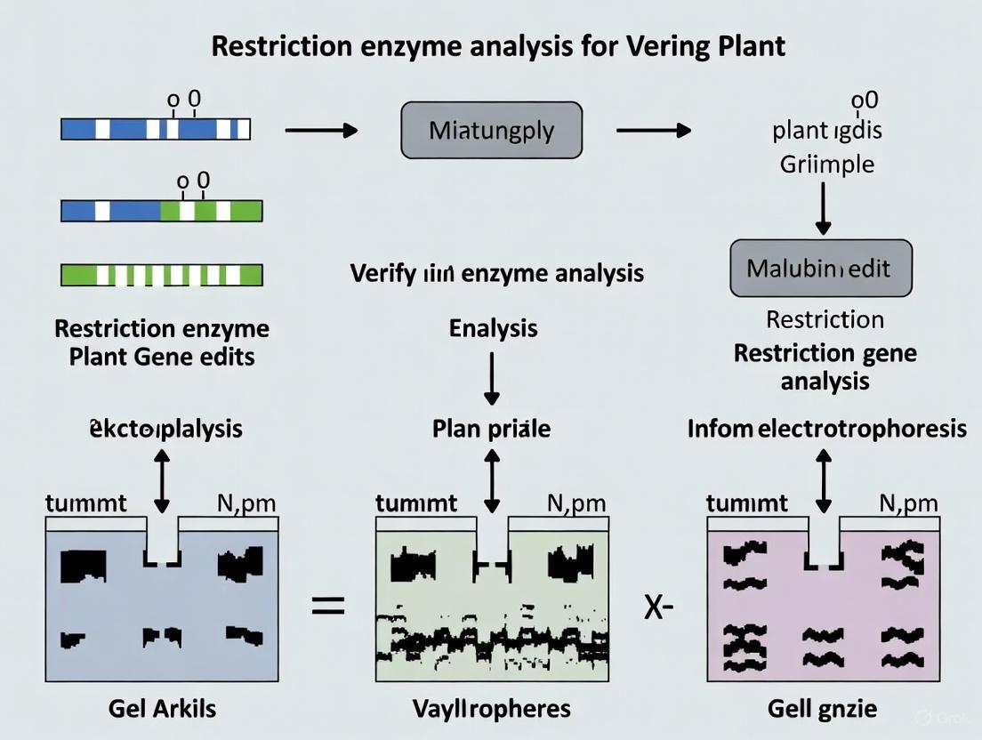
A Practical Guide to Restriction Enzyme Analysis for Verifying Plant Gene Edits
This article provides a comprehensive guide for researchers and scientists on using restriction enzyme analysis to verify CRISPR-Cas9 and other gene edits in plants.
A Practical Guide to Restriction Enzyme Analysis for Verifying Plant Gene Edits
Abstract
This article provides a comprehensive guide for researchers and scientists on using restriction enzyme analysis to verify CRISPR-Cas9 and other gene edits in plants. It covers foundational principles, step-by-step methodologies, advanced troubleshooting for common challenges, and a comparative analysis with modern sequencing-based validation techniques. By synthesizing current best practices and benchmarking data, this resource aims to equip professionals with the knowledge to accurately and efficiently confirm genome edits, thereby accelerating the development of improved crop traits and sustainable agricultural solutions.
The Role of Restriction Enzymes in the Modern Gene Editing Workflow
Restriction Fragment Length Polymorphism (RFLP) is a foundational molecular biology technique that exploits variations in homologous DNA sequences to detect genetic changes. In the context of modern plant genomics, RFLP analysis serves as a crucial verification tool for researchers using sequence-specific nucleases such as CRISPR/Cas9, TALENs, and ZFNs. These technologies create double-strand breaks at predetermined genomic locations, triggering DNA repair mechanisms that often result in small insertions or deletions (indels). RFLP provides a robust, cost-effective method for confirming the success of these editing procedures by detecting changes in restriction enzyme recognition sites caused by targeted mutations. This guide explores the core principles of RFLP analysis, its specific application in plant gene edit verification, and objectively compares its performance with contemporary alternatives, providing researchers with comprehensive experimental data to inform their methodological choices.
The Fundamental Principle of RFLP Analysis
The core principle of RFLP rests on detecting changes in the length of DNA fragments generated by restriction enzyme digestion. Restriction endonucleases are bacterial enzymes that recognize and cut DNA at specific palindromic sequences, typically 4-8 base pairs in length. When a DNA sequence is altered through genome editing—whether by natural mutation or targeted interventions—these changes can create, abolish, or modify restriction enzyme recognition sites. The technique is particularly effective for identifying single-nucleotide polymorphisms (SNPs) or small indels that affect restriction enzyme recognition sites [1].
In practice, RFLP analysis involves digesting DNA samples with specific restriction enzymes, separating the resulting fragments by size using gel electrophoresis, and visualizing them to identify length variations. For plant gene editing applications, this principle is often implemented through PCR-RFLP (also known as Cleaved Amplified Polymorphic Sequence or CAPS), where the target region is first amplified by PCR before restriction digestion [1]. This modification significantly enhances sensitivity and reduces the time and DNA quantity requirements compared to traditional RFLP, which directly digests genomic DNA.
RFLP Workflow in Plant Gene Edit Verification
The following diagram illustrates the standard RFLP workflow for detecting CRISPR/Cas9-induced mutations in plants, integrating both traditional and PCR-based approaches:
Detailed Experimental Protocol
Sample Preparation and DNA Extraction
- Extract genomic DNA from plant tissues (leaf punches approximately 100 mg) using CTAB or commercial kit methods [2] [3]
- Assess DNA quality and quantity using spectrophotometry (A260/A280 ratio of 1.8-2.0) and confirm integrity by gel electrophoresis
- For PCR-RFLP, dilute DNA to working concentration of 10-50 ng/μL for amplification
PCR Amplification (PCR-RFLP Protocol)
- Design primers flanking the target edit region with amplicon size of 300-1000 bp for optimal resolution
- Prepare 25-50 μL reaction mixtures containing: 1X PCR buffer, 1.5-2.5 mM MgCl₂, 0.2 mM dNTPs, 0.2-0.5 μM each primer, 1-2 units DNA polymerase, and 50-100 ng template DNA
- Use thermal cycling parameters: initial denaturation at 95°C for 3-5 min; 30-35 cycles of 95°C for 30s, primer-specific Tm for 30s, 72°C for 1 min/kb; final extension at 72°C for 5-10 min [2] [4]
- Verify amplification success by running 5 μL PCR product on agarose gel
Restriction Digestion and Analysis
- Select restriction enzymes whose recognition sites overlap or are adjacent to the target edit site [3]
- Prepare 20-30 μL digestion reactions containing: 1X restriction enzyme buffer, 5-10 units restriction enzyme, and 10-20 μL PCR product or 200-500 ng genomic DNA
- Incubate at enzyme-specific temperature (typically 37°C) for 2-4 hours to ensure complete digestion
- Separate fragments using 2-3% agarose gel electrophoresis (for fragments <1 kb) or 6-8% polyacrylamide gel electrophoresis (for higher resolution)
- Visualize using ethidium bromide, SYBR Safe, or silver nitrate staining [4]
- Compare fragment patterns to wild-type controls: successful edits eliminate restriction sites, resulting in different banding patterns
RFLP Performance Comparison with Alternative Edit Detection Methods
The table below summarizes experimental data comparing RFLP against other common genome edit detection techniques, based on systematic benchmarking studies in plant systems:
| Detection Method | Accuracy Range | Sensitivity Limit | Time to Result | Cost per Sample | Key Advantages | Key Limitations |
|---|---|---|---|---|---|---|
| PCR-RFLP | 85-95% [5] | 5-10% heterozygotes [5] | 6-8 hours [1] | Low [5] | Cost-effective, simple equipment needs, clear zygosity determination [1] [5] | Limited to edits affecting restriction sites, moderate sensitivity [6] [5] |
| T7 Endonuclease I (T7E1) | 75-90% [5] | 1-5% [6] [5] | 4-6 hours | Low | Site-agnostic, no requirement for restriction sites [6] | Cannot distinguish homozygous mutants, confounded by SNPs [6] |
| Sanger Sequencing | 95-99% [5] | 15-20% [6] [5] | 1-2 days | Medium | Provides complete sequence information, identifies precise mutations [6] [5] | Lower sensitivity, requires cloning for mixed samples, more expensive [6] |
| Amplicon Sequencing (AmpSeq) | ~99% [5] | 0.1-1% [5] | 2-4 days | High | Gold standard for accuracy and sensitivity, detects all mutation types [5] | Expensive, requires specialized equipment and bioinformatics [5] |
| PCR/RNP | 90-98% [6] | 1% [6] | 4-5 hours | Low-Medium | Highly sensitive, works without restriction sites, useful for polyploid plants [6] | Requires protein expression and purification [6] |
Case Study: RFLP Detection of CRISPR Edits in Rice and Arabidopsis
A landmark study demonstrated RFLP's effectiveness in verifying CRISPR/Cas9-induced mutations in both dicot (Arabidopsis) and monocot (rice) plants [3]. Researchers designed sgRNAs to target genes including BRASSINOSTEROID INSENSITIVE 1 (BRI1), JASMONATE-ZIM-DOMAIN PROTEIN 1 (JAZ1), and GIBBERELLIC ACID INSENSITIVE (GAI) in Arabidopsis, and Rice Outermost Cell-specific gene5 (ROC5), Stromal Processing Peptidase (SPP), and Young Seedling Albino (YSA) in rice.
The experimental design placed target sites within restriction enzyme recognition sequences, enabling RFLP to detect successful edits through disruption of these sites. Following Agrobacterium-mediated transformation, researchers genotyped T0 (rice) and T1 (Arabidopsis) plants by PCR amplification of target regions, restriction digestion with appropriate enzymes, and fragment analysis by gel electrophoresis. Clear undigested bands indicated mutant alleles with disrupted restriction sites [3].
The study reported remarkably high mutation frequencies: 26% to 84% in Arabidopsis and 5% to 84% in rice across different targets, with RFLP successfully identifying both heterozygous and homozygous mutants. Sequencing confirmed these results, revealing predominantly small indels (1-10 bp) at target sites. Notably, RFLP detected complex mutagenesis outcomes, including plants with 2+ different mutant alleles, demonstrating its utility in characterizing the complex mutation patterns often generated by CRISPR editing in plants [3].
The Scientist's Toolkit: Essential Reagents for RFLP Analysis
| Research Reagent | Function | Specific Examples | Application Notes |
|---|---|---|---|
| Restriction Enzymes | Recognize and cut specific DNA sequences | EcoRI, HindIII, BamHI, PstI [1] [7] | Select enzymes whose sites overlap target edit region; methylation-sensitive enzymes (PstI) enrich for single-copy sequences [1] |
| DNA Polymerase | Amplifies target DNA region for PCR-RFLP | High-fidelity polymerases | Reduces amplification errors in target sequence |
| Electrophoresis Matrix | Separates DNA fragments by size | Agarose, polyacrylamide [7] | Agarose (2-3%) for fragments 300-1000 bp; polyacrylamide for higher resolution [7] |
| Detection Reagents | Visualizes separated DNA fragments | Ethidium bromide, SYBR Safe, silver nitrate [4] | Silver nitrate staining increases sensitivity for low-abundance fragments [4] |
| Probe DNA (Traditional RFLP) | Hybridizes to specific sequences for detection | Single- or low-copy genomic DNA, cDNA clones [1] | 500-2000 bp fragments; labeled with radioactivity or fluorescence [1] |
Strategic Considerations for Method Selection
When incorporating RFLP into plant gene editing workflows, researchers should consider several strategic factors. For high-throughput screening or when analyzing edits not affecting restriction sites, T7E1 or PCR/RNP methods may be preferable [6] [5]. When precise sequence characterization is required or for analyzing highly heterogeneous editing outcomes, amplicon sequencing remains the gold standard despite higher costs [5]. RFLP excels in foundational research environments with budget constraints, for rapid preliminary screening, and when monitoring known edits specifically designed to alter restriction sites [1] [3].
The choice between traditional RFLP and PCR-RFLP depends on experimental needs. Traditional RFLP provides comprehensive genomic analysis but requires large DNA amounts (micrograms) and extended processing times (weeks) [8] [9]. PCR-RFLP offers significantly improved speed (hours), requires minimal DNA (nanograms), and provides sufficient sensitivity for most edit verification applications, making it particularly suitable for plant transformation pipelines where rapid genotyping of numerous transformants is essential [1] [3].
Restriction Enzymes as a Foundational Tool in a Multi-Method Validation Strategy
The advent of CRISPR/Cas9 technology has revolutionized plant genetics, enabling precise genome editing for gene validation and trait improvement. However, the success of these interventions hinges on accurate verification of the intended edits. Validation strategies often employ a multi-method approach to ensure reliability and comprehensiveness. Within this framework, restriction enzyme analysis emerges as a foundational, rapid, and cost-effective tool for the initial screening of editing events. This guide objectively compares the performance of restriction enzyme-based validation with other established alternatives, supported by experimental data and detailed protocols, providing plant gene edit researchers with a clear pathway for effective verification.
The Scientist's Toolkit: Essential Research Reagents
The following table details key reagents and their functions essential for performing genome editing validation using restriction enzymes and other comparative methods.
Table 1: Essential Research Reagents for Genome Edit Validation
| Reagent / Tool | Primary Function in Validation |
|---|---|
| Restriction Endonucleases | Enzyme that cuts DNA at specific sequences; used to detect edits that alter these sites [7]. |
| PCR Reagents | Amplifies the target genomic region from edited plant cells for subsequent analysis [10]. |
| Agarose Gel Electrophoresis System | Separates DNA fragments by size, allowing visualization of cleavage patterns or insertions/deletions [11]. |
| Sanger Sequencing Reagents | Determines the exact nucleotide sequence of the edited target locus to confirm the mutation [10]. |
| Next-Generation Sequencing (NGS) | Provides a comprehensive, quantitative assessment of editing efficiency and off-target effects across the genome [11]. |
| CRISPR/Cas9 Ribonucleoprotein (RNP) | Pre-complexed guide RNA and Cas9 protein used for in vitro cleavage assays to validate sgRNA efficiency before stable transformation [10]. |
Comparative Performance of Validation Methods
Validation methods offer different advantages in terms of information depth, throughput, and cost. The choice of method often depends on the nature of the edit and the stage of the research pipeline.
Table 2: Quantitative Comparison of Genome Edit Validation Methods
| Method | Best For | Key Metric: Editing Efficiency Assessment | Detection of Off-Target Effects | Relative Cost | Typical Workflow Time |
|---|---|---|---|---|---|
| Restriction Enzyme Analysis | Initial screening of small, specific knock-ins that create or destroy a restriction site [11]. | Semi-quantitative (band intensity on gel) [11]. | No | $ | 1-2 days |
| TIDE (Tracking of Indels by Decomposition) | Quantifying a spectrum of indel mutations in a bulk cell population [11]. | Quantitative (provides a precise percentage of editing efficiency and indel distribution) [11]. | No | $$ | 2-3 days |
| Fragment Analysis (Gel Electrophoresis) | Detecting large deletions or insertions when using a dual-guide CRISPR system [11]. | Semi-quantitative (presence/absence and size of bands) [11]. | No | $ | 1-2 days |
| Next-Generation Sequencing (NGS) | Comprehensive profiling of on-target edits and genome-wide off-target screening [11]. | Highly quantitative and precise (sequences thousands of alleles) [11]. | Yes [11] | $$$$ | 3-5 days (plus data analysis) |
Experimental Protocols for Key Validation Methods
Restriction Enzyme Screening for Knock-In Mutations
This protocol is ideal for verifying small, specific knock-in mutations, such as single nucleotide changes or small tags, that are designed to alter a restriction enzyme recognition site [11].
Detailed Methodology:
- Primer Design and PCR: Design primers that flank the edited genomic region, ensuring the amplicon is small enough to easily visualize a size shift (a good rule of thumb is to keep the product-to-knock-in size ratio below 10:1) [11]. Amplify the target region from both edited and wild-type plant DNA using a high-fidelity PCR enzyme.
- Restriction Digest: Digest the purified PCR products with the appropriate restriction enzyme. The edit should either confer resistance to digestion (if a site is destroyed) or create a new site (resulting in cleavage) [11].
- Analysis: Separate the digested fragments using agarose gel electrophoresis. Compare the banding pattern of the edited sample to the wild-type control. A successful edit will show a different pattern, such as an uncut band where the wild-type is cut, or vice-versa.
- Sequencing Confirmation: As with all methods, sequence confirmation is ultimately required to verify the correct sequence was inserted [11].
TIDE (Tracking of Indels by Decomposition) Analysis for Knockouts
TIDE is a powerful method for the rapid and quantitative analysis of knockout mutations in a bulk population of cells, providing insight into editing efficiency before moving to clonal isolation [11].
Detailed Methodology:
- PCR Amplification: Amplify the target region from both wild-type and edited cell populations. The amplicon should have at least ~200 base pairs of sequence flanking the edit site on either side [11].
- Sanger Sequencing: Sanger sequence the PCR products from both samples.
- Data Analysis: Upload the sequencing trace files from the wild-type and edited samples, along with the sgRNA sequence, to the online TIDE tool (https://tide.nki.nl). The software decomposes the complex trace file from the edited population and generates a graph representing all identified insertions and deletions and their frequencies, providing an estimated editing efficiency [11].
Next-Generation Sequencing for Comprehensive Analysis
NGS is the gold standard for a complete picture of editing outcomes, including precise mutation sequences and potential off-target effects [11].
Detailed Methodology:
- Library Preparation: Amplify the target region(s) from edited and control (untransformed) plant DNA. For off-target analysis, amplify genomic regions predicted to be the most likely off-target sites using in silico tools like CRISPOR or CRISPRitz [11].
- Sequencing and Data Analysis: Sequence the amplicon libraries on an NGS platform. Use specialized software, such as CRISPResso, to align the sequencing reads from your edited sample to the control sequence. This analysis quantifies the types and frequencies of on-target edits and can identify low-frequency mutations at off-target sites [11].
Visualizing the Validation Strategy Workflow
The following diagram illustrates a logical, multi-tiered validation workflow that integrates restriction enzyme analysis with other methods for comprehensive verification of plant gene edits.
In a robust, multi-method strategy for verifying plant gene edits, restriction enzyme analysis serves as a rapid and accessible foundational tool, particularly for specific knock-in mutations. Its strength lies in its low cost and simplicity for initial screening. However, as the comparative data shows, its limitations in quantification and scope mean it is best deployed as part of a sequential protocol. For a complete validation picture, restriction enzyme screening should be followed by more powerful quantitative methods like TIDE for knockouts or, ultimately, NGS for a comprehensive analysis of on-target and off-target effects, ensuring the highest confidence in genome editing outcomes.
Restriction Fragment Length Polymorphism (RFLP) represents one of the earliest molecular techniques for genetic analysis, pioneered in 1984 by Alec Jeffreys [9]. This method exploits variations in DNA sequences through differential digestion by restriction endonucleases, which cut DNA at specific recognition sites [9]. The resulting fragments are separated by gel electrophoresis to create unique fingerprint patterns that can distinguish between individuals, species, or genetic variants [9]. While largely superseded by newer technologies in many applications, RFLP maintains relevance in specific research contexts, including plant gene edit verification [5]. This guide objectively evaluates RFLP's performance against contemporary alternatives, providing experimental data and protocols to help researchers determine its appropriate role in modern plant genetic research.
The fundamental RFLP procedure involves several sequential steps. First, DNA is extracted and purified from the target organism [9]. Next, restriction endonucleases—enzymes that recognize and cut specific 4-6 base pair sequences—digest the purified DNA into fragments of varying lengths [9]. These fragments are then separated by size via gel electrophoresis, where an electric field propels negatively charged DNA molecules through a gel matrix, with smaller fragments migrating faster than larger ones [9]. Finally, the separated DNA fragments are visualized using luminescent dyes, creating a banding pattern that serves as the analytical readout [9].
In contemporary plant research, RFLP is often coupled with polymerase chain reaction (PCR) amplification, creating the PCR-RFLP (also known as cleaved amplified polymorphic sequences) method that improves sensitivity and reduces sample requirements [12] [13]. This modified approach begins with PCR amplification of target gene regions using specific primers, followed by restriction digestion of the amplicons and electrophoretic separation [12]. For example, an improved PCR-RFLP method was developed to identify 41 holotypes of cry1-type genes in Bacillus thuringiensis strains toxic to lepidoptera [12]. The workflow utilizes specific primer sets to divide cry1-type genes into subgroups, followed by digestion with the HinfI restriction enzyme and analysis of fragment patterns on agarose gels [12].
The following diagram illustrates a generalized PCR-RFLP workflow for plant gene edit verification:
Comparative Performance Analysis
When benchmarked against modern genome editing quantification techniques, RFLP demonstrates both strengths and limitations. A comprehensive 2025 study systematically evaluated methods for quantifying CRISPR edits in plants, providing contemporary performance data [5]. The research compared PCR-RFLP against targeted amplicon sequencing (AmpSeq), T7 endonuclease 1 (T7E1) assay, Sanger sequencing with decomposition algorithms, PCR-capillary electrophoresis/InDel detection by amplicon analysis (PCR-CE/IDAA), and droplet digital PCR (ddPCR) [5]. The findings revealed significant methodological differences in quantification accuracy, sensitivity, and practical implementation.
Table 1: Performance Benchmarking of Genome Edit Quantification Methods
| Method | Accuracy vs. AmpSeq | Sensitivity | Cost | Throughput | Technical Complexity |
|---|---|---|---|---|---|
| AmpSeq | Gold Standard [5] | High [5] | High [5] | Moderate [5] | High [5] |
| PCR-RFLP | Moderate [5] | Moderate [5] | Low [5] | High [5] | Low [5] |
| T7E1 | Low-Moderate [5] | Moderate [5] | Low [5] | High [5] | Low [5] |
| Sanger + ICE | Moderate (varies with base caller) [5] | Low for rare edits [5] | Moderate [5] | Moderate [5] | Moderate [5] |
| PCR-CE/IDAA | High [5] | High [5] | Moderate-High [5] | High [5] | Moderate [5] |
| ddPCR | High [5] | High [5] | High [5] | Moderate [5] | High [5] |
The same study highlighted that PCR-CE/IDAA and ddPCR methods demonstrated the highest accuracy when benchmarked against AmpSeq, while PCR-RFLP and T7E1 assays showed more variability in quantification, particularly for edits at lower frequencies [5]. However, the research also noted that base calling algorithms significantly impact the sensitivity of Sanger sequencing-based methods for low-frequency edits [5].
In applied settings, RFLP continues to demonstrate utility in specific research contexts. A novel PCR-RFLP method for detecting Mycobacterium tuberculosis complex in broth cultures achieved 98.18% sensitivity and 99.31% specificity, rivaling more expensive gene chip technologies [14]. Similarly, PCR-RFLP effectively identified genetically modified components in puffed cereal products, detecting CaMV P35S in 24.3% of samples, NPT II in 27%, and MON 810 in 38.8% of 384 tested samples [4].
Advantages of RFLP in Contemporary Research
Cost-Effectiveness and Accessibility
RFLP remains one of the most economically viable techniques for genetic analysis, particularly in resource-constrained environments [14]. The method requires only standard laboratory equipment—thermal cyclers, gel electrophoresis apparatus, and UV transilluminators—already available in most molecular biology laboratories [9]. This contrasts with advanced techniques like AmpSeq and ddPCR that necessitate specialized instrumentation and expensive reagents [5]. The recent development of novel PCR-RFLP methods for tuberculosis detection specifically highlighted the "economical advantage" and suitability for "resource-limited settings" [14].
Technical Simplicity and Reliability
The straightforward protocol of RFLP makes it particularly valuable for laboratories with limited molecular expertise [9]. The technique provides robust, visual results through gel banding patterns that are easily interpreted without complex bioinformatics pipelines [13]. This simplicity facilitates rapid implementation and troubleshooting. In plant research, PCR-RFLP of the plastid trnL UAA intron has been successfully used to identify root fragments from grass swards, enabling studies of rhizosphere ecology where morphological identification is impossible [13]. The method discriminated 14 common grassland species using length heterogeneity and a maximum of two restriction digests [13].
Established Validation and Reproducibility
With decades of use across various disciplines, RFLP protocols are thoroughly optimized and validated for numerous applications [12]. This extensive historical data provides strong reference standards for interpreting results. In crop breeding, RFLP linkage maps continue to provide valuable frameworks for selecting desirable genes via their linkage to easily detectable markers [15]. The technique enables researchers to "expedite the movement of desirable genes among varieties" and "analyze complex polygenic characters as ensembles of single Mendelian factors" [15].
Limitations and Methodological Constraints
Technical Limitations
Table 2: Key Technical Limitations of RFLP and Potential Workarounds
| Limitation | Impact on Research | Potential Workarounds |
|---|---|---|
| Requires specific restriction sites | Cannot detect edits that don't alter restriction sites [9] | Use multiple enzymes; combine with other methods |
| Lower sensitivity | Limited detection of edits <5% frequency [5] | Digital PCR or sequencing for low-frequency edits |
| Low throughput | Time-consuming for large-scale screens [9] | Reserve for validation; use for targeted analysis |
| Large DNA requirement | Challenging for limited samples [9] | PCR-RFLP reduces sample requirement [12] |
| Inability to detect precise sequence changes | Cannot characterize exact edit sequences [9] | Sanger sequencing for precise characterization |
Competition from Advanced Methodologies
Next-generation sequencing techniques, particularly targeted amplicon sequencing (AmpSeq), have become the gold standard for quantifying genome editing outcomes due to their high sensitivity, accuracy, and ability to characterize exact sequence changes [5]. While RFLP remains adequate for detecting homozygous edits or edits present at high frequency, it struggles with detecting low-frequency heterozygous edits in complex samples [5]. Digital PCR methods provide absolute quantification without reference standards and can detect rare mutations below 1% frequency, significantly outperforming RFLP for sensitive applications [5].
The multi-step RFLP process requires numerous manual manipulations and takes considerably longer than modern alternatives to yield results [9]. Additionally, the technique demands relatively large amounts of high-quality DNA compared to PCR-based methods that can amplify minute amounts of DNA [9]. These limitations have led to RFLP being largely replaced for many applications, with one source noting it has "become almost obsolete with the advent of relatively simple and less expensive DNA profiling technologies such as PCR" [9].
Essential Research Reagent Solutions
Table 3: Key Research Reagents for PCR-RFLP Experiments
| Reagent/Category | Specific Examples | Function in Experiment |
|---|---|---|
| Restriction Enzymes | HinfI [12], BfaI, NlaIV, DdeI, XmnI, Hsp92II [13] | Digest PCR amplicons at specific recognition sites to generate polymorphic fragments |
| PCR Components | Taq DNA polymerase, dNTPs, specific primers (e.g., CRY1P1/CRY1PR) [12] | Amplify target DNA regions of interest prior to restriction digestion |
| Electrophoresis Materials | Agarose, TBE buffer, DNA size ladders, fluorescent dyes (ethidium bromide, SYBR Safe) | Separate DNA fragments by size for visualization and analysis |
| DNA Extraction Kits | Commercial plant DNA extraction kits | Isolate high-quality DNA from plant tissues with minimal inhibitors |
| Positive Controls | Plasmids with known restriction sites, DNA from confirmed edited lines | Validate enzyme activity and provide reference fragment patterns |
Strategic Implementation in Plant Gene Edit Verification
RFLP finds optimal application in plant gene editing research as a secondary validation tool rather than a primary screening method. Its most appropriate uses include:
Rapid Validation of Homozygous Edits: For well-characterized edits that introduce or remove specific restriction sites, RFLP provides cost-effective confirmation of homozygous edits in stable plant lines [12]. The method efficiently distinguishes between wild-type, heterozygous, and homozygous genotypes when the edit alters a restriction enzyme recognition sequence.
Educational and Training Contexts: The technical simplicity and visual nature of RFLP make it ideal for teaching core concepts in molecular biology and genetic analysis [9]. Students benefit from the direct observation of genotype-phenotype relationships through gel banding patterns.
Preliminary Screening in Resource-Limited Settings: When budget constraints prohibit extensive sequencing, RFLP offers a viable alternative for initial screening, with positive results confirmed by more specific methods [14]. This tiered approach optimizes resource allocation while maintaining analytical rigor.
Quality Control in Large-Scale Production: For industrial applications involving known genetic constructs, such as GMO detection, RFLP provides reliable quality control at reduced costs [4]. The technique effectively monitors for specific genetic elements like the cauliflower mosaic virus 35S promoter (CaMV P35S) or neomycin phosphotransferase II (NPT II) in agricultural products [4].
RFLP maintains a niche role in the contemporary molecular biology toolkit, balancing clear limitations against distinct advantages of cost-effectiveness, technical simplicity, and reliability. While advanced methods like amplicon sequencing and digital PCR now provide superior sensitivity and precision for characterizing genome edits, RFLP retains value for specific applications in plant genetic research. Its optimal implementation involves strategic deployment as a validation tool for known edits, an educational resource, and a screening method in resource-constrained environments. As the field of plant gene editing continues to evolve, understanding the appropriate context for RFLP application ensures researchers can maximize experimental efficiency while maintaining scientific rigor.
The verification of genome edits in plants is a critical process that spans from initial screening of transformed cells to the final validation of stable, transgene-free lines. Within this pipeline, restriction enzyme analysis has long served as a foundational tool, prized for its accessibility, cost-effectiveness, and rapid turnaround. However, the evolving complexity of CRISPR-Cas9 applications, including multiplex editing and large DNA rearrangements, demands a clear understanding of where traditional methods excel and where next-generation techniques are required. This guide objectively compares the performance of restriction enzyme-based methods with contemporary alternatives, providing experimental data and protocols to inform researchers' choices for plant gene edit verification.
Performance Benchmarking of Edit Verification Methods
A 2025 systematic benchmarking study compared techniques for quantifying CRISPR edits in plants, using targeted amplicon sequencing (AmpSeq) as the reference standard. The following table summarizes key performance metrics across the evaluated methods [5].
Table 1: Performance Comparison of CRISPR Edit Quantification Methods
| Method | Accuracy vs. AmpSeq | Sensitivity | Cost & Throughput | Key Applications in Plant Workflow |
|---|---|---|---|---|
| PCR-RFLP | Moderate | Low (Limited by natural restriction sites) | Low cost, Medium throughput | Initial screening of T0/T1 generations; detecting biallelic mutations [5] [16] |
| T7 Endonuclease I (T7E1) | Moderate to Low | Low (Inefficient heteroduplex detection) | Low cost, Medium throughput | Initial mutation detection in pooled samples [5] |
| Sanger Sequencing + Deconvolution | Varies (Depends on algorithm and base caller) | Low to Medium (>5% allele frequency) | Medium cost, Low to Medium throughput | Quick assessment of editing efficiency; small sample sets [5] |
| Droplet Digital PCR (ddPCR) | High | High (Detects <1% allele frequency) | High cost, High throughput | Accurate zygosity determination; sensitive detection in complex samples [5] |
| PCR-Capillary Electrophoresis | High | High | High cost, High throughput | Precise indel size characterization; high-throughput genotyping [5] |
| Targeted Amplicon Sequencing | Gold Standard | Very High (Detects <0.1% allele frequency) | Highest cost, Highest throughput (multiplexed) | Comprehensive profiling of all edit types; off-target analysis; final validation [5] |
Experimental Protocols for Key Methods
PCR-Restriction Fragment Length Polymorphism (RFLP) Assay
This classic method detects edits that disrupt a native restriction enzyme site at the target locus [16].
Workflow:
- DNA Extraction: Isolate genomic DNA from control (wild-type) and putative edited plant tissue.
- PCR Amplification: Design primers flanking the CRISPR target site (amplicon size 500-1200 bp). Amplify the region from both wild-type and test samples [17].
- Restriction Digestion: Digest the purified PCR products with the appropriate restriction enzyme whose recognition site overlaps the target sequence.
- Gel Electrophoresis: Analyze the digested fragments by agarose gel electrophoresis.
- Wild-type result: The PCR product is cleaved, producing two or more smaller fragments.
- Edited result: A mutated allele loses the restriction site, resulting in an undigested PCR product of full length [16].
Data Interpretation: The presence of the undigested band confirms a mutation. Zygosity can be inferred: a homozygous mutant shows only the undigested band, a heterozygous mutant shows both digested and undigested bands, and a homozygous wild-type shows only digested bands [16].
Cas9-Based Restriction Assay for Genotyping
A modern adaptation uses purified SpCas9 protein and in vitro transcribed sgRNA as a programmable restriction enzyme to digest PCR amplicons, which is highly flexible as it does not rely on pre-existing restriction sites [18].
Workflow:
- RNP Complex Formation: Incubate SpCas9 nuclease with sgRNA targeting the original genomic sequence to form a ribonucleoprotein (RNP) complex.
- Target Amplification: PCR-amplify the target region from edited plants.
- In Vitro Digestion: Treat the purified PCR amplicons with the pre-formed RNP complex.
- Gel Analysis: Run the products on a gel. The wild-type amplicon is cleaved by Cas9, while a successfully edited amplicon (with a disrupted target sequence) remains uncut [18].
Advanced Validation with Long-Range Sequencing
For final, comprehensive validation—especially to detect large on-target rearrangements—targeted long-read sequencing is recommended. Methods like Xdrop indirect sequence capture can enrich ~100 kb regions using a single primer pair, enabling the discovery of unintended large deletions or insertions that are often missed by standard PCR-based assays [19].
Workflow:
- Design Primers: Design a single primer pair flanking a very large region (up to 100 kb) encompassing the edit site.
- Target Enrichment: Use the Xdrop system for microfluidic-based enrichment of this megabase-sized DNA fragment.
- Long-Read Sequencing: Sequence the enriched DNA on a platform like PacBio or Oxford Nanopore.
- Data Analysis: Assemble the long reads to identify not only small indels but also large structural variations and complex rearrangements at the target locus [19].
The Scientist's Toolkit: Essential Reagents and Materials
Table 2: Key Research Reagent Solutions for Edit Verification
| Reagent / Material | Function in Workflow | Specific Examples & Notes |
|---|---|---|
| Proofreading DNA Polymerase | High-fidelity amplification of target loci for sequencing and RFLP. | Q5 High-Fidelity DNA Polymerase; KOD FX Polymerase [17] [18]. Critical for reducing PCR errors. |
| Restriction Endonucleases | Core reagent for RFLP assays to detect site disruption. | Standard enzymes like Sal I, Xho I, Sbf I [16]. Select based on presence of site in target. |
| SpCas9 Nuclease (NEB) | Used for flexible, site-specific in vitro digestion of PCR amplicons for genotyping [18]. | Enables Cas9-based restriction assay without native restriction site. |
| T7 Endonuclease I | Detects mismatches in heteroduplexed DNA from edited samples [5]. | Part of T7E1 assay. Lower resolution than other methods. |
| ddPCR Supermix | Enables absolute quantification of edit frequency and zygosity without standard curves [5]. | Provides high sensitivity and precision for complex samples. |
| Modular CRISPR Vectors | Delivery of editing machinery for creating stable lines. | pGreen/pCAMBIA backbones with plant resistance markers [20]; RUBY marker systems for visual selection of transgene-free progeny [16]. |
The choice of verification method in plant genome editing is highly dependent on the experimental stage and required resolution. PCR-RFLP and Cas9-based assays offer robust, cost-effective solutions for initial screening and genotyping of early generations (T0/T1). In contrast, ddPCR and PCR-Capillary Electrophoresis provide higher accuracy for quantitative analysis and zygosity determination. For the final validation of stable, transgene-free lines, especially to rule out complex unintended edits, targeted amplicon or long-read sequencing remains the indispensable gold standard. A tiered approach, leveraging the strengths of each technique, creates an efficient and reliable pipeline from initial screening to the validation of edited stable lines.
A Step-by-Step Protocol for PCR-RFLP Analysis in Plants
In the context of verifying plant gene edits, the strategic design of primers to flank the target site and generate an amplicon of optimal size is a critical determinant for the success and accuracy of subsequent analytical methods, such as restriction enzyme analysis. The choice of validation technique directly dictates the required amplicon size, making primer design the foundational step in the experimental workflow.
Comparison of Genome Editing Validation Methods
The selection of a validation method dictates the necessary amplicon size and primer design strategy. The following table summarizes key techniques used for detecting CRISPR-Cas9-induced edits in plants.
| Method | Key Principle | Optimal Amplicon Size | Key Advantages | Key Limitations |
|---|---|---|---|---|
| PCR-Restriction Fragment Length Polymorphism (RFLP) [5] | Loss or gain of a restriction enzyme site due to editing; cleavage of PCR product reveals edit. | Varies by target | Inexpensive; simple protocol if a site is affected [5]. | Limited to edits that alter a specific restriction site [11]. |
| T7 Endonuclease I (T7E1) Assay [5] [21] | Enzyme cleaves heteroduplex DNA formed by reannealing of wild-type and indel-containing strands. | 400-800 bp [21] | Does not require a specific mutation; rapid and inexpensive [5] [21]. | Cannot identify the specific sequence change; sensitivity depends on optimization [11] [21]. |
| Sanger Sequencing + TIDE/TIDER Analysis [11] [5] | Decomposes Sanger sequencing trace files from a mixed population to quantify indel frequency and type. | Amplicon should have ~200 bp flanking the edit site on each side [11]. | Reveals specific indels and their percentages in a bulk population [11]. | Lower sensitivity for very low-frequency edits (<5%) compared to NGS [5]. |
| Targeted Amplicon Sequencing (AmpSeq) [5] [22] | Next-generation sequencing of the target region provides a comprehensive, base-pair-resolution view of all edits. | Varies; often kept within NGS platform read lengths (e.g., 150-500 bp). | "Gold standard" for sensitivity and accuracy; detects all edit types and their exact frequencies [5]. | Higher cost and longer turnaround time; requires specialized data analysis [5]. |
| PCR-Capillary Electrophoresis/IDAA [5] | Detects size shifts in fluorescently labeled amplicons caused by indels. | Varies by system | Accurate and sensitive for quantifying indel sizes; high-throughput capable [5]. | Does not provide sequence-level information, only indel size [5]. |
| Droplet Digital PCR (ddPCR) [5] | Absolute quantification of edited and wild-type alleles using endpoint PCR in thousands of water-in-oil droplets. | Varies by probe/assay design | High sensitivity and precision for quantifying editing efficiency without a standard curve [5]. | Requires specific fluorescent probe design; does not reveal the nature of large indels [5]. |
Experimental Protocols for Key Validation Methods
Here are detailed methodologies for two commonly used techniques in plant gene edit verification.
T7 Endonuclease I (T7E1) Mismatch Cleavage Assay
This protocol is used to detect small insertions or deletions (indels) at the target site [21].
- PCR Amplification: Amplify the target region from genomic DNA (isolated from edited and control plants) using primers designed to produce an amplicon between 400 and 800 base pairs, with the cut site located roughly in the center. Primers should bind approximately 250 bp upstream and downstream of the expected edit site to ensure cleavage products are large enough (>100 bp) to be resolved on a gel [21].
- Heteroduplex Formation: Purify the PCR product. To form heteroduplexes, denature the DNA by heating to 95°C for 5-10 minutes, and then slowly reanneal by ramping the temperature down to 25°C slowly (e.g., -0.1 to -2.0°C per second). This allows strands from different alleles (wild-type and indel-containing) to hybridize, creating mismatches at the site of mutation [21].
- T7E1 Digestion:
- Set up a digestion reaction in a small volume (e.g., 20 µL) containing:
- 100-500 ng of the reannealed PCR product
- 1X T7E1 reaction buffer
- 1-2 units of T7 Endonuclease I enzyme
- Incubate the reaction at 37°C for 15-90 minutes [21]. Optimization of incubation time and enzyme amount may be necessary for maximum cleavage efficiency.
- Set up a digestion reaction in a small volume (e.g., 20 µL) containing:
- Analysis by Gel Electrophoresis: Analyze the digestion products by agarose gel electrophoresis (e.g., 2% agarose gel). A successful edit will be indicated by the presence of two or more cleaved bands in addition to the intact, full-length PCR product. The editing efficiency can be estimated by comparing the band intensities of the cleavage products to the full-length product [21].
Sanger Sequencing and TIDE Analysis
This method uses standard Sanger sequencing followed by free, web-based software analysis to quantify and characterize edits in a heterogeneous cell population [11].
- PCR Amplification and Sequencing: Design primers to amplify the target region, ensuring there is at least ~200 base pairs of sequence flanking the editing site on either side to provide sufficient sequence context for the analysis algorithm. Purify the PCR product and submit it for Sanger sequencing using one of the PCR primers [11].
- Data Collection: Obtain the sequencing trace file (
.ab1file) from the edited plant population and, crucially, a control trace file from an unedited (wild-type) plant. - TIDE Analysis:
- Access the TIDE web tool (available from the developers).
- Upload the two sequencing trace files (wild-type and edited).
- Input the sgRNA target sequence exactly, ensuring it is correctly positioned relative to the sequencing chromatogram.
- The software will decompose the complex trace file from the edited sample and generate a graph showing the spectrum of insertion and deletion mutations, their sequences, and the overall editing efficiency in the population [11].
The Scientist's Toolkit: Research Reagent Solutions
The following table details essential reagents and their functions for experiments in plant gene editing validation.
| Reagent / Tool | Critical Function |
|---|---|
| High-Fidelity DNA Polymerase [23] | Ensures accurate amplification of the target locus from genomic DNA for downstream analysis, minimizing polymerase-introduced errors. |
| T7 Endonuclease I [21] | Recognizes and cleaves DNA heteroduplexes at mismatch sites, enabling the detection of indel mutations. |
| CRISPOR or CHOPCHOP [5] [24] | Bioinformatics tools for designing and selecting highly efficient guide RNAs (gRNAs) with minimized off-target potential for the editing step. |
| Primer3 & In-Silico PCR (ISPCR) [22] | Accessible bioinformatics tools (integrated in pipelines like CREPE) for designing specific PCR primers and evaluating their potential for off-target binding across the genome. |
| OligoAnalyzer Tool [25] | Analyzes oligonucleotide properties such as melting temperature ((T_m)), hairpins, self-dimers, and heterodimers to ensure high-quality primer design. |
Workflow for Validating Plant Gene Edits
The diagram below illustrates the core experimental workflow for verifying genome edits in plants, from initial design to final validation, highlighting how primer design and amplicon size are integral to each step.
Essential Guidelines for Primer Design
Adhering to established primer design rules is crucial for efficient and specific amplification, which underpins all validation methods [23] [26] [25].
| Design Parameter | Optimal Guideline | Rationale |
|---|---|---|
| Primer Length | 18 - 30 nucleotides [26] [25] | Balances specificity (longer) with hybridization efficiency and yield (shorter) [26]. |
| Melting Temperature ((T_m)) | 60-64°C; forward and reverse primers should be within 2°C [25] or 5°C [23] of each other. | Ensures both primers bind to the target simultaneously and efficiently during the annealing step. |
| GC Content | 40% - 60% [23] [26] [25] | Prevents overly weak (low GC) or strong (high GC) binding, which can lead to nonspecific amplification or secondary structures. |
| 3' End (GC Clamp) | Avoid runs of 3 or more G/C residues [26]. | Prevents non-specific binding at the 3' end, which is critical for primer extension. |
| Secondary Structures | Avoid self-dimers, cross-dimers, and hairpins [23] [25]. | Prevents primers from annealing to themselves or each other instead of the target template, which reduces PCR yield. |
In summary, the verification of plant gene edits through restriction enzyme analysis or any other molecular method is profoundly dependent on careful primer design. By flanking the target site to create an amplicon of the size optimal for the chosen validation technique and adhering to fundamental primer design principles, researchers can ensure robust, reliable, and interpretable results in their genome editing workflows.
In the context of restriction enzyme analysis for verifying plant gene edits, the isolation of high-quality, PCR-ready DNA is a foundational prerequisite. The accuracy of downstream analytical techniques, including PCR and enzymatic digestion, is entirely dependent on the purity and integrity of the extracted template DNA. Plant tissues present unique challenges for DNA extraction due to their high content of polysaccharides, polyphenols, and secondary metabolites that can co-purate with nucleic acids and inhibit enzymatic reactions essential for molecular analysis [27]. These contaminants can compromise restriction enzyme activity, leading to false negatives or inaccurate results during the verification of edited gene sequences. This guide provides an objective comparison of DNA extraction methods, supported by experimental data, to enable researchers to select the optimal approach for their plant gene editing verification workflow.
Method Comparison: CTAB vs. Commercial Kits
Selecting an appropriate DNA extraction method is critical for balancing yield, purity, practical efficiency, and cost. The following table summarizes the performance of common extraction methods based on comparative studies.
Table 1: Performance Comparison of Plant DNA Extraction Methods
| Method | Average Yield | Purity (A260/A280) | DNA Integrity | Cost per Sample | Best For |
|---|---|---|---|---|---|
| CTAB (Traditional) | High (e.g., 3-85 µg from 50 mg grapevine leaf) [27] | 1.5-1.8 [28] [29] | High (70-85% high-molecular-weight DNA) [27] | Low [27] | High-yield, high-purity needs; polyphenol-rich tissues; cost-sensitive projects [28] [27] |
| CTAB + PVP | Moderate (lower than CTAB) [27] | Improved purity for polyphenol-rich tissues [27] | High (comparable to CTAB) [27] | Low [27] | Tissues with high polyphenol content (e.g., mature leaves, woody stems) [27] |
| Qiagen DNeasy Kit | Moderate (e.g., 3-30 µg from 50 mg tissue) [30] | High, symmetrical A260 peak [30] | Moderate (40-60% high-molecular-weight DNA) [27] | High [27] | High-throughput applications; routine PCR; fast, reproducible results [27] [30] |
| SPINeasy MP Kit | High [27] | Variable [27] | Low (<10% high-molecular-weight DNA) [27] | High [27] | Applications where fragmentation is not a concern [27] |
| peqGOLD VWR Kit | Low [27] | Lower purity [27] | Information Missing | High [27] | Information Missing |
Key Experimental Findings from Comparative Studies
A 2025 study directly compared two CTAB-based methods and three commercial kits for DNA extraction from grapevine leaves, providing critical performance data [27]. The research concluded that while all protocols generated DNA sufficient for PCR amplification, CTAB-based methods provided the highest yields and purity at a low cost, with densitometry showing approximately 70–85% high-molecular-weight DNA (>20 kb) [27]. The Qiagen kit yielded reproducible results with moderate integrity (about 40–60% high-molecular-weight fraction), making it suitable for high-throughput applications where speed and consistency are prioritized [27]. Notably, the addition of PVP to the CTAB buffer significantly improved DNA purity when processing polyphenol-rich tissues, though it resulted in a reduced yield [27].
Detailed Experimental Protocols
The CTAB-Based Extraction Method
The CTAB method is a well-established, robust protocol for isolating high-quality DNA from a wide range of plant species.
Table 2: Key Reagents for CTAB DNA Extraction
| Reagent | Function |
|---|---|
| CTAB (Cetyltrimethylammonium bromide) Buffer | A detergent that disrupts cell and nuclear membranes, forming complexes with DNA in low-salt conditions [27]. |
| Polyvinylpyrrolidone (PVP) | Binds to and removes polyphenolic contaminants [27]. |
| Beta-Mercaptoethanol | A reducing agent that prevents oxidation of polyphenols, which can darken DNA and inhibit enzymes [27]. |
| Chloroform:Isoamyl Alcohol | Organic solvent used to separate DNA from proteins and polysaccharides after cell lysis [27]. |
| Isopropanol | Precipitates nucleic acids from the aqueous phase [27]. |
| Ethanol (70%) | Washes and de-salts the DNA pellet [27]. |
Procedure Steps [28] [29] [27]:
- Tissue Disruption: Grind approximately 1.0 cm² of fresh or frozen leaf tissue to a fine powder in liquid nitrogen using a mortar and pestle.
- Cell Lysis: Transfer the powder to a microfuge tube containing pre-warmed (65°C) CTAB extraction buffer (100 mM Tris-HCl, 100 mM EDTA, 250 mM NaCl, 3% CTAB) supplemented with 2% beta-mercaptoethanol and, for polyphenol-rich tissues, 3% PVP [27]. Incubate at 65°C for 30-60 minutes with occasional gentle mixing.
- Organic Extraction: Add an equal volume of chloroform:isoamyl alcohol (24:1), mix thoroughly, and centrifuge to separate the phases. The upper aqueous phase containing the DNA is transferred to a new tube.
- DNA Precipitation: Add 0.6-1.0 volumes of isopropanol to the aqueous phase to precipitate the DNA. Incubate at -20°C for 30 minutes and centrifuge to pellet the DNA.
- DNA Washing: Wash the pellet with 70% ethanol to remove residual salts and centrifuges again. Carefully decant the ethanol and air-dry the pellet.
- Rehydration: Dissolve the pure DNA pellet in TE buffer or nuclease-free water.
Silica Column-Based Kit Protocol
Commercial silica-membrane kits, such as the Qiagen DNeasy Plant Kit, offer a standardized and convenient alternative [30].
Procedure Steps [30]:
- Lysis: Disrupt plant tissue (up to 100 mg) and incubate in a proprietary lysis buffer. RNase A is often added at this stage to degrade RNA.
- Homogenization: Load the lysate onto a QIAshredder spin column and centrifuge. This step removes cell debris and homogenizes the sample, improving subsequent DNA binding.
- DNA Binding: Adjust the buffering conditions of the cleared lysate and load it onto a DNeasy Mini spin column. During a brief centrifugation, DNA selectively binds to the silica membrane, while contaminants pass through.
- Washing: Wash the membrane-bound DNA twice with different wash buffers to remove remaining impurities and enzyme inhibitors.
- Elution: Elute the pure, ready-to-use DNA in water or a low-salt buffer (e.g., AE buffer).
Decision Workflow for Method Selection
The following diagram illustrates the strategic decision-making process for selecting the most appropriate DNA extraction method based on your project's specific requirements.
The Scientist's Toolkit: Essential Reagents and Kits
Table 3: Essential Research Reagents and Kits for Plant DNA Extraction
| Item | Function & Application Notes |
|---|---|
| DNeasy Plant Mini Kit (Qiagen) | Silica-membrane technology for rapid purification without organic solvents. Ideal for PCR and other enzymatic applications from most plant tissues [30]. |
| CTAB Buffer | Core lysis reagent for the traditional method. Must be supplemented with beta-mercaptoethanol and potentially PVP immediately before use [27]. |
| Polyvinylpyrrolidone (PVP) | Essential additive for binding and removing polyphenols when extracting from challenging tissues like grapevine leaves, conifers, or medicinal plants [27]. |
| Beta-Mercaptoethanol | Critical reducing agent to prevent browning and degradation of DNA during lysis. Note: Handle in a fume hood due to toxicity [27]. |
| Chloroform:Isoamyl Alcohol (24:1) | Organic solvent for deproteinization and removal of lipid contaminants in CTAB protocols [28] [27]. |
| RNAse A | Enzyme used to digest RNA during extraction, ensuring the final product is genomic DNA free of RNA contamination [30]. |
| Taq DNA Polymerase | The workhorse enzyme for endpoint PCR, used for amplifying the target gene region from the extracted DNA prior to restriction analysis [31]. |
The verification of plant gene edits through restriction enzyme analysis demands a robust and reliable DNA extraction workflow. While commercial kits offer clear advantages in speed, safety, and reproducibility for high-throughput screening of standard tissues, the traditional CTAB method remains a powerful, cost-effective solution for obtaining high-yield, high-integrity DNA from even the most challenging plant samples. The decision between these methods should be guided by a clear understanding of project goals, sample nature, and resource constraints. By selecting the appropriate extraction strategy as outlined in this guide, researchers can ensure that their foundational template DNA is of the highest quality, thereby guaranteeing the accuracy and reliability of all subsequent gene editing verification analyses.
In plant genomics research, verifying successful gene edits—such as those introduced by CRISPR-Cas9—is a critical step. Among the various validation techniques available, restriction enzyme analysis remains a widely used method due to its accessibility, cost-effectiveness, and rapid turnaround time. This guide objectively compares the core components of a restriction digestion reaction—buffer selection, enzyme quantity, and incubation parameters—with alternative genome editing validation methods, providing supporting experimental data to inform researchers' choices.
Buffer Selection: A Comparative Analysis
The reaction buffer is a decisive factor for complete digestion. Its composition directly influences enzyme activity and specificity. Suppliers typically provide optimized buffers, but selection becomes complex in double digests or with specific DNA substrates.
Table 1: Buffer Selection and Compatibility
| Buffer Consideration | Impact on Reaction | Comparative Performance Data |
|---|---|---|
| Standard Single Buffer | Ensures 100% activity for a specific enzyme; supplied by manufacturer. | Optimal for single-enzyme digests; considered the baseline for performance [32]. |
| Universal Buffers (e.g., rCutSmart) | Enables simultaneous activity of multiple enzymes in one tube. | In double digests, saves significant time without sacrificing efficiency; a key advantage for high-throughput workflows [33]. |
| Non-Optimal Buffer | Can reduce enzymatic activity, leading to incomplete digestion. | Enzyme performance can drop below 50% in sub-optimal buffers, necessitating increased units or incubation time [33]. |
| Sequential Digestion | Used when no single buffer supports >50% activity for both enzymes. | Although it adds steps (incubation, potential purification, second incubation), it guarantees complete digestion for challenging pairs [33]. |
Enzyme Units and Incubation: Optimizing the Reaction
The definitions of enzyme units and incubation times are interconnected. Conventional wisdom defines one unit as the amount of enzyme needed to digest 1 µg of substrate DNA in one hour at 37°C in a 50 µL reaction [32]. However, "fast" enzymes can achieve this in 5-15 minutes. For complete digestion, suppliers often recommend a 5- to 20-fold excess of enzyme (or 1 µL per reaction) to account for variations in DNA quality and quantity [32].
Prolonged incubation is a common strategy to compensate for lower enzyme activity in non-optimal buffers. However, this can increase the risk of star activity, a phenomenon where the enzyme loses specificity and cleaves at non-canonical sites [32]. This is a significant drawback compared to more specific validation methods.
Table 2: Enzyme and Incubation Parameters
| Parameter | Standard Protocol | Effect of Deviation |
|---|---|---|
| Enzyme Units | 1 unit per µg DNA for 1 hour [32]. | Too little: Incomplete digestion. Too much: Risk of star activity, especially with high glycerol concentrations [32]. |
| Incubation Time | 1 hour for diagnostic digests; 4+ hours for cloning [34]. | Too short: Incomplete digestion. Too long: Risk of star activity and sample evaporation [32]. |
| Glycerol Concentration | Should be kept below 5% in the final reaction [33]. | Concentrations >5% can induce star activity; this is a critical consideration when adding multiple enzymes [32]. |
| Reaction Temperature | Typically 37°C; must be constant for "fast" enzymes [32]. | Non-optimal temperatures are a common cause of incomplete digestion [32]. |
Benchmarking Against Alternative Validation Methods
A 2025 systematic study compared techniques for quantifying CRISPR edits in plants, providing a robust benchmark for method performance [5]. The study assessed methods based on their accuracy, sensitivity, and cost, using targeted amplicon sequencing (AmpSeq) as the gold standard.
Table 3: Comparative Performance of Genome Editing Validation Methods
| Validation Method | Reported Accuracy/Sensitivity | Key Advantages | Key Limitations |
|---|---|---|---|
| PCR-RFLP | Lower accuracy compared to AmpSeq, PCR-CE/IDAA, and ddPCR [5]. | Low cost, simple, fast (hours), requires basic lab equipment [5]. | Limited to edits that alter a restriction site; sensitivity affected by reaction completeness [11]. |
| T7 Endonuclease I (T7E1) | Shows differences in quantified edit frequency vs. AmpSeq [5]. | Detects a range of heteroduplex indels without needing a specific site [35]. | Cannot identify the exact sequence change; overlooks single nucleotide changes; sensitivity requires optimization [35]. |
| Sanger Sequencing + TIDE | Accuracy affected by base-calling software; lower sensitivity for low-frequency edits [5]. | Provides sequence-level information; easy to set up with online tools [11]. | Less quantitative for mixed populations than NGS. |
| PCR-CE/IDAA | Highly accurate when benchmarked to AmpSeq [5]. | High sensitivity, quantitative, size-based indel detection. | Requires specialized capillary electrophoresis equipment. |
| Droplet Digital PCR (ddPCR) | Highly accurate when benchmarked to AmpSeq [5]. | Extreme sensitivity, absolute quantification without a standard curve. | Higher cost per reaction, requires specialized equipment. |
| AmpSeq (NGS) | Considered the "gold standard" for sensitivity and accuracy [5]. | Highly sensitive, detects all mutations quantitatively, identifies offtarget effects. | High cost, long turnaround, complex data analysis [5]. |
Experimental Protocols
Protocol 1: Standard Restriction Enzyme Digest for Edit Validation
This protocol is adapted for validating plant gene edits where a CRISPR edit has disrupted or created a restriction enzyme recognition site [11] [34].
- Design: After genome editing, amplify the target region by PCR. Pro-Tip: Ensure the amplicon is large enough (e.g., 400-800 bp) to easily resolve fragments after digestion [35].
- Reaction Setup:
- Combine ~500 ng of PCR product (for diagnostic digests) with the recommended 10x buffer and 1 µL (or a 5-20 fold excess) of the restriction enzyme [32] [34].
- Adjust the volume with nuclease-free water to a total of 30-50 µL.
- Add the enzyme last and mix by gently flicking the tube. Centrifuge briefly to collect the contents [32].
- Incubation: Incubate at the enzyme's optimal temperature (usually 37°C) for 1 hour. For diagnostic digests, this is often sufficient [34].
- Analysis: Run the entire reaction on an agarose gel. A successful edit is indicated by a change in the banding pattern compared to the wild-type control [11].
Protocol 2: T7 Endonuclease I (T7E1) Mismatch Cleavage Assay
This protocol offers an alternative when an edit does not affect a restriction site [35].
- PCR Amplification: Amplify the target region from both edited and control plant DNA using primers that produce a 400-800 bp amplicon. The target site should be roughly centered.
- Heteroduplex Formation: Denature the PCR amplicon at 95°C for 5-10 minutes and then slowly reanneal by ramping down to room temperature slowly. This allows strands from edited and unedited DNA to hybridize, forming heteroduplexes at mismatch sites.
- Digestion: Digest the reannealed DNA with T7E1 enzyme in a small reaction volume, often in a supplied buffer that may include MnCl₂ to enhance efficiency [35].
- Analysis: Resolve the cleavage products by gel electrophoresis. The cleavage fragments indicate the presence of indels, and band intensities can be used to estimate mutation frequency [35].
Workflow Diagram
The following diagram illustrates the logical workflow for selecting a validation method in plant gene editing research, based on project requirements and constraints.
The Scientist's Toolkit
Table 4: Essential Research Reagent Solutions for Restriction Enzyme Validation
| Reagent / Tool | Critical Function | Considerations for Plant Genomics |
|---|---|---|
| High-Fidelity (HF) Restriction Enzymes | Engineered for reduced star activity, even in prolonged incubations. | Essential for validating low-frequency edits in heterogeneous plant populations to avoid false positives from non-specific cleavage [32] [33]. |
| Universal Restriction Buffers | Allows simultaneous activity of multiple enzymes, simplifying double digests. | Saves time when screening multiple edits or using an internal control; compatible with a wide range of enzymes [33]. |
| Single Buffer System Enzymes | Enzymes designed to work in one optimal buffer regardless of DNA substrate. | Reduces optimization steps, which is valuable when processing many samples from different plant lines [32]. |
| Bovine Serum Albumin (BSA) | Stabilizes enzymes and prevents loss of activity on tube surfaces. | Often recommended by manufacturers; its use can be critical for complete digestion of plant genomic DNA [34]. |
| Online Buffer Selection Tools | Digital tools (e.g., NEBcloner, Double Digest Finder) to find optimal buffers for single or double digests. | Ensures reaction efficiency by simulating buffer compatibility, preventing wasted reagents and time [33]. |
| Methylation-Free E. coli Strains | Used for propagating plasmid controls where Dam/Dcm methylation could block digestion. | Critical for creating reliable positive controls, as some plant DNA methylation patterns may also inhibit certain enzymes [34]. |
In plant gene editing research, confirming the success of genetic modifications is a critical step following the application of technologies like CRISPR-Cas9. While advanced sequencing methods provide ultimate confirmation, gel electrophoresis remains a foundational, rapid, and accessible technique for the initial validation of edits. This guide focuses on interpreting DNA banding patterns on agarose gels to verify gene edits, specifically framing this within the established workflow of restriction enzyme analysis. We objectively compare the performance of this traditional method against modern PCR-based and computational tools, providing the experimental data and protocols necessary for researchers to select the optimal verification strategy for their projects.
Gel Electrophoresis and Restriction Analysis in Edit Verification
Restriction Enzyme Analysis (REA) leverages the principle that successful gene edits can alter or abolish restriction endonuclease recognition sites. By comparing digestion patterns of wild-type versus edited DNA, researchers can infer the presence of edits.
- Fundamental Principle: Type II restriction endonucleases are the primary tools for this application, as they recognize stereotypical palindromic sequences and produce a predictable cleavage pattern [36]. When a gene edit (e.g., an indel) disrupts the specific recognition sequence for a restriction enzyme, the enzyme can no longer cut at that site.
- Expected Banding Patterns: The verification relies on a clear shift in the DNA fragment sizes observed on the gel.
- Wild-Type Control: Digestion produces a specific banding pattern based on the known restriction sites.
- Successfully Edited Sample: The loss of a restriction site results in a novel, larger DNA fragment and the disappearance of the smaller fragments that would have been produced from a cut at that site. This produces a distinctly different banding pattern compared to the wild type [37].
- Advantages and Limitations: This method is cost-effective, rapid, and requires equipment standard in molecular biology labs. However, its utility is contingent on the edit fortuitously creating or destroying a restriction site, and it cannot determine the exact sequence of the indel.
Comparative Analysis of Gene Edit Verification Methods
While restriction analysis is a valuable tool, it is one of several methods available for edit confirmation. The table below provides a comparative overview of common techniques, highlighting their key performance metrics.
Table 1: Performance Comparison of Gene Edit Verification Methods
| Method | Principle | Edit Detection Capability | Throughput | Cost | Key Advantages | Key Limitations |
|---|---|---|---|---|---|---|
| Restriction Enzyme Analysis | Loss/gain of restriction site alters fragment size on a gel [37]. | Indirect detection via size change; cannot resolve complex edits. | Low | Low | Rapid, low-cost, uses standard lab equipment. | Limited to edits that alter restriction sites; low resolution. |
| PCR-Based Size Selection | PCR amplification across target site detects large deletions/insertions [38]. | Directly detects large edits visible as PCR product size shifts. | Medium | Low-Moderate | Simple PCR setup; excellent for detecting large, PCR-detectable deletions [38]. | Cannot detect single-base changes or small indels. |
| Computational Deconvolution (TIDE, ICE) | Deconvolutes Sanger sequencing traces from mixed edited/unedited populations [39]. | Quantifies indel frequency and distribution; identifies sequences for simple indels. | Medium | Moderate | Provides quantitative indel frequency from standard Sanger sequencing [39]. | Accuracy declines with highly complex indel mixtures [39]. |
| High-Throughput Sequencing (HTS) | Direct sequencing of PCR amplicons from edited samples. | Gold standard; detects all sequence changes with high accuracy. | High | High | Unparalleled accuracy and capacity to detect all edit types and off-target effects. | Expensive; requires complex data analysis and bioinformatics expertise. |
Experimental data from a systematic tool comparison supports this performance overview. When analyzing simple indels, tools like TIDE, ICE, and DECODR show acceptable agreement. However, as the complexity of the indel mixture increases, the variation in estimated indel frequencies between these tools becomes more pronounced [39]. This underscores the value of HTS for definitive validation when precise sequence data is critical.
Experimental Protocols for Key Verification Methods
Protocol 1: Verification by Restriction Fragment Length Analysis
This protocol is ideal for a quick, initial check of edits that are predicted to alter a known restriction site.
- PCR Amplification: Design primers flanking the edited genomic region and amplify the target site from both wild-type and putative edited plant DNA.
- Restriction Enzyme Digestion:
- Reaction Setup: Combine ~500 ng of purified PCR product, 1X appropriate restriction enzyme buffer, and 10 units of the selected restriction enzyme in a total volume of 20 µL [36].
- Incubation: Incubate at the enzyme's optimal temperature (typically 37°C) for 1 hour.
- Gel Electrophoresis:
- Gel Preparation: Cast a 1-2% agarose gel in 1X TAE or TBE buffer, containing a fluorescent nucleic acid stain.
- Sample Loading: Load the digested samples alongside an undigested PCR product control and an appropriate DNA ladder.
- Electrophoresis: Run the gel at 5-8 V/cm until sufficient separation is achieved.
- Analysis: Visualize the gel under UV light. A successful edit is indicated by the absence of the cleavage band(s) present in the wild-type digested sample and the appearance of a single, larger band corresponding to the uncut PCR product.
Protocol 2: Verification by PCR Screening for Large Deletions
This method is highly effective when using multiplex CRISPR systems designed to excise large segments of DNA [38].
- Primer Design: Design two PCR primers that bind outside the two outermost gRNA target sites in the genome.
- PCR Amplification: Perform PCR on genomic DNA from wild-type and edited plants using the external primers.
- Gel Electrophoresis:
- Gel Preparation: Use a 1% agarose gel for optimal separation of larger DNA fragments.
- Analysis: Compare the PCR product sizes. A significantly smaller PCR product in the edited sample compared to the wild-type indicates a successful large deletion between the two gRNA cut sites [38].
This workflow for detecting large deletions using PCR is illustrated below:
Workflow for PCR-Based Detection of Large Deletions
Essential Research Reagent Solutions
The following table details key reagents and their functions for the experiments described in this guide.
Table 2: Essential Research Reagents for Edit Verification
| Reagent / Tool | Function in Experiment | Specific Example / Note |
|---|---|---|
| Type II Restriction Enzymes | Cuts DNA at specific sequences to analyze edits that disrupt these sites [36]. | High-Fidelity (HF) versions are engineered to minimize star activity for cleaner digests [36]. |
| Agarose | Matrix for gel electrophoresis; separates DNA fragments by size. | Standard agarose for fragments >100 bp; high-resolution agarose for better separation of smaller fragments. |
| DNA Ladder | Molecular weight standard for estimating the size of DNA fragments on a gel. | Essential for confirming the expected size of restriction fragments or PCR products. |
| PCR Reagents | Amplifies the target genomic locus for downstream analysis (digestion or direct sizing). | Use high-fidelity polymerases to minimize PCR-induced errors during amplification. |
| Computational Tools | Analyzes Sanger sequencing data to quantify editing efficiency and indel spectra [39]. | DECODR was shown to provide among the most accurate estimations for a majority of samples in a comparative study [39]. |
Gel electrophoresis, particularly when coupled with restriction enzyme analysis or PCR sizing, provides a robust and cost-effective first pass for verifying gene edits in plants. While it lacks the resolution of high-throughput sequencing, its speed and accessibility make it an indispensable tool for the initial screening and validation of transgenic lines. The choice of verification method should be guided by the experimental needs: restriction analysis for quick checks of specific edits, PCR for large deletions, computational tools for quantitative indel analysis from Sanger data, and high-throughput sequencing for comprehensive, base-precision validation. By understanding the strengths and limitations of each method, researchers can design efficient and reliable workflows to confirm their gene edits.
In the field of plant molecular biology and genetic engineering, the verification of successful gene edits, such as gene knockouts, is a critical step in functional genomics studies. While CRISPR-Cas9 and other gene-editing technologies have revolutionized the creation of targeted genetic modifications in plants, including tobacco (Nicotiana tabacum), confirming these edits requires robust, accessible, and reliable analytical methods [40]. Among the available techniques, Polymerase Chain Reaction-Restriction Fragment Length Polymorphism (PCR-RFLP) stands out as a powerful tool for the validation of gene-specific edits without requiring advanced instrumentation [41] [4].
This case study explores the application of PCR-RFLP to verify a targeted knockout of the tobacco phosphoribulokinase (PRK) gene, a candidate reference gene identified for its high expression stability [42]. We detail the experimental workflow, present comparative performance data against alternative verification methods, and discuss the integration of PCR-RFLP within a broader research strategy for plant gene editing validation.
Background and Rationale
The Role of Verification in Gene Editing
The success of gene editing projects in crop plants extends beyond the delivery of editing reagents. Functional validation of the resulting genetic changes is essential for correluting genotype to phenotype, a requirement for both basic research and crop improvement programs [40]. The verification process typically involves confirming that the intended DNA sequence alteration has occurred, which can include small insertions or deletions (indels) introduced by non-homologous end joining or specific base changes via homology-directed repair.
PCR-RFLP as an Accessible Verification Method
PCR-RFLP leverages the sequence-specific activity of restriction enzymes to detect genetic variations. When a gene edit alters a natural restriction site or creates a new one, digestion of PCR-amplified fragments yields distinct banding patterns that differentiate wild-type from edited alleles [41]. This method provides several advantages for resource-limited settings: it requires only standard PCR and gel electrophoresis equipment, offers rapid turnaround, and generates easily interpretable results. Recent studies have successfully employed PCR-RFLP for detecting single-nucleotide mutations in human mitochondrial diseases and genetically modified organisms in food products [41] [4].
Experimental Design and Workflow
Target Gene Selection and gRNA Design
This case study focuses on knocking out the tobacco phosphoribulokinase (PRK) gene, which was previously identified as a superior reference gene through genome-wide screening approaches [42]. A guide RNA (gRNA) was designed to target exon 2 of the PRK gene, with the specific objective of disrupting the gene's coding sequence and catalytic function. The selection of this target was strategic, as knocking out a stable reference gene provides a clear phenotypic benchmark for validation.
PCR-RFLP Verification Workflow
Table 1: Key Research Reagent Solutions for PCR-RFLP Gene Edit Verification
| Reagent/Material | Function in Experiment | Specification/Notes |
|---|---|---|
| Plant Genomic DNA | Template for PCR amplification | Extracted from wild-type and edited tobacco leaves using CTAB method |
| Sequence-Specific Primers | Amplification of target gene region | Designed to flank the edited region with ~500 bp product |
| Restriction Enzyme MaeIII | Digests PCR products at specific recognition sites | Recognizes ↓GTnAC; selected based on edit disrupting natural site [41] |
| Agarose Gel Electrophoresis System | Separation and visualization of DNA fragments | 2-3% agarose gels stained with ethidium bromide or safer alternatives |
| DNA Size Marker | Reference for fragment size determination | 100 bp ladder suitable for resolving expected fragments |
| Thermal Cycler | Nucleic acid amplification | Standard PCR equipment |
| Water Bath/Incubator | Restriction digestion | Maintains optimal temperature for enzyme activity (often 37°C) |
The following diagram illustrates the complete experimental workflow for verifying a gene knockout using PCR-RFLP:
Diagram 1: PCR-RFLP workflow for knockout verification.
Detailed Experimental Protocols
DNA Extraction and PCR Amplification
Genomic DNA was extracted from wild-type and putative edited tobacco plants using a modified CTAB protocol [42]. The target region surrounding the edit site was amplified using sequence-specific primers (PRK-F: 5'-ATGGCGATCACCTTCAACAC-3' and PRK-R: 5'-TCGATTGGCTTCCTTCTCAG-3') designed to produce a 512-bp amplicon. The PCR reaction mixture (25 μL total volume) contained approximately 50 ng of template DNA, 1X PCR buffer, 1.5 mM MgCl₂, 0.2 mM dNTPs, 0.4 μM of each primer, and 1 unit of Taq DNA polymerase [43]. The thermal cycling conditions consisted of an initial denaturation at 95°C for 5 minutes; followed by 35 cycles of 95°C for 30 seconds, 58°C for 30 seconds, and 72°C for 45 seconds; with a final extension at 72°C for 7 minutes.
Restriction Digestion and Electrophoresis
A 10 μL aliquot of each PCR product was subjected to restriction digestion using MaeIII (5 units) in a 20 μL reaction volume with the appropriate buffer, incubated at 55°C for 2 hours [41]. The digestion products were separated on a 2.5% agarose gel stained with ethidium bromide and visualized under UV light. The wild-type PRK allele contains a natural MaeIII recognition site (GTnAC) within the amplified region, which is disrupted by successful knockout, resulting in differentiable banding patterns.
Results and Comparative Analysis
PCR-RFLP Verification Outcomes
The PCR-RFLP analysis successfully distinguished between wild-type and edited tobacco plants. The wild-type PRK allele produced two fragments (328 bp and 184 bp) after MaeIII digestion, while a successfully edited allele, with the restriction site disrupted, remained as a single 512 bp undigested fragment. Heterozygous plants showed a mixed pattern with all three fragments (512 bp, 328 bp, and 184 bp).
Table 2: Performance Comparison of Gene Edit Verification Methods
| Method | Principle | Key Equipment | Detection of Heterozygotes | Cost per Sample | Throughput | Key Advantages |
|---|---|---|---|---|---|---|
| PCR-RFLP | Restriction site alteration | Standard thermocycler, gel electrophoresis | Yes | Low | Medium | Accessible, rapid, clear visual result [41] [4] |
| Sanger Sequencing | Direct nucleotide determination | Sequencing facility/instrument | Yes (requires analysis software) | Medium | Low | Gold standard for sequence confirmation [4] |
| Real-Time PCR (TaqMan) | Allele-specific fluorescence | Real-time PCR system | Yes | High | High | Quantitative, high throughput, closed-tube system [42] [4] |
| CRISPR-Edited Allele Assay | Mismatch cleavage assay | Standard thermocycler, gel electrophoresis | Yes | Low | Medium | No restriction site requirement, design flexibility |
Integration with Complementary Methods
While PCR-RFLP efficiently confirmed the presence of the edit, we employed complementary techniques to fully characterize the knockout plants. Real-time PCR analysis demonstrated the absence of PRK transcript in homozygous knockout lines, confirming functional gene disruption at the transcriptional level [42]. This multi-tiered verification approach—using PCR-RFLP for initial screening and more resource-intensive methods like sequencing for final confirmation—optimizes both reliability and efficiency in the gene editing pipeline.
Discussion
Strategic Application in Plant Gene Editing Research
PCR-RFLP represents a strategically important tool in the plant gene editor's toolkit, particularly suitable for high-throughput initial screening of edited plants. Its primary strength lies in detecting edits that naturally alter or create restriction enzyme recognition sites, a consideration that can be incorporated during gRNA design. The method's accessibility makes it particularly valuable for laboratories with standard molecular biology equipment but without access to advanced instrumentation like sequencers or real-time PCR systems [41] [4].
In our case study, PCR-RFLP enabled rapid identification of homozygous knockout tobacco lines for further phenotypic characterization. The disruption of the phosphoribulokinase gene, which plays a critical role in the Calvin cycle, resulted in measurable physiological alterations that correlated perfectly with the RFLP patterns, demonstrating the functional impact of the verified knockout.
Limitations and Considerations
The principal limitation of PCR-RFLP is its dependence on naturally occurring or introduced restriction sites, which may not be available for all target sequences. While silent mutations can sometimes be introduced to create diagnostic sites, this requires additional design consideration. Furthermore, the method typically detects only the presence of an edit rather than providing comprehensive sequence context, which may be insufficient for characterizing complex edits or ruling off-target effects.
For comprehensive validation, we recommend a hierarchical approach where PCR-RFLP serves as an efficient initial screening tool, followed by Sanger sequencing of selected lines to confirm the exact sequence alteration, and eventually real-time PCR or functional assays to validate the phenotypic consequences [42] [4].
This case study demonstrates that PCR-RFLP is a robust, accessible, and cost-effective method for verifying gene knockouts in tobacco and other plant species. When integrated within a broader analytical framework that may include real-time PCR and sequencing, it provides researchers with a powerful approach to validate gene edits efficiently. As plant genome editing continues to advance crop improvement efforts, methods like PCR-RFLP that balance technical sophistication with practical accessibility will remain essential for laboratories worldwide, accelerating the transition from gene discovery to functional characterization.
Solving Common Problems: A Troubleshooting Guide for Reliable RFLP Results
Incomplete restriction digestion is a frequent obstacle in molecular biology, particularly in applications like verifying plant gene edits, where precise DNA analysis is paramount. This guide objectively compares the performance of standard restriction enzyme protocols against modified approaches and modern alternatives for overcoming three common causes of digestion failure: methylation, salt inhibition, and slow restriction sites.
# Comparative Analysis of Digestion Challenges and Solutions
The table below summarizes the root causes and tested solutions for each major challenge, providing a direct performance comparison.
| Challenge | Primary Cause | Standard Protocol Performance | Modified/Alternative Approach & Performance | Key Experimental Data & Evidence |
|---|---|---|---|---|
| Methylation Blocking Cleavage | Overlap of recognition site with DAM/DCM (E. coli) or CpG (eukaryotic) methylation patterns. [44] [45] | Ineffective cleavage of methylated DNA, leading to false-negative results in diagnostic digests. [45] | 1. Use methylation-insensitive isoschizomers: Performance varies by enzyme but effectively restores complete digestion. [44]2. Propagate plasmid in dam-/dcm- E. coli strains (e.g., GM2163): Prevents methylation, enabling complete digestion with standard enzymes. [44] [45] | Gel evidence: Control digest with methylated vs. demethylated plasmid shows full digestion only in demethylated sample. [45] |
| Salt Inhibition | High salt concentration from DNA storage buffers (e.g., TE) or evaporation during incubation, denaturing enzymes. [44] [45] | Significantly reduced or absent enzyme activity, manifesting as incomplete or no digestion. [44] | Clean up DNA prior to digestion using spin columns or ethanol precipitation to remove salts. Performance: Restores >90% activity. [44] [45] | Unit activity assay: Enzyme tested on lambda DNA in high-salt vs. cleaned conditions shows complete digestion only after clean-up. [45] |
| Slow/Restricted Sites | Too few flanking bases (PCR fragments) or steric hindrance from proximal cuts (double digests), impeding enzyme binding. [45] | Incomplete or inefficient digestion, resulting in a mixture of cut and uncut DNA on gels. [45] | 1. Optimize enzyme order in double digests: Cut the sterically hindered site first. Performance: Increases efficiency from ~20% to near 100%. [45]2. Use engineered "FastDigest" enzymes: All function in a single buffer, simplifying reactions. Performance: Complete digestion in 5-15 minutes. [45] | Efficiency data: Sequential digest of pUC19 MCS: XbaI first, then SalI = 20% efficiency; SalI first, then XbaI = 100% efficiency. [45] |
# Detailed Experimental Protocols for Reliable Results
Protocol 1: Overcoming Methylation Blocking
Method: Plasmid Propagation in Methylation-Deficient Strains [44] [45]
- Transform your plasmid of interest into an E. coli strain deficient in DAM and DCM methylases (e.g., GM2163).
- Isolate plasmid DNA from this culture using a standard miniprep kit.
- Set up the restriction digest using the standard protocol with the previously inhibited enzyme.
- Analyze the results via agarose gel electrophoresis. The expected complete digestion pattern should now be visible.
- DNA Clean-up: Use a commercial PCR clean-up or DNA extraction kit according to the manufacturer's instructions. Elute the purified DNA in molecular biology-grade water or a low-EDTA TE buffer.
- Reaction Setup:
- Combine 1 µg of cleaned DNA, 5 µL of the recommended 10X reaction buffer, and molecular biology-grade water to a final volume of 50 µL.
- Add 3-5 units of restriction enzyme per µg of DNA, ensuring the glycerol concentration from the enzyme stock is <5% of the total reaction volume.
- Incubate at the optimal temperature for 1 hour.
- Control: Always include a control reaction with a standard DNA like lambda to confirm enzyme activity.
This protocol is essential when performing double digests where one site is slow or two sites are too close.
- Determine Enzyme Order: Consult supplier data on cleavage efficiency near DNA ends. The enzyme that cuts less efficiently near an end should be used first.
- First Digestion: Set up the reaction with the first enzyme, its specific buffer, and DNA. Incubate to completion.
- Buffer Exchange: Clean up the digested DNA using a spin column kit to remove the first enzyme and its buffer.
- Second Digestion: Set up a new reaction with the cleaned DNA, the second enzyme's buffer, and the second enzyme. Incubate to completion.
- Analysis: Run the final product on a gel to confirm complete double digestion.
# The Scientist's Toolkit: Research Reagent Solutions
The following reagents are fundamental for diagnosing and solving incomplete digestion problems.
| Research Reagent | Function in Experiment |
|---|---|
| dam-/dcm- E. coli Strains | Host strains for propagating plasmid DNA without DAM/DCM methylation, allowing subsequent cleavage at sensitive restriction sites. [45] |
| Methylation-Insensitive Isoschizomers | Restriction enzymes that recognize the same DNA sequence as a methylation-sensitive enzyme but are able to cleave the methylated DNA. [44] |
| DNA Clean-up Kits | Spin-column or precipitation-based kits for removing contaminants like salts, ethanol, or SDS from DNA samples that can inhibit enzyme activity. [44] [45] |
| Lambda DNA Control | Standardized DNA substrate with known cleavage patterns used to verify the activity and fidelity of a restriction enzyme batch. [45] |
| "FastDigest" Enzymes | Engineered restriction enzymes from various suppliers designed to work in a universal buffer and complete digestion in very short timeframes, reducing the chance of evaporation-related issues. [45] |
# Troubleshooting Workflow for Incomplete Digestion
The logical flow for diagnosing and resolving incomplete digestion is summarized in the following workflow. This structured approach helps efficiently identify the root cause and apply the correct solution.
Workflow for Diagnosing Incomplete Digestion
Successful restriction enzyme analysis for verifying plant gene edits relies on complete and specific DNA cleavage. Methylation, salt inhibition, and slow restriction sites are key challenges that can be systematically addressed. By employing targeted strategies such as using alternative enzymes, purifying DNA, and optimizing reaction order, researchers can ensure reliable digestion results. Integrating CRISPR-Cas9 as a complementary tool further enhances flexibility for genotyping edited plant lines.
In plant gene editing research, verifying edits through restriction analysis demands absolute precision. Unexpected cleavage patterns can mislead researchers, leading to false conclusions about genetic modifications. Star activity, the relaxation of specificity in restriction enzymes, is a fundamental phenomenon that can introduce such errors, compromising experimental integrity [46] [47]. Under standard conditions, enzymes like EcoRI cleave with high fidelity at their canonical site (e.g., 5´-GAATTC-3´), with cleavage at a single-base substitution site being orders of magnitude slower [47]. However, under suboptimal conditions, this fidelity can erode, leading to cleavage at degenerate, "star" sites (e.g., 5´-NAATTC-3´ for EcoRI*) and the appearance of unexpected bands on gels [46].
For researchers using restriction enzyme analysis to verify CRISPR-Cas edits in plants, understanding and preventing star activity is paramount. This guide objectively compares enzyme performance under various conditions and provides actionable protocols to ensure the reliability of your restriction analysis data.
Defining the Problem: What is Restriction Enzyme Star Activity?
Star activity is the altered activity of a restriction enzyme that causes it to cleave DNA at non-canonical recognition sequences [46]. This is not a rare artifact but an inherent property of many enzymes that can be unmasked by specific reaction conditions [48]. The molecular basis involves a disruption of the precise enzyme-DNA interactions that confer specificity. Proposed mechanisms include changes in solvation, altered communication with catalytic metal ions, and the use of non-standard divalent cations [46] [47].
The most common manifestations of star activity are single-base substitutions, truncation of the outer bases in the recognition sequence, and single-strand nicking [49]. For example, while BamHI normally recognizes GGATCC, its star activity (BamHI*) can lead to cleavage at sequences like GGNTCC or GNATCC [46]. The products from star activity are often less predictable and can complicate analysis in applications from conventional cloning to the verification of plant gene edits.
Conditions Inducing Star Activity and Strategic Prevention
Star activity is predominantly triggered by deviations from optimal reaction conditions. Understanding these factors is the first step toward prevention. The table below synthesizes the primary inducers and their mitigating strategies.
Table 1: Conditions That Induce Star Activity and Recommended Prevention Strategies
| Inducing Factor | Effect on Enzyme Specificity | Prevention Strategy |
|---|---|---|
| High Glycerol (>5% v/v) [46] [48] [49] | Disrupts water-mediated interactions critical for specific binding [46]. | Keep enzyme volume ≤10% of total reaction volume [46]. |
| High Enzyme Concentration (>100 U/μg DNA) [48] [49] | Increases probability of enzyme binding to and cleaving secondary sites. | Use the minimum enzyme amount required for complete digestion [49]. |
| Low Ionic Strength (<25 mM salt) [48] [49] | Reduces electrostatic shielding, potentially stabilizing non-specific enzyme-DNA complexes. | Increase ionic strength to 100-150 mM NaCl (if tolerated by the enzyme) [49]. |
| Non-Standard pH (High pH >8.0) [48] [49] | May alter the ionization state of amino acids critical for sequence recognition. | Use a neutral pH buffer, such as pH 7.0 [49]. |
| Organic Solvents (e.g., DMSO, Ethanol, Ethylene Glycol) [48] [49] [50] | Can denature the enzyme or alter the DNA structure, relaxing specificity. | Ensure the reaction is free of contaminants like alcohols from DNA preps [49]. |
| Incorrect Divalent Cations (e.g., Mn2+, Co2+, Zn2+ instead of Mg2+) [48] [49] | Mn2+ supports catalysis but often with severely compromised specificity [47]. | Use Mg2+ as the sole divalent cation [49]. |
The relationship between these factors and the resulting enzyme behavior can be visualized in the following pathway.
Comparative Enzyme Performance: Fidelity and Star Propensity
Not all restriction enzymes are equally susceptible to star activity. The Fidelity Index (FI) provides a systematic, quantitative measure of this property. The FI is defined as the ratio of the highest amount of an enzyme showing no star activity (HNS) to the lowest amount needed for complete digestion at cognate sites (LCC) [48]. A higher FI indicates a more robust enzyme that can tolerate a greater excess before exhibiting star activity.
Table 2: Fidelity and Star Activity Propensity of Common Restriction Enzymes
| Restriction Enzyme | Normal Recognition Sequence | Reported Star Sequences | Common Inducing Conditions |
|---|---|---|---|
| EcoRI [46] [50] | G↓AATTC | NAATTN [46] | High glycerol, Mn2+, low ionic strength, organic solvents [50] |
| BamHI [46] [50] | G↓GATCC | GGNTCC, GGANCC, GNATCC [46] | High glycerol, Mn2+, low ionic strength [50] |
| EcoRV [47] [50] | GAT↓ATC | RATATC, GNTATC, GANATC [50] | Mn2+, organic solvents [50] |
| HindIII [48] [50] | A↓AGCTT | RAGCTT, ANGCTT, AAGCTY [50] | High glycerol, Mn2+ [50] |
| PstI [48] [50] | CTGCA↓G | Not specified | High glycerol, organic solvents [50] |
| PvuII [48] [50] | CAG↓CTG | NAGCTG, CAGCTN [50] | High glycerol, organic solvents [50] |
| TaqI [48] [50] | T↓CGA | Not specified | High glycerol, Mn2+, high ionic strength [50] |
For critical applications like verifying plant gene edits, selecting enzymes with a high inherent FI is advisable. Furthermore, many suppliers now offer High-Fidelity (HF) engineered enzymes that are specifically designed to minimize star activity, even under mild stress conditions, providing a more reliable tool for sensitive workflows [49].
Experimental Protocol: Assessing Enzyme Fidelity
When validating a restriction enzyme for a new construct or a critical gene editing verification, it is prudent to empirically determine the conditions that prevent star activity. The following protocol, adapted from systematic studies, allows for a practical assessment of an enzyme's fidelity [48].
Materials and Reagents
Table 3: Research Reagent Solutions for Star Activity Assessment
| Item | Function / Description |
|---|---|
| Restriction Enzyme | The enzyme being tested, at the highest available concentration. |
| DNA Substrate | Typically lambda DNA or the specific plasmid/target being used in the main experiment. |
| 10X Reaction Buffer | The manufacturer's recommended buffer, without contaminants. |
| Enzyme Diluent | A suitable buffer (e.g., with 50% glycerol) provided by the manufacturer for making serial dilutions. |
| Agarose Gel Electrophoresis System | For visualizing and comparing digestion products. |
Step-by-Step Methodology
- Prepare Enzyme Dilutions: Perform a series of 2-fold dilutions of the restriction enzyme using the recommended diluent, creating a range of concentrations (e.g., from 1x to 0.5x, 0.25x, etc.) [48].
- Set Up Digestion Reactions: In a series of tubes, combine a fixed amount of DNA substrate (e.g., 0.6 μg) with the different enzyme dilutions in the appropriate 1X reaction buffer. Ensure the total reaction volume is consistent (e.g., 30 μL) [48].
- Incubate and Terminate: Incubate all reactions at the optimal temperature (usually 37°C) for 1 hour, or for the time required for the main experiment. Heat-inactivate the enzymes if possible.
- Analyze Products: Resolve the digestion products by agarose gel electrophoresis. Include a lane with uncut DNA and a lane digested with the minimum recommended enzyme units as controls.
- Interpret Results:
- Complete Digestion (LCC): Identify the lane with the most dilute enzyme that still shows a complete, clean cleavage pattern matching the expected fragments from the canonical site(s).
- Star Activity (HNS): Identify the lane with the most concentrated enzyme that shows no extra or unexpected bands compared to the complete digestion pattern.
- Calculate Fidelity Index (Optional): The FI can be calculated as FI = HNS / LCC. A high FI indicates a wider "safe" window for the enzyme without star activity [48].
This experimental workflow is summarized in the diagram below.
Application in Plant Gene Editing Research
In the context of plant biotechnology, precise restriction analysis is frequently used to screen CRISPR-Cas9 edits, such as identifying successful knock-outs where a mutation disrupts a restriction site [51] [52]. Star activity can generate false-positive or false-negative results in this screening process. An enzyme displaying star activity might cleave a supposedly mutated site, leading to a false conclusion that the edit was unsuccessful. Conversely, spurious cleavage could be misinterpreted as a specific edit.
Advanced plant research methodologies, such as the Comparative Restriction Enzyme Analysis of Methylation (CREAM) used in epigenetics, also rely on the predictable activity of methylation-sensitive restriction enzymes. In these protocols, star activity would create background noise and spurious bands, complicating the interpretation of methylome profiles [53].
Therefore, incorporating the fidelity testing protocol outlined above into the workflow for verifying plant gene edits adds a critical layer of validation. It ensures that the banding pattern observed truly reflects the intended genetic modification and not an artifact of enzyme misbehavior.
Star activity is a well-defined yet often overlooked variable that can significantly impact the reliability of restriction enzyme analysis in plant gene editing research. A proactive approach—involving the selection of high-fidelity enzymes, strict adherence to optimal buffer conditions, and empirical validation of enzyme performance—is essential for eliminating unexpected bands and ensuring data integrity. By integrating these practices, researchers can confidently use restriction analysis as a robust tool for verifying genetic edits.
In the pipeline for verifying plant gene edits, restriction enzyme analysis often serves as a critical, intermediate step to confirm the presence of intended genetic modifications before proceeding to more costly sequencing. The accuracy of this and other PCR-based verification techniques is highly dependent on the quality and conformation of the DNA template used. Among the most challenging templates are supercoiled plasmid DNA and PCR amplicons, each presenting unique amplification obstacles that can severely compromise quantification accuracy and experimental reproducibility.
Research has consistently demonstrated that the physical conformation of DNA significantly influences its amplification efficiency in polymerase chain reaction (PCR) [54]. Supercoiled plasmid DNA, a common vector in molecular biology and a precursor for in vitro transcription of guide RNAs for editing systems, is particularly problematic. Its closed circular, superhelical structure can suppress PCR amplification, leading to substantial underestimation of copy number in digital PCR (dPCR) and serious overestimation in quantitative real-time PCR (qPCR) when used as a standard [55] [56] [57]. Similarly, PCR amplicons, while generally easier to amplify, can introduce biases when used as standards without proper validation. Within the context of plant gene editing research—where accurately quantifying editor components or verifying successful integration events is paramount—understanding and optimizing for these challenging templates becomes essential for generating reliable, reproducible data.
Comparative Analysis of Template Challenges
The structural characteristics of supercoiled DNA and PCR amplicons present distinct advantages and limitations for molecular applications. The table below summarizes their key properties and associated challenges in PCR-based analysis.
Table 1: Comparison of Supercoiled DNA and PCR Amplicons as Challenging Templates
| Characteristic | Supercoiled DNA | PCR Amplicons |
|---|---|---|
| Primary Structure | Closed circular, negatively supercoiled plasmid [54] | Linear, double-stranded DNA fragment |
| Common Source | Bacterial propagation or in vitro synthesis [58] | In vitro amplification from template DNA |
| Key Challenge in PCR | Amplification suppression due to supercoiled conformation [55] [54] | Potential for non-specific amplification and primer-dimer formation |
| Impact on qPCR Quantification | Significant overestimation of target copy number (2.65-4.38 Ct difference) when used as standard [56] [57] | Generally reliable, but sequence-dependent secondary structures can cause inaccuracies |
| Impact on dPCR Quantification | Significant underestimation of copy number concentration [55] | Typically accurate for copy number determination |
| Recommended Solution | Enzymatic linearization prior to PCR [55] [56] | Purification and verification of amplicon size and sequence |
The Supercoiled DNA Problem: A Dual Challenge in qPCR and dPCR
The issue with supercoiled DNA manifests differently in qPCR versus dPCR, but stems from the same root cause: the topological constraint of the supercoil impedes the DNA polymerase's progress during amplification.
In qPCR, this results in a delayed amplification profile and a higher threshold cycle (Ct) value compared to an equimolar amount of linear DNA. When a supercoiled plasmid is used to generate a standard curve, this delay causes a severe overestimation of the target copy number in unknown samples. One study documented that circular plasmids required 2.65 to 4.38 more PCR cycles than linearized standards, leading to an estimate of 7.77 copies of a gene per genome in a diatom where the true value was 1.02 [57]. This overestimation occurs because the inefficient early amplification of the supercoiled template goes undetected, skewing the entire quantification [56].
Conversely, in dPCR, an enumeration-based method, the supercoiled conformation can lead to complete amplification failure or significant delay in a subset of partitions. Since dPCR calculates concentration based on the number of positive partitions, any partition containing a supercoiled molecule that fails to amplify is miscounted as negative, leading to a systemic underestimation of the true copy number [55]. An international comparison (CCQM P154) confirmed this effect, with most participating laboratories significantly underestimating supercoiled plasmid DNA concentration by dPCR [55].
Optimized Experimental Protocols
Protocol 1: Enzymatic Linearization of Plasmid DNA
Linearizing supercoiled DNA is the most effective method to overcome amplification bias [55] [56]. This protocol describes the procedure using a restriction enzyme.
Principle: A restriction enzyme that cuts once within the plasmid backbone, but outside the region of interest (e.g., the insert containing the plant gene edit), is used to convert the supercoiled circular DNA into a linear molecule. This relaxes the topological constraints and allows for efficient polymerase binding and amplification.
Reagent Solutions:
- Supercoiled Plasmid DNA: The template to be linearized.
- Restriction Enzyme (e.g., EcoRI): Select an enzyme with a single cut site in the plasmid backbone.
- 10x Reaction Buffer: Supplied with the enzyme.
- Nuclease-Free Water: To adjust reaction volume.
Procedure:
- Prepare the following reaction mix on ice:
- Plasmid DNA (1 µg): X µL
- 10x Reaction Buffer: 5 µL
- Restriction Enzyme (10 U/µL): 1 µL
- Nuclease-Free Water: to 50 µL
- Mix gently and centrifuge briefly to collect the reaction at the bottom of the tube.
- Incubate at the enzyme-specific optimal temperature (e.g., 37°C for EcoRI) for 1 hour.
- Optional: Heat-inactivate the enzyme according to the manufacturer's instructions (e.g., 15 min at 65°C for EcoRI).
- Verify complete linearization by agarose gel electrophoresis. The linearized plasmid should migrate as a single band at the expected size, distinct from the supercoiled and nicked circular forms.
- The linearized DNA can now be used directly as a template or standard in PCR or quantified and diluted as needed.
Protocol 2: Optimized Digital PCR for Supercoiled DNA
When pre-analytical linearization is not desirable, optimizing the dPCR assay conditions can enable accurate quantification of supercoiled DNA [55].
Principle: Specific choices of PCR master mix and fluorescent probe chemistry can enhance the amplification efficiency of supercoiled templates, minimizing the partition failure rate and providing a accurate copy number count.
Reagent Solutions:
- 16S PCR Master Mix (e.g., Molzym): Found to avoid poor amplification of supercoiled DNA better than standard mixes [55].
- HEX-Labeled Probe: Using a HEX label on the dPCR probe, rather than FAM, resulted in more robust amplification curves [55].
- Primers/Probes: Designed to target the gene of interest.
- dPCR System (e.g., Fluidigm BioMark): For partition generation and amplification.
Procedure:
- Assay Design: Design primers and a HEX-labeled probe targeting the sequence of interest. The position of the primers can influence efficiency and should be evaluated [55].
- Reaction Setup: Prepare the dPCR reaction mix gravimetrically to minimize pipetting error.
- 2x 16S PCR Master Mix
- Forward/Reverse Primer (optimized concentrations)
- HEX-labeled Probe (optimized concentration)
- ROX Reference Dye
- Supercoiled DNA Template
- Nuclease-Free Water
- Partitioning: Load the reaction mix into the dPCR chip or droplet generator according to the manufacturer's instructions.
- PCR Amplification: Run the thermal cycling profile. A typical profile may include:
- Initial Denaturation: 10 min at 95°C
- 50 Cycles of:
- Denaturation: 15 s at 95°C
- Annealing/Extension: 60 s at 60°C
- Data Analysis: Read the plate and analyze the data using the system's software. The optimized conditions should yield a clear separation between positive and negative partitions, allowing for accurate copy number calculation based on Poisson statistics.
Research Reagent Solutions for Template Optimization
Successful analysis of challenging templates requires a toolkit of reliable reagents and materials. The following table outlines essential solutions for working with supercoiled DNA and PCR amplicons.
Table 2: Key Research Reagent Solutions for Template-Specific PCR
| Reagent/Material | Function & Importance | Application Context |
|---|---|---|
| Restriction Enzymes (e.g., EcoRI) | Linearizes supercoiled plasmid DNA by cutting at a specific site, relieving topological constraints that inhibit PCR [55]. | Essential pre-treatment for accurate qPCR standard curve generation with plasmid DNA. |
| 16S PCR Master Mix | A specialized PCR mix that was demonstrated to avoid poor amplification efficiency of supercoiled DNA in dPCR [55]. | Optimal for dPCR quantification of supercoiled DNA without prior linearization. |
| HEX-Labeled Probes | Using HEX as the fluorophore instead of FAM was found to produce more robust amplification curves for supercoiled templates in dPCR [55]. | Probe-based dPCR assays targeting supercoiled DNA. |
| DNA Topoisomerase I | Enzyme that relaxes supercoiled DNA by introducing transient single-strand breaks, an alternative to linearization for relaxing DNA [54]. | Can be used to study the effect of supercoiling on PCR or as a relaxation method. |
| T5 Exonuclease | Digests unwanted linear DNA molecules, helping to purify circular DNA products during in vitro synthesis of supercoiled DNA [58]. | Production of high-purity supercoiled DNA for transfection or as PCR templates. |
| Agarose Gel Electrophoresis System | Critical for verifying DNA conformation (supercoiled vs. linear vs. nicked) and assessing amplicon size/purity post-amplification [55]. | Quality control check post-linearization and post-PCR. |
The verification of plant gene edits through restriction analysis and other PCR-based methods is a cornerstone of reliable genome engineering research. The conformation of the DNA template used in these assays is not a trivial detail but a fundamental factor that can dictate success or failure. As demonstrated, supercoiled plasmid DNA, a ubiquitous tool in molecular biology, can introduce severe inaccuracies in both qPCR and dPCR if not properly managed.
The strategic approaches outlined—primarily the enzymatic linearization of plasmids prior to use as standards and the careful optimization of dPCR master mixes and probes—provide a clear path to overcoming these challenges. By integrating these template-specific optimizations into the standard workflow for plant gene edit verification, researchers can significantly enhance the accuracy and reproducibility of their results, ensuring that the observed data truly reflect the underlying genetic modifications.
Critical Controls and Best Practices for Reproducible Assays
In plant gene editing research, the verification of precise genetic modifications is a critical step following the application of technologies like CRISPR-Cas9. Restriction enzyme analysis serves as an accessible, rapid, and cost-effective method for initial screening of editing success, particularly when edits are designed to alter specific restriction sites. Unlike sequencing which provides comprehensive sequence data, restriction analysis offers a high-throughput approach for identifying potential edited events before committing to more resource-intensive confirmation methods. The reproducibility of these assays, however, is contingent upon strict adherence to standardized protocols and critical control measures that account for enzyme specificity, reaction conditions, and substrate quality.
The fundamental principle involves exploiting the sequence specificity of restriction enzymes. When a gene edit creates, destroys, or modifies a restriction enzyme recognition site, the digestion pattern of PCR-amplified target fragments will change accordingly, detectable through gel electrophoresis or other fragment analysis methods. This technique is especially valuable in plant research where handling large populations of primary transformants is common. However, challenges such as enzyme star activity, incomplete digestion, and DNA quality can compromise results, making the implementation of robust controls and best practices essential for generating reliable, reproducible data.
Comparative Analysis of Restriction Enzymes and Alternative Verification Methods
Performance Comparison of Verification Methods
The selection of an appropriate verification method depends on the research goals, resources, and required level of precision. The table below summarizes key characteristics of restriction enzyme analysis and other common gene editing verification techniques.
Table 1: Comparison of Gene Edit Verification Methods in Plant Research
| Method | Key Principle | Typical Time to Result | Cost Consideration | Key Advantages | Key Limitations |
|---|---|---|---|---|---|
| Restriction Enzyme Analysis | Detection of altered restriction patterns in PCR-amplified target sites [59] | 4-8 hours (post-PCR) | Low | Rapid, cost-effective for high-throughput initial screening; simple data interpretation | Only detects edits that affect restriction sites; potential for false positives/negatives due to partial digestion |
| Sanger Sequencing | Direct determination of DNA sequence of PCR-amplified target regions [10] | 1-2 days | Medium | Provides complete sequence information; confirms exact edit sequence | Lower sensitivity for detecting mosaic edits in pooled samples |
| Next-Generation Sequencing (NGS) | High-throughput sequencing of target regions or whole genomes [10] | 3-7 days | High | Detects off-target effects; identifies complex edits; highly sensitive | Higher cost and computational requirements |
| In Vitro CRISPR-Cas9 RNP Assay | Pre-validation of sgRNA efficiency using purified Cas9-gRNA complex on PCR-amplified target [10] | 3-6 hours | Low-Medium | Validates sgRNA functionality before stable transformation; reduces false negatives | Does not confirm edits in plant genomic context |
Restriction Enzyme Types and Characteristics
Understanding restriction enzyme properties is crucial for appropriate selection. The table below categorizes common enzyme types and their characteristics relevant to gene edit verification.
Table 2: Restriction Enzyme Types and Properties for Gene Edit Verification
| Enzyme Type | Recognition Sequence Features | Cleavage Position | Cofactor Requirements | Considerations for Edit Verification |
|---|---|---|---|---|
| Type II (Standard) | Palindromic or interrupted palindromic sequences [59] | Within recognition site | Mg²⁺ [59] | Most commonly used; predictable fragment sizes |
| Type IIs | Non-palindromic, asymmetric sequences [59] | Outside recognition site (defined distance) [59] | Mg²⁺ | Useful for specialized applications; creates unique overhangs |
| High-Fidelity (HF) Variants | Enhanced specificity versions of Type II enzymes [60] | Same as parent enzyme | Mg²⁺ | Reduced star activity; preferred for verification assays |
Experimental Protocols for Restriction Analysis in Plant Gene Editing
Workflow for Restriction Enzyme-Based Verification of Plant Gene Edits
The following diagram illustrates the complete workflow for using restriction enzyme analysis in plant gene edit verification, integrating critical control points to ensure reproducibility.
Detailed Restriction Digest Protocol
The following protocol outlines the specific steps for performing reproducible restriction enzyme digests for plant gene edit verification, based on established best practices and manufacturer guidelines [32] [60].
Reaction Setup:
- DNA Preparation: Use purified PCR products or genomic DNA free of contaminants (phenol, chloroform, alcohol, EDTA, detergents, or excessive salts). Include extra wash steps during DNA purification [60].
- Reaction Components:
- DNA: 0.1-1 μg (typically 200-500 ng of PCR product)
- 10X Reaction Buffer: 1X final concentration (use manufacturer-recommended buffer)
- Restriction Enzyme: 5-10 units per μg DNA [60]
- Nuclease-free water to final volume
- Controls:
- Wild-type DNA control: Verifies expected digestion pattern
- No enzyme control: Detects potential contamination or non-specific amplification
- Enzyme activity control: DNA with known restriction sites to verify enzyme viability [60]
Optimal Conditions:
- Reaction Volume: 20-50 μL to prevent evaporation while maintaining proper component concentrations [60]
- Enzyme Addition: Add enzyme last; do not exceed 10% of total reaction volume to prevent star activity from glycerol [32] [60]
- Mixing: Mix by pipetting or flicking tube; avoid vortexing. Centrifuge briefly to collect reaction mixture [60]
- Incubation: 1 hour at enzyme-specific temperature (typically 37°C). For difficult digests, incubation can be extended to 16 hours with potential enzyme reduction [60]
- Termination: Use stop solution or heat inactivation depending on downstream applications [60]
Troubleshooting Common Issues:
- Incomplete Digestion: Increase enzyme amount (up to 20 units/μg for genomic DNA), extend incubation time, or add enzyme cofactors (DTT, SAM) if required [32]
- Star Activity: Resulting from non-optimal conditions [32]:
- Reduce enzyme amount to minimize excess
- Avoid prolonged incubation beyond recommended time
- Ensure glycerol concentration <5% in final reaction
- Maintain optimal salt concentrations and pH
- Eliminate organic solvents (DMSO, ethanol)
- No Digestion: Verify enzyme viability with control DNA, check for DNA methylation inhibiting cleavage, and confirm recognition site presence in target [32] [60]
The Scientist's Toolkit: Essential Research Reagent Solutions
Successful restriction analysis requires specific high-quality reagents and materials. The table below details essential components for reproducible restriction enzyme assays in plant gene editing research.
Table 3: Essential Research Reagents for Restriction Enzyme Analysis of Plant Gene Edits
| Reagent/Material | Function/Purpose | Key Quality Considerations | Example Applications |
|---|---|---|---|
| High-Fidelity Restriction Enzymes | Sequence-specific cleavage of DNA at target sites [59] | Minimal star activity; high specificity; unit definition consistency; single-buffer compatibility [32] | Detection of edited restriction sites in PCR amplicons; genotyping |
| Proofreading DNA Polymerase | High-fidelity amplification of target regions from plant genomic DNA [10] | Low error rate; high processivity; compatibility with GC-rich plant genomes [10] | Amplification of target loci for restriction analysis; minimal introduction of mutations |
| DNA Purification Kits | Removal of inhibitors and contaminants from PCR products and plant genomic DNA [60] | Efficient removal of salts, enzymes, primers; high DNA recovery; minimal inhibitor carryover [32] | Pre-digest DNA cleanup; post-PCR purification for clean restriction patterns |
| Standardized Reaction Buffers | Provide optimal chemical environment for restriction enzyme activity [60] | Maintain pH and ionic strength; compatible with multiple enzymes; contain essential cofactors [60] | Single and double digests; predictable enzyme performance |
| DNA Molecular Weight Markers | Size reference for digested DNA fragments [32] | Evenly spaced bands; accurate size determination; appropriate range for expected fragments | Fragment size analysis in gel electrophoresis; verification of expected digestion patterns |
| Electrophoresis Systems | Separation of DNA fragments by size [32] | Consistent field strength; cooling capability; clear visualization of bands | Analysis of restriction digest completeness; fragment pattern documentation |
| Methylation-Sensitive Enzymes | Detection of epigenetic modifications or methylation-specific edits [32] | Sensitivity to CpG, Dam, or Dcm methylation; predictable inhibition patterns [32] | Analysis of epigenetic changes in edited plants; verification of methylation status |
Data Interpretation and Reproducibility Considerations
Expected Results and Analysis
Proper interpretation of restriction analysis results requires understanding expected patterns and potential pitfalls:
- Successful Edit Verification: Compared to wild-type digestion patterns, successfully edited samples will show altered banding patterns consistent with the predicted change in restriction sites (loss or gain of sites) [59].
- Complete vs. Incomplete Digestion: Complete digestion shows clear, sharp bands of expected sizes, while incomplete digestion reveals residual undigested DNA and partial fragments above the expected smallest band [32].
- Star Activity Identification: Distinguished from incomplete digestion by unexpected bands that increase with longer incubation and typically appear at different sizes than partial digestion products [32].
- Quantitative Considerations: For reproducible results, ensure all comparative samples are digested in parallel using the same enzyme batch and reaction conditions [60].
Ensuring Reproducibility Across Experiments
- Enzyme Quality Control: Source enzymes from manufacturers with established quality control processes, including batch-to-batch consistency testing and clear activity specifications [32].
- Standardized Protocols: Follow manufacturer recommendations for reaction conditions and avoid protocol modifications that might introduce variability [60].
- Proper Enzyme Storage: Maintain enzymes at -20°C in non-frost-free freezers, aliquot to minimize freeze-thaw cycles, and use before expiration dates [32] [60].
- Reproducibility Documentation: Maintain detailed records of enzyme lot numbers, reaction conditions, and incubation times to facilitate troubleshooting and protocol replication [61].
While restriction enzyme analysis provides a valuable initial screening method for identifying potential gene edits in plants, it should be integrated within a comprehensive verification pipeline. For complete characterization of edits, restriction-positive samples should proceed to Sanger sequencing to confirm the exact DNA change [10]. In cases where complex edits or off-target effects are concerns, next-generation sequencing provides the most thorough assessment of editing outcomes [10].
The reproducibility of restriction enzyme-based verification depends fundamentally on rigorous attention to reaction conditions, appropriate controls, and understanding enzyme properties. By implementing the critical controls and best practices outlined here, researchers can confidently employ this classical technique alongside modern genome editing technologies to accelerate plant genetic research and trait development.
Beyond RFLP: Benchmarking Against Sequencing and Other Gold Standards
In the rapidly advancing field of plant genome editing, the accurate verification of genetic modifications is paramount. Restriction Fragment Length Polymorphism (RFLP) analysis represents a foundational technique in this verification process, yet it operates within a diverse ecosystem of analytical methods including T7 endonuclease 1 (T7E1) assays and Sanger sequencing. Each method brings distinct advantages and limitations to the critical task of detecting CRISPR-induced mutations, with significant implications for research efficiency and reliability. This guide provides an objective comparison of these three established techniques—RFLP, T7E1, and Sanger sequencing—drawing on recent experimental data to delineate their performance characteristics within plant genomics research. By examining their relative accuracy, sensitivity, cost-effectiveness, and technical requirements, we aim to equip researchers with the evidence necessary to select optimal validation strategies for their specific experimental contexts.
Understanding the fundamental working principles of each method is essential for contextualizing their performance differences and applications in plant gene editing research.
Restriction Fragment Length Polymorphism (RFLP)
Principle: RFLP detection relies on the presence or absence of specific restriction enzyme recognition sites at the target locus. Successful CRISPR-Cas9 editing disrupts the native sequence, potentially eliminating a restriction site and altering the fragment pattern after enzymatic digestion [5] [62].
Experimental Protocol:
- PCR Amplification: Amplify the target genomic region surrounding the CRISPR cut site using gene-specific primers.
- Restriction Digestion: Incubate the purified PCR products with appropriate restriction enzymes that recognize sites within the original target sequence.
- Electrophoresis: Separate the digested fragments using agarose or polyacrylamide gel electrophoresis.
- Analysis: Visualize fragment patterns under UV light. Edited alleles lack the restriction site and produce larger, undigested fragments, while wild-type alleles are cleaved into smaller predictable fragments [4].
T7 Endonuclease 1 (T7E1) Assay
Principle: The T7E1 assay detects mismatches in heteroduplex DNA formed when wild-type and edited DNA strands hybridize. The T7 endonuclease enzyme recognizes and cleaves these mismatched structures, producing discrete fragments that indicate editing activity [5] [62].
Experimental Protocol:
- PCR Amplification: Amplify the target region from both edited and control samples.
- Heteroduplex Formation: Denature and reanneal PCR products using a thermal cycler program (e.g., 95°C for 5 minutes, ramp down to 85°C at -2°C/second, then to 25°C at -0.1°C/second) to form heteroduplexes between wild-type and mutated strands.
- T7E1 Digestion: Incubate the reannealed DNA with T7 endonuclease I (typically 0.5-1 unit) at 37°C for 15-60 minutes.
- Electrophoresis & Quantification: Separate cleavage products via gel electrophoresis. Editing efficiency can be estimated using band intensity ratios with the formula: % indel = 100 × (1 - [1 - (a + b)/(c + d)]^1/2), where a and b are cleaved band intensities and c and d are uncleaved band intensities [62].
Sanger Sequencing with Computational Analysis
Principle: Traditional Sanger sequencing provides nucleotide-level resolution of edited regions. While direct sequencing of PCR products from heterogeneous edited populations produces complex chromatograms, specialized computational tools deconvolute these signals to quantify editing efficiencies and identify specific indel patterns [5] [39].
Experimental Protocol:
- PCR Amplification: Amplify the target region using high-fidelity DNA polymerase.
- Purification: Clean PCR products to remove primers and enzymes.
- Sequencing Reaction: Perform cycle sequencing with fluorescently labeled dideoxynucleotides.
- Capillary Electrophoresis: Separate extension products by size to generate sequencing chromatograms.
- Computational Analysis: Process .ab1 or .seq files through specialized algorithms:
- ICE (Inference of CRISPR Edits): User-friendly interface that identifies all indels and their relative contributions, with a reported accuracy comparable to NGS (R² = 0.96) [62].
- TIDE (Tracking of Indels by Decomposition): Decomposes sequencing traces using wild-type sequence as reference, providing statistical significance for identified indels [39] [62].
- DECODR (Deconvolution of Complex DNA Repair): Particularly effective for complex indel patterns and accurate frequency estimation [5] [39].
Comparative Performance Analysis
Recent systematic benchmarking studies provide quantitative insights into how these three methods perform across critical parameters for plant genome editing applications.
Accuracy and Sensitivity Benchmarking
A comprehensive 2025 study systematically evaluated techniques for quantifying plant genome editing across a wide efficiency range, using targeted amplicon sequencing (AmpSeq) as the benchmark [5]. The findings reveal significant methodological differences in quantification accuracy and detection sensitivity.
Table 1: Comparative Accuracy and Sensitivity of Genome Editing Detection Methods
| Method | Accuracy Relative to AmpSeq | Effective Detection Range | Key Limitations |
|---|---|---|---|
| RFLP | Variable (high only when restriction site is effectively disrupted) | ~1.5% to 100% [6] | Limited to targets with restriction sites; confounded by natural sequence polymorphisms |
| T7E1 | Moderate correlation; tends to underestimate efficiency with single dominant indels [5] [39] | ~2% to 100% [6] | Association with indel complexity rather than frequency; reduced accuracy with single dominant indels |
| Sanger + Computational Tools | High correlation (e.g., ICE R² = 0.96 vs NGS) [62] | ~1% to 100% [6] | Base caller software affects low-frequency edit sensitivity [5] |
The PCR/RNP method, a variation of RFLP that uses CRISPR nucleases instead of restriction enzymes, demonstrates superior sensitivity compared to traditional RFLP and T7E1, effectively detecting mutations at frequencies as low as 1% in controlled mixtures [6].
Practical Implementation Considerations
Beyond pure performance metrics, practical considerations significantly influence method selection for plant genomics research.
Table 2: Practical Implementation Characteristics
| Parameter | RFLP | T7E1 Assay | Sanger Sequencing |
|---|---|---|---|
| Equipment Requirements | Standard molecular biology equipment (PCR, gel electrophoresis) | Same as RFLP | Requires capillary sequencer; access often through core facilities |
| Technical Complexity | Low; straightforward protocol | Low; simple workflow | Moderate; requires bioinformatic analysis for decomposition |
| Time to Results | 1-2 days | 1-2 days | 2-4 days (including sequencing and analysis) |
| Cost per Sample | Low | Low | Moderate |
| Information Richness | Binary (edited/not edited) | Semi-quantitative efficiency estimate | Nucleotide-level resolution; identifies specific indel sequences |
| Throughput Capacity | Medium | Medium | Low to medium |
Applications in Plant Genomics
The comparative advantages of each method lend themselves to specific applications in plant gene editing research:
RFLP excels in high-throughput screening of large plant populations when a restriction site is conveniently positioned at the target locus and natural polymorphisms are absent [6]. Its simplicity makes it ideal for initial transformant screening.
T7E1 provides a rapid, cost-effective assessment of editing activity across multiple targets during gRNA validation in transient expression systems, particularly in diploid plants with minimal sequence complexity issues [5].
Sanger sequencing with computational analysis offers the optimal balance of cost and information content for detailed characterization of edited lines, providing nucleotide-level resolution without NGS expenses. It reliably identifies specific indel sequences and their frequencies in heterogeneous plant populations [5] [39].
Experimental Design and Workflow
The following diagram illustrates the decision pathway for selecting the appropriate verification method based on experimental requirements:
Essential Research Reagent Solutions
Successful implementation of these verification methods requires specific reagent systems optimized for plant genomics applications.
Table 3: Essential Research Reagents for Genome Editing Verification
| Reagent Category | Specific Examples | Application Notes |
|---|---|---|
| Restriction Enzymes | Traditional restriction enzymes (e.g., EcoRI, BamHI); CRISPR RNPs (SpCas9, FnCpf1) [6] | CRISPR RNPs overcome restriction site limitation; suitable for polyploid plants |
| Mismatch Cleavage Enzymes | T7 Endonuclease I; Surveyor Nuclease [5] [39] | T7E1 more cost-effective; Surveyor offers similar functionality |
| PCR Reagents | High-fidelity DNA polymerases (e.g., KOD One, Q5) [39] | Essential for accurate amplification; reduce PCR errors in sequencing |
| Computational Tools | ICE, TIDE, DECODR, SeqScreener [5] [39] [62] | DECODR shows superior accuracy for complex indels; ICE user-friendly |
| Electrophoresis Systems | Agarose gel systems; capillary electrophoresis instruments | Standard gel systems suffice for RFLP/T7E1; capillary needed for Sanger |
The comparative analysis of RFLP, T7E1, and Sanger sequencing reveals a clear paradigm: method selection must align with specific experimental requirements within plant gene editing research. RFLP provides targeted efficiency for high-throughput screening when appropriate restriction sites are available. The T7E1 assay offers a rapid, accessible option for initial editing confirmation despite its limitations in quantitative accuracy and sensitivity to indel complexity. Sanger sequencing coupled with modern computational tools delivers the optimal balance of information content and practical implementation for most research scenarios, approaching NGS-level resolution at substantially lower cost. As plant genome editing continues to advance toward increasingly complex applications—including base editing, multiplex editing, and polyploid modification—researchers must remain informed about both the capabilities and limitations of these verification methodologies to ensure accurate characterization of genetic modifications.
In plant gene editing research, verifying edit specificity and efficiency is paramount. While traditional methods like restriction fragment length polymorphism (RFLP) have been widely used, technological advancements have established targeted amplicon sequencing (AmpSeq) as the modern benchmark for quantification. This guide objectively compares AmpSeq against established alternatives, presenting experimental data to help researchers select the optimal verification method for their specific applications.
The plant gene editing workflow, whether employing stable transformation or transient expression systems, produces heterogeneous populations of edited and non-edited alleles. This heterogeneity is further complicated in polyploid species, where multiple gene copies (homeologs) may be edited at different frequencies [5]. Accurately quantifying these genome editing outcomes is crucial for evaluating guide RNA (gRNA) performance, determining edit zygosity in stable transformants, and advancing new editing tools. Despite this critical need, current plant research employs vastly different quantification techniques, limiting the comparability and repeatability of results across studies [5]. This comparison guide evaluates the leading quantification methods, with a specific focus on establishing when AmpSeq becomes the indispensable tool.
Method Comparison: Performance Benchmarking Data
A systematic 2025 benchmarking study compared techniques for quantifying CRISPR-SpCas9 edits across 20 targets in Nicotiana benthamiana [5]. The following tables summarize the key performance metrics, using AmpSeq as the benchmark due to its high sensitivity and accuracy.
Table 1: Quantitative Comparison of Genome Editing Quantification Methods
| Method | Reported Accuracy vs. AmpSeq | Sensitivity | Key Technical Limitations |
|---|---|---|---|
| Targeted Amplicon Sequencing (AmpSeq) | Benchmark (Gold Standard) | Very High (detects low-frequency edits) | Higher cost, longer turnaround, specialized facilities needed [5] |
| PCR-Capillary Electrophoresis (PCR-CE/IDAA) | Accurate [5] | High | Limited in detecting complex heterozygous indels and single-nucleotide variants [5] |
| Droplet Digital PCR (ddPCR) | Accurate [5] | High | Requires specific probe/assay design for each target; not ideal for discovering unknown edits [5] |
| Sanger Sequencing + Deconvolution Algorithms (ICE, TIDE) | Variable; accuracy decreases for low-frequency edits and is affected by base-calling software [5] | Moderate | Lacks sensitivity for low-frequency edits (<5-10%) in heterogeneous samples [5] |
| PCR-Restriction Fragment Length Polymorphism (RFLP) | Lower accuracy, tends to overestimate efficiency [5] | Low to Moderate | Dependent on presence of restriction site; cannot detect all edit types (e.g., in-frame edits) [5] |
| T7 Endonuclease 1 (T7E1) Assay | Lower accuracy, tends to overestimate efficiency [5] | Low to Moderate | Low sensitivity; cannot identify specific sequence changes [5] |
Table 2: Operational and Practical Considerations
| Method | Cost (Relative) | Turnaround Time | Throughput | Primary Application Sweet Spot |
|---|---|---|---|---|
| AmpSeq | High | Long (days-weeks) | High | Final, comprehensive analysis; low-frequency edit detection; R&D requiring high precision [5] |
| PCR-CE/IDAA | Medium | Medium (hours-days) | Medium | Rapid, accurate sizing of indel distributions [5] |
| ddPCR | Medium | Medium (hours-days) | Medium | Absolute quantification without standard curves; validation of specific edits [5] |
| Sanger + Tools | Low | Short (hours) | Low to Medium | Early-stage screening and quick checks with clear, dominant edits [5] |
| PCR-RFLP / T7E1 | Low | Short (hours) | Low to Medium | Initial, low-cost binary check for editing activity [5] |
Experimental Protocols: How the Key Methods Work
Understanding the underlying protocols is essential for interpreting data and selecting the appropriate method.
Targeted Amplicon Sequencing (AmpSeq) Protocol
This NGS-based method is lauded for its sensitivity and accuracy [5] [63].
- PCR Amplification: Design primers to flank the target genomic region. Amplify this region from the purified genomic DNA sample.
- Library Preparation: Attach sequencing adapters and sample-specific barcodes (indexes) to the amplicons. This allows multiple samples to be pooled ("multiplexed") in a single sequencing run [63].
- Sequencing: The pooled library is sequenced on a next-generation sequencing platform, generating thousands to millions of reads per amplicon.
- Bioinformatic Analysis: Process the raw sequence data. Align reads to a reference sequence to identify and quantify insertion/deletion (indel) mutations and single nucleotide variants (SNVs) with high precision [5].
PCR-Restriction Fragment Length Polymorphism (RFLP) Protocol
A classic method that relies on the loss or gain of a restriction enzyme site due to the edit [5].
- PCR Amplification: Amplify the target region from genomic DNA.
- Restriction Digest: Incubate the PCR product with a restriction enzyme that cuts the wild-type sequence but not the edited sequence (or vice-versa).
- Gel Electrophoresis: Separate the digested fragments by size using agarose or polyacrylamide gel electrophoresis. The presence and intensity of the cleaved vs. uncut bands are used to estimate editing efficiency.
T7 Endonuclease 1 (T7E1) Assay Protocol
This method detects mismatches in heteroduplex DNA formed between wild-type and edited strands [5].
- PCR Amplification & Denaturation/Renaturation: Amplify the target region. The PCR products are then denatured and slowly reannealed. This allows wild-type and edited DNA strands to hybridize, forming heteroduplexes with bulges at the mismatch sites.
- T7E1 Digestion: The heteroduplex DNA is treated with T7 Endonuclease I, which cleaves at the mismatch sites.
- Gel Electrophoresis: The cleavage products are separated by gel electrophoresis, and the pattern of fragments is used to infer editing activity.
Decision Framework: Selecting Your Method
The choice of quantification method depends on the research question, project stage, and available resources. The following workflow diagrams the decision-making process.
Research Reagent Solutions
Essential materials and their functions for implementing these quantification protocols are listed below.
Table 3: Key Research Reagents for Edit Quantification
| Reagent / Material | Function / Description | Example Application |
|---|---|---|
| High-Fidelity DNA Polymerase | PCR amplification of target locus with minimal errors. | Essential for all PCR-based methods (AmpSeq, RFLP, T7E1). |
| Restriction Enzymes | Cuts DNA at specific recognition sequences. | Core reagent for the PCR-RFLP assay [5]. |
| T7 Endonuclease I | Recognizes and cleaves non-perfectly matched DNA. | Core reagent for the T7E1 assay [5]. |
| Next-Generation Sequencer | Platform for high-throughput parallel sequencing. | Required for AmpSeq (e.g., Illumina, PacBio, Oxford Nanopore) [5] [64]. |
| Barcoded Sequencing Adapters | Unique molecular identifiers for sample multiplexing. | Allows pooling of samples in a single AmpSeq run [63]. |
| ddPCR Probe/Primer Sets | Sequence-specific assays for absolute quantification. | Required for droplet digital PCR (ddPCR) [5]. |
| Capillary Electrophoresis System | High-resolution fragment size separation. | Required for PCR-CE/IDAA analysis [5]. |
No single method is universally superior; the choice hinges on the specific research context. PCR-RFLP and T7E1 remain viable for initial, low-cost binary checks of editing activity. Sanger sequencing with deconvolution tools offers a balance of cost and sequence information for early-stage screening. PCR-CE/IDAA and ddPCR provide robust, accurate quantification for validation phases.
However, when the research demands the highest possible accuracy, sensitivity for low-frequency edits, and comprehensive sequence-level detail—particularly for final characterization, publication, or critical developmental steps—Targeted Amplicon Sequencing (AmpSeq) stands as the unequivocal gold standard [5]. Its performance and data richness justify the higher cost and complexity, making it an indispensable tool for rigorous plant gene editing research.
Restriction Fragment Length Polymorphism (RFLP) has long served as a fundamental tool in molecular biology for analyzing genetic variations. In plant gene editing research, this technique is routinely employed to verify the success of CRISPR/Cas9 experiments by detecting site-specific mutations. RFLP analysis functions by identifying changes in restriction enzyme cleavage patterns resulting from small insertion-deletion mutations (indels) introduced during non-homologous end joining (NHEJ) repair of CRISPR-induced DNA double-strand breaks [1] [65]. However, as research advances, scientists are uncovering significant limitations of RFLP methodology, particularly its inability to detect large, complex genetic rearrangements that frequently occur during genome editing. This article examines the technical constraints of RFLP analysis and compares its performance with modern methods for comprehensive characterization of CRISPR editing outcomes in plant systems.
How RFLP Analysis Works in Genome Editing Verification
The standard RFLP workflow for verifying CRISPR edits involves extracting genomic DNA from edited plant tissue, amplifying the target region via polymerase chain reaction (PCR), digesting the amplicons with appropriate restriction enzymes, and separating the resulting fragments by gel electrophoresis [1] [7]. Successful editing is indicated by the presence of digestion-resistant fragments due to disruption of the restriction enzyme recognition site.
Experimental Protocol: PCR-RFLP for Plant Gene Editing
- DNA Extraction: Isolate high-quality genomic DNA from CRISPR-treated plant leaves or protoplasts using established protocols [5].
- Target Amplification: Design primers flanking the CRISPR target site and amplify a 300-800 bp fragment using PCR.
- Restriction Digestion: Digest purified PCR products with the appropriate restriction enzyme (e.g., EcoRI, HindIII) whose recognition site overlaps the Cas9 cut site.
- Fragment Separation: Resolve digested fragments by agarose gel electrophoresis (2-3% agarose).
- Analysis: Identify undigested PCR products as evidence of successful gene editing [5] [66].
This method, also known as Cleaved Amplified Polymorphic Sequence (CAPS) analysis, provides a cost-effective and technically straightforward approach for detecting small indels at the target site [1].
Technical Limitations of RFLP in Detecting Complex Edits
Inability to Detect Large Structural Variations
The most significant limitation of RFLP analysis is its fundamental inability to detect large genetic rearrangements beyond the scope of the amplified region. CRISPR/Cas9 editing frequently generates unintended large on-target modifications including:
- Large deletions (≥200 bp) extending beyond the amplified region [67]
- Chromosomal rearrangements including inversions and translocations [67]
- Complex DNA repair patterns involving multiple breakpoints [67]
- Megabase-scale copy-neutral losses of heterozygosity [67]
Standard short-range PCR amplification for RFLP typically covers only 300-800 bp surrounding the target site, completely missing larger structural variations that extend beyond these boundaries [67]. Even within the amplified region, RFLP may fail to detect moderate-sized deletions if they do not affect the specific restriction enzyme recognition site being analyzed.
Limited Resolution and Quantitative Capability
RFLP analysis provides limited quantitative data about editing efficiency, particularly in heterogeneous plant tissues where edited and non-edited cells coexist. The method struggles to accurately quantify editing frequencies in complex populations, a common scenario in transient transformation systems used for rapid testing of CRISPR reagents [5]. Furthermore, RFLP cannot comprehensively characterize the spectrum of mutation types present in a polyclonal editing population, as it primarily reveals whether the restriction site was disrupted rather than providing sequence-level information about the specific mutations [5].
Comparative Performance of Genome Editing Detection Methods
Extensive benchmarking studies have systematically compared RFLP against other methods for quantifying CRISPR edits in plants, with targeted amplicon sequencing (AmpSeq) serving as the gold standard [5].
Table 1: Comparison of CRISPR Editing Detection Methods
| Method | Detection Capability | Quantitative Accuracy | Throughput | Cost | Key Limitations |
|---|---|---|---|---|---|
| RFLP | Small indels at restriction sites | Moderate | Low | Low | Cannot detect large rearrangements; limited to specific sequence changes |
| T7 Endonuclease 1 (T7E1) | Small indels (1-50 bp) | Moderate | Low | Low | Limited detection efficiency for complex edits; not quantitative |
| Sanger Sequencing | Small indels and point mutations | Low | Low | Medium | Requires clonal isolation; struggles with heterogeneous populations |
| Droplet Digital PCR (ddPCR) | Known specific edits | High | Medium | Medium | Requires prior knowledge of expected edits; probe-dependent |
| PCR-Capillary Electrophoresis | Small indels (<100 bp) | High | Medium | Medium | Limited to smaller indels; cannot detect large rearrangements |
| Targeted Amplicon Sequencing | All mutation types including complex patterns | High | High | Medium-High | Comprehensive but requires specialized equipment and analysis |
Table 2: Capability to Detect Different Edit Types Across Methods
| Edit Type | RFLP | T7E1 | ddPCR | Amplicon Sequencing |
|---|---|---|---|---|
| Small indels (<20 bp) | ✓ | ✓ | ✓ | ✓ |
| Point mutations | ✓ (if affecting restriction site) | ✗ | ✓ | ✓ |
| Large deletions (>200 bp) | ✗ | ✗ | ✗ | ✓ |
| Large insertions | ✗ | ✗ | ✗ | ✓ |
| Complex rearrangements | ✗ | ✗ | ✗ | ✓ |
| Chromosomal translocations | ✗ | ✗ | ✗ | ✓ |
Data from comprehensive benchmarking studies in plants reveal that RFLP and T7E1 assays consistently underestimate true editing frequencies compared to amplicon sequencing, particularly for targets with moderate to high efficiency [5]. The discrepancy arises because these methods miss a substantial proportion of edited alleles, especially those with complex mutation patterns that do not disrupt the restriction enzyme recognition site or create detectable heteroduplex mismatches.
Advanced Detection Strategies for Comprehensive Edit Characterization
Long-Range PCR and Sequencing
To overcome the limitations of RFLP, researchers employ long-range PCR amplification spanning several kilobases around the target site, followed by sequencing or other analysis methods. This approach enables detection of large deletions and other structural variations that extend beyond the typical short amplicons used in conventional RFLP [67].
Next-Generation Sequencing Approaches
Next-generation sequencing (NGS) provides the most comprehensive solution for characterizing CRISPR editing outcomes:
- Targeted amplicon sequencing: Sequences the target region to identify all mutation types at single-nucleotide resolution [5] [68]
- Whole genome sequencing (WGS): Enables genome-wide detection of off-target effects and large structural variations [67] [68]
- Long-read sequencing: Platforms from PacBio and Oxford Nanopore facilitate detection of large, complex rearrangements [67]
Digital PCR and Quantitative Genotyping
Droplet digital PCR (ddPCR) provides absolute quantification of specific edits with high sensitivity and precision, outperforming RFLP for accurate frequency measurement of known edits in mixed populations [5]. Quantitative genotyping PCR (qgPCR) offers another alternative for detecting large deletions and zygosity status in edited plants [67].
Experimental Workflows for Comprehensive Edit Analysis
The following workflow diagrams illustrate the key processes for RFLP analysis and more comprehensive editing assessment:
Diagram 1: Comparison of RFLP and comprehensive analysis workflows for detecting CRISPR edits in plants.
Research Reagent Solutions for Genome Editing Analysis
Table 3: Essential Reagents and Tools for CRISPR Edit Detection
| Reagent/Tool | Application | Key Features | Considerations for Plant Research |
|---|---|---|---|
| Restriction Enzymes | RFLP analysis | Sequence-specific cleavage | Select enzymes with sites overlapping target region |
| PCR Reagents | Target amplification | High-fidelity polymerases | Optimized for plant genomic DNA |
| Gel Electrophoresis Systems | Fragment separation | Standard agarose gels | Resolution limits for size detection |
| Next-Generation Sequencers | Comprehensive edit characterization | High-throughput sequencing | Specialized facilities required |
| Digital PCR Systems | Absolute quantification of edits | High sensitivity and precision | Suitable for complex plant samples |
| Bioinformatics Tools | Sequencing data analysis | Edit characterization algorithms | Customized for plant genomes |
RFLP analysis remains a valuable initial screening method for detecting small CRISPR-induced indels in plant gene editing research due to its simplicity, low cost, and technical accessibility. However, its significant limitations in detecting large genetic rearrangements and complex editing outcomes necessitate complementary approaches for comprehensive characterization. As plant genome editing advances toward precise applications in crop improvement, researchers must select appropriate detection methods based on their specific needs, recognizing that RFLP provides only a partial view of the complete spectrum of editing outcomes. Integration of RFLP with more powerful methods like amplicon sequencing and long-range PCR represents the most robust strategy for fully validating CRISPR edits in plant systems, ensuring both efficacy and safety of edited lines for agricultural applications.
The advent of advanced genome editing technologies, particularly CRISPR-Cas systems, has revolutionized plant molecular biology, enabling precise modifications to address global challenges in food security and climate adaptation [69]. However, the successful implementation of these technologies hinges on robust verification methods to confirm intended genetic alterations. Restriction enzyme analysis stands as a fundamental, cost-effective, yet powerful technique within the verification pipeline. As research projects vary in scope, budget, and required sensitivity, selecting the appropriate analytical method becomes a critical strategic decision.
This guide provides an objective comparison of verification methodologies—restriction enzyme analysis, Sanger sequencing, and next-generation sequencing—framed within the practical constraints of cost, throughput, and sensitivity. By applying a structured decision matrix approach, researchers can systematically evaluate these alternatives to optimize their experimental design and resource allocation for confirming plant gene edits.
Restriction Enzyme Analysis
Restriction enzyme analysis (REA) leverages the specific cleavage patterns of restriction enzymes to detect sequence changes at target loci. When a genome editing event alters a restriction site—either disrupting an existing site or creating a new one—the fragment pattern generated by enzyme digestion changes detectably. The core principle involves PCR amplification of the target region, digestion with appropriate restriction enzymes, and fragment separation by gel electrophoresis. Successful editing is indicated by a deviation from the wild-type banding pattern, such as the absence of cleavage or appearance of novel fragments.
This method provides a rapid, binary assessment of edit presence but cannot characterize the exact sequence change or detect complex rearrangements. Its utility is maximized when editing strategies are specifically designed to introduce or disrupt known restriction sites.
Alternative Verification Methods
While REA offers simplicity and low cost, more comprehensive methods exist for characterizing edits. Sanger sequencing provides definitive base-pair resolution of the modified sequence, revealing the exact nature of indels or substitutions but at higher cost and lower throughput. Next-generation sequencing (NGS) delivers unparalleled sensitivity by sequencing thousands of molecules simultaneously, enabling detection of mosaic edits and precise quantification of editing efficiency in complex samples, though with substantially higher resource requirements.
Experimental Protocols for Key Verification Methods
Restriction Enzyme Analysis Protocol
The following protocol is adapted from high-throughput screening approaches used in plant genome editing verification [70].
Materials Required:
- PCR reagents (polymerase, dNTPs, primers)
- Thermostable DNA polymerase
- Restriction enzymes specific to target site
- Agarose gel electrophoresis system
- DNA extraction kit
Procedure:
- DNA Extraction: Isolate genomic DNA from edited plant tissue using a CTAB-based method or commercial kit.
- PCR Amplification: Design primers flanking the target edit region (150-300 bp). Perform PCR with touchdown protocol: initial denaturation at 95°C for 3 min; 10 cycles of 95°C for 30s, 65°C (-1°C/cycle) for 30s, 72°C for 45s; 25 cycles of 95°C for 30s, 55°C for 30s, 72°C for 45s; final extension at 72°C for 5 min.
- Restriction Digest: Digest PCR products with 5-10U of appropriate restriction enzyme in recommended buffer for 2 hours at optimal temperature.
- Electrophoresis: Separate fragments on 2.5-3% agarose gel at 100V for 45-60 minutes.
- Analysis: Visualize banding patterns under UV light; compare to undigested control and wild-type digest pattern.
Sanger Sequencing Verification Protocol
For precise characterization of edits, Sanger sequencing provides nucleotide-level resolution [70].
Procedure:
- PCR Amplification: Amplify target region as above, using primers with added M13 tails.
- Purification: Clean PCR products with magnetic beads or spin columns.
- Sequencing PCR: Perform sequencing reaction with M13 primers and fluorescent terminators.
- Capillary Electrophoresis: Run products on sequencer.
- Sequence Analysis: Align sequences to reference using tools like CRISPResso2 or TIDE.
Next-Generation Sequencing Protocol
For comprehensive analysis of editing outcomes, including complex rearrangements [70] [52].
Procedure:
- Library Preparation: Amplify target regions with barcoded primers for multiplexing.
- Quality Control: Validate library quality and quantity.
- Sequencing: Run on Illumina, PacBio, or Oxford Nanopore platform.
- Bioinformatic Analysis: Process data with specialized pipelines (CRISPResso2, Hi-TOM) to quantify editing efficiency and characterize variants.
Decision Matrix: Comparative Analysis of Verification Methods
The following decision matrix provides a structured comparison of the three primary verification methods across critical parameters for plant gene edit verification. Weight assignments reflect typical priorities in academic research settings.
Table 1: Decision Matrix for Gene Edit Verification Methods
| Criterion | Weight | Restriction Enzyme Analysis | Sanger Sequencing | Next-Generation Sequencing |
|---|---|---|---|---|
| Cost per Sample | 30% | 9 | 6 | 3 |
| Throughput | 25% | 8 | 5 | 9 |
| Sensitivity | 20% | 4 | 7 | 10 |
| Ease of Implementation | 15% | 9 | 7 | 4 |
| Information Depth | 10% | 3 | 8 | 10 |
| Total Weighted Score | 100% | 7.25 | 6.20 | 6.05 |
Scoring Scale: 1-10 (1=Lowest performance, 10=Highest performance)
Scoring Justification
- Cost per Sample: REA scores highest due to minimal reagent requirements (~$2-5/sample). Sanger sequencing is intermediate ($10-15/sample), while NGS is most expensive ($20-50/sample) despite decreasing costs [70].
- Throughput: REA enables rapid processing of dozens of samples daily. Sanger sequencing is limited by capillary capacity, while NGS excels at multiplexing thousands of samples despite longer turnaround [52].
- Sensitivity: REA detects only edits affecting restriction sites with limited detection threshold (~5-10% editing efficiency). Sanger sequencing detects ~15-20% variant frequency, while NGS can detect edits at <1% frequency [70].
- Ease of Implementation: REA requires only standard molecular biology equipment. Sanger sequencing needs specialized instrumentation, and NGS demands both advanced equipment and bioinformatics expertise [71].
- Information Depth: REA provides simple presence/absence data. Sanger sequencing reveals exact sequence changes, while NGS offers comprehensive characterization of complex editing outcomes [70] [52].
Workflow Visualization for Method Selection
The following diagram illustrates the decision pathway for selecting the appropriate verification method based on project requirements and experimental constraints:
Figure 1: Gene Edit Verification Method Selection
The Scientist's Toolkit: Essential Research Reagents
Successful verification of plant gene edits requires specific reagents and tools. The following table details essential materials and their functions in the verification workflow.
Table 2: Essential Research Reagents for Gene Edit Verification
| Reagent/Tool | Function | Application Examples |
|---|---|---|
| Restriction Enzymes | Recognize and cleave specific DNA sequences | Detecting edit-induced changes in restriction sites |
| DNA Polymerase | Amplifies target DNA regions | PCR amplification of edit locus for analysis |
| Gel Electrophoresis System | Separates DNA fragments by size | Visualizing restriction fragment patterns |
| Sequence Alignment Software | Compares sequences to reference | Identifying variations in Sanger/NGS data |
| High-Sensitivity DNA Assay Kits | Quantifies DNA concentration and quality | Ensuring sample quality for sequencing |
| CRISPR Analysis Tools | Quantifies editing efficiency from NGS data | CRISPResso2, Hi-TOM for edit characterization |
The verification of plant gene edits requires careful matching of methodological capabilities to experimental needs. Restriction enzyme analysis emerges as the optimal choice for high-throughput screening scenarios with budget constraints, where edits are designed to affect specific restriction sites. Sanger sequencing provides greater sequence resolution for detailed characterization of smaller sample sets, while next-generation sequencing offers unparalleled sensitivity and comprehensive variant detection for complex editing outcomes and low-frequency events.
By applying the decision framework presented here, researchers can make informed choices that balance practical constraints with scientific requirements, ultimately accelerating the development of improved crop varieties through precise genome editing. As the field evolves with new technologies like flow-guided biolistic transformation enhancing editing efficiency [52], verification methods will continue to play an essential role in validating these genetic improvements.
Conclusion
Restriction enzyme analysis, particularly PCR-RFLP, remains a valuable, cost-effective, and accessible method for the initial screening and verification of plant gene edits. Its utility is maximized when researchers understand its specific limitations regarding sensitivity and its inability to detect large, complex rearrangements. As the field advances, a robust validation pipeline will strategically integrate RFLP with more sensitive, sequencing-based methods like amplicon sequencing (AmpSeq) or droplet digital PCR (ddPCR) for final confirmation. This multi-tiered approach ensures accuracy while managing resources, ultimately supporting the reliable development of edited crops with enhanced traits for food security and sustainable agriculture. Future directions will involve further integration with automated systems and the continued refinement of methods for detecting transgene-free edited plants.
References
- 1. Restriction Fragment Length Polymorphism (RFLP) - NCBI - NIH [https://www.ncbi.nlm.nih.gov/probe/docs/techrflp/]
- 2. Development of a low-cost, simple, and rapid identification ... [https://pmc.ncbi.nlm.nih.gov/articles/PMC12620321/]
- 3. Efficient genome editing in plants using a CRISPR/Cas ... [https://www.nature.com/articles/cr2013114]
- 4. Discovering Genetic Insights by PCR-RFLP, Real-Time ... [https://www.sciencedirect.com/science/article/pii/S0023643825013210]
- 5. Comprehensive benchmarking of genome editing ... [https://pmc.ncbi.nlm.nih.gov/articles/PMC12144422/]
- 6. Genotyping genome‐edited mutations in plants using ... [https://pmc.ncbi.nlm.nih.gov/articles/PMC6230946/]
- 7. Restriction Endonucleases in Genome Mapping and Analysis [https://www.thermofisher.com/us/en/home/life-science/cloning/cloning-learning-center/invitrogen-school-of-molecular-biology/molecular-cloning/restriction-enzymes/restriction-enzymes-genome-mapping.html]
- 8. Restriction fragment length polymorphism [https://en.wikipedia.org/wiki/Restriction_fragment_length_polymorphism]
- 9. Restriction Fragment Length Polymorphism (RFLP) ... [https://www.news-medical.net/life-sciences/Restriction-Fragment-Length-Polymorphism-(RFLP)-Technique.aspx]
- 10. Guidelines for Performing CRISPR/Cas9 Genome Editing ... [https://www.mdpi.com/2223-7747/12/20/3564]
- 11. CRISPR 101: Validating Your Genome Edit [https://blog.addgene.org/crispr-101-validating-your-genome-edit]
- 12. An Improved PCR-Restriction Fragment Length ... [https://pmc.ncbi.nlm.nih.gov/articles/PMC3811518/]
- 13. Identification of roots from grass swards using PCR-RFLP and ... [https://bmcecol.biomedcentral.com/articles/10.1186/1472-6785-3-8]
- 14. Development of the novel gene chip and restriction ... [https://www.sciencedirect.com/science/article/pii/S1684118224001816]
- 15. RFLP Mapping in Plant Breeding: New Tools for an Old ... [https://www.nature.com/articles/nbt0389-257]
- 16. RUBY-Reported Seed Marker Streamlines CRISPR-Clean ... [https://pmc.ncbi.nlm.nih.gov/articles/PMC12495005/]
- 17. Guidelines for Performing CRISPR/Cas9 Genome Editing ... [https://pmc.ncbi.nlm.nih.gov/articles/PMC10610170/]
- 18. Cas9 endonuclease: a molecular tool for in vitro cloning ... [https://www.frontiersin.org/journals/genome-editing/articles/10.3389/fgeed.2025.1565297/full]
- 19. Verification of CRISPR editing and finding transgenic ... [https://www.sciencedirect.com/science/article/pii/S104620232100027X]
- 20. A CRISPR/Cas9 toolkit for multiplex genome editing in plants [https://bmcplantbiol.biomedcentral.com/articles/10.1186/s12870-014-0327-y]
- 21. Genome Editing Validation using T7 Endonuclease I [https://www.goldbio.com/blogs/articles/genome-editing-validation-using-t7-endonuclease?srsltid=AfmBOooxQfCGe5USppke8uOtEgx3-ObQmJpptl2ec04Lfm0m3u502GG3]
- 22. A Computational Tool for Large-Scale Primer Design and ... [https://pmc.ncbi.nlm.nih.gov/articles/PMC12469620/]
- 23. Proven tips for PCR primer design [https://www.neb.com/en-us/nebinspired-blog/proven-tips-for-pcr-primer-design?srsltid=AfmBOorff3KtTv01N2ql47bXxRvaf-6TI13-paCT9p2C24GqlQRERoDp]
- 24. Unlocking the potential of CRISPR tools and databases for ... [https://www.frontiersin.org/journals/plant-science/articles/10.3389/fpls.2025.1563711/full]
- 25. Rules and Tips for PCR & qPCR Primer Design | IDT [https://www.idtdna.com/page/support-and-education/decoded-plus/how-to-design-primers-and-probes-for-pcr-and-qpcr/]
- 26. Primer design guide - 5 tips for best PCR results [https://the-dna-universe.com/2022/09/05/primer-design-guide-the-top-5-factors-to-consider-for-optimum-performance/]
- 27. Comparison of Different DNA Isolation Methods from ... [https://www.mdpi.com/2297-8739/12/11/316]
- 28. High-quality plant DNA extraction for PCR: an easy approach - PubMed [https://pubmed.ncbi.nlm.nih.gov/19433907/]
- 29. High-quality plant DNA extraction for PCR: an easy approach | Journal of Applied Genetics [https://link.springer.com/article/10.1007/BF03195661]
- 30. DNeasy Plant Kits [https://www.qiagen.com/us/products/discovery-and-translational-research/dna-rna-purification/dna-purification/genomic-dna/dneasy-plant-kits]
- 31. Guidelines for PCR Optimization with Taq DNA Polymerase [https://www.neb.com/en-us/tools-and-resources/usage-guidelines/guidelines-for-pcr-optimization-with-taq-dna-polymerase?srsltid=AfmBOoq5jgwF5QfdP9s3U0yVRro8wHbrU-b_H4eRh1KlhBoFvT_PYXoF]
- 32. Restriction Enzyme Digestion Key Considerations [https://www.thermofisher.com/us/en/home/life-science/cloning/cloning-learning-center/invitrogen-school-of-molecular-biology/molecular-cloning/restriction-enzymes/restriction-enzyme-key-considerations.html]
- 33. Double Digests [https://www.neb.com/en-us/tools-and-resources/usage-guidelines/double-digests?srsltid=AfmBOoqHKq3ZIdN93Dsp2rsBZrJe-e8-gOv66EgYumGhRgeO6e_q5jtS]
- 34. Protocols Restriction Digest of Plasmid DNA [https://www.addgene.org/protocols/restriction-digest/]
- 35. Genome Editing Validation using T7 Endonuclease I [https://www.goldbio.com/blogs/articles/genome-editing-validation-using-t7-endonuclease?srsltid=AfmBOoomDNEzJRR123__Bmmiz8zSws0-h98s7H2SQqr8Rx4IKoyMA4fF]
- 36. Restriction Enzyme Digestion [https://www.neb.com/en-us/applications/cloning-and-synthetic-biology/dna-analysis/restriction-enzyme-digestion?srsltid=AfmBOop3La8ZwORPqiuJC9Zv9loLIJzCPZGDcUa2u9vDbeqjKlLD88mC]
- 37. 10: Gel Electrophoresis, Restriction Enzymes, and DNA ... [https://bio.libretexts.org/Courses/Harrisburg_Area_Community_College/BIOL_101%3A_Lab_Manual-_Version_2/10%3A_Gel_Electrophoresis_Restriction_Enzymes_and_DNA_Fingerprinting]
- 38. A simple and reliable method for creating PCR-detectable ... [https://link.springer.com/article/10.1007/s11738-019-2961-3]
- 39. Systematic Comparison of Computational Tools for Sanger ... [https://pmc.ncbi.nlm.nih.gov/articles/PMC10854981/]
- 40. Functional Allele Validation by Gene Editing to Leverage the ... [https://pmc.ncbi.nlm.nih.gov/articles/PMC9223900/]
- 41. Development and validation of a novel PCR-RFLP based ... [https://pmc.ncbi.nlm.nih.gov/articles/PMC4657363/]
- 42. CN103642907A - 利用 基 芯片结合Real time 因 ... - Google Patents PCR [https://patents.google.com/patent/CN103642907A/zh]
- 43. Investigating Tobacco's Impact on DNA Repair Genes and ... [https://pmc.ncbi.nlm.nih.gov/articles/PMC11303620/]
- 44. Fix Failed DNA Digests: Restriction Enzyme Troubleshooting [https://go.zageno.com/blog/restriction-enzyme-digestion-troubleshooting]
- 45. Troubleshooting Common Issues with Restriction Digestion ... [https://www.thermofisher.com/us/en/home/brands/thermo-scientific/molecular-biology/molecular-biology-learning-center/molecular-biology-resource-library/spotlight-articles/7-common-issues-with-restriction-digestion-reactions-and-how-to-avoid-them.html]
- 46. Star Activity - Restriction Enzymes [https://www.sciencedirect.com/topics/biochemistry-genetics-and-molecular-biology/star-activity]
- 47. What is restriction enzyme star activity [https://www.promega.com/resources/pubhub/enotes/what-is-restriction-enzyme-star-activity/]
- 48. The Fidelity Index provides a systematic quantitation of star ... [https://pmc.ncbi.nlm.nih.gov/articles/PMC2396408/]
- 49. FAQ: What is Star Activity and how can it be avoided? [https://www.neb.com/en-us/faqs/0001/01/01/what-is-star-activity-and-how-can-it-be-avoided?srsltid=AfmBOooCP7HbHHIBhKds2HXRhpXHsd3FdaxL9mnGogTypAcgJ4vwVFFN]
- 50. Compensating for star activity of restriction enzymes [https://www.takarabio.com/us/products/molecular_biology_tools/restriction_enzymes/star_activity?srsltid=AfmBOoofGt7m8aEfDPdyeDpj7DisrwNTIK_UrX4FykGKvZymXpvPzqEI]
- 51. Special Issue: Latest Research on Plant Genomics and ... [https://pmc.ncbi.nlm.nih.gov/articles/PMC12562773/]
- 52. Enhancing biolistic plant transformation and genome ... [https://www.nature.com/articles/s41467-025-60761-x]
- 53. Comparative restriction enzyme analysis of methylation ... [https://www.frontiersin.org/journals/plant-science/articles/10.3389/fpls.2024.1381154/full]
- 54. DNA supercoiling suppresses real-time PCR: a new approach ... [https://pmc.ncbi.nlm.nih.gov/articles/PMC1851651/]
- 55. Accurate quantification of supercoiled DNA by digital PCR [https://www.nature.com/articles/srep24230]
- 56. Serious Overestimation in Quantitative PCR by Circular ... [https://pmc.ncbi.nlm.nih.gov/articles/PMC2832698/]
- 57. Serious Overestimation in Quantitative PCR by Circular ... [https://journals.plos.org/plosone/article?id=10.1371/journal.pone.0009545]
- 58. Synthesizing supercoiled circular DNA molecules in vitro [https://pmc.ncbi.nlm.nih.gov/articles/PMC12421384/]
- 59. How to Use Restriction Enzymes: A Resource Guide [https://www.promega.com/resources/guides/nucleic-acid-analysis/restriction-enzyme-resource/]
- 60. Optimizing Restriction Endonuclease Reactions [https://www.neb.com/en-us/tools-and-resources/usage-guidelines/optimizing-restriction-endonuclease-reactions?srsltid=AfmBOorAQbVHLXB8cLV0ePV-QDQ6HNu3RXesRhN0TT5mYJ2Vs_EWDxrF]
- 61. Breaking the reproducibility barrier with standardized ... [https://pmc.ncbi.nlm.nih.gov/articles/PMC12416739/]
- 62. How to Pick the Best CRISPR Data Analysis Method for ... [https://www.synthego.com/guide/how-to-use-crispr/analysis-methods/]
- 63. Targeted NGS Amplicon Sequencing | IDT [https://www.idtdna.com/pages/technology/next-generation-sequencing/dna-sequencing/targeted-sequencing/amplicon-sequencing]
- 64. Plasmidsaurus – Amplicon Sequencing [https://plasmidsaurus.com/amplicon]
- 65. CRISPR/Cas9-gene editing approaches in plant breeding [https://pmc.ncbi.nlm.nih.gov/articles/PMC10512805/]
- 66. Target Selection, Homokaryotic Isolation, and Screening ... [https://www.mdpi.com/2037-0164/16/1/22]
- 67. Detection and quantification of unintended large on-target ... [https://www.sciencedirect.com/science/article/abs/pii/S246845112300034X]
- 68. CRISPR Genome Editing & NGS | Off-target analysis and ... [https://www.illumina.com/techniques/popular-applications/genome-editing.html]
- 69. Programmable genome engineering and gene ... [https://www.sciencedirect.com/science/article/pii/S2590346225001890]
- 70. Genome Editing of the NF-YA8 Gene Modifies Tomato Plant ... [https://pmc.ncbi.nlm.nih.gov/articles/PMC12196555/]
- 71. Review and Validation of Plant Gene Function Research ... [https://www.mdpi.com/2073-4395/15/3/603]