2D vs. 3D Plant Phenotyping: A Comparative Guide for Researchers on Trait Extraction Accuracy and Applications
This article provides a comprehensive comparison of 2D and 3D plant phenotyping methodologies for trait extraction, tailored for researchers and professionals in drug development and biomedical sciences.
2D vs. 3D Plant Phenotyping: A Comparative Guide for Researchers on Trait Extraction Accuracy and Applications
Abstract
This article provides a comprehensive comparison of 2D and 3D plant phenotyping methodologies for trait extraction, tailored for researchers and professionals in drug development and biomedical sciences. It explores the foundational principles of both approaches, detailing key 3D imaging technologies like LiDAR, stereo vision, and structured light. The content delves into methodological applications for extracting critical morphological traits, addresses common troubleshooting and optimization challenges, and presents a rigorous validation of 3D methods against traditional 2D techniques through recent case studies. By synthesizing performance metrics and emerging trends, including AI and deep learning, this guide aims to inform strategic decisions in adopting phenotyping technologies for enhanced research and development outcomes.
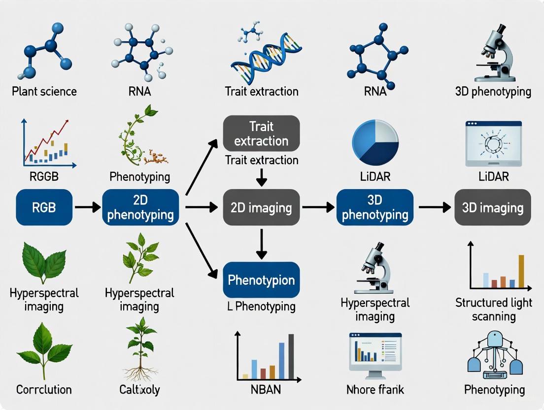
From Flat Images to Spatial Data: Understanding the Core Principles of 2D and 3D Phenotyping
Plant phenotyping, the quantitative assessment of plant traits, serves as a critical bridge between genomics and plant performance in agriculture and drug development. For decades, scientific research has relied heavily on two-dimensional (2D) projection methods for trait extraction due to their simplicity and low cost. However, a paradigm shift is underway toward three-dimensional (3D) approaches that capture the complex spatial architecture of biological structures. This comparison guide objectively examines the fundamental limitations of 2D projection against emerging 3D technologies, providing researchers with experimental data and methodological insights to inform their experimental designs. The transition from 2D to 3D phenotyping represents more than a technical upgrade—it constitutes a fundamental reimagining of how we quantify biological form and function across diverse domains from crop improvement to preclinical drug testing.
Theoretical Foundations: The Inherent Constraints of 2D Projection
The Spatial Information Deficit
The primary limitation of 2D projection lies in its fundamental inability to capture spatial depth. When complex 3D structures are projected onto a 2D plane, depth information is permanently lost, leading to measurement inaccuracies and structural ambiguities. In plant phenotyping, this manifests as an inability to accurately characterize root system architecture or canopy structure, where spatial arrangement directly correlates with function [1]. Similarly, in biomedical research, 2D cell cultures fail to recapitulate the three-dimensional tissue architecture that governs cellular behavior and drug response in vivo [2].
The Occlusion Problem
A second fundamental constraint arises from occlusion, where foreground structures obscure elements behind them. In complex biological systems like plant root systems or tissue models, this results in incomplete data capture and systematic measurement errors. Studies demonstrate that 2D root imaging frequently fails to capture the full root system architecture, with one study noting that field-based approaches often extract "only the top portion of the root system" [1]. The occlusion problem is particularly limiting for traits like leaf area index, branching patterns, and vascularization in tissue models.
Structural Simplification
Biological systems possess intricate 3D geometries that are inherently simplified when reduced to 2D representations. This structural simplification distorts critical phenotypic traits including surface area-to-volume ratios, spatial orientation, and mechanical properties. In cancer research, for instance, this simplification has profound implications, as 2D cultured cells "lose the peculiar signals coming from their niches" and are "constantly exposed to high levels of nutrients and oxygen" unlike the gradient conditions found in vivo [2].
Experimental Comparisons: Quantitative Performance Assessment
Accuracy Metrics in Trait Measurement
Table 1: Comparison of 2D vs. 3D Phenotyping Accuracy Across Domains
| Application Domain | Trait Measured | 2D Method Accuracy | 3D Method Accuracy | Citation |
|---|---|---|---|---|
| Soybean Root Phenotyping | Root Tip Counting | 79% correlation with manual counts | 95% correlation with manual counts (with background correction) | [1] |
| Plant Morphology Extraction | Plant Height | R² not reported for 2D | R² > 0.92 with manual measurements | [3] |
| Plant Morphology Extraction | Crown Width | R² not reported for 2D | R² > 0.92 with manual measurements | [3] |
| Leaf Parameter Extraction | Leaf Length/Width | Significant information loss from projection | R² = 0.72-0.89 with manual measurements | [3] |
| Organ Segmentation | Training Efficiency | Required 25 annotated plants for comparable performance | Achieved similar performance with only 5 annotated plants | [4] [5] |
Comprehensive Trait Extraction Capabilities
Table 2: Trait Extraction Capabilities of 2D vs. 3D Phenotyping Platforms
| Phenotypic Trait Category | 2D Projection Capability | 3D Reconstruction Capability | Research Implications |
|---|---|---|---|
| Root System Architecture | Limited to basic morphology (length, tips) | Comprehensive analysis (volume, distribution, spatial arrangement) | Enables identification of genes for deeper root systems [1] |
| Plant Biomass Estimation | Indirect estimation with occlusion errors | Direct volume calculation from 3D models | More accurate yield prediction and growth monitoring [6] |
| Gravotropic Responses | Basic directional assessment | Quantitative analysis of root and shoot angles over time | Enables study of developmental plasticity [7] |
| Drug Response Prediction | Poor clinical translation (5% efficacy) | Better recapitulation of in vivo conditions | Improved drug development success rates [2] [8] |
| Multi-Organ Interactions | Limited to separate analysis | Simultaneous tracking of 6+ plant structures [7] | Holistic understanding of plant development |
Methodological Approaches: Experimental Protocols
2D Phenotyping Workflow
The standard 2D phenotyping protocol for root system architecture analysis involves several established steps. Plants are typically grown in pouch growth systems with a transparent viewing surface, employing a black filter paper background to enhance contrast between roots and background [1]. Image acquisition uses standard RGB cameras under consistent lighting conditions. The critical image processing phase involves binary segmentation to separate roots from background, followed by skeletonization to extract topological attributes. Trait extraction typically includes basic morphological parameters like total root length, number of tips, and projection area. This approach has been widely adopted in high-throughput systems like the original ChronoRoot platform, which provided temporal analysis but was limited to binary segmentation of root structures alone [7].
Advanced 3D Reconstruction Methodologies
Multi-View Stereo Reconstruction
Advanced 3D phenotyping employs sophisticated reconstruction workflows. The integrated two-phase plant 3D reconstruction workflow begins with bypassing integrated depth estimation modules on standard binocular cameras. Instead, researchers apply Structure from Motion (SfM) and Multi-View Stereo (MVS) techniques to high-resolution images from multiple viewpoints, producing high-fidelity, single-view point clouds that effectively avoid distortion and drift [3]. The second phase addresses self-occlusion through precise registration of point clouds from six viewpoints into a complete plant model. This involves rapid coarse alignment using a marker-based Self-Registration method, followed by fine alignment with the Iterative Closest Point algorithm [3]. The resulting 3D models enable extraction of volumetric traits, surface areas, and spatial distribution parameters unavailable through 2D methods.
AI-Enhanced Temporal Phenotyping
Modern platforms like ChronoRoot 2.0 combine 3D imaging with artificial intelligence for comprehensive temporal analysis. The system employs infrared imaging with Raspberry Pi-controlled cameras and LED backlighting to eliminate variations from day/night cycles [7]. At the core of the analysis pipeline is an nnUNet segmentation module that performs simultaneous multi-class segmentation of six distinct plant structures: main root, lateral roots, seed, hypocotyl, leaves, and petiole [7]. This enables comprehensive tracking of plant development from seed to mature seedling, capturing intricate relationships between different organs during growth. The system incorporates Functional Principal Component Analysis for time series comparison across different experimental groups, enabling discovery of new data-driven phenotypic parameters.
2D-to-3D Projection Segmentation
An innovative hybrid approach has emerged that leverages well-established 2D segmentation algorithms for 3D analysis. This method involves reprojecting 2D predictions to 3D point clouds and using a majority vote algorithm to merge multiple predictions [4] [5]. Research demonstrates that this 2D-to-3D method achieves comparable performance to state-of-the-art 3D segmentation algorithms like Swin3D-s and Point Transformer v3, while offering significantly higher training efficiency [4]. With only five annotated plants, the 2D-to-3D approach achieved similar performance to training Swin3D-s on 25 plants, demonstrating remarkable efficiency gains [5].
The Scientist's Toolkit: Essential Research Reagents and Platforms
Table 3: Key Research Reagent Solutions for 2D and 3D Phenotyping
| Tool/Platform | Category | Function | Research Application |
|---|---|---|---|
| 2D Pouch System [1] | Growth Platform | Provides flat plane for root growth against transparent surface | High-throughput root screening in controlled conditions |
| ChronoRoot 2.0 [7] | Imaging Hardware | Automated infrared imaging system with AI analysis | Temporal phenotyping of plant development with multi-organ tracking |
| ZED 2 & ZED Mini Binocular Cameras [3] | 3D Capture Device | Capture stereo image pairs for 3D reconstruction | Generation of high-resolution point clouds for plant models |
| nnUNet Segmentation [7] | AI Software | Self-configuring neural network for image segmentation | Precise identification of plant organs in complex images |
| ResDGCNN [9] | Specialized Algorithm | Point cloud segmentation integrating residual learning with dynamic graph convolution | Cotton organ segmentation across entire growth cycle |
| Structure from Motion (SfM) [3] [10] | Reconstruction Algorithm | 3D point cloud generation from multiple 2D images | Non-destructive plant modeling from multi-view images |
| 3D Gaussian Splatting (3DGS) [10] | Emerging Technique | Represents geometry through Gaussian primitives | Efficient and scalable reconstruction of plant structures |
The experimental evidence clearly demonstrates that 3D phenotyping approaches overcome fundamental limitations of 2D projection across biological research domains. The spatial information captured by 3D methods enables more accurate trait measurement, better prediction of in vivo behavior, and discovery of previously inaccessible phenotypic relationships. However, 2D methods retain value for high-throughput screening scenarios where cost and simplicity are primary considerations. The emerging 2D-to-3D projection methods represent a promising intermediate approach, leveraging well-established 2D computer vision algorithms while capturing essential 3D structural information. As the field advances, the integration of dimensional approaches will continue to evolve, enabling researchers to select appropriate phenotyping strategies based on their specific accuracy requirements, throughput needs, and resource constraints. This dimensional integration promises to accelerate discoveries across fundamental plant science, agricultural improvement, and pharmaceutical development.
Plant phenotyping, the science of quantitatively measuring plant traits, serves as the critical link between genetics, environment, and observable characteristics. Traditional phenotyping has relied heavily on manual measurements and 2D imaging techniques, which project complex three-dimensional plant structures onto a flat plane. This process inevitably loses depth information and fails to accurately capture crucial architectural traits such as leaf curvature, stem inclination, and volumetric distribution [3]. These limitations have constrained our understanding of plant morphology and its functional implications.
The emergence of 3D phenotyping technologies addresses these fundamental shortcomings. By preserving spatial relationships and volumetric data, 3D methods enable researchers to move beyond simple length and area measurements to extract complex traits related to plant architecture, biomass distribution, and structural complexity [10] [11]. This paradigm shift offers unprecedented opportunities for advancing breeding programs and precision agriculture. This guide objectively compares the performance of 2D and 3D phenotyping approaches, supported by experimental data and methodological insights from current research.
Technical Comparison: 2D vs. 3D Phenotyping Performance
The transition from 2D to 3D phenotyping represents more than just technological advancement—it fundamentally enhances the quality, scope, and accuracy of trait extraction. Quantitative comparisons across multiple studies demonstrate clear advantages for 3D approaches, particularly for architectural traits.
Table 1: Performance Comparison of 2D vs. 3D Phenotyping for Key Plant Traits
| Plant Trait | 2D Method Limitations | 3D Method Advantages | Experimental Validation (R²) |
|---|---|---|---|
| Plant Height | Perspective distortion; reference scaling required | Direct measurement from 3D point clouds | 0.92-0.95 [3] |
| Crown Width | Single-view projection underestimates actual volume | Multi-view reconstruction captures true extent | >0.92 [3] |
| Leaf Area | Projection errors; inability to account for curvature | Surface area calculation from 3D mesh | 0.72-0.89 [3] |
| Internode Length | Destructive dissection often required | Non-destructive extraction from skeletonized models | Enabled via 3D skeletonization [11] |
| Leaf Angle | Manual protractor measurements; single point sampling | Automated calculation from 3D normals/vectors | Included in TomatoWUR dataset [11] |
| Organ Segmentation | Overlap obscures individual organs; loss of spatial context | 3D spatial separation enables instance segmentation | mIoU: 83.05-89.21% across species [12] |
The data reveal that 3D methods particularly excel in capturing traits that require spatial context. For example, while 2D approaches struggle with leaf area estimation due to their inability to account for curvature, 3D reconstructions can accurately measure actual surface area, achieving R² values of 0.72-0.89 compared to manual measurements [3]. Similarly, architectural traits like internode length and leaf angle, which traditionally required destructive sampling or cumbersome manual tools, can now be extracted automatically from 3D skeletonized models [11].
Experimental Approaches in Modern 3D Plant Phenotyping
2D-to-3D Reprojection Segmentation
A innovative approach that leverages well-established 2D computer vision for 3D segmentation has been developed and compared against native 3D methods. The experimental protocol involves:
- Image Segmentation: Applying the Mask2Former model (pre-trained on diverse 2D datasets) to segment individual plant organs from 2D images [4].
- Reprojection to 3D: Projecting the 2D segmentation predictions back onto the corresponding 3D point cloud using known camera parameters [4].
- Majority Vote Fusion: Employing a majority vote algorithm to merge multiple predictions from different viewpoints into a consensus 3D segmentation [4].
This method demonstrated no significant performance difference compared to state-of-the-art 3D segmentation algorithms like Swin3D-s and Point Transformer v3, while achieving higher training efficiency. Remarkably, training on just five annotated plants with the 2D-to-3D method yielded similar performance to training Swin3D-s on 25 plants, highlighting its data efficiency [4].
Table 2: Comparison of 3D Segmentation Method Performance
| Algorithm | Principle | Key Advantage | Training Efficiency |
|---|---|---|---|
| 2D-to-3D Reprojection | Projects 2D segmentation to 3D space | Leverages well-developed 2D models; high data efficiency | 5 plants for comparable performance [4] |
| Swin3D-s | Voxel-based 3D transformer | State-of-the-art on structured data | Required 25 plants for comparable performance [4] |
| Point Transformer v3 | Point-based neural network | Directly processes point clouds | Similar accuracy but lower efficiency than 2D-3D [4] |
| MinkUNet34C | Sparse convolutional networks | Memory efficient for large scenes | Lower performance than other 3D methods [4] |
| PointNeXt | Point-based deep learning | High accuracy across species | mIoU: 83.05-89.21% across crops [12] |
Multi-View 3D Reconstruction with Fine Registration
A comprehensive two-phase workflow for high-fidelity plant reconstruction addresses common challenges in 3D plant phenotyping:
Phase 1: High-Fidelity Single-View Reconstruction
Phase 2: Multi-View Registration
This workflow was validated on Ilex verticillata and Ilex salicina, demonstrating high correlation with manual measurements (R² > 0.92 for plant height and crown width) and successfully extracting fine-scale traits like leaf length and width, which are rarely addressed in multi-view fusion studies [3].
Figure 1: Workflow for multi-view 3D plant reconstruction and trait extraction.
Two-Stem Deep Learning for Organ Segmentation
A two-stage deep learning approach addresses the challenge of organ-level segmentation across multiple crop species:
Stage 1: Semantic Segmentation
Stage 2: Instance Segmentation
This method consistently outperformed four state-of-the-art networks (ASIS, JSNet, DFSP, and PSegNet), achieving average values of 93.32% precision, 85.60% recall, 87.94% F1, and 81.46% mIoU across all tested crops [12].
The Scientist's Toolkit: Essential Research Reagents & Materials
Successful implementation of 3D plant phenotyping requires specific hardware, software, and datasets. The following table summarizes key resources referenced in the experimental studies.
Table 3: Essential Research Reagents and Materials for 3D Plant Phenotyping
| Category | Specific Product/Method | Function/Application | Research Context |
|---|---|---|---|
| Imaging Hardware | ZED 2 & ZED Mini binocular cameras | Capture stereo image pairs for 3D reconstruction | Multi-view plant reconstruction [3] |
| Annotation Software | TomatoWUR dataset | Comprehensive benchmark for algorithm development | Includes point clouds, skeletons, manual measurements [11] |
| Segmentation Models | Mask2Former | 2D segmentation for reprojection methods | 2D-to-3D segmentation pipeline [4] |
| 3D Deep Learning | PointNeXt framework | Point-based semantic segmentation | Two-stage organ segmentation [12] |
| Reconstruction Algorithms | SfM + MVS pipeline | Generate 3D point clouds from 2D images | High-fidelity plant reconstruction [3] |
| Registration Tools | ICP algorithm + Marker-based SR | Align multi-view point clouds | Complete plant model creation [3] |
| Synthetic Data | AI-generated leaf point clouds | Augment training data; algorithm benchmarking | Trait estimation without manual labeling [13] |
| Evaluation Metrics | mIoU, Precision, Recall | Standardize algorithm performance assessment | Cross-study method comparison [4] [12] |
Emerging Frontiers and Future Directions
The field of 3D plant phenotyping continues to evolve with several promising technological developments:
- 3D Gaussian Splatting (3DGS): An emerging technique that represents plant geometry through Gaussian primitives, offering potential benefits in both efficiency and scalability compared to traditional point clouds [10].
- AI-Generated Synthetic Data: Generative models that produce realistic 3D leaf point clouds with known geometric traits are reducing the bottleneck caused by limited labeled data [13].
- Neural Radiance Fields (NeRF): This recent advancement enables high-quality, photorealistic 3D reconstructions from sparse viewpoints, though computational cost remains a challenge [10].
These technologies collectively address current limitations in 3D plant phenotyping, particularly regarding computational efficiency, annotation requirements, and applicability in field conditions.
Figure 2: Complete workflow from image acquisition to 3D trait extraction.
The comparative evidence presented in this guide demonstrates the clear advantage of 3D phenotyping over traditional 2D approaches for capturing plant architecture and complex traits. While 2D methods remain valuable for specific applications, 3D technologies enable precise, non-destructive measurement of structural traits that are fundamental to understanding plant growth and function.
The integration of advanced computer vision, deep learning, and high-fidelity reconstruction algorithms has transformed plant phenotyping from a primarily manual, destructive process to an automated, quantitative science. As these technologies continue to mature and become more accessible, they promise to accelerate crop improvement programs and enhance our understanding of gene-environment interactions in plants.
For researchers adopting 3D phenotyping, the choice between specific approaches (2D-to-3D reprojection vs. native 3D algorithms, SfM vs. depth cameras) should be guided by the target traits, available resources, and scale of operation. The methodologies and performance benchmarks provided in this guide offer a foundation for making these critical decisions in experimental design.
Three-dimensional (3D) imaging has emerged as a powerful tool in plant phenotyping, moving beyond the limitations of traditional two-dimensional approaches. While 2D imaging captures only intensity and color across a flat plane, 3D imaging technologies capture spatial depth and geometric structure, creating a comprehensive three-dimensional representation of objects and environments by measuring the X, Y, and Z coordinates of each point in the observed space [14]. This capability is revolutionizing trait extraction research by enabling a more accurate understanding of spatial relationships, plant dimensions, and environmental structures.
In the specific context of plant phenotyping, 3D reconstruction models provide significant advantages for morphological classification and growth tracking [15]. These technologies allow researchers to resolve occlusions and crossings of plant structures by reconstructing distance, orientation, and illumination from multiple viewing angles—capabilities that are hard or impossible to achieve with 2D models alone [15]. As the field advances, understanding the fundamental distinction between active and passive 3D imaging approaches becomes essential for selecting appropriate methodologies for specific research applications in trait extraction.
Fundamental Principles: Active vs. Passive 3D Imaging
3D imaging methods can be broadly classified into active and passive approaches, each with distinct operating principles, advantages, and limitations [15]. This fundamental distinction lies in their use of controlled illumination sources to interact with the subject being measured.
Active 3D imaging approaches use active sensors that rely on radiometric interaction with the object by using structured light or laser to directly capture a 3D point cloud representing the coordinates of each part of the subject in 3D space [15]. These systems project their own controlled energy emissions onto the scene and measure how this projected energy interacts with the objects. Because they rely on emitted energy rather than ambient light, active approaches can overcome several problems associated with passive methods, such as correspondence problems (ascertaining which parts of one image correspond to which parts of another image) [15]. Common active techniques include structured light, laser scanners (LiDAR), Time-of-Flight (ToF) cameras, and laser triangulation systems.
Passive 3D imaging approaches, in contrast, rely on ambient light or naturally available illumination to form images without projecting any energy onto the scene [15]. These systems typically use multiple cameras or sensors to capture images from different viewpoints and then apply computational algorithms to reconstruct the 3D structure. Because they don't require specialized illumination equipment, passive techniques tend to be more cost-effective as they typically use commodity or off-the-shelf hardware, but may result in comparatively lower-quality data that often require significant computational processing to be useful [15]. Common passive methods include stereo vision, photogrammetry, and structure from motion.
The following diagram illustrates the fundamental working principles of both active and passive 3D imaging approaches:
Active 3D Imaging Modalities: Technologies and Protocols
Laser Triangulation
Experimental Protocol: Laser triangulation involves shining a laser beam to illuminate the object of interest and using a sensor array to capture the laser reflection [15]. The deformation of the laser line or point on the target surface reveals topological information through triangulation geometry. As either the object or sensor moves, a sequence of profiles is collected and stitched together to form a high-resolution 3D reconstruction [16]. This method excels in precision applications and has been successfully implemented in laboratory experiments for producing 3D point clouds of barley plants [15], wheat canopies [15], and rapeseed [15].
System Requirements: Low-cost setup typically including a laser source (line or point), a camera positioned at a known angle to the laser, and a precision translation stage for moving either the sensor or object. The system requires precise calibration to determine the exact spatial relationship between the laser and camera [16].
3D Laser Scanning (LiDAR)
Experimental Protocol: LiDAR (Light Detection and Ranging) systems use laser pulses to create detailed point clouds representing 3D space with high precision [14]. These high-precision instruments require calibration objects or repeated scanning to accomplish point cloud registration and stitching [15]. Terrestrial laser scanners (TLS) allow for large volumes of plants to be measured with relatively high accuracy and are therefore mostly used for determining parameters of plant canopies and fields of plants [15]. Chebrolu et al. used a laser scanner to record time-series data of tomato and maize plants over a period of two weeks, while Paulus et al. used a 3D laser scanner to create point clouds of grapevine and wheat [15].
System Requirements: Laser scanner (from high-precision research grade to low-cost consumer versions like Microsoft Kinect), calibration objects, and substantial computational resources for processing large data volumes [15] [16].
Time of Flight (ToF)
Experimental Protocol: ToF cameras use light emitted by a laser or LED source and measure the roundtrip time between the emission of a light pulse and the reflection from thousands of points to build up a 3D image [15]. The time delay is translated directly into distance measurements for each pixel in the image [16]. Applications in plant phenotyping include the work of Chaivivatrakul et al. on maize plants, Baharav et al. on sorghum plants, and Kazmi et al. on various plants including cyclamen, hydrangea, and orchidaceae [15].
System Requirements: ToF sensor or camera, controlled lighting conditions, and onboard correction mechanisms for multipath interference, temperature drift, and ambient light noise [16].
Structured Light
Experimental Protocol: Structured light systems project a predefined pattern, such as stripes, dot matrices, or fringe patterns, onto a target scene [16]. The deformation of this pattern, as observed from a separate camera viewpoint, reveals surface topology by analyzing how the structured light distorts over varying depths [15]. This method offers excellent spatial resolution and is particularly useful for detailed scanning tasks. The approach requires very accurate correspondence between images for optimal performance [15].
System Requirements: Pattern projector (often using DLP technology), camera positioned at a known angle, and computational resources for analyzing pattern deformation [16].
Table 1: Technical Specifications of Active 3D Imaging Modalities
| Modality | Depth Sensing Principle | Resolution Potential | Operating Range | Scanning Speed |
|---|---|---|---|---|
| Laser Triangulation | Triangulation geometry | High (sub-millimeter) | Short (cm to few m) | Medium |
| 3D Laser Scanning (LiDAR) | Laser pulse measurement | Very High | Long (m to km) | Slow to Fast (depends on system) |
| Time of Flight (ToF) | Light pulse roundtrip time | Medium to High | Medium (cm to tens of m) | Very Fast (real-time) |
| Structured Light | Pattern deformation analysis | Very High | Short to Medium (cm to m) | Fast |
Passive 3D Imaging Modalities: Technologies and Protocols
Stereo Vision
Experimental Protocol: Stereo vision leverages two or more cameras to mimic human binocular perception [16]. By identifying corresponding points between the captured images and calculating their disparity, a stereo system can triangulate depth and generate dense depth maps [16]. This approach works well in environments with sufficient texture and lighting and is commonly used in robotic navigation and real-time inspection [16]. For plant phenotyping applications, the method requires multiple synchronized cameras positioned at different angles to capture the plant structure from various viewpoints.
System Requirements: Two or more calibrated and synchronized cameras, precise knowledge of camera positions and orientations, and significant computational power for disparity map calculation (often implemented on FPGAs or GPUs) [16].
Photogrammetry
Experimental Protocol: Photogrammetry reconstructs 3D geometry from a series of overlapping 2D images taken from different viewpoints [16]. Using algorithms such as structure-from-motion (SfM) and multi-view stereo (MVS), it identifies shared features across images and estimates their position in 3D space [16]. While photogrammetry requires more post-processing than other methods, it can produce highly detailed models using off-the-shelf cameras [16]. It is widely used in digital archiving of plant specimens and growth monitoring.
System Requirements: Standard digital camera (multiple cameras optional but not required), controlled lighting for consistent image capture, and substantial computational resources for processing overlapping images [16].
Neural Radiance Fields (NeRF)
Experimental Protocol: NeRF is a recent advancement that enables high-quality, photorealistic 3D reconstructions from sparse viewpoints [10]. This method uses a fully-connected deep network to represent a continuous volumetric scene function, optimizing the network to minimize the error between rendered and true images from various viewing directions. The network input is a continuous 5D coordinate (spatial location and viewing direction), and the output is the volume density and view-dependent emitted radiance at that location [10].
System Requirements: Multiple images from different viewpoints, significant GPU resources for training the neural network, and specialized software implementations of the NeRF algorithm [10].
3D Gaussian Splatting (3DGS)
Experimental Protocol: The emerging 3D Gaussian Splatting technique introduces a new paradigm in reconstructing plant structures by representing geometry through Gaussian primitives, offering potential benefits in both efficiency and scalability [10]. This approach represents scenes as a collection of 3D Gaussians that have controllable properties including position, covariance, and color. The optimization process involves adapting these Gaussians to best represent the scene, followed by a tile-based rasterization approach for real-time rendering [10].
System Requirements: Multiple calibrated images or video frames, modern GPU with support for CUDA, and implementation of the 3DGS algorithm which is computationally intensive during training but enables real-time rendering [10].
Table 2: Technical Specifications of Passive 3D Imaging Modalities
| Modality | Depth Sensing Principle | Resolution Potential | Texture Dependency | Computational Demand |
|---|---|---|---|---|
| Stereo Vision | Binocular disparity | Medium to High | High (requires texture) | High (real-time processing) |
| Photogrammetry | Multi-view feature matching | High | High (requires texture) | Very High (post-processing) |
| Neural Radiance Fields (NeRF) | Neural volume rendering | Very High | Medium | Extremely High (training) |
| 3D Gaussian Splatting (3DGS) | Gaussian primitive optimization | Very High | Medium | High (training), Low (rendering) |
Comparative Analysis: Performance Metrics in Plant Phenotyping
When selecting 3D imaging modalities for plant phenotyping applications, researchers must consider multiple performance metrics to align technological capabilities with research objectives. The following comparative analysis highlights key considerations for trait extraction research.
Accuracy and Resolution Requirements: Active methods generally provide higher accuracy and resolution for detailed morphological studies. Structured light systems offer excellent spatial resolution for capturing fine plant structures [16], while laser triangulation provides high (sub-millimeter) precision suitable for laboratory experiments on individual plants [15]. Passive methods like photogrammetry can also achieve high resolution but are more dependent on optimal lighting conditions and surface texture [16].
Data Acquisition and Processing Speed: For dynamic growth studies and high-throughput phenotyping, acquisition speed is critical. Time-of-Flight systems can operate in real time, making them ideal for capturing plant movement or rapid scans [16]. Stereo vision also enables real-time processing when implemented with appropriate hardware [16]. In contrast, methods like photogrammetry and NeRF require significant post-processing time but can produce more detailed models [10] [16].
Cost and Accessibility Considerations: Passive techniques tend to be more cost-effective as they typically use commodity or off-the-shelf hardware [15], making them accessible to research groups with limited budgets. Active methods generally require specialized and often expensive equipment [15], creating higher barriers to entry but potentially offering superior performance for specific applications.
Scalability to Different Plant Architectures: The complexity and diversity of plant structures present unique challenges for 3D reconstruction. Methods like 3D Gaussian Splatting show promise for efficiently representing complex plant geometries [10], while stereo vision and photogrammetry struggle with occlusions in dense canopies. Laser scanning approaches have proven effective for various architectures from barley plants to wheat canopies [15].
The following workflow diagram illustrates a recommended decision-making process for selecting appropriate 3D imaging modalities based on specific research requirements:
The Researcher's Toolkit: Essential Solutions for 3D Plant Phenotyping
Implementing successful 3D imaging protocols for plant phenotyping requires specific hardware and software solutions. The following table details essential research reagent solutions and their functions in trait extraction studies.
Table 3: Essential Research Reagent Solutions for 3D Plant Phenotyping
| Solution Category | Specific Products/Technologies | Primary Function | Application Examples |
|---|---|---|---|
| Active 3D Sensors | Microsoft Kinect, Terrestrial Laser Scanners (TLS), Structured Light Projectors | Direct 3D data capture via projected energy | Root architecture studies, canopy density measurement, growth monitoring |
| Multi-Camera Systems | Synchronized industrial cameras, Stereo vision setups, Smartphone arrays | Multi-view image capture for passive 3D reconstruction | Leaf area index estimation, plant volume calculation, morphological analysis |
| Computational Hardware | GPU clusters (NVIDIA), FPGAs, Edge computing devices | Processing large 3D datasets and reconstruction algorithms | Real-time plant growth tracking, neural network training for NeRF/3DGS |
| Software Platforms | Photogrammetry software (Agisoft Metashape), NeRF implementations, 3DGS codebase | 3D model reconstruction and analysis | Creating digital plant models, quantifying trait variations over time |
| Calibration Tools | Checkerboard patterns, calibration spheres, reference objects | System calibration and accuracy validation | Ensuring measurement precision across imaging sessions |
| Data Management Solutions | Cloud-based PACS, High-capacity storage arrays | Storing and managing large 3D datasets | Long-term growth studies, multi-institutional collaboration |
The selection between active and passive 3D imaging modalities for plant phenotyping involves careful consideration of research objectives, environmental constraints, and available resources. Active methods like structured light and laser scanning provide high accuracy for controlled laboratory environments, while passive approaches like photogrammetry and stereo vision offer flexibility and cost-effectiveness for field applications. Emerging technologies such as Neural Radiance Fields and 3D Gaussian Splatting represent promising directions for capturing complex plant architectures with unprecedented detail.
As the field advances, the integration of artificial intelligence with both active and passive 3D imaging modalities will likely enhance automated trait extraction capabilities, potentially overcoming current limitations in processing speed and accuracy. Researchers should consider establishing multimodal imaging platforms that leverage the complementary strengths of both approaches to address the diverse challenges in plant phenotyping across different species, growth stages, and environmental conditions.
Methodologies in Action: A Practical Guide to 3D Trait Extraction Technologies
The transition from traditional 2D image analysis to advanced 3D sensing represents a paradigm shift in plant phenotyping and agricultural research. While 2D methods project complex 3D plant structures onto a plane, causing loss of depth information and inaccurate morphological capture, 3D sensing technologies provide comprehensive structural data essential for precise trait extraction [3]. This evolution is particularly crucial for pharmaceutical development from plant sources, where accurate morphological and structural phenotyping directly impacts understanding of plant physiology, stress responses, and compound production [10] [9].
Active sensing technologies, particularly LiDAR and laser triangulation, have emerged as powerful tools for generating high-fidelity 3D structural data. Unlike passive imaging systems, these active methods project their own energy sources (typically laser light) and measure returned signals, enabling precise 3D mapping regardless of ambient lighting conditions. This capability is revolutionizing how researchers quantify plant architecture, monitor growth dynamics, and extract phenotypic traits with unprecedented accuracy [3] [10]. The integration of these technologies into research pipelines provides the robust, quantitative structural data necessary for advancing pharmaceutical development from plant resources.
Technology Fundamentals: Operating Principles and Mechanisms
LiDAR (Light Detection and Ranging)
LiDAR operates on the time-of-flight principle, emitting laser pulses and measuring the time taken for reflected light to return to the sensor. By calculating this interval and knowing the speed of light, precise distance measurements are obtained. Through rapid scanning, LiDAR systems generate dense 3D point clouds representing surface geometry [17]. Modern LiDAR systems achieve remarkable precision, with range accuracies between 0.5 to 10mm relative to the sensor, though environmental factors, target surface properties, and measurement distance can affect this precision [17].
LiDAR Technology Variants:
- Terrestrial LiDAR: Tripod-mounted systems providing millimeter-level accuracy for detailed structural documentation [17]
- Mobile LiDAR: Wearable or vehicle-mounted systems utilizing SLAM (Simultaneous Localization and Mapping) algorithms for rapid data capture over larger areas [17]
- Aerial LiDAR: UAV-mounted systems covering extensive territories efficiently, particularly valuable for canopy penetration and topographic mapping [17]
Laser Triangulation
Laser triangulation sensors employ geometric principles to determine distance measurements. A laser diode projects a visible spot or line onto a target surface, while a camera positioned at a known angle captures the reflection. Displacement of the laser point in the camera's field of view corresponds directly to distance changes, enabling high-precision measurements through trigonometric calculations [18]. These sensors are categorized by their measurement ranges, from ultra-precise 0-2μm systems for micro-assembly to 101-500μm range sensors for larger-scale applications [18].
The global laser triangulation sensor market, estimated at $1.5 billion in 2025, reflects the growing adoption of this technology across research and industrial applications, with particularly strong utilization in automotive, aerospace, and electronics manufacturing where precision measurement is critical [18].
Comparative Performance Analysis: Quantitative Data Comparison
Table 1: Performance Specifications of LiDAR and Laser Triangulation Systems
| Parameter | Terrestrial LiDAR | Mobile LiDAR | Aerial LiDAR | Laser Triangulation |
|---|---|---|---|---|
| Accuracy | 1-3mm (at optimal range) [17] | 5-20mm [17] | 10-50mm (depends on altitude) [17] | Varies by range: 0-2μm to 101-500μm [18] |
| Measurement Range | Up to 350m [17] | Several hundred meters [17] | 1600-3900m AGL [19] | Limited to sensor range (typically <1m) [18] |
| Data Capture Speed | Moderate (stationary setup required) [17] | High (walking speed operation) [17] | Very High (aerial coverage) [17] | Very High (thousands of measurements/second) [18] |
| Point Density | Very High (stationary scanning) [17] | Moderate (depends on movement speed) [17] | Variable (depends on altitude) [17] | Extremely High (focused measurement area) [18] |
| Best Applications | Architectural documentation, high-precision plant phenotyping [17] | Corridor mapping, large facility documentation [17] | Topographical mapping, forestry, environmental monitoring [17] [19] | Micro-scale plant organ measurement, leaf surface analysis [18] |
Table 2: Cost Analysis and Implementation Considerations
| Factor | Terrestrial LiDAR | Mobile LiDAR | Aerial LiDAR | Laser Triangulation |
|---|---|---|---|---|
| Equipment Cost | $35,000-$80,000 [17] | $10,500-$60,000 [17] | Varies widely by platform | Varies by precision requirements [18] |
| Operational Workflow | Multiple scan positions requiring registration [17] | Continuous scanning with SLAM processing [17] | Flight planning, GPS/IMU integration [19] | Single-point or line scanning |
| Processing Complexity | High (manual registration often needed) [17] | Moderate (automated SLAM with potential manual correction) [17] | Moderate (specialized photogrammetry software) [17] | Low to Moderate (direct measurement or basic profiling) |
| Environmental Limitations | Atmospheric conditions, temperature variations [17] | Limited features areas cause SLAM drift [17] | Weather, flight regulations, lighting [17] | Sensitive to surface properties, ambient light [18] |
Experimental Protocols for Plant Phenotyping Applications
Multi-View 3D Reconstruction Workflow for Plant Phenotyping
Protocol Objective: Generate complete 3D models of plants by integrating multiple viewpoints to overcome occlusion issues common in plant structures [3].
Materials and Equipment:
- Binocular stereo vision camera (e.g., ZED 2 or ZED mini) [3]
- Controlled rotation platform or U-shaped rotating arm [3]
- Calibration objects (spheres/markers for registration) [3]
- High-resolution computing workstation for processing
Methodology:
- Multi-View Image Acquisition: Position plant specimen on rotation platform. Capture images from six viewpoints around the plant, with additional captures from varying heights at each position. For each viewpoint, acquire 8 RGB images with 2208×1242 resolution [3].
- Structure from Motion (SfM) Processing: Apply SfM algorithms to captured high-resolution images to generate initial 3D point clouds, bypassing integrated depth estimation modules to avoid distortion and drift [3].
- Multi-View Stereo (MVS) Enhancement: Implement MVS techniques to produce high-fidelity, single-view point clouds with improved density and accuracy [3].
- Point Cloud Registration:
- Phenotypic Trait Extraction: From the complete 3D model, extract key phenotypic parameters including plant height, crown width, leaf length, and leaf width using automated measurement algorithms [3].
Validation: Experimental validation on Ilex species demonstrated strong correlation with manual measurements, with coefficients of determination (R²) exceeding 0.92 for plant height and crown width, and ranging from 0.72 to 0.89 for leaf parameters [3].
High-Throughput Cotton Phenotyping Using 3D Point Cloud Segmentation
Protocol Objective: Achieve automated organ-level segmentation and phenotypic extraction across the complete growth cycle of cotton plants [9].
Materials and Equipment:
- Smartphone camera or structured light scanner for data acquisition
- Digital calipers for manual validation measurements
- GPU-enabled computing system for deep learning processing
- Controlled growth environment (greenhouse with maintained temperature 25-27°C) [9]
Methodology:
- Data Collection: Capture video of cotton plants using smartphone, recording approximately 40-second durations yielding 1200 frames. Include full rotations from top-down, horizontal, and bottom-up perspectives. Move camera slowly to prevent motion blur [9].
- 3D Reconstruction: Generate point clouds from video frames using SfM and MVS approaches, creating comprehensive 3D models of cotton plants throughout growth cycle [9].
- Deep Learning Segmentation: Implement ResDGCNN network architecture integrating residual learning with dynamic graph convolution to address significant structural variations in cotton organs across growth stages [9].
- Organ-Level Segmentation Optimization: Apply improved region-growing algorithm incorporating point distance mapping with curvature-based normal vectors to address overlapping regions between different cotton organs [9].
- Phenotypic Parameter Calculation: Extract plant height, stem length, leaf dimensions, and innovatively calculate bell drop rate - a critical phenotypic trait for cotton yield estimation [9].
Validation: The method achieved a segmentation accuracy of 67.55% with 4.86% improvement in mIoU compared to baseline models. In overlapping leaf segmentation, the model achieved R² of 0.962 and RMSE of 2.0. Average relative error in stem length estimation was 0.973 [9].
Essential Research Reagent Solutions for 3D Phenotyping
Table 3: Research Reagent Solutions for Active Sensing in Phenotyping
| Reagent Category | Specific Product/Technology | Research Application & Function |
|---|---|---|
| Terrestrial LiDAR Systems | FARO Focus Premium Max [19] | High-precision outdoor plant scanning with 266-megapixel resolution for detailed structural phenotyping |
| Mobile LiDAR Systems | Wearable SLAM-based systems [17] | Rapid phenotyping of large agricultural fields or greenhouse facilities with centimeter-level accuracy |
| Aerial LiDAR Systems | RIEGL VQ-1560 III-S [19] | Large-scale crop monitoring, canopy structure analysis, and environmental interaction studies |
| Laser Triangulation Sensors | KEYENCE, SICK, Panasonic sensors [18] | Micro-scale measurement of plant organs, leaf surface topography, and detailed morphological analysis |
| Binocular Stereo Cameras | ZED 2 and ZED Mini [3] | Cost-effective 3D reconstruction for plant phenotyping using multi-view stereo approaches |
| Registration Algorithms | Iterative Closest Point (ICP) [3] | Alignment of multi-view point clouds into complete 3D plant models |
| Segmentation Algorithms | ResDGCNN with residual learning [9] | Organ-level segmentation of complex plant structures across full growth cycles |
| Validation Instruments | Digital calipers, manual measurement tools [9] | Ground-truth validation of automated phenotypic extraction algorithms |
The strategic selection between LiDAR and laser triangulation technologies depends fundamentally on the specific requirements of the phenotyping research. LiDAR systems offer superior range and flexibility for macroscopic plant architecture studies, while laser triangulation provides exceptional precision for microscopic organ-level analysis.
For comprehensive phenotyping pipelines, integrated approaches often yield optimal results. Terrestrial LiDAR captures overall plant architecture with millimeter precision, while laser triangulation sensors can be deployed for detailed analysis of specific organs. Mobile LiDAR systems enable high-throughput phenotyping of large populations, essential for breeding programs and pharmaceutical source selection [17].
The integration of these active sensing technologies with advanced computational methods—including deep learning segmentation, multi-view registration, and automated trait extraction—represents the future of high-throughput 3D plant phenotyping. As these technologies continue evolving with improvements in speed, accuracy, and accessibility, they will play an increasingly vital role in advancing pharmaceutical development from plant resources and addressing challenges in sustainable agriculture [10] [9].
Researchers should consider establishing technology stacks that combine the strengths of both LiDAR and laser triangulation, creating complementary phenotyping workflows that capture both macroscopic architectural traits and microscopic morphological features. This integrated approach provides the comprehensive structural data necessary for breakthroughs in plant-based pharmaceutical research and development.
Plant phenotyping, the quantitative assessment of complex plant traits, has traditionally relied on two-dimensional (2D) imaging and manual measurements [9]. However, projecting complex three-dimensional plant structures onto a 2D plane results in significant information loss, particularly depth information, making it difficult to accurately capture morphological features [3]. This limitation is especially pronounced for complex plant architectures with issues such as shading, overlapping leaves, and multiple branches [9]. In drug discovery, traditional 2D cell cultures face a similar challenge, as they cannot accurately illustrate and mimic the complex environment found in living organisms [20] [21].
The emergence of three-dimensional (3D) reconstruction technologies represents a paradigm shift, enabling non-destructive, high-throughput analysis of biological specimens. Among these technologies, image-based methods utilizing Structure-from-Motion (SfM) and Multi-View Stereo (MVS) have gained prominence due to their ability to generate highly detailed 3D models from standard 2D images [22]. These methods are revolutionizing phenotyping by providing access to accurate morphological and structural data that was previously difficult or impossible to obtain with conventional 2D approaches [3]. This guide provides a comprehensive comparison of SfM and MVS methodologies, their performance relative to alternative 3D reconstruction techniques, and their specific applications in trait extraction research.
Understanding the Core Technologies: SfM and MVS
Structure-from-Motion (SfM)
Structure-from-Motion is a photogrammetric technique that estimates three-dimensional structures from two-dimensional image sequences [23] [24]. A key advantage of SfM is its ability to handle unordered image sets, making it suitable for scenarios where image capture is less controlled [24]. The SfM process begins by detecting distinctive feature points across multiple images, matching these features across different views, and then simultaneously calculating camera parameters (positions, orientations, and intrinsic calibration) and a sparse 3D point cloud through a process called bundle adjustment [22] [23]. This results in a geometrically consistent configuration of the image set and an initial sparse reconstruction of the scene [22].
Multi-View Stereo (MVS)
Multi-View Stereo builds upon the output of SfM to create highly detailed 3D models [24]. Once camera parameters and sparse point clouds are established through SfM, MVS algorithms perform dense image matching to compute pixel-wise correspondences between images, generating a dense and detailed surface model [22]. The primary strength of MVS is its ability to generate dense point clouds and detailed textures, making it indispensable for projects requiring high-resolution 3D models with fine surface geometry [24]. Unlike SfM, MVS typically requires more controlled image capture with systematic coverage of the subject from various angles to ensure comprehensive overlapping views [24].
The Integrated SfM-MVS Workflow
In practice, SfM and MVS are complementary techniques often deployed in an integrated pipeline [22] [24]. The SfM stage derives the initial camera parameters and sparse point clouds, setting the stage for MVS to generate a dense and detailed 3D model [24]. This combination leverages the strengths of both techniques, allowing for robust reconstructions even in complex scenarios [24]. The following diagram illustrates this integrated workflow:
SfM-MVS Reconstruction Workflow
Comparative Performance Analysis of 3D Reconstruction Techniques
SfM-MVS vs. Emerging Neural Approaches
Recent studies have compared traditional SfM-MVS photogrammetry with emerging neural rendering approaches like Neural Radiance Fields (NeRF) and Gaussian Splatting (GS). The table below summarizes key quantitative findings from comparative studies:
Table 1: Performance Comparison of 3D Reconstruction Techniques
| Technique | Geometric Accuracy | Processing Time | Completeness | Fine Detail Capture | Primary Strengths |
|---|---|---|---|---|---|
| SfM-MVS | High (RMS error: ~0.1-0.5 mm for small artifacts) [25] | Moderate to High [26] | Minor gaps possible [26] | Excellent for fine geometric details [25] | Highest geometric precision [26] |
| NeRF | Lower than SfM [25] [26] | Fast [26] | High completeness [26] | Lower for fine geometry [25] | Superior rendering, good completeness [26] |
| Gaussian Splatting | Lower than SfM [25] | Fast (real-time capable) [25] | Moderate | Outperformed by NeRF geometrically [25] | Real-time rendering [25] |
| LiDAR | High [3] | Fast acquisition | Limited by occlusion [3] | Good for structural features | Direct distance measurement [3] |
| Binocular Stereo | Moderate (prone to distortion) [3] | Fast | Limited by occlusion [3] | Challenging for low-texture surfaces [3] | Real-time acquisition |
For archaeological artifact reconstruction, SfM demonstrated superior geometric fidelity with root mean square (RMS) error measurements significantly lower than both NeRF and Gaussian Splatting alternatives [25]. Similarly, in architectural heritage documentation, SfM-MVS provided the highest geometric precision despite minor gaps in reconstruction, while NeRF and GS fell short of the accuracy required for precise geometric documentation [26].
Application-Specific Performance in Phenotyping
In plant phenotyping applications, SfM-MVS has demonstrated remarkable accuracy for trait extraction. Studies on Ilex species showed that key phenotypic parameters extracted from SfM-MVS models exhibited a strong correlation with manual measurements, with coefficients of determination (R²) exceeding 0.92 for plant height and crown width, and ranging from 0.72 to 0.89 for leaf parameters [3]. Another study on cotton plants achieved a mean relative error of only 0.973 for stem length estimation using SfM-MVS based reconstruction [9].
Experimental Protocols for High-Fidelity 3D Reconstruction
Image Acquisition Protocol for Plant Phenotyping
A standardized image acquisition protocol is fundamental for high-quality 3D reconstruction. The following methodology has been validated for plant phenotyping applications:
- Equipment Setup: Use a robotic arm to control an industrial RGB camera for maximum flexibility in image acquisition [22]. This mobility ensures comprehensive coverage of plants of different sizes and architectures while minimizing occlusions [22].
- Camera Parameters: Set exposure time to 50 milliseconds, camera-to-object distance of 16 centimeters, and use an optimized parameter tweak value of 0.9 to improve reconstruction of thin and delicate plant parts [22].
- Acquisition Configuration: Employ multiple height levels (3 levels recommended) with systematic coverage at each level [22]. Capture approximately 40 frames per position to balance processing time and model quality [22].
- Lighting Conditions: Use diffuse and uniform lighting to minimize shadows and reflections that can interfere with feature matching [22]. Overcast weather conditions provide ideal natural lighting for outdoor acquisition [27].
- Background Separation: Apply background separation techniques such as chroma keying or use plain, low-texture surfaces to simplify feature detection [22].
SfM-MVS Processing Pipeline with Optimized Parameters
Recent research has identified key optimization parameters that significantly enhance reconstruction quality:
- Minimum Triangulation Angle: Set a minimum triangulation angle of 3° to improve geometric stability [23].
- Bundle Adjustment: Reduce overall re-projection error by simultaneously optimizing all camera poses and 3D points in the bundle adjustment step [23].
- Tiling Configuration: Use a tiling buffer size of 1024 × 1024 pixels for processing high-resolution images [23].
- Feature Matching: Ensure thorough feature detection and matching across images, potentially facilitated by placing distinctive multicolored texture objects in the scene [22].
- Dense Reconstruction: Apply MVS algorithms to the aligned images to generate dense point clouds, followed by surface reconstruction and texturing [22].
The following diagram illustrates a complete experimental setup for plant phenotyping:
3D Plant Phenotyping Workflow
Essential Research Toolkit for SfM-MVS Implementation
Equipment and Software Solutions
Table 2: Essential Research Toolkit for SfM-MVS 3D Reconstruction
| Category | Specific Tools | Specifications/Functions | Application Context |
|---|---|---|---|
| Acquisition Hardware | DSLR/Mirrorless Camera (Canon EOS R) [25] | High-resolution sensor with macro lens | Laboratory-controlled artifact digitization [25] |
| Smartphone (iPhone 13 Pro) [25] [27] | Consumer-grade RGB sensor with video capability | Field documentation and rapid scanning [25] | |
| Binocular Stereo Camera (ZED 2) [3] | Simultaneous multi-view capture | Automated plant phenotyping systems [3] | |
| Software Platforms | Meshroom [23] | Open-source pipeline with customizable features | Academic research, educational use [23] |
| Agisoft Metashape [22] [23] | Commercial-grade with automated processing | Professional documentation, high-precision requirements [22] | |
| Pix4Dmapper [27] [23] | Commercial solution with robust algorithms | Field mapping, architectural documentation [27] | |
| Processing Parameters | Minimum Triangulation Angle [23] | 3° minimum for geometric stability | All reconstruction scenarios |
| Bundle Adjustment [23] | Simultaneous optimization of all parameters | Improving overall model accuracy | |
| Tiling Buffer Size [23] | 1024 × 1024 pixels for memory management | Handling high-resolution datasets | |
| Accessory Equipment | Robotic Positioning Arm [22] | Precise camera positioning | Automated laboratory systems |
| Rotation Stage [3] | Controlled object rotation | Multi-view acquisition for small specimens | |
| Calibration Objects [3] | Known dimensions for scale reference | Metric accuracy validation |
Protocol Selection Guide
The choice of acquisition protocol depends on specific research requirements:
- High-Precision Laboratory Studies: Implement the optimized robotic arm system with controlled lighting and parameter tweak (0.9) for maximum reconstruction fidelity of delicate structures [22].
- Field Documentation and Rapid Scanning: Utilize smartphone-based video acquisition with subsequent frame extraction, suitable for time-sensitive documentation or when professional equipment is unavailable [25] [9].
- High-Throughput Phenotyping: Employ integrated systems with stereo cameras and automated rotation stages, capturing images from six viewpoints with marker-based registration for complete 3D model reconstruction [3].
SfM-MVS photogrammetry remains the most reliable image-based method for generating high-fidelity 3D models when geometric accuracy is the primary requirement [26]. While emerging neural approaches like NeRF and Gaussian Splatting offer advantages in processing speed and visual rendering quality, they currently cannot match the geometric precision of SfM-MVS for scientific applications requiring metric accuracy [25] [26]. In plant phenotyping and drug development research, where quantitative trait extraction is essential, SfM-MVS provides the necessary balance of accuracy, accessibility, and non-destructive analysis, enabling researchers to move beyond the limitations of 2D imaging while maintaining scientific rigor in 3D data acquisition.
The shift from traditional 2D imaging to advanced 3D vision represents a fundamental transformation in phenotyping for trait extraction research. While 2D methods excel at structure, color analysis, and character recognition, they project the complex 3D spatial structure of a subject onto a 2D plane, resulting in an inherent loss of depth information [28]. This limitation makes accurate capture of morphological features, such as volume, shape, and spatial orientation, challenging [28]. Depth cameras, particularly Time-of-Flight (ToF) and Stereo Vision systems, address this gap by providing precise depth perception, thereby enabling high-fidelity 3D reconstruction and quantitative analysis essential for modern phenotyping [29] [30]. This guide provides an objective comparison of these two dominant 3D imaging technologies, framing their performance within the context of plant phenotyping research.
Time-of-Flight (ToF) Cameras
ToF is an active 3D sensing technology that measures distance by calculating the time taken for emitted light to travel to an object and back to the sensor. The core components of a ToF camera include a computing unit, a light source (typically near-infrared), a control unit, and a ToF sensor [31]. The camera emits modulated light pulses from an integrated source; these pulses hit the object and are reflected back. By precisely measuring the phase shift or time delay for the light's round trip, the camera determines the distance for each pixel, generating a depth map or point cloud in real-time [28] [31]. A significant advantage of this method is that it does not rely on ambient light contrast or surface textures for 3D capture [28].
Stereo Vision Cameras
Stereo vision is a passive technology that mimics human binocular vision. It uses two or more 2D cameras separated by a known distance (baseline) to capture synchronous images of a scene from slightly different viewpoints [32]. Depth information is calculated through a process called triangulation. First, the images are rectified. Then, a matching algorithm searches for corresponding pixels in the left and right images. The disparity—the difference in the horizontal location of these corresponding pixels—is inversely proportional to the distance of the object from the camera [32] [28]. Using the camera's calibration parameters (both intrinsic and extrinsic), this disparity map is converted into a dense depth image [28].
The diagram below illustrates the core workflow of a stereo vision system.
Performance Comparison: Quantitative Data and Analysis
A direct comparison of key performance metrics is crucial for selecting the appropriate technology for a specific phenotyping application. The following table summarizes the characteristic differences between Stereo Vision and ToF.
Table 1: Characteristic Performance Comparison of Stereo Vision vs. Time-of-Flight
| Performance Metric | Stereo Vision | Time-of-Flight (ToF) |
|---|---|---|
| Typical Working Distance | ≤ 2 m [32] | 0.4 - 5 m [32] |
| Depth Accuracy | 5 - 10% of distance [32] | ≤ 0.5% of distance [32] |
| Depth Data Resolution | Medium [32] | Low [32] |
| Low-Light Performance | Poor (requires ambient light) [32] [28] | Excellent (active illumination) [32] [28] |
| Performance on Homogeneous Surfaces | Poor (requires texture) [28] | Excellent (texture-independent) [28] |
| Frame Rate | High [32] | Variable [32] |
| Power Consumption | Comparatively High [32] | Medium [32] |
| Susceptibility to Ambient Light | Suitable for bright ambient light [28] | Requires optimization (e.g., 940nm filter) [28] [31] |
The data in Table 1 highlights a fundamental trade-off. ToF cameras generally provide superior absolute accuracy and perform reliably in various lighting conditions and on textureless surfaces. In contrast, stereo vision systems can achieve higher spatial resolution but are dependent on ambient light and surface texture to generate accurate depth maps.
Beyond these general characteristics, performance in real-world phenotyping is best evaluated through application-specific experimental data. The table below synthesizes findings from recent research studies that have employed these technologies for precise trait extraction.
Table 2: Experimental Performance in Plant Phenotyping Applications
| Study Focus | Technology Used | Experimental Results & Correlation with Manual Measurements |
|---|---|---|
| 3D Fine-Grained Plant Reconstruction [29] | Binocular Stereo Vision (ZED 2 / ZED mini) with SfM-MVS processing | Plant Height & Crown Width: R² > 0.92Leaf Parameters (Length/Width): R² = 0.72 - 0.89 |
| Tomato Fruit Phenotypic Recognition [30] | ToF Depth Camera (Azure Kinect 3.0) | Fruit Transverse/Longitudinal Diameter: High correlation (R²), validated by a Hybrid Depth Regression Model (HDRM). |
| General Morphological Phenotyping [29] | ToF Cameras | Effective for measuring plant height and leaf area, but lower resolution can miss fine details like stalks and petioles. |
The experimental data in Table 2 demonstrates that both technologies, when coupled with advanced processing algorithms, can achieve high correlation with manual measurements. The choice between them depends on the specific traits of interest: ToF is well-suited for gross morphological measurements, while stereo vision, with specialized processing, can capture more fine-grained details.
Experimental Protocols for Phenotyping
Protocol 1: High-Fidelity 3D Plant Reconstruction Using Stereo Vision
This protocol, adapted from a 2025 study on Ilex species, bypasses the onboard depth estimation of binocular cameras to achieve higher accuracy through photogrammetric processing [29].
Workflow Diagram: Multi-View 3D Plant Reconstruction
Key Methodology:
- Image Acquisition: A stereo camera (e.g., ZED 2) is mounted on a rotational arm to capture high-resolution RGB images from six viewpoints (0°, 60°, 120°, 180°, 240°, 300°) around the plant [29].
- 3D Reconstruction: Instead of using the camera's built-in depth calculation, the high-resolution 2D images are processed using Structure from Motion (SfM) and Multi-View Stereo (MVS) algorithms. This produces high-fidelity, single-view point clouds, effectively avoiding the distortion and drift common in standard stereo vision [29].
- Point Cloud Registration: To overcome plant self-occlusion, point clouds from all six viewpoints are merged. This involves a rapid coarse alignment using a marker-based Self-Registration (SR) method, followed by a precise fine alignment using the Iterative Closest Point (ICP) algorithm, resulting in a complete 3D plant model [29].
- Trait Extraction: Morphological parameters such as plant height, crown width, leaf length, and leaf width are automatically extracted from the unified 3D model [29].
Protocol 2: Tomato Fruit Phenotyping Using a ToF Depth Camera
This protocol leverages the active sensing of a ToF camera to automate the extraction of complex phenotypic traits from tomato fruits [30].
Key Methodology:
- Image Acquisition: Transverse and longitudinal sections of tomato fruits are placed on a uniform background. A ToF depth camera (e.g., Azure Kinect 3.0) is fixed in a top-down view to capture synchronized RGB and depth (RGB-D) images under consistent illumination [30].
- Segmentation: An improved deep learning model (SegFormer-MLLA) is used to accurately segment complex structures within the fruit, such as locules (the gel-filled cavities) and stem scars [30].
- Depth Optimization and Trait Extraction: A key challenge with ToF sensors is depth error from optical interference. This protocol uses a designed Hybrid Depth Regression Model (HDRM) to optimize depth estimation by modeling parameter errors and applying random forest-based residual correction [30]. This refined depth data, fused with RGB information, enables the high-accuracy measurement of traits like fruit longitudinal/transverse diameter, mesocarp thickness, and stem scar depth and width [30].
The Researcher's Toolkit: Essential Research Reagents and Materials
Selecting the appropriate hardware and software is critical for establishing a reliable phenotyping workflow. The following table details key solutions used in the featured research.
Table 3: Essential Research Toolkit for Depth Camera-Based Phenotyping
| Item Name & Example | Function / Key Characteristics | Representative Use Case |
|---|---|---|
| Stereo Vision Camera(e.g., ZED 2, Basler Stereo) | Captures synchronized image pairs for depth perception via triangulation. Often includes onboard software for robotics. | High-accuracy 3D reconstruction of plant architecture when used with multi-view SfM processing [29] [28]. |
| ToF Depth Camera(e.g., Azure Kinect, Basler ToF) | Active sensor using infrared light to measure distance directly for each pixel. Real-time depth image output. | High-throughput phenotyping of fruit dimensions and volume, effective in varied lighting [30] [28]. |
| FoundationStereo Model [33] | A foundation model for stereo depth estimation designed for strong zero-shot generalization, trained on a large-scale synthetic dataset. | Generating accurate dense disparity maps from rectified stereo images without application-specific fine-tuning [33]. |
| SegFormer-MLLA Model [30] | A deep learning model for image segmentation, enhanced with a linear attention mechanism to reduce computational cost. | Precise segmentation of fine anatomical structures in tomato fruits, such as locules and stem scars [30]. |
| Calibration Spheres/Markers | Passive markers with known dimensions and non-reflective surfaces. | Providing reference points for coarse alignment and registration of multi-view point clouds [29]. |
| Iterative Closest Point (ICP) | An algorithm for the fine alignment of 3D point clouds by minimizing the distance between points in two clouds. | Precisely merging point clouds from different viewpoints into a single, complete 3D model [29]. |
Both Time-of-Flight and Stereo Vision depth cameras are powerful tools that have moved 3D phenotyping from a manual, labor-intensive process to an automated, high-throughput endeavor. The choice between them is not a matter of which is universally better, but which is more appropriate for the specific research context.
ToF technology, with its active illumination, offers robustness in varying light conditions and on textureless surfaces, providing excellent absolute accuracy for gross morphological measurements at a medium working distance. In contrast, Stereo Vision systems, particularly when their high-resolution RGB outputs are processed with advanced algorithms like SfM-MVS and foundation models, can achieve exceptional resolution and accuracy for fine-grained structural analysis, albeit with a greater dependency on ambient light and surface texture.
The ongoing integration of these sensing technologies with sophisticated AI and machine learning algorithms, as demonstrated by the cited protocols, is continuously pushing the boundaries of what is possible in trait extraction research. This synergy ensures that 3D depth sensing will remain a cornerstone technology in the quest for precise, non-invasive, and high-throughput phenotyping.
The accurate measurement of plant phenotypic traits is fundamental to advancing plant breeding, enhancing crop yields, and understanding plant responses to environmental stresses. Traditional phenotyping has relied heavily on manual, two-dimensional (2D) measurements, which are often labor-intensive, destructive, and limited in their ability to capture the full complexity of plant architecture. The transition to three-dimensional (3D) phenotyping, powered by technologies that generate detailed point clouds, represents a paradigm shift. This guide provides a comparative analysis of 2D and 3D phenotyping methodologies for extracting key morphological traits—plant height, crown width, and leaf dimensions—framed within the broader thesis of evaluating the performance and applicability of these contrasting approaches in research settings. We objectively compare the underlying technologies, present supporting experimental data, and detail the protocols that enable researchers to move from raw point cloud data to accurate phenotypic parameters.
The core distinction between 2D and 3D phenotyping lies in the dimensionality of the data and the subsequent completeness of the plant representation. 2D phenotyping projects the complex 3D structure of a plant onto a single plane, inevitably losing depth information and leading to occlusions, which can result in the underestimation of traits like leaf area and total root length [3] [15]. In contrast, 3D phenotyping constructs a spatial model of the plant, typically represented as a point cloud—a set of data points in a 3D coordinate system. This allows for the accurate measurement of plant geometry, the resolution of occlusions, and the extraction of traits that are impossible to measure reliably in 2D, such as leaf angle and 3D root system architecture [34] [15].
Table 1: Fundamental Comparison of 2D and 3D Phenotyping Approaches
| Feature | 2D Phenotyping | 3D Phenotyping |
|---|---|---|
| Data Foundation | 2D images (RGB) | 3D point clouds, meshes |
| Depth Information | Lost due to projection | Preserved |
| Occlusion Handling | Poor; leads to missing data | Good; can be resolved via multi-view fusion [3] |
| Trait Scope | Limited to planar traits (e.g., projected leaf area) | Comprehensive (e.g., 3D leaf area, stem volume, leaf angle) |
| Typical Technologies | Standard photography, flatbed scanners | LiDAR, stereo imaging, Structure from Motion (SfM), depth cameras [15] |
| Root Phenotyping | Grown in rhizoboxes with transparent media; does not mimic soil conditions [34] | X-ray CT scanning in soil; provides in-situ, realistic 3D architecture [34] |
The choice between these systems involves a clear trade-off: 2D methods often offer higher throughput and lower computational cost, while 3D methods provide superior accuracy and a more complete phenotypic profile, which is critical for correlating morphology with function.
Technologies for 3D Point Cloud Acquisition
The first step in 3D phenotyping is acquiring the point cloud. The technologies for this purpose can be broadly categorized into active and passive sensing methods, each with distinct advantages and limitations [15].
Active Sensing Methods
Active sensors emit energy (e.g., laser or patterned light) and measure the returned signal to calculate 3D coordinates directly.
- LiDAR (Light Detection and Ranging): LiDAR sensors, such as the Velodyne VLP-16, emit laser pulses and measure their return time to create high-precision point clouds. They offer high resolution, long range, and strong anti-interference capabilities, making them suitable for field use [35] [15]. A key application is the accurate measurement of plant height, leaf width, and leaf angle in maize, with reported accuracies of 99%, 86%, and 97%, respectively [35].
- Time-of-Flight (ToF) Cameras: ToF cameras project a light source and measure the round-trip time for each point in the scene. Consumer-grade ToF cameras (e.g., Microsoft Kinect) offer a cost-effective solution, though their resolution is lower and they can be sensitive to outdoor sunlight [15].
- Structured Light: These systems project a known pattern (e.g., a grid) onto the plant and use camera(s) to capture the deformation of the pattern to reconstruct 3D shape. They are highly accurate at close range but perform poorly in bright, ambient light [15].
Passive Sensing Methods
Passive methods rely on ambient light and multiple 2D images to reconstruct the 3D structure computationally.
- Stereo Vision: Mimicking human binocular vision, stereo cameras (e.g., ZED camera) use two or more lenses to capture slightly different images. The 3D structure is calculated from pixel disparities. Challenges include distortion and feature matching errors on low-texture surfaces [3].
- Structure from Motion (SfM) with Multi-View Stereo (MVS): SfM is a photogrammetric technique that reconstructs a 3D point cloud by identifying and matching feature points across multiple overlapping 2D images taken from different viewpoints [3]. This method can produce highly detailed models using low-cost equipment (RGB cameras) but is computationally intensive and time-consuming, limiting its use in high-throughput scenarios [3] [15].
Table 2: Comparison of 3D Point Cloud Acquisition Technologies
| Technology | Operating Principle | Key Advantages | Key Limitations |
|---|---|---|---|
| LiDAR | Laser pulse time-of-flight | High accuracy, long range, works in various light conditions [35] | High cost, complex data processing [3] |
| ToF Camera | Measures round-trip time of projected light | Low cost, real-time capability | Lower resolution, sensitive to sunlight [15] |
| Structured Light | Projects and analyzes a deformed light pattern | High accuracy at close range | Poor performance in ambient light |
| Stereo Vision | Calculates depth from image disparities of multiple cameras | Lower cost than LiDAR, can use high-res RGB cameras | Prone to distortion; struggles with low-texture surfaces [3] |
| SfM/MVS | Feature matching from multiple images | High detail from low-cost RGB cameras [3] | Computationally intensive, slower [3] |
Experimental Data: A Head-to-Head Comparison
Direct comparisons between 2D and 3D systems reveal significant differences in the phenotypic data obtained, underscoring the impact of the chosen methodology.
Case Study: Soybean Root System Architecture
A compelling comparison was conducted on soybean cultivars (Casino and OAC Woodstock) known to have contrasting root systems. The plants were phenotyped using both a 2D rhizobox system and a 3D X-ray Computed Tomography (CT) system in pots filled with sand [34].
- Total Root Length: In the 2D system, OAC Woodstock showed a total root length of 530.4 cm, vastly greater than Casino's 21.2 cm. This stark contrast, however, is influenced by the 2D growth environment, which may not reflect the true 3D architecture [34].
- Fractal Dimension (FD): The FD is a measure of structural complexity. The study found that while both systems could differentiate the cultivars, the absolute FD values and the magnitude of difference between cultivars changed between the 2D and 3D frameworks. This indicates that the perceived architectural complexity is sensitive to the phenotyping modality [34].
Table 3: Comparison of Soybean Root Phenotyping in 2D vs. 3D Systems [34]
| Cultivar & System | Total Root Length (cm) | Fractal Dimension (FD) |
|---|---|---|
| Casino (2D) | 21.2 | 1.31 ± 0.16 |
| OAC Woodstock (2D) | 530.4 | 1.48 ± 0.16 |
| Casino (3D) | Data not fully reported in source | 1.24 ± 0.13 |
| OAC Woodstock (3D) | Data not fully reported in source | 1.52 ± 0.14 |
Case Study: Fine-Grained Shoot Phenotyping
For shoot phenotyping, a workflow using stereo imaging and multi-view point cloud alignment was validated on Ilex species. The process involved using SfM/MVS on high-resolution images from a ZED 2 stereo camera to create high-fidelity single-view point clouds, which were then registered into a complete model using a marker-based self-registration and the Iterative Closest Point (ICP) algorithm [3]. The phenotypic parameters extracted from these 3D models showed a strong correlation with manual measurements:
- Plant height and crown width: R² > 0.92 [3]
- Leaf parameters (length and width): R² ranging from 0.72 to 0.89 [3] This demonstrates that 3D reconstruction can achieve high accuracy not only for overall plant structure but also for finer-scale leaf traits.
From Point Cloud to Parameter: Essential Processing Workflows
The journey from a raw point cloud to extracted phenotypes involves a multi-step computational pipeline. The following diagram and description outline a generalized workflow for shoot phenotyping, integrating common steps from several studies [3] [35].
Detailed Experimental Protocols
Workflow for Maize Phenotyping using LiDAR [35]:
- Data Acquisition: Capture 3D point cloud data of maize plants during middle-late growth stages using a VLP-16 LiDAR sensor.
- Point Cloud Registration: Employ a Gaussian Mixture Model (GMM) to register and fuse point clouds from multiple frames, enhancing plant morphological features.
- Pre-processing: Apply filtering techniques to remove background noise and weeds, isolating the plant point cloud of interest.
- Stem-Leaf Segmentation: Use a combined method of point cloud projection and Euclidean clustering to separate stem and leaf point clouds.
- Trait Extraction:
- Plant Height (PH): Calculate the vertical distance from the highest point of the plant to its base (root crown).
- Leaf Width (LW): For a segmented leaf, project its point cloud and perform linear fitting of the midvein. The leaf width is measured by finding intersections of perpendicular lines on the projected leaf contours.
- Leaf Angle (LA): Construct a plant skeleton diagram via linear fitting to identify the stem apex, stem-leaf junctions, and midrib points. The leaf angle is derived from the vectors of the stem and leaf midrib.
Workflow for High-Fidelity Plant Reconstruction using Stereo Imaging [3]:
- Image Acquisition: Capture high-resolution RGB images from multiple viewpoints (e.g., six) around the plant using a binocular stereo camera (e.g., ZED 2).
- Single-View 3D Reconstruction: Bypass the camera's integrated depth estimation. Instead, apply Structure from Motion (SfM) and Multi-View Stereo (MVS) algorithms to the captured images to produce a high-fidelity, distortion-free point cloud for each viewpoint.
- Multi-View Registration:
- Coarse Alignment: Use a marker-based Self-Registration (SR) algorithm for the initial alignment of the multiple point clouds into a common coordinate system.
- Fine Alignment: Apply the Iterative Closest Point (ICP) algorithm to refine the alignment, resulting in a unified and complete 3D plant model.
- Phenotypic Extraction: Based on the complete 3D model, automatically extract parameters such as plant height, crown width, leaf length, and leaf width.
Protocol for 2D-to-3D Projection-Based Segmentation [4]:
- 2D Image Segmentation: Use a powerful 2D neural network (e.g., Mask2Former) pre-trained on large diverse datasets to segment plant organs (leaves, main stem, side stem) in individual 2D images.
- Reprojection to 3D: Reproject the 2D segmentation predictions back onto the 3D point cloud.
- Fusion of Predictions: For points visible in multiple images, use a majority vote algorithm to merge the multiple 2D predictions into a single, consistent 3D segmentation label for each point in the cloud. This method was shown to achieve a similar performance to state-of-the-art 3D segmentation algorithms like Swin3D-s and Point Transformer v3 but with significantly higher training efficiency [4].
The Scientist's Toolkit: Key Reagents and Research Solutions
The following table details essential hardware, software, and algorithmic solutions that form the backbone of modern 3D plant phenotyping research.
Table 4: Research Reagent Solutions for 3D Plant Phenotyping
| Item Name | Category | Function / Application | Example Use Case |
|---|---|---|---|
| Velodyne VLP-16 LiDAR | Hardware (Active Sensor) | Acquires high-precision 3D point clouds outdoors; measures traits like plant height and leaf angle [35]. | Field-based phenotyping of maize [35]. |
| ZED 2 Stereo Camera | Hardware (Passive Sensor) | Captures high-resolution RGB images from multiple perspectives for 3D reconstruction via SfM/MVS [3]. | Fine-grained reconstruction of tree seedlings [3]. |
| X-ray CT Scanner | Hardware (Active Sensor) | Non-destructively acquires 3D images of root systems growing in soil [34]. | In-situ analysis of soybean root system architecture (RSA) [34]. |
| Iterative Closest Point (ICP) | Algorithm | Precisely aligns multiple point clouds into a complete 3D model through fine registration [3]. | Merging multi-view point clouds of plants [3]. |
| Euclidean Clustering | Algorithm | Segments a plant point cloud into individual organs (e.g., leaves, stems) based on spatial distance [35]. | Stem-leaf segmentation in maize [35]. |
| Bézier Surface Fitting | Algorithm | Estimates leaf surface area from a 3D point cloud with high robustness and accuracy [36]. | Leaf area estimation from point clouds of trees and crops [36]. |
| Mask2Former | Software (AI Model) | A state-of-the-art 2D image segmentation model; can be used in a 2D-to-3D reprojection pipeline for segmenting plant organs [4]. | Segmenting tomato plant point clouds into leaves and stems [4]. |
| Gaussian Mixture Model (GMM) | Algorithm | Used for point cloud registration, fusing multiple scans to enhance morphological features [35]. | Splicing maize point clouds from multiple frames [35]. |
The evidence demonstrates that 3D phenotyping, based on point cloud technologies, provides a more accurate, comprehensive, and reliable approach for extracting plant phenotypic traits compared to traditional 2D methods. While 2D systems retain a role in high-throughput, low-cost applications where approximate values suffice, 3D systems are indispensable for capturing the true spatial complexity of plants, from the intricate architecture of roots in soil to the subtle angles and areas of leaves. The continued development of more accessible, efficient, and automated 3D processing workflows, including the promising integration of advanced AI and novel techniques like 3D Gaussian Splatting [10], is set to further solidify 3D phenotyping as the standard for rigorous plant trait analysis in forward-looking research.
Overcoming Implementation Hurdles: Troubleshooting and Optimizing 3D Phenotyping Pipelines
This guide compares advanced technological strategies designed to overcome the challenge of occlusion, which obstructs data capture and hinders accurate analysis. By objectively evaluating multi-view registration and fusion methods against traditional single-view and hardware-based alternatives, we provide a framework for selecting optimal approaches in 2D and 3D phenotyping for trait extraction research.
Competitive Performance Analysis
The table below summarizes the performance of various occlusion-handling strategies, highlighting their relative strengths and limitations.
| Strategy | Core Principle | Reported Performance Metrics | Key Advantages | Primary Limitations |
|---|---|---|---|---|
| Uncertainty-Constrained Single/Multi-view Fusion [37] [38] | Fuses single-view depth priors with multi-view geometry, constrained by confidence maps. | - ~23.9% ↓ in AbsRel vs. multi-view baseline [38]- ~28% ↓ in RMSE [38]- +5.43 pp ↑ in δ1 accuracy [38]- +7.13% ↑ in boundary IoU for occlusion [38] | High robustness in dynamic scenes; sharper occlusion boundaries. | Complex pipeline design; requires training. |
| Differentiable X-ray Rendering with Cross-View Constraints [39] | Uses differentiable rendering and dual cross-view losses for multi-view 2D/3D alignment. | - Mean TRE: (0.79 \pm 2.17) mm [39] | High accuracy and robustness in multi-view medical imaging. | Specialized for medical imaging; requires known camera models. |
| Multi-view POI Tracking & Triangulation [40] | Tracks Points of Interest (POI) across views and triangulates them for 3D spatial mapping. | - Fiducial Registration Error (FRE): ~6 mm [40]- 96.67% registration success rate [40] | Effective with limited clinical data; does not require large datasets. | Accuracy limited by POI detection and matching. |
| Dual-line Registration Network (DBR-Net) [41] | Uses a bilinear encoder and multi-level features for point cloud registration under low overlap. | Outperforms existing techniques in feature extraction and registration accuracy [41]. | Addresses low overlap and perspective occlusion in complex parts. | Performance metrics not quantitatively detailed. |
| Hardware Depth Sensors | Directly measures depth information using specialized hardware (e.g., LiDAR, RGB-D cameras). | (Baseline for comparison) | High precision under ideal conditions; computationally efficient. | Limited by hardware cost, deployment scope, and ambient noise. |
Experimental Protocols and Methodologies
A deep dive into the experimental procedures of the cited studies reveals the methodologies behind the data.
Protocol for Uncertainty-Constrained Depth Fusion
This protocol, designed for augmented reality, is highly relevant for achieving robust 3D reconstruction in dynamic plant environments [37] [38].
- Baseline Model: The method builds upon SimpleRecon, a multi-view depth estimation model [37].
- Feature Augmentation: The multi-view cost volume is augmented with features extracted from a single-view depth estimation network. This injects semantic and local detail priors into the geometric multi-view framework [37].
- Dynamic Masking: A dynamic mask, predicted by the single-view branch, is used to identify and suppress moving regions in non-target frames. This mitigates the negative impact of dynamic objects that violate multi-view consistency assumptions [37].
- Uncertainty Estimation: A Bayesian convolution-based module is incorporated into the decoding stage. This outputs multi-scale confidence maps, which are used to weight feature fusion, thereby suppressing low-confidence predictions at boundaries and occluded regions [37] [38].
- Evaluation Metrics: Performance was evaluated on the ScanNet v2 dataset using standard depth accuracy metrics (AbsRel, RMSE, δ1) and a boundary Intersection over Union (IoU) metric specifically for occlusion handling quality [37] [38].
Protocol for Differentiable Multi-view 2D/3D Registration
This protocol is pivotal for medical surgery navigation and exemplifies high-precision alignment [39].
- Two-Stage Framework:
- Stage 1 - Coarse Registration: A combined loss function is used to train a network. It includes pose prediction loss, image dissimilarity loss (e.g., normalized cross-correlation), and critical dual cross-view loss terms. These cross-view terms explicitly enforce consistency between the predicted poses and the simulated images from different viewpoints [39].
- Stage 2 - Test-Time Optimization: The poses estimated in the first stage are further refined through a test-time optimization process that aligns the simulated images with the observed intraoperative images [39].
- Differentiable Renderer: The framework employs a differentiable X-ray renderer (DRR), which allows gradients to flow back from the image dissimilarity loss to the pose estimation parameters, enabling end-to-end learning and optimization [39].
- Evaluation: The method was validated on the DeepFluoro dataset, with performance measured by the Target Registration Error (TRE) in millimeters [39].
Protocol for Multi-view Point Cloud Registration in Botany
This protocol addresses the specific challenge of creating accurate 3D models from robotic scans of complex plants [41].
- Feature Extraction: A Dual-line Registration Network (DBR-Net) uses a bilinear encoder and facilitates multi-level feature interactions between point-wise local features and global features [41].
- Correspondence Sampling: The extracted features are sampled using a unanimous voting scheme to find reliable feature correspondences between different point cloud views [41].
- Pose Estimation: The robust RANSAC (Random Sample Consensus) algorithm is used on the established correspondences to estimate the final transformation (rotation and translation) that aligns the multi-view point clouds [41].
The Scientist's Toolkit: Essential Research Reagents & Materials
Successful implementation of the strategies described above relies on a suite of key software and data components.
| Item / Solution | Function / Application Context | Specific Examples / Notes |
|---|---|---|
| ScanNet v2 Dataset [37] [38] | A benchmark dataset of 3D reconstructed indoor scenes with rich depth annotations; used for training and evaluating depth estimation models. | Contains 1,201 training, 312 validation, and 100 test scans. Ideal for developing and benchmarking occlusion-handling methods [37]. |
| DeepFluoro Dataset [39] | A medical imaging dataset used for developing and testing 2D/3D registration algorithms. | Serves as the benchmark for evaluating multi-view registration accuracy in a clinical context [39]. |
| Differentiable DRR Renderer [39] | A module that simulates X-ray (or other projective) images from 3D data in a way that allows gradient computation, enabling learning-based registration. | Crucial for end-to-end training in multi-view 2D/3D registration frameworks [39]. |
| RANSAC Algorithm [41] | A robust iterative method for estimating model parameters (like geometric transformations) from a set of data points containing outliers. | Used in the final pose estimation stage of point cloud registration to handle imperfect feature correspondences [41]. |
| Segments.ai Platform [42] | An online tool for annotating and segmenting 3D point cloud data, creating ground-truth data for training AI models. | Used for providing organ-level segmentation labels in plant phenotyping datasets [42]. |
| PyTorch [37] | An open-source machine learning framework that provides the foundational tools for building and training deep learning models. | Serves as the underlying implementation platform for many learning-based registration and fusion models [37]. |
Workflow Visualization
The following diagram illustrates the logical flow and key components of a state-of-the-art multi-view fusion strategy for handling occlusion.
Logical workflow for multi-view fusion with uncertainty constraints [37] [38]
In plant phenotyping and biomedical research, the transition from 2D to 3D imaging represents a significant technological shift, offering unprecedented capabilities for trait extraction research. However, this transition introduces complex data quality challenges including point cloud distortion, spatial drift, and sensor-specific noise that can compromise phenotypic measurements. These artifacts differ fundamentally from the occlusion and perspective limitations that plague 2D methodologies [3] [15]. The accuracy of downstream analyses—whether for quantifying plant architectural traits or cellular structures—depends critically on robust correction pipelines that address these 3D-specific artifacts. This guide examines the performance of various approaches for ensuring 3D data quality, providing researchers with experimentally-validated methodologies for reliable trait extraction.
Comparative Analysis of 3D Data Correction Performance
The table below summarizes the performance characteristics of different 3D imaging and correction approaches as validated in experimental studies:
Table 1: Performance comparison of 3D data acquisition and correction methods
| Method | Typical Equipment | Common Artifacts | Correction Approaches | Reported Accuracy/Performance |
|---|---|---|---|---|
| Binocular Stereo Vision | ZED 2 camera, stereo rigs | Point cloud distortion, boundary drift, layered noise on edges [3] | SfM+MVS processing; multi-view registration with ICP [3] | R² > 0.92 for plant height/crown width; 0.72-0.89 for leaf parameters [3] |
| Structure from Motion (SfM) | Standard RGB cameras | Incomplete models due to occlusion; feature matching errors [3] | Multi-view image capture (60-80 images); marker-based registration [3] | High fidelity but computationally intensive; time-consuming processing [3] |
| LiDAR/Laser Scanning | Terrestrial/portable laser scanners | Multi-view stitching errors; large data volumes [15] | Multi-site scanning; point cloud fusion; calibration objects [15] | High precision for plant height/main stem length; comparable to manual methods [3] |
| Time of Flight (ToF) | Microsoft Kinect, ToF cameras | Low resolution; missing fine details [3] [15] | Real-time processing (KinectFusion); depth image refinement [15] | Effective for plant height/leaf area; limited for delicate structures [3] |
| 2D-to-3D Reprojection | RGB cameras + Mask2Former | Projection errors; segmentation boundaries [4] | Majority vote algorithms; virtual cameras [4] | Similar accuracy to Swin3D/Point Transformer; higher training efficiency [4] |
The table reveals a consistent trade-off between equipment cost, computational demands, and measurement accuracy across methods. For instance, while binocular stereo approaches achieve high correlation with manual measurements (R² > 0.92), they require sophisticated distortion correction pipelines [3]. The 2D-to-3D reprojection method demonstrates that leveraging advanced 2D segmentation models can achieve comparable accuracy to native 3D approaches while significantly reducing training data requirements—with just five annotated plants matching the performance of training Swin3D-s on 25 plants [4].
Experimental Protocols for 3D Data Quality Assurance
Multi-View Stereo Reconstruction with SfM-MVS
Protocol Objective: To overcome distortion in binocular stereo cameras by implementing Structure from Motion (SfM) and Multi-View Stereo (MVS) techniques for high-fidelity plant reconstruction [3].
- Equipment Setup: Employ a ZED 2 binocular camera system mounted on a U-shaped rotating arm with synchronous belt wheel lifting mechanism for vertical movement. Capture high-resolution RGB images (2208×1242) from multiple heights and viewpoints [3].
- Image Acquisition: For each viewpoint, capture 8 RGB images (using both ZED 2 and ZED mini cameras simultaneously). Acquire images from six viewpoints around the plant specimen to address self-occlusion [3].
- Processing Workflow:
- Bypass the camera's integrated depth estimation module
- Apply SfM to estimate camera positions and sparse point cloud
- Implement MVS to generate dense single-view point clouds
- Register point clouds from six viewpoints using marker-based self-registration for coarse alignment
- Apply Iterative Closest Point (ICP) algorithm for fine alignment
- Merge into a unified 3D plant model [3]
- Quality Metrics: Validate against manual measurements of plant height, crown width, leaf length, and leaf width. Correlation coefficients (R²) should exceed 0.92 for plant-level traits [3].
2D-to-3D Reprojection for Efficient Segmentation
Protocol Objective: To leverage advanced 2D segmentation models for 3D point cloud segmentation while minimizing annotation requirements [4].
- Equipment Setup: Standard RGB camera systems capable of capturing multiple viewpoints of the specimen.
- Image Acquisition: Capture overlapping images from multiple viewpoints around the plant or specimen.
- Processing Workflow:
- Segment 2D images using Mask2Former or similar advanced 2D segmentation network
- Reproject 2D predictions to 3D point cloud using camera calibration parameters
- Apply majority vote algorithm to merge multiple predictions from different viewpoints
- Incorporate virtual cameras to enhance performance [4]
- Validation: Compare segmentation accuracy against voxel-based (Swin3D-s) and point-based (Point Transformer v3) methods using metrics including IoU (Intersection over Union) and per-class accuracy for leaves, main stem, side stem, and support structures [4].
Table 2: Research reagent solutions for 3D phenotyping
| Item | Function | Example Applications |
|---|---|---|
| ZED 2 Binocular Camera | Captures stereoscopic RGB imagery | Plant 3D reconstruction [3] |
| Mask2Former | 2D image segmentation model | 2D-to-3D reprojection method [4] |
| Iterative Closest Point Algorithm | Point cloud registration | Fine alignment of multi-view point clouds [3] |
| Calibration Spheres/Markers | Reference objects for spatial alignment | Marker-based self-registration [3] |
| Structure from Motion Software | 3D reconstruction from 2D images | Generating point clouds from multi-view images [3] [15] |
Visualization of 3D Data Processing Workflows
Multi-View 3D Reconstruction Pipeline
2D-to-3D Reprojection Workflow
The optimal approach for correcting distortion, drift, and sensor noise in 3D phenotyping depends on research constraints and objectives. For highest accuracy in morphological trait extraction, SfM-MVS pipelines with multi-view registration provide superior results despite greater computational demands [3]. When training efficiency and annotation scalability are priorities, 2D-to-3D reprojection methods leverage advanced 2D segmentation models to achieve competitive performance with significantly reduced annotation requirements [4]. As 3D phenotyping continues to evolve, integration of AI-driven correction workflows and standardized validation protocols will be essential for ensuring data quality across diverse applications from plant phenotyping to biomedical research.
In modern plant phenotyping research, a fundamental trade-off exists between the computational cost and resource investment of a method and the accuracy of the resulting phenotypic model. On one end of the spectrum, traditional 2D imaging offers high throughput and lower operational complexity. On the other, advanced 3D reconstruction techniques capture intricate structural details but demand greater computational resources and financial investment. This guide objectively compares the performance of predominant phenotyping technologies by synthesizing current experimental data, providing researchers with a framework for selecting methodologies that optimally balance throughput with model accuracy for their specific trait extraction goals. The following analysis is situated within the broader thesis of comparing 2D and 3D phenotyping, focusing on their respective efficiencies in computational demand, cost structure, and output accuracy.
Performance Comparison of Phenotyping Technologies
The table below summarizes the key performance characteristics of different phenotyping technologies, based on recent experimental studies and platform evaluations.
Table 1: Performance Comparison of Plant Phenotyping Technologies
| Technology | Typical Accuracy (vs. Manual) | Relative Throughput | Hardware Cost | Computational Demand | Key Applications |
|---|---|---|---|---|---|
| 2D Photography | Moderate (Visual traits only) | Very High | Low | Low | Projected leaf area, color analysis, basic morphology [43] [44] |
| Photogrammetry (SfM) | High (R² > 0.92 for plant height/crown width) [3] | Medium | Low to Medium ($3,000 CAD system) [45] | High | Plant height, crown width, leaf angle, convex hull [3] [45] |
| Binocular Stereo Vision | Variable (Prone to distortion on low-texture surfaces) [3] | High | Medium | Medium | Morphological phenotyping (plant scale) [3] |
| LiDAR | Very High (Comparable to manual for stem traits) [3] [44] | Medium to High | High | Medium | Biomass estimation, canopy structure, 3D architecture [3] [46] |
| X-ray CT | High (3D root architecture in soil) [43] | Low | Very High | Very High | Root system architecture, soil-root interactions [43] |
| NeRF (Neural Radiance Fields) | Potentially High (Photorealistic) [10] | Low | Medium (depends on sensors) | Very High | Detailed plant morphology research [10] |
Table 2: Quantitative Trait Extraction Accuracy Across Methods
| Trait | 2D Method Performance | 3D Method Performance | Validation Source |
|---|---|---|---|
| Plant Height | Indirect estimation only | R² > 0.92 with manual measurement [3] | Stereo Imaging & Multi-view Point Cloud [3] |
| Crown Width | Projected area only | R² > 0.92 with manual measurement [3] | Stereo Imaging & Multi-view Point Cloud [3] |
| Total Root Length | 21.2 cm (Casino) vs. 530.4 cm (OAC Woodstock) in rhizobox [43] | Requires 3D reconstruction (e.g., X-ray CT) [43] | Soybean RSA Study [43] |
| Fractal Dimension (Complexity) | 1.48 ± 0.16 vs. 1.31 ± 0.16 (2D analysis) [43] | 1.52 ± 0.14 vs. 1.24 ± 0.13 (3D analysis) [43] | Soybean RSA Study [43] |
| Leaf Parameters (Length/Width) | Challenging due to occlusion | R² = 0.72 to 0.89 with manual measurement [3] | Stereo Imaging & Multi-view Point Cloud [3] |
Experimental Protocols for Key Methodologies
Low-Cost Photogrammetry for 3D Plant Modeling
Objective: To create accurate 3D models of plants using a structure-from-motion (SfM) approach with a low-cost, automated system [45].
Hardware Setup:
- Imaging System: Raspberry Pi 4 controller with four 64MP autofocus cameras (Arducam kit) [45].
- Turntable: Motorized rotation platform (Ortery PhotoCapture 360) with ±1 degree precision [45].
- Lighting: Two 45W all-purpose LED grow lights for consistent illumination [45].
- Backdrop: Matte blue fabric to provide a featureless background [45].
- Total Hardware Cost: < $3,000 CAD [45].
Workflow:
- Image Capture: The system automatically captures images from multiple heights and viewing angles as the turntable rotates.
- Point Cloud Generation: Apply SfM and Multi-View Stereo (MVS) algorithms to the high-resolution RGB images to produce high-fidelity single-view point clouds.
- Point Cloud Registration: Register point clouds from six viewpoints into a complete plant model using a marker-based Self-Registration method for coarse alignment, followed by the Iterative Closest Point algorithm for fine alignment [3].
- Trait Extraction: Algorithms process the complete 3D point cloud to compute phenotypic traits such as plant height, radius, leaf angles, and convex hull measurements [45].
2D vs. 3D Root System Architecture Phenotyping
Objective: To compare root system architecture characterization between 2D and 3D phenotyping systems [43].
2D System Protocol:
- Growing Container: Rhizobox (40.6 H × 25.4 L × 1.5 W cm³) with two acrylic plates [43].
- Growing Medium: Vermiculite [43].
- Imaging: Photography with Nikon D3000 camera after 10 days of growth (V1 stage) [43].
- Analysis: Automatic Root Image Analysis (ARIA) software for root system quantification [43].
3D System Protocol:
- Growing Container: Plastic pots (15 cm top diameter, 13 cm height) [43].
- Growing Medium: Non-sieved sand (mineral soil) [43].
- Imaging: X-ray computed tomography (CT) scanning with Canon CT Aquilion Prime SP at V1 stage [43].
- Analysis: MATLAB R2023b and ImageJ Fiji for processing and analyzing CT images to reconstruct 3D root architecture [43].
Key Comparative Findings:
- Differences in RSA traits like total root length and fractal dimension observed in 2D can change in 3D [43].
- Fractal dimension values showed similar trends but different absolute values between 2D and 3D systems, indicating variations in RSA complexity measurement [43].
- The physical constraints of the growing environment significantly impact root architecture development and measurement [43].
Workflow and Technology Positioning
The following diagram illustrates the typical workflows for 2D and 3D phenotyping, highlighting their fundamental differences in process complexity and output.
The diagram above illustrates how 2D phenotyping employs a simpler, more direct workflow suited for high-throughput applications, while 3D phenotyping involves more complex reconstruction and registration steps to capture comprehensive structural data.
The Technology Spectrum: Accuracy vs. Cost
The positioning of different phenotyping technologies along dimensions of cost and accuracy reveals clear trade-offs that researchers must navigate.
This visualization demonstrates how technologies are distributed across the cost-accuracy spectrum, helping researchers identify appropriate solutions based on their specific precision requirements and resource constraints.
The Scientist's Toolkit: Essential Research Reagents and Materials
Table 3: Essential Research Reagents and Solutions for Plant Phenotyping
| Item | Function | Example Applications | Considerations |
|---|---|---|---|
| Vermiculite Growth Medium | Low-resistance medium for 2D root phenotyping in rhizoboxes [43] | 2D root system architecture studies [43] | Does not reproduce physical constraints of mineral soil [43] |
| Non-sieved Sand/Mineral Soil | Realistic growth medium for 3D root studies [43] | X-ray CT root phenotyping [43] | Provides natural soil structure and penetration resistance [43] |
| Chroma Key Backdrop | Featureless background for improved image segmentation [45] | Photogrammetry and 2D imaging systems [45] | Matte blue fabric recommended to minimize reflections [45] |
| Calibration Spheres/Markers | Reference objects for point cloud registration [3] | Multi-view 3D reconstruction [3] | Essential for accurate alignment of multiple point clouds [3] |
| Turntable with Precision Control | Automated multi-view image capture [45] | Photogrammetry systems [45] | Requires ±1 degree precision for consistent results [45] |
| LED Growth Lights | Consistent, controllable illumination [45] | All imaging-based phenotyping [45] | Minimizes shadows and provides uniform lighting conditions [45] |
The selection between 2D and 3D phenotyping methodologies involves navigating a complex trade-off space where computational efficiency and cost must be balanced against model accuracy and structural insight. For high-throughput screening of simple morphological traits where relative comparisons suffice, 2D approaches offer compelling advantages in speed and resource requirements. However, for studies requiring precise volumetric measurements, understanding complex root-soil interactions, or capturing intricate canopy architecture, 3D technologies provide qualitatively superior data despite their greater computational and financial costs. Emerging technologies like 3D Gaussian Splatting and NeRF present promising avenues for future efficiency gains in photorealistic plant reconstruction [10]. The optimal approach depends critically on specific research questions, trait complexity, and available resources, with hybrid strategies often providing the most pragmatic solution for comprehensive phenotyping studies.
Leveraging Synthetic Data and AI to Overcome Labeling Bottlenecks
A significant challenge in plant phenotyping research is the reliance on large, manually annotated datasets. This "labeling bottleneck" impedes the development of advanced models for quantifying plant traits. This guide compares how 2D and 3D phenotyping approaches are leveraging synthetic data and artificial intelligence to overcome this constraint, providing an objective analysis of their performance and methodologies.
Accurate plant phenotyping—the quantitative assessment of plant traits such as height, leaf area, and biomass—is crucial for understanding plant growth, health, and response to environmental stresses [3] [47]. Traditional methods often rely on manual measurements, which are labor-intensive, time-consuming, and subjective [3] [48]. While imaging technologies like visible-light, spectral, and depth cameras have improved data acquisition efficiency, the need for extensive manual labeling to train deep learning models remains a major bottleneck [49] [50].
This challenge is particularly pronounced in 3D phenotyping, where obtaining complete, non-occluded plant models and annotating complex 3D structures like point clouds require significant expertise and effort [3] [10]. In response, researchers are turning to AI-powered solutions, particularly synthetic data generation, to create scalable, accurately labeled datasets that can train robust phenotyping models without manual annotation.
Comparative Analysis: 2D vs. 3D Phenotyping Approaches
The integration of synthetic data varies significantly between 2D and 3D phenotyping pipelines, each with distinct strengths and applications. The table below summarizes the core methodologies and performance of recent advances in both domains.
Table 1: Performance Comparison of 2D and 3D Phenotyping with Synthetic Data
| Aspect | 3D Phenotyping with Generative AI | 2D Phenotyping with Advanced Segmentation |
|---|---|---|
| Core Innovation | Generative 3D convolutional neural network (3D U-Net) producing labeled leaf point clouds from skeletons [49]. | 2D-to-3D reprojection for segmentation; Vision Transformer (ViT) adapters with detail-enhancing upsamplers [4] [50]. |
| Primary Use Case | Fine-grained trait estimation (leaf length, width) for crops like sugar beet, maize, and tomato [49]. | High-throughput organ-level segmentation (leaf, stem, head) and counting, e.g., in wheat and tomato [4] [50]. |
| Key Metric Results | Models fine-tuned with synthetic data showed improved accuracy and lower error variance in estimating leaf length and width [49]. | mIoU of 0.75 on wheat stem segmentation (GWFSS challenge); Error rates of 6.9% for tomato plant height and 10.12% for petiole count [47] [50]. |
| Advantages | Creates biologically accurate 3D structures; dramatically reduces need for manual 3D measurements [49]. | Leverages mature, pre-trained 2D models; higher training efficiency; excellent for high-throughput applications [4] [47]. |
| Limitations | May struggle with highly complex plant morphologies (e.g., compound leaves); requires initial real-world data for skeleton extraction [49]. | Struggles with fine structures (e.g., stems); relies on 2D projections that can lose 3D spatial information [4] [50]. |
Experimental Protocols and Methodologies
Protocol for 3D Synthetic Data Generation
A groundbreaking study detailed a rigorous protocol for generating realistic 3D leaf point clouds to overcome the scarcity of labeled 3D data [49]. The methodology can be broken down into four key stages:
- Data Acquisition and Skeletonization: The process begins with real 3D point cloud datasets of plants (e.g., sugar beet, maize, tomato). The "skeleton" of each leaf is extracted, comprising the central petiole and the main and lateral veins that define its fundamental shape and geometry.
- Skeleton Expansion via Gaussian Model: These skeletal structures are then expanded into preliminary, dense point clouds using a Gaussian Mixture Model (GMM), which adds volume around the skeleton.
- Neural Network Refinement: A 3D convolutional neural network, specifically a 3D U-Net architecture, is trained to predict per-point offsets. This refines the preliminary point cloud into a complete, high-fidelity leaf structure that accurately represents the curvature and surface geometry of a real leaf.
- Validation and Training: The quality of the synthetic leaves is validated against real-world data using metrics like Fréchet Inception Distance (FID) and CLIP Maximum Mean Discrepancy (CMMD). The validated synthetic dataset is then used to fine-tune existing trait estimation algorithms, such as those based on polynomial fitting, leading to improved performance on real data.
Protocol for 2D-to-3D Segmentation
An efficient alternative to full 3D reconstruction is a 2D-to-3D reprojection method, which leverages advanced 2D models for 3D trait extraction [4]. This protocol involves:
- Multi-View Image Capture: A series of 2D images of the plant are captured from multiple viewpoints to cover the entire structure.
- 2D Semantic Segmentation: Each 2D image is processed by a state-of-the-art 2D segmentation model (e.g., Mask2Former) that has been pre-trained on large, diverse datasets. The model predicts pixel-level labels for plant organs (e.g., leaf, main stem, side stem) in each image.
- Reprojection to 3D: The 2D segmentation masks and their corresponding camera pose information are used to reproject the predictions into 3D space, creating a labeled 3D point cloud.
- Majority Vote Fusion: For 3D points that are visible in multiple 2D images, a majority vote algorithm is applied to merge the multiple predictions into a single, consensus label for each point in the 3D cloud. This method was shown to achieve a performance similar to state-of-the-art 3D segmentation algorithms but with significantly higher training efficiency [4].
Workflow Visualization
The following diagram illustrates the logical workflow and key decision points for selecting between 2D and 3D phenotyping approaches with synthetic data, integrating the methodologies discussed.
Diagram 1: AI-driven phenotyping workflow.
The Scientist's Toolkit: Essential Research Reagents & Materials
Building and applying these AI-driven phenotyping solutions requires a suite of computational and physical tools. The following table details key components of the modern phenotyping toolkit.
Table 2: Essential Research Reagents & Solutions for AI-Powered Phenotyping
| Tool Category | Specific Examples | Function & Application |
|---|---|---|
| Imaging Hardware | Binocular Stereo Cameras (e.g., ZED 2) [3]; Smartphones with Cameras [48] | Captures high-resolution RGB images and depth information for 3D reconstruction or large-scale 2D image collection. |
| AI Models & Architectures | 3D U-Net [49]; Vision Transformer (ViT) [51] [50]; YOLOv11 [47] | Core neural networks for generating synthetic data, segmenting plant parts, and detecting objects. |
| Software & Algorithms | Structure from Motion (SfM)/Multi-View Stereo (MVS) [3]; 2D-to-3D Reprojection [4]; Gaussian Splatting (3DGS) [10] | Algorithms for reconstructing 3D models from 2D images or representing 3D geometry efficiently. |
| Validation Metrics | Fréchet Inception Distance (FID) [49]; Mean Intersection over Union (mIoU) [50]; Coefficient of Determination (R²) [3] | Quantitatively assesses the quality of synthetic data and the accuracy of model predictions against ground truth. |
The integration of synthetic data and AI is decisively tackling the labeling bottleneck in plant phenotyping. The choice between 3D and 2D approaches is not a matter of superiority but of strategic alignment with research goals. For studies demanding high-fidelity geometric analysis of complex plant structures, such as quantifying subtle leaf curvature, 3D generative models provide an unparalleled, biologically accurate solution [49]. Conversely, for high-throughput applications requiring efficient organ segmentation and counting across large populations, 2D-based pipelines leveraging advanced models like ViT-adapters and YOLO offer a more practical and immediately scalable path forward [47] [50]. As both trajectories continue to evolve, they collectively empower researchers to move beyond manual constraints, accelerating discovery in plant science and precision agriculture.
Data-Driven Decisions: Validating 3D Phenotyping and Comparing Performance with 2D
The transition from traditional two-dimensional (2D) to three-dimensional (3D) measurement techniques represents a paradigm shift in multiple scientific fields, from medical diagnostics to agricultural phenotyping. While 2D methods have long been the standard for data collection, they often fail to capture the complex spatial relationships and structural intricacies that 3D technologies can reveal. This evolution necessitates rigorous benchmarking to establish the correlation and accuracy between conventional manual measurements and emerging 3D approaches. Within trait extraction research, understanding the degree of alignment between these methodologies is fundamental for validating new technologies and interpreting comparative results. The central thesis of this guide posits that 3D phenotyping provides a more comprehensive and physiologically relevant dataset than 2D analysis, yet the correlation between manual and 3D measurements is highly dependent on the specific protocols, technologies, and alignment methods employed. This article provides an objective comparison of product performance across various applications, supported by experimental data, to guide researchers, scientists, and drug development professionals in their methodological decisions.
Quantitative Correlation Analysis Across Disciplines
The correlation between manual and 3D measurements varies significantly across different applications and technological implementations. The following tables summarize key quantitative findings from recent studies, providing a comparative overview of measurement accuracy and reliability.
Table 1: Accuracy Benchmarks of 3D Measurement Systems
| Application Field | Measurement System | Reported Accuracy | Key Measured Parameters | Citation |
|---|---|---|---|---|
| Body Measurement Software | 3D Measure Up | >99.9% (linear distances), >99.9% (free-fall), >99.9% (girth/circumference) | Straight distance, free-fall distance, girth on primitive shapes | [52] |
| Dental Scan Alignment | Reference Best-Fit Alignment | 22 μm (translation error), 0.26° (angular error) | Translation error, angular error, volume change measurement | [53] |
| Dental Scan Alignment | Landmark-Based Alignment | 139 μm (translation error), 2.52° (angular error) | Translation error, angular error, volume change measurement | [53] |
| Depth Camera Performance | Kinect System | 2.80 cm (average error) | Euclidean distances between symmetrical body landmarks | [54] |
| Depth Camera Performance | RealSense System | 5.14 cm (average error) | Euclidean distances between symmetrical body landmarks | [54] |
Table 2: Comparative Performance Between 2D and 3D Phenotyping Systems
| Phenotyping System | Soybean Cultivar | Fractal Dimension (FD) | Total Root Length | Citation |
|---|---|---|---|---|
| 2D System | Casino | 1.31 ± 0.16 | 21.2 cm | [34] |
| 2D System | OAC Woodstock | 1.48 ± 0.16 | 530.4 cm | [34] |
| 3D System | Casino | 1.24 ± 0.13 | Not specified | [34] |
| 3D System | OAC Woodstock | 1.52 ± 0.14 | Not specified | [34] |
The data reveal several critical patterns. In controlled software environments, 3D measurement systems can achieve exceptional accuracy (>99.9%) when assessing primitive shapes and geometries [52]. However, in biological applications, the alignment methodology becomes a crucial determinant of accuracy. In dental research, reference best-fit alignment significantly outperformed landmark-based approaches, reducing translation error from 139 μm to 22 μm and angular error from 2.52° to 0.26° [53]. This highlights how technical implementation dramatically affects measurement correlation.
Similarly, in root system architecture phenotyping, the complexity differences between cultivars remained consistent in direction between 2D and 3D systems, but the absolute values of fractal dimension changed, suggesting that 3D systems capture structural complexity differently [34]. The disparity between depth camera systems (Kinect vs. RealSense) further underscores how technological underpinnings affect measurement fidelity, with the Kinect system demonstrating superior accuracy (2.80 cm vs. 5.14 cm average error) in body landmark estimation [54].
Detailed Experimental Protocols
3D Hand Scanning Accuracy Protocol
A comprehensive study evaluating 3D hand scanning accuracy employed a structured protocol to assess the impact of training on measurement reliability [55].
- Participant Groups: The study enrolled 87 participants divided into an experimental group (n=45) receiving structured 3D scanning training and a control group (n=42) without training.
- Scanning Equipment: Researchers used a Structure Sensor Pro 3D scanner for iPad with a resolution of 1280 × 960 pixels, 59° × 46° field of view, and recommended scanning range of 0.3 to 5 m.
- Scanning Process: Each participant performed three scans of a standardized 3D-printed hand replica, generating 261 total scans for analysis.
- Training Intervention: The experimental group received a 15-minute structured training session covering sensor setup, calibration, optimal hand positioning, and scanning execution with real-time feedback.
- Data Analysis: Scans were evaluated using Meshmixer software, analyzing key parameters including surface area, volume, number of vertices, triangles, and gap count.
- Additional Assessments: The protocol incorporated the Northstar Digital Literacy Assessment to establish baseline digital proficiency and a User Experience Questionnaire (UEQ) to evaluate participant interaction with the scanning equipment.
The experimental results demonstrated that the trained group outperformed the control group across all measured parameters with large effect sizes, confirming that even minimal training significantly improves measurement correlation between operators [55].
Dental Scan Alignment Methodology
Research investigating dental scan alignment accuracy employed a rigorous protocol to compare three alignment techniques [53].
- Sample Preparation: Ten lower molar teeth were scanned using a dental model scanner (Rexcan DS2) with stated accuracy of <10 μm.
- Defect Creation: Digital duplicates were created, and a standardized 300 μm layer was removed from the occlusal surface using Meshlab software, leaving a 1 mm intact perimeter.
- Alignment Techniques: Three alignment methods were compared:
- Landmark-based alignment: Manual selection of common landmarks by operators.
- Best-fit alignment: Automated iterative closest point (ICP) algorithm without operator intervention.
- Reference best-fit alignment: ICP algorithm restricted to operator-identified unchanged surfaces.
- Error Quantification: Alignment accuracy was assessed through translation error (μm), angular error (degrees), and multiple measurement metrics including maximum profilometric change, mean profilometric loss, volume change, and percentage of surface change.
This protocol established that reference best-fit alignment significantly outperformed other methods, producing the lowest translation error (22 μm) and most accurate defect size measurements [53].
Soybean Root System Architecture Phenotyping
A comparative study of 2D versus 3D root phenotyping systems implemented the following methodology [34]:
- Plant Material: Two soybean cultivars (Casino and OAC Woodstock) with contrasting root system architectures were selected based on previous 2D analysis.
- 2D Phenotyping System:
- Plants grown in rhizoboxes (40.6 H × 25.4 L × 1.5 W cm³) with vermiculite growing medium.
- Root systems photographed at V1 vegetative stage using a NIKON D3000 camera.
- Automatic Root Image Analysis software used for primary and secondary root identification.
- 3D Phenotyping System:
- Plants grown in pots filled with non-sieved sand.
- CT scanning performed at V1 stage using a Canon CT Aquilion Prime SP scanner.
- Scanning parameters: 135 kV, 150 mA, voxel size 0.21 × 0.21 × 0.2 mm³.
- MATLAB R2023b and ImageJ Fiji used for image processing and analysis.
- Fractal Dimension Analysis: FD estimates calculated from skeletonized 2D photographs (box-counting method) and skeletal 3D images (cube-counting method) to quantify structural complexity.
This protocol revealed significant differences in root architecture characterization between 2D and 3D systems, highlighting the methodological impact on measurement outcomes [34].
Comparative Workflow Analysis: 2D vs. 3D Phenotyping
The following diagram illustrates the fundamental differences in workflow and data output between 2D and 3D phenotyping systems, highlighting critical divergence points that affect measurement correlation.
This workflow visualization demonstrates that 2D and 3D phenotyping systems diverge at the initial sample preparation stage and maintain fundamentally different pathways through to final parameter output. The 2D pathway involves sample flattening or compression with photography capture, resulting in projected geometric measurements. In contrast, the 3D pathway preserves natural structure through techniques like CT scanning, generating spatial architectural data. This fundamental difference in capture methodology explains the varying correlation coefficients observed between manual and 3D measurements across different applications [34].
The Scientist's Toolkit: Essential Research Reagents and Materials
Table 3: Key Research Reagent Solutions for 2D/3D Comparison Studies
| Item Category | Specific Product/Model | Application Function | Experimental Context |
|---|---|---|---|
| 3D Scanner | Structure Sensor Pro for iPad | High-resolution 3D capture of anatomical structures | Hand scanning accuracy research [55] |
| Scanning Software | Meshmixer (Autodesk) | 3D model analysis and geometric parameter quantification | Evaluation of scan quality parameters [55] |
| Dental Scanner | Rexcan DS2 | High-precision (<10 μm) dental model digitization | Dental scan alignment accuracy study [53] |
| Depth Cameras | Kinect System (Microsoft) | Marker-less body landmark location estimation | Ergonomics and motion tracking research [54] |
| Depth Cameras | RealSense D435i (Intel) | Stereoscopic 3D shape reconstruction | Comparative depth camera accuracy [54] |
| CT Scanner | Canon CT Aquilion Prime SP | Non-destructive 3D imaging of root systems | Soybean root architecture phenotyping [34] |
| Analysis Software | ImageJ Fiji | Image processing and analysis of CT scan data | Root system reconstruction and measurement [34] |
| Cell Culture Plates | Nunclon Sphera U-bottom plates | Scaffold-free 3D spheroid formation | Cancer drug development research [56] |
The correlation between manual and 3D measurements is influenced by a complex interplay of technological factors, methodological choices, and application-specific requirements. The evidence presented demonstrates that while 3D measurement systems can achieve exceptional accuracy in controlled environments, their correlation with traditional methods varies significantly across disciplines. Key findings indicate that implementation factors such as operator training, alignment algorithms, and sample preparation methods critically influence measurement outcomes. The transition from 2D to 3D phenotyping represents more than merely a technical upgrade—it constitutes a fundamental shift in how biological structures are quantified and understood. Researchers should carefully consider their specific measurement requirements, the structural complexity of their samples, and the methodological rigor needed when selecting between 2D and 3D approaches. As 3D technologies continue to evolve and become more accessible, they offer unprecedented opportunities for capturing the spatial complexity of biological systems, ultimately enhancing the accuracy and predictive power of trait extraction research across multiple scientific domains.
In the field of plant phenotyping, the accurate segmentation of 3D plant data is crucial for extracting meaningful architectural traits that inform breeding programs and agricultural management. A central challenge lies in selecting the optimal computational approach to transform raw sensor data into segmented 3D structures. This case study objectively evaluates the performance of a 2D-to-3D reprojection method against leading voxel-based (Swin3D-s) and point-based (Point Transformer v3, MinkUNet34C) algorithms for segmenting plant point clouds into constituent parts such as leaves, main stems, and side stems [5] [57]. The analysis is framed within the broader thesis of comparing 2D and 3D phenotyping methodologies, providing researchers with the empirical data and methodological details necessary to select an approach that balances accuracy, data efficiency, and computational demand.
Experimental Protocols and Methodologies
2D-to-3D Reprojection Method
The core hypothesis of this approach is that leveraging advanced, pre-trained 2D segmentation models can yield higher accuracy than native 3D algorithms [5]. The implemented pipeline involves several stages [5]:
- Image Acquisition and Point Cloud Generation: Multiple images of a plant are used to reconstruct a 3D point cloud.
- 2D Segmentation: A pre-trained 2D segmentation model, Mask2Former, is used to segment each 2D image into target classes (e.g., leaf, main stem).
- Reprojection to 3D: The 2D segmentation predictions are reprojected back onto the 3D point cloud using the camera parameters.
- Fusion via Majority Vote: For points visible in multiple images, a majority vote algorithm is used to merge the multiple 2D predictions into a single, consolidated 3D label. The method's performance can be further enhanced by including virtual cameras to increase the number of viewpoints [5].
Voxel-Based Algorithms (Swin3D-s, MinkUNet34C)
Voxel-based methods convert irregular point clouds into a regular 3D grid (voxels), enabling the use of well-established 3D convolutional neural networks [58] [59].
- Swin3D-s is a transformer-based architecture adapted for 3D data, known for its ability to model long-range dependencies [5].
- MinkUNet34C utilizes a U-Net-like architecture with sparse convolutions, making it highly efficient for processing the sparse voxel grids typical of 3D plant data [5].
Point-Based Algorithms (Point Transformer v3)
Point-based algorithms operate directly on raw point clouds, preserving their inherent geometric structure. Point Transformer v3 is a state-of-the-art model that uses self-attention mechanisms to learn features directly from the unordered set of points, effectively capturing complex local and global contexts [5] [59].
Performance Comparison
All algorithms were trained and tested on the same dataset of 3D point clouds of tomato plants, with the task of segmenting points into categories: leaves, main stem, side stem, and pole [5]. The following table summarizes the key quantitative and qualitative findings.
Table 1: Comprehensive Performance Comparison of 3D Segmentation Algorithms
| Algorithm | Type | Key Metric: Data Efficiency | Key Metric: Training Efficiency | Key Metric: Segmentation Performance | Strengths | Weaknesses |
|---|---|---|---|---|---|---|
| 2D-to-3D Reprojection | Projection-based | Achieved similar performance with only 5 annotated plants compared to 25 for Swin3D-s [5]. | High; leverages pre-trained 2D models [5]. | No significant difference from top 3D methods [5]. | High data efficiency, leverages mature 2D vision, improved with virtual cameras [5]. | Performance depends on view coverage and reprojection accuracy. |
| Swin3D-s | Voxel-based | Required ~5x more data (25 plants) to match 2D method performance [5]. | Not specifically highlighted. | No significant difference from 2D method and Point Transformer v3 [5]. | Strong performance with sufficient data [5]. | Lower data efficiency; voxelization can lose fine details [58]. |
| Point Transformer v3 | Point-based | Not specifically reported. | Not specifically highlighted. | No significant difference from 2D method and Swin3D-s [5]. | Operates directly on points, preserving geometry [59]. | Can be computationally intensive for large point clouds. |
| MinkUNet34C | Voxel-based | Not specifically reported. | Not specifically highlighted | Lower performance compared to the other three methods [5]. | Efficient sparse convolutions [5]. | Lower segmentation accuracy in this study [5]. |
| PVCNN (Cotton Study) | Hybrid (Point & Voxel) | Not applicable (different study). | ~0.88s average inference time; faster than PointNet/++ [60]. | mIoU: 89.12%, Accuracy: 96.19% [60]. | Balances efficiency and detail; suitable for segmenting similarly shaped parts [60]. | Requires a voxelization step. |
The primary conclusion from the direct comparison is that there was no statistically significant difference in segmentation performance between the 2D-to-3D method, Swin3D-s, and Point Transformer v3 [5]. This indicates that state-of-the-art voxel or point-based methods can perform on par with the projection-based approach. However, the 2D-to-3D method distinguished itself with superior data efficiency and training efficiency, achieving robust results with far less annotated data by leveraging pre-trained 2D models [5].
Workflow Visualization
The following diagram illustrates the logical workflow and key differences between the three core approaches compared in this case study.
The Researcher's Toolkit
Selecting and implementing these algorithms requires a suite of computational tools and datasets. The table below details essential "research reagent solutions" for this field.
Table 2: Essential Tools and Resources for 3D Plant Phenotyping Research
| Tool/Resource | Type | Function & Application | Relevant Citation |
|---|---|---|---|
| Mask2Former | Software Model | A pre-trained 2D segmentation model used within the 2D-to-3D pipeline to generate initial predictions from images. | [5] |
| PlantCloud | Annotation Software | A custom 3D point cloud annotation tool designed for efficient semantic segmentation of high-resolution plant data, reducing memory consumption. | [60] |
| PVCNN | Hybrid Deep Learning Model | A network architecture that combines point- and voxel-based representations to achieve efficient and accurate segmentation of similarly shaped plant parts (e.g., stem and branches). | [60] |
| Point Transformer v3 | Deep Learning Model | A state-of-the-art point-based network that uses transformer architecture for feature learning, offering high performance in direct point cloud processing. | [5] [59] |
| Annotated 3D Plant Datasets | Dataset | Curated datasets of plant point clouds (e.g., tomato, cotton) with labeled plant parts, essential for training and validating segmentation models. | [5] [60] |
This case study demonstrates that while the segmentation accuracy of a sophisticated 2D-to-3D reprojection method is statistically equivalent to modern voxel-based (Swin3D-s) and point-based (Point Transformer v3) algorithms, its defining advantage lies in superior data and training efficiency. For research projects where acquiring large volumes of annotated 3D plant data is a bottleneck, the 2D-to-3D approach provides a compelling solution by effectively leveraging mature 2D computer vision. Conversely, native 3D methods remain powerful and capable alternatives. The choice of algorithm should therefore be guided by the specific constraints of the phenotyping project, prioritizing annotation resources, available computational infrastructure, and the specific architectural traits of interest.
The choice between two-dimensional (2D) and three-dimensional (3D) deep learning models is pivotal in phenotyping research, directly impacting data requirements, computational costs, and ultimately, the accuracy of trait extraction. This guide provides an objective comparison of these approaches, focusing on their training efficiency and performance. While 2D models, which process individual image slices, often have a lower computational entry point, 3D models that analyze entire volumetric data can achieve superior accuracy at the cost of greater resource demands [61] [62]. Framed within the broader context of phenotyping for trait extraction—where the goal is to quantitatively measure plant or biological characteristics—this analysis synthesizes findings from medical imaging and plant science to offer researchers a clear, data-driven perspective. Understanding these trade-offs is essential for scientists and drug development professionals to allocate resources effectively and design robust analysis pipelines.
Performance and Data Requirement Comparison
The following tables summarize key quantitative comparisons between 2D and 3D deep learning models, drawing on experimental results from imaging studies.
Table 1: Comparative Performance Metrics for Image Segmentation Tasks
| Metric | 3D Models | 2D Models | Notes & Context |
|---|---|---|---|
| Segmentation Accuracy (Dice Score) | 79% [61] | 73% [61] | Segmentation of consolidation and ground-glass opacities in CT scans. |
| Higher Dice scores across all models (CapsNets, UNets, nnUNets) [62] | Lower Dice scores across all models [62] | Auto-segmentation of brain structures (third ventricle, thalamus, hippocampus) on MRIs. | |
| Inference Speed | ~5x faster than 2D counterpart [61] | Baseline | Inference time on CT scans. |
| 30% to 50% faster during deployment [62] | Slower during deployment [62] | Deployment time for brain MRI auto-segmentation. | |
| Training Convergence Speed | 20% to 40% faster convergence during training [62] | Slower convergence [62] | Training on brain MRIs. |
| Computational Memory | Requires ~20x more memory [62] | Lower requirement [62] | Memory usage for brain MRI segmentation. |
Table 2: Data Efficiency and General Experimental Conditions
| Aspect | 3D Models | 2D Models | Experimental Context |
|---|---|---|---|
| Performance with Limited Data | Maintains higher Dice scores as training set size decreases [62] | Performance drops more significantly with less data [62] | Tested by reducing training set from 3199 to 60 brain MRIs. |
| Typical Input Data | 3D image volumes (e.g., 64x64x64 voxels) [62] | 2D image slices [62] or five consecutive slices (2.5D) [62] | Brain MRI patches. |
| Key Advantage | Superior accuracy and speed; better data efficiency [61] [62] | Lower computational memory footprint [62] |
Detailed Experimental Protocols
The comparative data presented above are derived from rigorous experimental protocols in different domains. Below, we detail the key methodologies from the cited studies to provide context for the results.
Protocol 1: Semantic Segmentation in CT Scans
This study directly compared 2D and 3D techniques for segmenting pathologies in CT scans [61].
- Model Architecture: The core of the 3D approach was a 3D stack-based deep learning technique. The specific architecture details were not fully elaborated, but it was designed to process volumetric data.
- Training Configuration: The models were trained and evaluated on 3D Computed Tomography (CT) scans. The study introduced the "area-plot", a metric to visualize the slice-wise areas of predicted pathology regions, which was used for comparison against ground truth data.
- Evaluation Method: The primary metric for evaluation was the Dice similarity coefficient, used to quantify the overlap between the model's segmentation and the ground truth. The study also compared the contextual information retained by each model and measured the inference time.
Protocol 2: Brain MRI Auto-Segmentation
This research provided a comprehensive comparison of 2D, 2.5D, and 3D approaches across multiple model architectures for segmenting brain structures [62].
- Dataset: A large cohort of 3430 T1-weighted brain MRIs from 841 patients across 19 institutions (from the Alzheimer's Disease Neuroimaging Initiative) was used. The data was split at the patient level into training (3199 MRIs), validation (117 MRIs), and test (114 MRIs) sets.
- Structures Segmented: Three brain structures with varying segmentation difficulty were chosen: the third ventricle (easy), thalamus (medium), and hippocampus (difficult).
- Model Architectures: Three models were trained for each approach:
- 3D Models: Processed a 3D patch of the image (e.g., 64X64X64 voxels). All feature maps and parameter tensors were 3D.
- 2.5D Models: Processed five consecutive 2D slices as input channels to produce a segmentation of the middle slice. The internal features and tensors were 2D.
- 2D Models: Processed a single 2D slice. All components were 2D.
- Training Details: Models were trained for 50 epochs using Dice loss and the Adam optimizer. The learning rate was dynamically scheduled. To test data efficiency, models were also trained on subsets of the data, reducing the training set size from 3199 down to 60 MRIs.
Protocol 3: 3D Plant Phenotyping Workflow
This study offers an alternative perspective on 3D data acquisition, which is a critical precursor to 3D deep learning in phenotyping [3] [29].
- Imaging System: A self-developed system used ZED 2 and ZED mini binocular cameras to capture high-resolution RGB images. The system featured a U-shaped rotating arm and a lifting mechanism to capture images from multiple heights and six viewpoints around the plant (0°, 60°, 120°, 180°, 240°, 300°).
- 3D Reconstruction Workflow:
- Phase 1 - Single-view point cloud generation: The system bypassed the cameras' built-in depth estimation. Instead, it applied Structure from Motion (SfM) and Multi-View Stereo (MVS) algorithms to the captured high-resolution images to produce high-fidelity, single-view point clouds, avoiding distortion.
- Phase 2 - Multi-view point cloud registration: To create a complete 3D model, point clouds from six viewpoints were aligned. This involved:
- Coarse Alignment: A rapid, marker-based Self-Registration (SR) method using calibration spheres.
- Fine Alignment: The Iterative Closest Point (ICP) algorithm was used for precise registration.
- Trait Extraction: Phenotypic parameters (plant height, crown width, leaf length, leaf width) were automatically extracted from the unified 3D model and showed a strong correlation (R² > 0.92 for plant height and crown width) with manual measurements.
Workflow and Relationship Visualizations
3D Plant Phenotyping and Reconstruction Workflow
2D vs 3D Deep Learning Model Comparison
The Scientist's Toolkit: Research Reagent Solutions
Table 3: Essential Materials and Tools for 2D/3D Phenotyping Research
| Item | Function/Application | Relevance to Model Type |
|---|---|---|
| Binocular Stereo Cameras (e.g., ZED 2) | Capture synchronized image pairs for 3D reconstruction via stereo vision; provide high-resolution RGB data [3]. | 3D Phenotyping |
| Structure from Motion (SfM) & Multi-View Stereo (MVS) Software | Algorithms that reconstruct 3D point clouds from multiple 2D images, creating high-fidelity models without built-in camera depth estimation [3] [29]. | 3D Phenotyping |
| Iterative Closest Point (ICP) Algorithm | A point cloud registration algorithm used for fine alignment of multi-view point clouds into a unified 3D model [3] [29]. | 3D Phenotyping |
| Calibration Spheres/Markers | Passive markers with known dimensions placed in the scene to enable rapid, coarse alignment (self-registration) of point clouds from different viewpoints [3]. | 3D Phenotyping |
| Medical Image Datasets (e.g., ADNI) | Large, multi-institutional cohorts of volumetric scans (MRIs, CT) essential for training and benchmarking deep learning models [62]. | 2D & 3D DL Models |
| Deep Learning Frameworks (PyTorch, TensorFlow/Keras) | Provide libraries and utilities for building, training, and visualizing complex 2D and 3D model architectures (e.g., 3D CNNs, UNets) [63]. | 2D & 3D DL Models |
| Model Visualization Tools (PyTorchViz, Keras plot_model) | Generate graphs of model architecture to inspect data flow, layer connections, and parameter counts, which is critical for debugging and optimization [63]. | 2D & 3D DL Models |
| High-Memory GPUs (e.g., NVIDIA GeForce RTX 3080Ti) | Graphics processing units with substantial video memory are crucial for handling the high computational load and massive parameter count of 3D models [62] [29]. | 3D DL Models |
Plant phenotyping, the quantitative assessment of plant characteristics, stands at the forefront of plant research and breeding. While traditional 2D imaging methods have provided valuable insights, they project the complex three-dimensional architecture of plants onto a two-dimensional plane, inevitably losing critical structural information [3]. The emergence of 3D phenotyping technologies addresses this fundamental limitation, offering unprecedented capabilities for capturing plant morphology and structure. However, the adoption of 3D approaches requires significant investment in equipment, computational resources, and expertise. This guide objectively examines when the transition from 2D to 3D phenotyping is scientifically and economically justified, providing researchers with evidence-based criteria for making this crucial technological decision.
The core value proposition of 3D phenotyping lies in its ability to overcome the inherent constraints of 2D projection. As noted in research comparing these approaches, "2D image-based analysis methods project the 3D spatial structure of the plant onto a 2D plane, which results in the loss of depth information and fails to accurately capture the plant's morphological features" [3]. This limitation becomes particularly problematic when measuring complex plant architectures where organs overlap and occlude one another, or when seeking to quantify volume-based traits rather than projected area. The investment in 3D technology becomes justifiable when research objectives require precise geometric measurements that 2D methodologies cannot adequately provide.
Technical Comparison: 2D vs. 3D Phenotyping Capabilities
Fundamental Limitations of 2D Approaches
Two-dimensional phenotyping approaches, while cost-effective and computationally efficient, suffer from several inherent constraints that impact their utility for advanced research applications. The primary limitation involves projection distortion, where three-dimensional structures are compressed into a two-dimensional representation, resulting in the loss of critical spatial information [3]. This distortion particularly affects measurements of plant volume, canopy structure, and organ orientation. Additionally, occlusion problems prevent comprehensive analysis in dense canopies or complex architectures, as overlapping structures cannot be distinguished in a single 2D projection [15]. The inability to accurately measure volumetric parameters represents another significant constraint, as 3D traits like biomass, organ volume, and complex surface areas cannot be derived from 2D images alone [43].
Research comparing root system architecture in soybeans demonstrated that fundamental traits like fractal dimension (FD), which quantifies structural complexity, showed significant differences between 2D and 3D measurements. In one study, the mean FD values in 2D were 1.48 ± 0.16 versus 1.31 ± 0.16 for different cultivars, whereas in 3D, they were 1.52 ± 0.14 versus 1.24 ± 0.13 for the same cultivars, indicating that spatial dimensionality affects even basic structural assessments [43]. These discrepancies highlight how 2D projections provide an incomplete representation of plant morphology, potentially leading to erroneous conclusions in comparative studies.
Advantages of 3D Phenotyping Technologies
Three-dimensional phenotyping technologies overcome the fundamental limitations of 2D approaches by capturing the complete spatial geometry of plants. The key advantages include:
- Complete spatial representation: 3D models preserve the actual geometry of plants, enabling accurate measurement of organ orientation, curvature, and spatial relationships [15]
- Volumetric trait extraction: Direct measurement of plant volume, organ volume, surface area, and complex structural parameters becomes possible [9]
- Occlusion resolution: By combining multiple viewpoints, 3D approaches can reconstruct complete structures even when self-occlusion occurs [3]
- Growth tracking: The ability to precisely track plant movement, growth, and architectural development over time provides insights unavailable from 2D time-series [15]
Studies validating 3D reconstruction workflows demonstrate their remarkable accuracy, with extracted phenotypic parameters showing "a strong correlation with manual measurements, with coefficients of determination (R²) exceeding 0.92 for plant height and crown width, and ranging from 0.72 to 0.89 for leaf parameters" [3]. This level of precision enables researchers to detect subtle phenotypic variations that would be lost in 2D projections.
Table 1: Quantitative Comparison of 2D vs. 3D Phenotyping Performance for Specific Plant Traits
| Phenotypic Trait | 2D Measurement Accuracy | 3D Measurement Accuracy | Improvement with 3D |
|---|---|---|---|
| Plant Height | Limited by single viewpoint | R² > 0.92 [3] | >40% increase in reliability |
| Crown Width | Projection distortion effects | R² > 0.92 [3] | >45% increase in accuracy |
| Leaf Parameters | Area estimation only | R² = 0.72-0.89 [3] | Enables 3D curvature measurement |
| Organ Volume | Not measurable | Direct volumetric calculation [64] | Infinite improvement |
| Structural Complexity | FD: 1.31-1.48 [43] | FD: 1.24-1.52 [43] | Altered complexity assessment |
| Root System Architecture | Restricted by growth media | 3D in situ non-destructive [43] | Enables natural growth observation |
3D Imaging Technologies: Methodologies and Applications
Active 3D Imaging Approaches
Active 3D imaging methods utilize controlled emissions of energy to directly capture spatial coordinates of plant surfaces, generating precise point clouds without extensive computational processing. These technologies include:
Laser Scanning/LiDAR: These systems employ laser beams to measure distances through triangulation or time-of-flight calculations, creating high-precision point clouds [15]. Terrestrial laser scanners (TLS) allow large volumes of plants to be measured with relatively high accuracy, making them suitable for canopy-level phenotyping, though acquisition and processing of TLS data is time-consuming and costly due to large data volumes [15]. Low-cost alternatives like the Microsoft Kinect sensor provide lower resolutions but may be sufficient for less demanding applications [15] [65].
Structured Light Systems: These approaches project specific light patterns onto plants and use camera systems to detect pattern deformation, enabling 3D reconstruction through triangulation [15]. The David laser scanning system represents a low-cost structured light solution, comprising a line laser pointer, calibration panel, and camera, achieving accuracy of approximately 0.1% of object size [65].
Time-of-Flight (ToF) Cameras: ToF systems measure the roundtrip time of light pulses between the camera and plant surfaces to construct 3D images [15]. These cameras are widely used for morphological phenotyping to measure plant height and leaf area, though their relatively low resolution can miss fine details, especially for smaller plants or delicate structures [3].
Passive 3D Imaging Approaches
Passive 3D imaging techniques rely on ambient light and multiple 2D images to reconstruct 3D models through computational methods, typically offering more cost-effective solutions:
Structure from Motion (SfM) with Multi-View Stereo (MVS): This approach reconstructs 3D models by identifying corresponding features across multiple 2D images taken from different viewpoints [3]. The method can produce detailed point clouds with low-cost equipment (standard cameras) but is computationally intensive and time-consuming, potentially limiting application in high-throughput phenotyping [3]. Research indicates that smaller plants may require about 60 images for quality reconstruction, while taller plants may need up to 80 images [3].
Binocular Stereo Vision: Using two or more lenses with separate image sensors, stereo cameras capture slightly different images allowing 3D reconstruction by calculating pixel disparities [3]. However, inherent limitations in hardware and texture-based matching can lead to point cloud distortions, particularly on low-texture or smooth surfaces [3].
Table 2: Technical Specifications of Major 3D Phenotyping Technologies
| Technology | Resolution | Accuracy | Cost Category | Best Application Context |
|---|---|---|---|---|
| LiDAR/Terrestrial Laser Scanning | Sub-millimeter to centimeter | High precision | High (>$50,000) | Canopy architecture, field phenotyping |
| MRI/CT Scanning | Micrometer to millimeter | Ultra-high precision | Very High (>$100,000) | Root architecture, internal tissue analysis [43] [64] |
| Structured Light (Commercial) | ~0.1% of object size | ~0.1% of object size | Medium-High ($10,000-$50,000) | Laboratory-based organ geometry |
| Time-of-Flight Cameras | ~0.2% of object size [65] | Distance-dependent | Medium ($1,000-$10,000) | Growth monitoring, height estimation |
| SfM/MVS (Image-Based) | Depends on camera resolution | R² = 0.72-0.92 for key traits [3] | Low (<$1,000) | Research with limited budget, non-high-throughput |
| Binocular Stereo Vision | Varies with baseline and sensors | Prone to distortion on low-texture surfaces [3] | Low-Medium ($500-$5,000) | Robotics integration, mobile platforms |
Experimental Protocols for 3D Phenotyping
Implementing robust 3D phenotyping requires standardized methodologies to ensure reproducible and comparable results across experiments. The following protocols represent common approaches cited in the literature:
Multi-View Reconstruction Workflow:
- Image Acquisition: Capture images from multiple viewpoints around the plant. For comprehensive reconstruction, research suggests 60-80 images depending on plant size and complexity [3].
- Point Cloud Generation: Apply SfM and MVS algorithms to generate 3D point clouds from the image set [3].
- Point Cloud Registration: Align point clouds from different viewpoints into a unified coordinate system using initial coarse alignment (e.g., marker-based methods) followed by fine alignment with algorithms like Iterative Closest Point (ICP) [3].
- Model Processing: Clean the registered point cloud by removing outliers and noise, then optionally convert to mesh models for further analysis.
- Trait Extraction: Implement algorithms to measure specific phenotypic parameters from the 3D model.
Low-Cost Laser Scanning Protocol:
- System Setup: Configure a structured light system such as the David laser scanning system, consisting of laser pointer, calibration panel, and camera [65].
- Calibration: Perform camera calibration using the provided calibration corner to establish spatial relationships [65].
- Scanning: Illuminate the plant with the laser line at approximately 45° to the viewing direction while capturing images from multiple perspectives.
- 3D Coordinate Calculation: Compute 3D coordinates for each pixel illuminated by the laser line based on triangulation principles [65].
- Model Integration: Combine multiple scans to create a complete 3D model of the plant.
Diagram 1: 3D Phenotyping Multi-View Reconstruction Workflow
Decision Framework: When to Invest in 3D Phenotyping
Research Questions Justifying 3D Investment
The decision to implement 3D phenotyping should be driven by specific research requirements that cannot be adequately addressed by 2D methods. The investment is justified when:
- Volumetric Traits Are Primary Outcomes: Research focusing on biomass accumulation, organ volume, or complex surface areas requires 3D approaches, as these parameters cannot be accurately derived from 2D projections [9] [64].
- Architectural Complexity Impacts Biological Processes: Studies investigating light interception efficiency, nutrient distribution, or hydraulic conductance benefit immensely from 3D structural data that captures spatial relationships between organs [15].
- Dynamic Growth Processes Are Being Quantified: Research tracking plant movement, tropic responses, or developmental plasticity over time requires the temporal 3D tracking capabilities that 2D methods cannot provide [15].
- Internal Structures Are Critical: Investigations of root system architecture, seed internal composition, or tissue differentiation necessitate 3D technologies like CT or MRI that can visualize internal structures non-destructively [43] [64].
- Occlusion-Prone Structures Are Being Studied: Plants with dense canopies, complex branching patterns, or overlapping organs require multi-view 3D approaches to overcome occlusion problems inherent in 2D imaging [3].
Cost-Benefit Analysis Across Scenarios
The financial investment in 3D phenotyping ranges from under $1,000 for basic SfM setups to over $100,000 for high-end CT or laser scanning systems [65]. This investment must be weighed against the scientific return across different research scenarios:
Table 3: Cost-Benefit Analysis of 3D Phenotyping Implementation
| Research Scenario | Recommended Technology | Approximate Cost | Justification | Expected ROI |
|---|---|---|---|---|
| Student research projects / preliminary studies | SfM/MVS with consumer camera | < $1,000 | Minimal equipment cost, high flexibility | High for training, moderate for throughput |
| Laboratory-based organ-level phenotyping | Structured light systems / ToF cameras | $1,000 - $10,000 | Balance of accuracy and cost for controlled environments | High for specific trait extraction |
| High-throughput canopy phenotyping | Multi-view stereo systems / LiDAR | $10,000 - $50,000 | Automated data collection, processing pipelines | Medium to high based on scale |
| Internal structure analysis (roots, seeds) | X-ray CT, MRI systems | > $100,000 | Unique capability for non-destructive internal visualization | Variable based on application criticality |
| Field-based phenotyping | Terrestrial LiDAR, UAV-based systems | $50,000 - $150,000 | Ability to capture 3D structure in field conditions | High for breeding programs, ecological studies |
Hybrid Approaches: Integrating 2D and 3D Methodologies
In many research contexts, a hybrid approach that strategically combines 2D and 3D methodologies provides the optimal balance of comprehensive data collection and resource efficiency. This integrated framework might employ:
- High-frequency 2D monitoring with periodic 3D assessment for validation and detailed structural analysis
- 2D screening of large populations followed by 3D characterization of selected variants
- 2D for longitudinal growth tracking combined with 3D for architectural assessment at key developmental stages
Research demonstrates that segmentation of point clouds using 2D-to-3D reprojection methods can achieve accuracy comparable to native 3D segmentation algorithms while offering higher training efficiency [4]. This integration of 2D computer vision advances with 3D structural data represents a promising direction for maximizing research output while managing computational costs.
Implementation Guide: The Researcher's Toolkit
Essential Research Reagent Solutions
Successful implementation of 3D phenotyping requires careful selection of equipment and computational tools based on research objectives and budget constraints:
Table 4: Essential 3D Phenotyping Research Reagents and Tools
| Tool Category | Specific Examples | Function | Approximate Cost |
|---|---|---|---|
| Image Acquisition Hardware | ZED 2 binocular camera [3], Microsoft Kinect [65], PlantEye F600 [42] | Capture 2D images or 3D point clouds of plants | $500 - $50,000 |
| 3D Reconstruction Software | David Laser Scanning Software [65], ReconstructMe [65], Open3D library [64] | Process raw data into 3D models and point clouds | Free - $5,000 |
| Segmentation & Analysis Platforms | Segments.ai [42], CornSeger [64], ResDGCNN [9] | Separate plant organs, classify structures, extract traits | Free - $10,000 |
| Reference Objects | Calibration spheres [3], marker boards [65] | Scale and align 3D models accurately | $100 - $1,000 |
| Data Annotation Tools | Segments.ai platform [42], Custom MATLAB codes [43] | Label training data for machine learning algorithms | Free - $2,000 |
Workflow Integration and Data Management
Implementing an efficient 3D phenotyping pipeline requires careful attention to workflow integration and data management strategies:
Diagram 2: 3D Phenotyping Implementation Workflow
The decision to invest in 3D phenotyping technologies should be guided by specific research needs that cannot be adequately addressed by conventional 2D approaches. The evidence indicates that 3D phenotyping is justified when research requires: (1) accurate volumetric measurements rather than projected areas; (2) quantification of complex architectural traits subject to occlusion in 2D projections; (3) analysis of internal structures or spatial relationships between plant organs; or (4) tracking of dynamic growth processes in three dimensions.
The rapidly evolving landscape of 3D phenotyping offers solutions across a wide budget spectrum, from low-cost image-based reconstruction to high-end CT scanning systems. By carefully matching technology selection to specific research objectives and throughput requirements, researchers can maximize the return on investment while advancing plant science through more precise and comprehensive phenotypic characterization. As the field continues to develop, integration of 3D phenotyping with genomic and environmental data will undoubtedly unlock new insights into plant growth, development, and adaptation.
Conclusion
The comparative analysis conclusively demonstrates that 3D phenotyping offers significant advantages over 2D methods for precise trait extraction, particularly for complex architectural features, with validation studies showing high correlation (R² > 0.92) for parameters like plant height and crown width. While 3D technologies face challenges in computational cost and data processing, advancements in AI, deep learning, and multi-view registration are steadily mitigating these barriers. The emergence of hybrid approaches, such as 2D-to-3D reprojection, and AI-generated synthetic data offers a path to greater efficiency and scalability. For biomedical and clinical research, particularly in phenotypic drug discovery, the adoption of robust 3D phenotyping can provide deeper, more quantifiable insights into plant-based compound effects and complex biological interactions, ultimately driving more informed and data-driven decisions in therapeutic development. Future directions will likely focus on standardizing benchmark datasets, developing lightweight models, and integrating multimodal data for a holistic understanding of plant function and its biomedical applications.
References
- 1. Application of an Improved 2-Dimensional High- ... [https://pmc.ncbi.nlm.nih.gov/articles/PMC10538525/]
- 2. Advanced Cellular Models for Preclinical Drug Testing [https://pmc.ncbi.nlm.nih.gov/articles/PMC9367322/]
- 3. Accurate plant 3D reconstruction and phenotypic traits ... [https://www.frontiersin.org/journals/plant-science/articles/10.3389/fpls.2025.1642388/full]
- 4. Comparing a 2D-to-3D segmentation method with state-of- ... [https://www.sciencedirect.com/science/article/pii/S1537511025000832]
- 5. Comparing a 2D-to-3D segmentation method with state-of- ... [https://research.wur.nl/en/publications/3d-plant-segmentation-comparing-a-2d-to-3d-segmentation-method-wi/]
- 6. Complementary Phenotyping of Maize Root System ... [https://www.sciencedirect.com/science/article/pii/S2643651524000566]
- 7. ChronoRoot 2.0: An Open AI-Powered Platform for 2D ... [https://arxiv.org/html/2504.14736v1]
- 8. Human disease models in drug development [https://www.nature.com/articles/s44222-023-00063-3]
- 9. A Three-Dimensional Phenotype Extraction Method Based ... [https://pmc.ncbi.nlm.nih.gov/articles/PMC12157811/]
- 10. A Survey on 3D Reconstruction Techniques in Plant ... [https://www.sciencedirect.com/science/article/pii/S2643651525001438]
- 11. TomatoWUR: An annotated dataset of tomato plants to ... [https://pmc.ncbi.nlm.nih.gov/articles/PMC12296535/]
- 12. Two-stage deep learning boosts crop phenotyping across ... [https://www.eurekalert.org/news-releases/1101810]
- 13. AI-Generated 3D leaf models advance precision plant ... [https://www.alphagalileo.org/en-gb/Item-Display?ItemId=264472]
- 14. The Global 3D Imaging and Sensing Market 2025-2035 [https://www.futuremarketsinc.com/the-global-3d-imaging-and-sensing-market-2025-2035/]
- 15. How to make sense of 3D representations for plant phenotyping [https://plantmethods.biomedcentral.com/articles/10.1186/s13007-023-01031-z]
- 16. Understand 3D imaging techniques and hardware [https://www.activesilicon.com/news-media/news/tech-focus-3d-imaging/]
- 17. LiDAR Equipment Comparison Guide 2025 [https://iscano.com/laser-scanning-lidar-technology/lidar-equipment-comparison-guide/]
- 18. Laser Triangulation Sensors 2025 Trends and Forecasts ... [https://www.archivemarketresearch.com/reports/laser-triangulation-sensors-502755]
- 19. What's New in Lidar in 2025? [https://www.geoweeknews.com/blogs/lidar-roundup-new-sensors-leica-riegl-faro-teledyne]
- 20. Role of in vitro two-dimensional (2D) and ... - PubMed Central [https://pmc.ncbi.nlm.nih.gov/articles/PMC9909725/]
- 21. Comparative analysis of 2D and 3D tumor models for drug ... [https://www.nature.com/articles/srep28951]
- 22. Implementation of an SfM-MVS-based photogrammetry ... [https://pmc.ncbi.nlm.nih.gov/articles/PMC12512472/]
- 23. Improved 3D reconstruction pipeline for enhancing the ... [https://www.nature.com/articles/s41598-025-18318-x]
- 24. Multi-View Stereo (MVS) vs. SfM: When to Use Each ... [https://eureka.patsnap.com/article/multi-view-stereo-mvs-vs-sfm-when-to-use-each-technique]
- 25. Comparison of NeRF- and SfM-Based Methods for Point ... [https://www.mdpi.com/2072-4292/17/14/2535]
- 26. a comparative analysis of NeRF, Gaussian Splatting, and ... [https://isprs-archives.copernicus.org/articles/XLVIII-2-W8-2024/93/2024/]
- 27. A Case Study from an Area of Sinkhole Related Subsidence [https://www.mdpi.com/2072-4292/14/20/5187]
- 28. 3D Technologies: Time-of-Flight Versus Stereo Vision [https://www.baslerweb.com/en-us/learning/time-of-flight-stereovision/]
- 29. Accurate plant 3D reconstruction and phenotypic traits ... [https://pmc.ncbi.nlm.nih.gov/articles/PMC12518288/]
- 30. Depth Imaging-Based Framework for Efficient Phenotypic ... [https://www.mdpi.com/2223-7747/14/22/3434]
- 31. Optimizing Time-of-Flight (ToF) Camera Performance vs. Cost [https://industry.goermicro.com/blog/tech-briefs/optimizing-time-of-flight-tof-camera-performance-vs-cost-key-techniques-for-3d-sensing-applications.html]
- 32. ToF (Time of Flight) vs Stereo Vision - 3D Imaging ... [https://www.seeedstudio.com/blog/2020/07/03/3d-imaging-technology-comparison-tof-time-of-flight-vs-stereo-vision-m/?srsltid=AfmBOooEjifcUHf-4y8aj1d0Pfcrr27IUHwHGi9l58iRT4SL6QAbdL24]
- 33. [CVPR 2025 Best Paper Nomination] FoundationStereo [https://github.com/NVlabs/FoundationStereo]
- 34. Comparing Results from 2-D and 3-D Phenotyping ... [https://www.mdpi.com/2223-7747/13/23/3369]
- 35. Measurement of Maize Leaf Phenotypic Parameters Based ... [https://pmc.ncbi.nlm.nih.gov/articles/PMC12074292/]
- 36. Geometric models for plant leaf area estimation from 3D ... [https://www.sciencedirect.com/science/article/pii/S2666629422000109]
- 37. Uncertainty-Constrained Fusion of Single-View and Multi- ... [https://www.sciencedirect.com/science/article/abs/pii/S0893608025011001]
- 38. Uncertainty-constrained fusion of single-view and multi- ... [https://pubmed.ncbi.nlm.nih.gov/41145050/]
- 39. Robust and Accurate Multi-view 2D/3D Image Registration ... [https://arxiv.org/abs/2506.22191]
- 40. Multiview 2D/3D image registration in minimally invasive ... [https://www.nature.com/articles/s41598-025-10884-4]
- 41. A fast registration method for multi-view point clouds with ... [https://www.sciencedirect.com/science/article/pii/S2667379724000536]
- 42. Annotated 3D Point Cloud Dataset of Broad-Leaf Legumes ... [https://www.nature.com/articles/s41597-025-06049-7]
- 43. Comparing Results from 2-D and 3-D Phenotyping ... [https://pmc.ncbi.nlm.nih.gov/articles/PMC11644466/]
- 44. Unlocking plant secrets: A systematic review of 3D imaging ... [https://www.sciencedirect.com/science/article/abs/pii/S0168169924004241]
- 45. A Low-Cost Photogrammetry System for 3D Plant Modeling ... [https://arxiv.org/html/2504.16840v1]
- 46. A Comprehensive Review of High Throughput ... [https://pmc.ncbi.nlm.nih.gov/articles/PMC9590503/]
- 47. Deep learning-based approach for phenotypic trait ... [https://pmc.ncbi.nlm.nih.gov/articles/PMC12477189/]
- 48. Driving a New Era in Breeding: AI-Powered Phenotyping at ... [https://www.cimmyt.org/blogs/driving-a-new-era-in-breeding-ai-powered-phenotyping-at-cimmyt/]
- 49. AI-Generated 3D leaf models advance precision plant ... [https://www.eurekalert.org/news-releases/1102920]
- 50. First Place Solution to the MLCAS 2025 GWFSS Challenge [https://arxiv.org/html/2508.17305v1]
- 51. Killing Redundancy in Multi-View Plant Phenotyping [https://arxiv.org/html/2509.08550v1]
- 52. Benchmarking Body Measurements [https://3dmeasureup.ai/benchmarking-body-measurements-a-deep-dive-into-3d-measure-ups-accuracy-tests/]
- 53. Investigation into the accuracy and measurement methods ... [https://www.sciencedirect.com/science/article/abs/pii/S010956411830472X]
- 54. Static upright posture as a measurement example | PLOS One [https://journals.plos.org/plosone/article?id=10.1371/journal.pone.0254814]
- 55. Evaluating 3D Hand Scanning Accuracy Across Trained and ... [https://pmc.ncbi.nlm.nih.gov/articles/PMC12292462/]
- 56. Comparative analysis between 2D and 3D colorectal ... [https://www.nature.com/articles/s41598-023-45144-w]
- 57. Comparing a 2D-to-3D segmentation method with state-of ... [https://www.bohrium.com/paper-details/3d-plant-segmentation-comparing-a-2d-to-3d-segmentation-method-with-state-of-the-art-3d-segmentation-algorithms/1120003958086041603-1388]
- 58. Deep Learning for 3D Reconstruction, Augmentation, and ... [https://www.mdpi.com/1099-4300/26/3/235]
- 59. Voxel vs Point Based Approaches in 3D Deep Learning [https://www.thinkautonomous.ai/blog/voxel-vs-points/]
- 60. Cotton plant part 3D segmentation and architectural trait ... [https://plantmethods.biomedcentral.com/articles/10.1186/s13007-023-00996-1]
- 61. Comparative Evaluation of 3D and 2D Deep Learning ... [https://arxiv.org/abs/2101.07612]
- 62. Comparing 3D, 2.5D, and 2D Approaches to Brain Image Auto ... [https://pmc.ncbi.nlm.nih.gov/articles/PMC9952534/]
- 63. How to Visualize Deep Learning Models [https://neptune.ai/blog/deep-learning-visualization]
- 64. Precise 3D geometric phenotyping and ... [https://www.frontiersin.org/journals/plant-science/articles/10.3389/fpls.2025.1438594/full]
- 65. Low-Cost 3D Systems: Suitable Tools for Plant Phenotyping [https://pmc.ncbi.nlm.nih.gov/articles/PMC3958231/]